

Peptide research · 6 min · 1,401 words
How a chromatogram is actually read
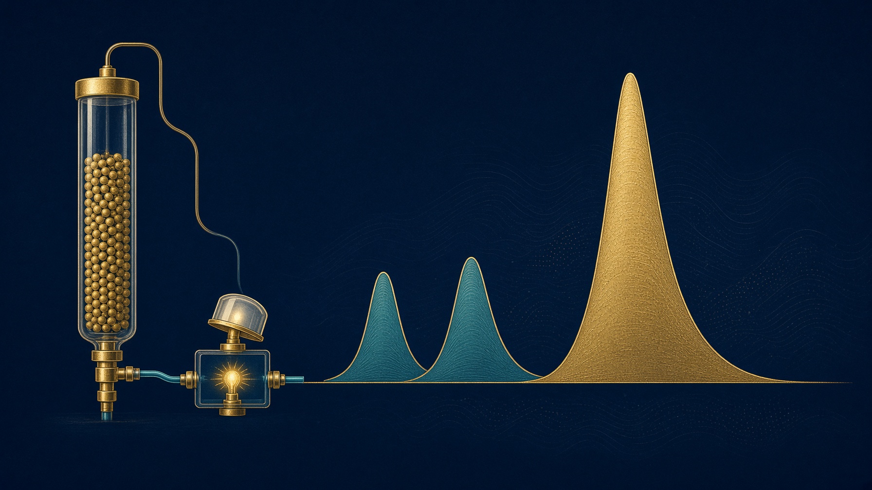
A peptide HPLC trace is time against ultraviolet absorbance. ≥98% is the main peak's share of the area. It is not the sequence. A chain missing one amino acid can hide in that peak. The mass is the other half of the sentence.
What this essay actually tells you
- A peptide HPLC trace is retention time against ultraviolet absorbance, usually at 214 nm where peptide bonds absorb. ≥98% is the main peak's share of the counted area.
- A chain missing one amino acid still absorbs that light and can hide in the main peak or on a shoulder. The mass is what shows the letter is gone. HPLC-MS is both measurements.
- Peak purity is not net peptide content. Salt and water never appear as peaks. A clean trace describes the solid. It is not a use instruction.
What this actually means
An HPLC chromatogram shows when a sample came off a column, measured by ultraviolet light. The purity percent is how much of that light sat in the main peak. A chain missing one amino acid can hide there, because it still absorbs light. Weighing the peak, by mass spectrometry, is how you find out. Ninety-eight percent is an area. It is not a name, and it is not a permission to use the vial.
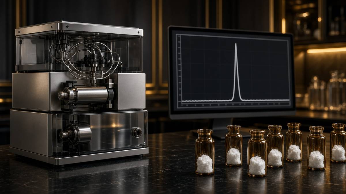
A peptide HPLC chromatogram is a plot of detector signal against time. The sample is pushed through a column, usually a C18 reversed-phase column, and the molecules come off in an order set by how tightly they stuck to the packing. What you are looking at is a separation, not a portrait. A single tall peak means most of the ultraviolet-absorbing material left the column together. It does not, by itself, prove that the material is the sequence printed on the label.
In short. The picture shows when things came off the column. A tall peak means most of the material arrived together. It does not spell the sequence.
What the axes actually are
The horizontal axis is retention time, in minutes. The vertical axis, for almost every peptide certificate, is ultraviolet absorbance. Peptide bonds absorb strongly near 214 nanometres, so 214 nm is the usual wavelength, with 280 nm added when the chain contains tryptophan or tyrosine and you want a second look. The area under a peak is proportional to how much of that absorbing material was there, if the detector was in its linear range and the wavelength suits the molecule. Percent purity on a certificate is that main-peak area divided by the sum of the peak areas the method chose to count. ≥98% HPLC means the main peak was at least ninety-eight percent of the counted area. It is an area ratio. It is not a molar guarantee, and it is not a mass.
In short. Time runs along the bottom. Height is how strongly the sample absorbed ultraviolet light. Ninety-eight percent means the main peak held almost all of that area. It is not a weighing of the sequence.
Retention time is a behaviour, not an identity card. Change the column, the temperature, the acetonitrile gradient, or the ion-pairing acid — trifluoroacetic acid is the usual one — and the same peptide can arrive a minute earlier or later. A matching retention time against a reference, run in the same method, is useful. A number on its own, copied from a different laboratory's method, is not the name of the chain.
In short. The minute a peak appears depends on the method. The same peptide can move if you change the column or the solvent. A time is not a name.
UV is not identity
The dangerous impurity in a synthetic peptide is often an N-1 deletion: the chain is missing one amino acid because a coupling failed and the growing chain was not properly capped. That shorter chain is still a peptide. It still absorbs at 214 nm. If the missing residue was not very hydrophobic, it can sit under the main peak or on its shoulder, and a quick glance at a pretty trace will call the vial clean. Area at one wavelength cannot see a missing residue. Mass spectrometry can, because the mass is short by that residue's weight. This is why a certificate that only shows a UV trace has answered a purity question and ducked an identity question.
In short. A chain missing one amino acid still absorbs ultraviolet light, and it can hide under the main peak. Only the mass tells you a letter is gone.
The same trap exists for a deletion, an insertion, a racemised residue, or a truncated chain that happens to have a similar hydrophobicity. Diastereomers — the same sequence with one amino acid in the wrong handedness — are a particular nuisance, because the mass is identical and only the shape, and sometimes the retention, has changed. HPLC can separate some of them. Mass spectrometry cannot, because the mass did not move. The two methods are not substitutes. They close different doors. HPLC-MS on one certificate is the UV separation and the mass of what came off, taken together.
In short. Ultraviolet light asks how much came off together. The mass asks what it weighs. A wrong-handed amino acid weighs the same, so you need both.
How to read the picture without lying to yourself
Start with the baseline. A drifting or noisy baseline makes small peaks look larger or smaller than they are, and the area percent moves with it. Then look at the main peak's shape. A shoulder, a split top, or a tail that does not come back to baseline is a second species trying to hide, or a column that is tired. Integration matters: where the software drew the start and the end of the peak changes the percent. A certificate that prints 98.4% without showing the trace is asking you to trust the arithmetic you cannot see.
In short. Look at the floor of the trace first, then at whether the main peak is clean or has a shoulder. A percentage without the picture is an arithmetic you cannot check.
Then ask what was not counted. Some methods ignore peaks below a threshold. Solvent, the counter-ion — trifluoroacetate is common on a synthetic peptide — and water do not show as peptide peaks at 214 nm in the way people hope, so a beautiful chromatogram can sit on a cake that is only seventy percent peptide by weight. That is net peptide content, a different number from chromatographic purity. Weighing the cake and calling it all peptide is how a concentration in a dish becomes fiction. The HPLC percent and the peptide content are both real. They are not the same sentence.
In short. A clean trace can still sit on a cake that is part salt and water. Purity of the peak is not the same as how much of the powder is peptide.
- The picture
- UV at 214 nm
- The percent
- main peak ÷ counted area
- The impostor
- N-1 deletion
- The name
- the mass
Peptide bonds absorb here. Area is how much absorbing material arrived together.
≥98% is that ratio. It is not a sequence and not a weight.
One residue short. Similar stickiness. Often a shoulder, sometimes invisible to UV alone.
HPLC-MS puts a mass on the peak. Handedness still needs the separation.
What this means for a vial
If you are comparing research peptides in the UK, read the certificate as two claims. Chromatographic purity is the area of the main peak. Identity is the mass, ideally taken on that peak, matching the calculated mass of the named sequence. A lot number, a date, and the method belong on the same page, because a percent without a method is a number from nowhere. The longer account of how a chain is built, residue by residue, is the synthesis essay. This page is only how to look at the picture that comes back.
In short. Purity is the size of the main peak. Identity is the mass of what that peak actually is. Both should be on the certificate, with the method and the lot.
The vials on this site are lyophilised research reagents, for laboratory use only, not for human consumption. A clean chromatogram is a description of a solid. It is not a permission.
In short. A clean trace describes the powder. It is not an instruction to take it.
Questions the essay actually answers
- What does a peptide HPLC chromatogram show?
- It shows when ultraviolet-absorbing material came off a column, usually C18, usually watched at 214 nm where peptide bonds absorb. The main peak's share of the counted area is the purity percent. Retention time is a behaviour in that method, not the name of the sequence.
- Does one tall peak prove the sequence?
- No. A deletion that is missing one amino acid still absorbs ultraviolet light and can sit under the main peak or on a shoulder. Identity wants the mass of that peak, which is what HPLC-MS adds.
- What does ≥98% HPLC purity mean?
- The main peak was at least 98% of the peak area the method counted. It is an area ratio at one wavelength. It is not a weighing of the cake, and it does not include salt and water that never appeared as peaks.
- What is an N-1 deletion?
- A synthetic chain missing one residue because a coupling failed. The mass is short by that amino acid. If the residue was not very hydrophobic, reversed-phase HPLC may only show a shoulder, or nothing you can see.
- Why is mass spectrometry on the same certificate?
- HPLC separates. Mass spectrometry weighs what came off. Together they catch the impurity that looks right in time and wrong in mass. They still do not see a wrong-handed amino acid, because that mass does not change.
Hypothetical research reconstitution
How these vials are typically mixed
Hypothetical research reconstitution for the named catalogue vial. Not a protocol, not medical advice, not a use instruction. These amounts sit in published and commonly cited laboratory ranges. The vial is labelled for research use only — not for human or veterinary administration.
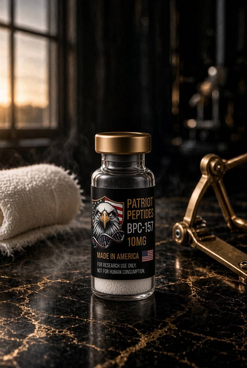
BPC-157
10mg
Mix with 2 ml bacteriostatic water → 5 mg/ml · 5,000 mcg/ml
- Hypothetical aliquot
- 250 mcg
- 0.05 ml · 5 units on a U-100 syringe
- How often
- Once or twice daily
- 2–4 weeks in the papers that actually run a course
Bench steps
- Let the vial sit until it is no longer cold to the touch.
- Wipe the stopper with 70% isopropyl alcohol. Let it dry.
- Draw 2 ml bacteriostatic water (0.9% benzyl alcohol).
- Run the water slowly down the inside glass — do not blast the cake.
- Roll between finger and thumb until the cake is gone. Do not shake.
- Label the date. Store the solution at 2–8 °C. Do not freeze. Use within 30 days unless the note below says otherwise.
Stable in bacteriostatic water in the fridge. 500 mcg is the upper end of what most bench notes call a daily aliquot; 250 mcg is the usual starting mark.
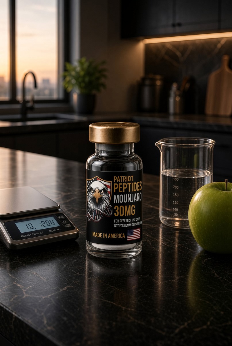
Mounjaro
30mg
Mix with 3 ml bacteriostatic water → 10 mg/ml
- Hypothetical aliquot
- The SURMOUNT arms used 5, 10 and 15 mg
- At 10 mg/ml those amounts are 0.50, 1.00 and 1.50 ml
- How often
- Once weekly in the published trials
- SURMOUNT-1 ran 72 weeks.
Bench steps
- Let the vial sit until it is no longer cold to the touch.
- Wipe the stopper with 70% isopropyl alcohol. Let it dry.
- Draw 3 ml bacteriostatic water (0.9% benzyl alcohol).
- Run the water slowly down the inside glass — do not blast the cake.
- Roll between finger and thumb until the cake is gone. Do not shake.
- Label the date. Store the solution at 2–8 °C. Do not freeze. Use within 30 days unless the note below says otherwise.
Mounjaro 30mg. Tirzepatide, LY3298176. The milligram figures are what the papers used. They are not a use instruction for this vial.
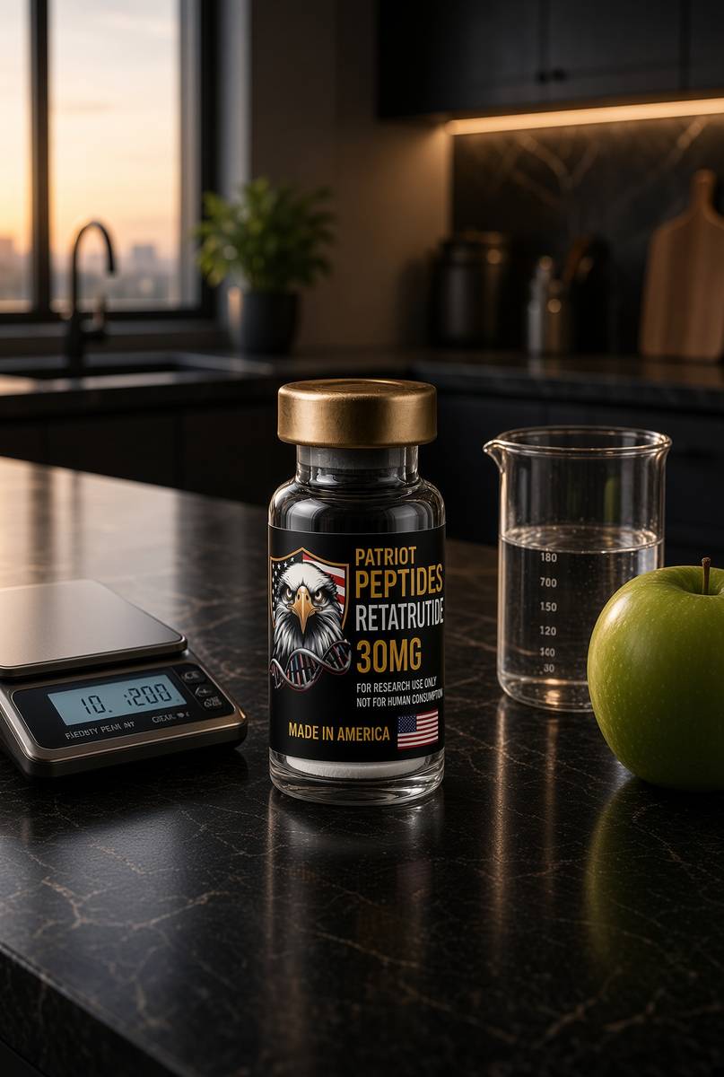
Retatrutide
30mg
Mix with 3 ml bacteriostatic water → 10 mg/ml
- Hypothetical aliquot
- 1–2 mg to start; published trial arms ran higher by week
- 0.10–0.20 ml · 10–20 units on a U-100 syringe (at 1–2 mg)
- How often
- Once weekly
- The Jastreboff NEJM 2023 arms ran 48 weeks. That is a trial, not a shop protocol.
Bench steps
- Let the vial sit until it is no longer cold to the touch.
- Wipe the stopper with 70% isopropyl alcohol. Let it dry.
- Draw 3 ml bacteriostatic water (0.9% benzyl alcohol).
- Run the water slowly down the inside glass — do not blast the cake.
- Roll between finger and thumb until the cake is gone. Do not shake.
- Label the date. Store the solution at 2–8 °C. Do not freeze. Use within 30 days unless the note below says otherwise.
LY3437943 architecture. Weekly, not daily. Those milligram figures are what the papers used on the investigational medicine — they are not a use instruction for this reagent.
Bacteriostatic water and sterile syringes ship with peptide orders over £75. Kit details · 10 ml bacteriostatic water
The vials this essay sits on
Named sequences the essay maps — BPC-157, Mounjaro, Retatrutide. Hypothetical research neighbourhood, not a protocol, not a medicine. One press puts every in-stock vial in the bag.
 Research only
 Research only
 Made in USA
Incretin
Retatrutide
One chain that copies three hormones you release after a meal: fullness, insulin, and how the liver spends fuel.
4.6(609)
6 browsing this now · 3 purchased in the last 24 hours
30mg · In stock
£120.00
Research use only. Not a combined-use instruction.
Read next
53 min · long read · Peptide research
How research peptides are made — and why HPLC actually matters
Solid-phase peptide synthesis builds a chain one residue at a time. HPLC then asks whether the main peak is what you think it is. ≥98% is not a slogan. It is a chromatogram.
43 min · long read · Peptide research
How to read a Phase 2 paper without lying to yourself
Jastreboff et al., NEJM 2023, is a public Phase 2 trial of retatrutide: 338 adults, placebo, 48 weeks, 24.2% mean weight loss at 12 mg. n, randomisation, adverse events, and why a research vial of the published structure is not the investigational medicine.
48 min · long read · Peptide research
What are peptides? Short chains that run the body
Peptides are amino acids joined by peptide bonds — shorter than proteins, more specific than most small-molecule drugs. Insulin was the first. GLP-1 agonists are the current proof.
More in this desk

52 min · long read · Peptide research
Recovery, GH pulses and the CJC / ipamorelin pair
Somatotrophs have two 'go' receptors. CJC without DAC is a slightly longer GHRH pulse. Ipamorelin is a selective ghrelin-receptor key that does not yank ACTH. Together they are the pair Bowers already showed is more than additive — not a gym protocol.
51 min · long read · Peptide research
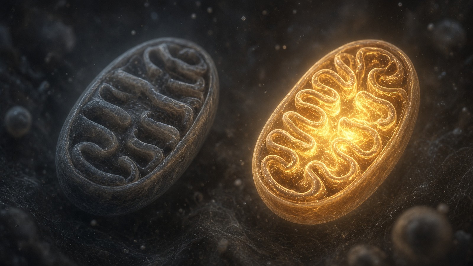
Fatigue, cellular energy and the NAD+ / MOTS-c neighbourhood
NAD+ is the rechargeable chip every cell spends on fuel and DNA repair — and the pool shrinks with age. MOTS-c is a mitochondrial 16-mer sent out under metabolic stress. Two answers to 'I have no energy' that are not coffee.
50 min · long read · Peptide research
Melanocortin circuits: pigment, appetite and PT-141
Melanotan II lights MC1, MC3, MC4 and MC5 — pigment plus the rest of the sheet. PT-141 is the free-acid cousin pointed at MC3 and MC4, the circuitry papers use for desire and energy, not skin colour. Same Arizona family. Different question.
50 min · long read · Peptide research
GHRH and ghrelin mimetics: two keys on the same pituitary lock
Somatotrophs have two ‘go’ receptors — GHRHR (Gs–cAMP) and GHSR (Gq). Bowers showed that occupying both releases more GH than the sum of either. That is synergy as a receptor fact, not a smoothie.
The page is the mechanism: the protein, what it does in the cell, and the drug or peptide that talks to it.