

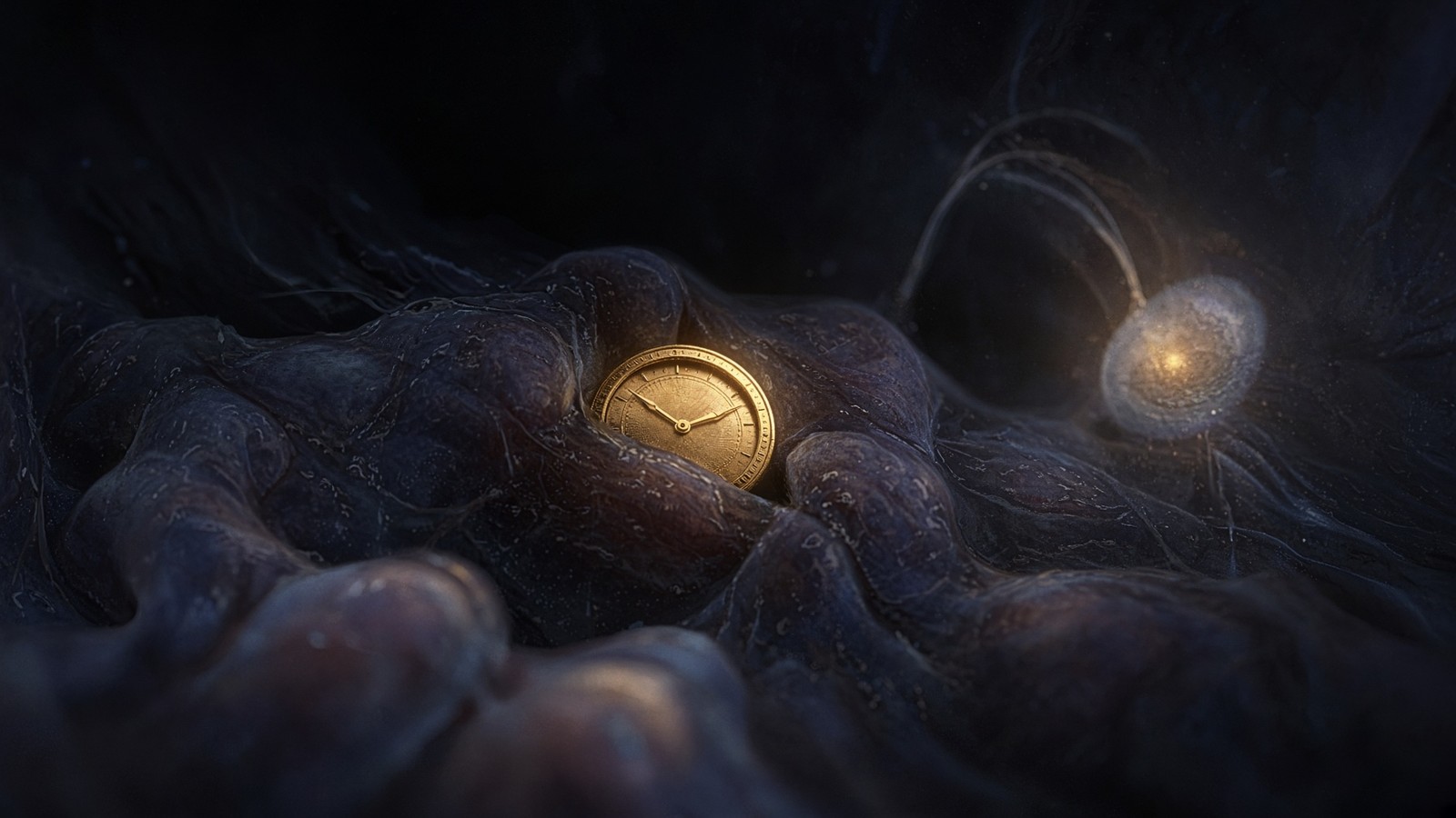
Peptide research · 49 min · 10,860 words
The pineal, melatonin and the 24-hour gene
The SCN is a transcription-translation loop that keeps ~24-hour time. The pineal converts its night signal into melatonin via AANAT. Peripheral clocks then decide when liver may make glucose. Aging flattens the peak.
· updated
What this essay actually tells you
- The suprachiasmatic nucleus is the master clock. The pineal gland converts its night signal into melatonin. Light in, hormone out, via a surprisingly small nucleus.
- CLOCK/BMAL1 and PER/CRY are the transcriptional loop that makes ~24-hour gene expression in almost every tissue. Almost every tissue. Not just sleep.
- Aging flattens the nocturnal melatonin peak. Epithalon papers from the Khavinson school sit at that oscillator. A research question. We didn't file it under sleep hygiene.
What this actually means
A cluster of about 20,000 neurons in the hypothalamus, the suprachiasmatic nucleus, keeps a 24-hour clock. Light on intrinsically photosensitive retinal ganglion cells (melanopsin, peak sensitivity around 480 nm) is the daily reset. In darkness, the SCN lets the pineal gland, via a spinal and superior-cervical-ganglion path, release melatonin. Melatonin is not sleep in a pill; it is a darkness certificate: N-acetyl-5-methoxytryptamine, synthesised from serotonin when AANAT is induced by nocturnal norepinephrine. Clock genes in liver, pancreas and muscle then decide when those tissues may make glucose, secrete insulin, or take it up. Ageing flattens the nocturnal melatonin peak even in the dark. Shift work, late phones and polar winter are versions of the same insult: a transcriptional programme running on the wrong photoperiod. IARC classifies night-shift work as a probable carcinogen in part because of that mismatch. Khavinson's tetrapeptide Epithalon was studied as a pineal-adjacent molecule that could restore some melatonin amplitude in old animals, and, in the stronger claims, talk to TERT. Those are two assays. They are not a substitute for a dark night.
You've got a master clock sitting just above where the optic nerves cross. The suprachiasmatic nucleus — about twenty thousand neurons in the anterior hypothalamus — keeps roughly 24-hour time, and the pineal gland turns the night half of that signal into melatonin. Light in, hormone out. The photopigment is melanopsin, sitting in a sparse set of intrinsically photosensitive retinal ganglion cells whose peak sensitivity is around 480 nanometres. The hormone is N-acetyl-5-methoxytryptamine, written from serotonin when arylalkylamine N-acetyltransferase, AANAT, is induced by nocturnal norepinephrine. Between those two sentences sits a transcription-translation loop, a multi-synapse sympathetic path, and a set of peripheral clocks that decide when a hepatocyte may make glucose. Ageing flattens the nocturnal melatonin peak even when the room is dark. Shift work, a phone at midnight and a polar winter are versions of the same insult: a transcriptional programme running on the wrong photoperiod. We'll name the loop, the factory and the hormone, and we'll put the tetrapeptide where the papers sat it — at one ageing oscillator, not in place of daylight.
In short. Your brain has a cluster of cells that keep 24-hour time. The pineal turns night into melatonin. Light in, hormone out.
CLOCK and BMAL1 are transcription factors — gene switches — that turn on Period and Cryptochrome. PER and CRY proteins accumulate, return to the nucleus, and shut CLOCK/BMAL1 off. Degradation of PER and CRY then lets the cycle start again. The delay is about a day. That loop, not a metaphor for one, is how almost every mammalian tissue keeps local time: liver, pancreas, muscle, adipose, immune cells, skin. Sleep is one downstream behaviour of a programme that also times gluconeogenesis, insulin secretion, DNA-repair transcripts, xenobiotic enzymes and the NAD+ salvage enzyme NAMPT. Takahashi, Tei, Reppert and Weaver spent the 1990s proving the loop is the clock. Hall, Rosbash and Young took the 2017 Nobel for the fly version of the same idea. It's tempting to file all this under sleep, but your liver is keeping time at four in the morning whether you're asleep or not. The SCN is special because it's the only clock that sees the outside world, via the retinohypothalamic tract, and then tells the others the time.
In short. A pair of gene switches turns a second pair on, then those products switch the first pair off, about once a day, in almost every tissue.
Epithalon is Ala-Glu-Asp-Gly, a synthetic tetrapeptide from Vladimir Khavinson's pineal-peptide programme in St Petersburg. It isn't melatonin. Melatonin is an indoleamine pinealocytes write when AANAT fires. We hold the tetrapeptide because the Khavinson school reported a restored night-time melatonin curve in old animals, and, in a neighbouring literature, a TERT conversation in fibroblasts. Those are two assays. They're a research question sitting at an oscillator ageing has flattened, and they're not a substitute for a dark night. We'll name the chemistry, the anatomy, the peripheral clocks, the shift-work epidemiology and the peptide papers in that order. A dark room still costs nothing. A characterised four-mer is how one literature proposed to study what the years took off the peak. Mixing the two into a sleep story is how a chromatogram gets replaced by a caption. Keep the hormone, the tetrapeptide and the photoperiod as three objects and the night stays interesting.
In short. Epithalon is four amino acids, not the night hormone. The papers sit on a flattened melatonin peak in old animals. Darkness still costs nothing.
What follows is physiology, named, rather than a protocol. Named nuclei, named enzymes, named photopigments, named papers, and a legal class stated once at the close. The sleep-architecture essay takes DSIP and the scored night; the Epithalon essay takes telomeres, shelterin and TRAP. This page takes the clock as a transcriptional object, the pineal as a factory, the liver as a peripheral oscillator that times glucose, and the tetrapeptide as a pineal-adjacent reagent rather than a hypnotic. IARC classifies night-shift work as a probable carcinogen in part because internal time and solar time can be pulled apart. Van Cauter showed that one restricted night is already a glucose insult in healthy adults. Ramsey and Bass put NAMPT, the NAD+ salvage bottleneck, on the same loop. A phone at 23:30 is an input to AANAT, not an entertainment footnote. Morning outdoor light is the correctly timed version of the same photon, and it's still the cheapest experiment you can run.
In short. This page is the clock, the pineal factory, liver glucose timing and a four-residue peptide. It's physiology, not a bedtime story.
The SCN is a transcription-translation loop that keeps ~24-hour time. The pineal converts its night signal into melatonin via AANAT. Almost every tissue runs a version of that loop. Not just sleep.
A clock made of transcription, not gears
The mammalian transcription-translation feedback loop is a delay line built from transcription factors. CLOCK — circadian locomotor output cycles kaput, named from a mutagenesis screen in mice — heterodimerises with BMAL1, also called ARNTL. The heterodimer occupies E-boxes in the promoters of Period genes (PER1, PER2, PER3) and Cryptochrome genes (CRY1, CRY2) and drives their transcription. PER and CRY proteins accumulate in the cytoplasm, are phosphorylated by casein kinase 1δ and 1ε, form complexes, and after a lag of hours re-enter the nucleus to repress CLOCK/BMAL1. When PER and CRY are degraded — the proteasome, again — repression lifts and the cycle restarts. The delay is the clock. Without a delay you have an equilibrium, not a day. Additional loops stabilise the period: REV-ERBα and REV-ERBβ (NR1D1, NR1D2) repress BMAL1; ROR nuclear receptors activate it. The positive and negative limbs together give a limit cycle whose period, in a human SCN, sits near 24.2 hours in the absence of light, a number Czeisler and colleagues spent careers measuring in forced-desynchrony protocols.
In short. CLOCK and BMAL1 turn on Period and Cryptochrome genes. Those proteins later switch CLOCK and BMAL1 off. The delay of about a day is the clock.
Joseph Takahashi's laboratory cloned the mammalian Clock gene from a chemically mutagenised mouse that had a long free-running period. The 1994 Science paper with Vitaterna, and the 1997 Cell cloning papers, are the mammalian birth certificates. Tei, Sakaki and colleagues cloned human and mouse PER independently. Reppert and Weaver, at Massachusetts General and then elsewhere, mapped the SCN as the site where those genes have to run for the animal to keep time, and wrote the reviews a generation of students still learn from. In Drosophila, period had already been a Konopka and Benzer mutant in 1971; Hall, Rosbash and Young then found that the fly protein cycles, that timeless is its partner, and that a similar feedback loop is the fly clock. The 2017 Nobel recognised that insect work. The mammalian expansion is Takahashi's, and it's why a hepatocyte and a neuron can be said to run the same kind of oscillator with tissue-specific accessories. That's why a mouse knockout of Bmal1 is arrhythmic, and why a human PER2 mutation — familial advanced sleep-phase syndrome, the Toh and Ptáček work — can shift a life by hours.
In short. The mouse Clock gene was cloned from a mutant that ran on a long day. Fly genetics had already shown a similar loop. Humans can inherit a shifted clock.
Panda, Hogenesch, Kay and the microarray era that followed showed that a large fraction of the protein-coding genome is circadian in at least one tissue. Not a handful of clock genes. Thousands of transcripts, tissue-by-tissue, with the overlap between liver and SCN smaller than you'd guess from a headline. Zhang, Kay and colleagues later put numbers on that census: a majority of genes are circadian somewhere, and the liver is one of the louder peripheral clocks. That's the molecular reason this page refuses to live under a sleep heading. A gene that times cytochrome P450 expression in a hepatocyte is a clock gene for the purpose of xenobiotic handling. A gene that times GLUT4 trafficking in a myocyte is a clock gene for glucose uptake. Sleep is one output. The transcriptional programme is the thing. When people say the body has a clock, they're usually picturing the SCN. The more accurate picture is a clock shop, with the SCN as the shopfront that looks at the street and the other clocks as the work going on in the back rooms, coupled by hormones, feeding and autonomic tone.
In short. Thousands of genes, not a handful, rise and fall across 24 hours, and different organs time different sets. Sleep is only one output of that programme.
Post-translational timing is as much the clock as transcription is. Casein kinase 1δ and 1ε phosphorylate PER and thereby set how long the protein lasts; the tau mutation in the Syrian hamster, which Ralph and Menaker used for the famous SCN-transplant experiments, is a CK1ε lesion, a short period written as a kinase rather than as a missing transcription factor. AMPK phosphorylates CRY1 and can shorten its half-life, which is one door between energy state and period. GSK3β phosphorylates REV-ERB. F-box proteins (FBXL3, FBXL21) ubiquitinylate CRYs and send them to the proteasome. None of that is decoration. A clock that was only transcription wouldn't have a four-hour kinase lever, and a peptide paper that reports a clock-gene qPCR without ever mentioning phosphorylation has reported a transcript, not a period. Temperature compensation, Pittendrigh's old demand that a true circadian oscillator keep roughly the same period across a physiological temperature range, is partly written in these modifications. The SCN is also electrically a clock: neuronal firing rate itself cycles, and vasoactive intestinal peptide couples the twenty thousand cells so that a noisy set of single-cell oscillators becomes a tissue that can shout a time of day.
In short. Kinases and protein breakdown set how long clock proteins last, which sets the length of the day. Individual clock cells also fire as a tissue, not as twenty thousand private watches.
A closed promoter and an open one are different objects at this loop, which is why the transcription diagram belongs on this page rather than only on a TERT essay. CLOCK/BMAL1 occupancy at an E-box is a chromatin event: histone acetylation, a promoter that can be found, a polymerase that is allowed to run. SIRT1, an NAD+-dependent deacetylase, has been reported to deacetylate BMAL1 and to sit on the same neighbourhood as CLOCK, which is how a cofactor budget and a 24-hour loop became one conversation in the Sassone-Corsi and Imai papers. NAMPT, the rate-limiting enzyme of NAD+ salvage, is itself a CLOCK/BMAL1 target. The cofactor that sirtuins spend is therefore both an input to the loop and an output of the loop. That's why the NAD+ essay and this one are allowed to point at each other without being the same essay. Transcription here isn't a generic machine. It's the delay line. If a tetrapeptide literature claims to have moved Per2 or Bmal1, the claim is that something in this chromatin-and-kinase neighbourhood moved. Name the transcript, the time point, and the photoperiod. A single daytime qPCR isn't a circadian result.
In short. Clock genes are turned on and off at chromatin, and the NAD+ recycling enzyme is one of their targets. A single daytime gene reading is not a 24-hour result.
Diagram
Closed chromatin (H3K27me3, DNA methylation) hides the promoter. Pioneer factors and histone acetyltransferases open it.
PIC: TFIID, TFIIH, Mediator, Pol II. Ser5 phosphorylation of the CTD lets the polymerase leave the promoter.
Elongation ~20–40 nt/s. Capping, splicing, cleavage and polyadenylation happen on the still-growing RNA.
Human genes are islands in 3.1 billion base pairs of mostly noncoding sequence. Promoter, enhancers, chromatin state and the Mediator complex decide whether Pol II is allowed to fire. Epithalon’s literature sits on TERT and pineal clocks — two of the rare promoters anyone bothers to name in a peptide essay.
The SCN is the only clock that sees the sun
The SCN sits above the optic chiasm, which is how it got its name and how it got its light. Two nuclei, one per side, each a few tens of thousands of small neurons, GABAergic in the main, with a ventral core rich in vasoactive intestinal peptide and a dorsal shell rich in arginine vasopressin. The retinohypothalamic tract, a minority branch of the optic nerve, lands on the core. Moore and Eichler, and independently Stephan and Zucker, lesioned this nucleus in the early 1970s and lost behavioural circadian rhythmicity; the animal still slept, still ate, but the timing fell apart. Ralph, Foster, Davis and Menaker then transplanted a tau-mutant SCN into a lesioned wild-type host and the host ran at the donor period, which is as close to a transplantable clock as mammalian neurobiology has offered. The SCN isn't a sleep centre. It's a timing centre whose outputs, humoral and synaptic, tell sleep centres, endocrine axes and peripheral organs what time it is. Destroy it and you don't create an insomniac. You create an animal whose days have no agreed noon.
In short. The master clock sits above where the optic nerves cross. Lesion it and timing falls apart. Transplant a mutant one and the animal runs on the donor's day.
Melanopsin, encoded by Opn4, is the photopigment of the reset. Provencio found it first in frog skin; Berson, Dunn and Takao, in 2002, showed that a sparse set of retinal ganglion cells in the rat still fired to light when rods and cones were blocked, and that those cells were the melanopsin cells. Peak sensitivity sits in the blue-rich neighbourhood around 480 nanometres. That's why a 4000 K screen at 23:30 is an input to the SCN, and why a warm, dim lamp is a weaker one. The cells are intrinsically photosensitive, which means they don't need rods or cones, although rods and cones do talk to them and contribute to the full irradiance code. Their axons form the retinohypothalamic tract and release glutamate and PACAP onto SCN neurons. Calcium rises. Per1 is induced. The loop is phase-shifted. A light pulse in the early night delays the clock; a pulse in the late night advances it. That phase-response curve is older than melanopsin as a molecule — Pittendrigh drew it for flies and Aschoff for humans — and melanopsin is the mammalian photopigment that makes the curve a retinal fact rather than a mystery.
In short. A blue-sensitive pigment in rare retinal cells tells the master clock about light, even without ordinary vision. Late-night light delays the clock; late-dark-night light advances it.
Czeisler's human work, and Lewy's melatonin-suppression work before and beside it, put numbers on what a photon does to a person. Bright light at night suppresses melatonin and can shift the pacemaker; the dose, spectrum and prior light history all move the curve. A phone isn't a 10,000-lux light box. It isn't nothing either, particularly in a dark-adapted observer whose melanopsin cells have been sitting in a dim room. Morning outdoor light, even under British cloud, is often hundreds to thousands of lux of a spectrum that includes the melanopsin band. That's the free, correctly timed reset. People still ask which peptide replaces a walk. None of them does. The tetrapeptide in the catalogue was studied against a flattened nocturnal melatonin peak in aged animals, which is a different experiment from a phase shift in a healthy student. Photoperiod is the control that costs nothing. A characterised four-mer is a research object for the ageing-pineal question. Use the light either way. Your eyes already know how.
In short. Morning daylight is the free, correctly timed reset. A phone at midnight is a weaker, mistimed version of the same signal. No peptide replaces a walk outside.
Single SCN neurons are already oscillators. Welsh, Logothetis, Meister and Reppert showed that in culture: dispersed SCN cells keep firing-rate rhythms, with a spread of periods, and the tissue-level precision comes from coupling. VIP, acting at VPAC2, is the coupling peptide the field keeps coming back to, because mice lacking the receptor have a desynchronised SCN and a weak behavioural rhythm. GABA, gap junctions and other peptides contribute. Coupling is how a noisy set of cellular clocks becomes a master that the body can trust. Ageing has been reported to weaken that coupling, to thin some of the peptidergic signals, and to flatten amplitude at the tissue level even when single cells still tick. A noisier master is a poorer teacher of the periphery. That's one of the candidate mechanisms for a flattened melatonin peak in an old pineal: the factory is still there, the night shift is receiving a less decisive instruction. Whether the lesion is in the SCN, in the sympathetic path, or in the pinealocyte's competence to induce AANAT is exactly the sort of question a 24-hour melatonin curve, a clock-gene panel and a carefully timed light stimulus can still argue about.
In short. Individual clock cells already keep time. They stay in step by talking, and ageing can make that conversation quieter, so the night signal to the pineal is less decisive.
- SCN census
- ~20,000 neurons
- Melanopsin peak
- ~480 nm
- Human free-run
- ~24.2 h
- AANAT
- night enzyme
- Melatonin
- N-acetyl-5-methoxytryptamine
- Epithalon
- AEDG, ~390 Da
Paired nuclei above the optic chiasm. VIP core, AVP shell. The only clock that sees the street.
Opn4, ipRGCs. Berson, Dunn, Takao, 2002. A 4000 K screen at 23:30 is an input.
Forced desynchrony. Czeisler. Slightly long, so morning light is the daily correction.
Serotonin → N-acetylserotonin. Tens-fold protein at night; minutes to collapse in light. Klein.
Nanomolar darkness certificate at MT1/MT2 (Gi). A different molecule from AEDG; a milligram tablet is not physiological pineal output.
Ala-Glu-Asp-Gly. Khavinson's defined stand-in for pineal epithalamin. Catalogue sequence, not melatonin.
The pineal is a factory. AANAT is the night shift.
The path from SCN to pineal is a mammalian joke about anatomy. Light doesn't hit the pineal. It hits the retina. The SCN computes night. A descending path then runs to the paraventricular nucleus of the hypothalamus, down the intermediolateral column of the spinal cord, out to the superior cervical ganglion, and back up as post-ganglionic sympathetic fibres that release norepinephrine onto pinealocytes. Only when that path is firing as night does the factory start. Cut the superior cervical ganglia and melatonin rhythmicity collapses, a classical result. The pineal in a mammal is therefore an endocrine output of a neural computation, not a photoreceptor. Some non-mammalian pineals still see. Ours doesn't. David Klein's biochemistry is the reference for what happens next: norepinephrine, via β1-adrenergic receptors (Gs, cAMP, protein kinase A) and α1-adrenergic receptors (Gq, calcium), induces and stabilises arylalkylamine N-acetyltransferase. AANAT is the night enzyme. Protein can rise tens-fold after dark and collapse in minutes if you turn a light on, because proteasomal degradation is the off-switch and cAMP is what holds it back.
In short. In mammals the pineal does not see light. The brain clock certifies night, a sympathetic nerve fires, and only then does the melatonin factory start.
Serotonin is the substrate. Pinealocytes are stuffed with it. AANAT acetylates the amine to N-acetylserotonin. Hydroxyindole-O-methyltransferase, also called acetylserotonin O-methyltransferase, ASMT, then methylates the 5-hydroxyl to give melatonin, N-acetyl-5-methoxytryptamine. AANAT is rate-limiting at night in the species that have been properly measured, which is why a light pulse that destroys AANAT protein destroys the melatonin signal even if ASMT is still sitting there. The chemistry is an acetyl transfer from acetyl-CoA and a methyl transfer from S-adenosylmethionine. Named cofactors, named enzymes, a two-step path from a neurotransmitter to a darkness hormone. Tryptophan hydroxylase and aromatic L-amino-acid decarboxylase sit upstream, which is why a tryptophan story and a melatonin story get mashed together in supplement aisles; the night-gated step is still AANAT. During the day the enzyme is transcribed less, phosphorylated less helpfully, and eaten by the proteasome. The factory isn't dismantled. The night shift is sent home.
In short. Melatonin is made from serotonin in two steps. The first enzyme, AANAT, is the night switch: abundant in the dark, destroyed within minutes if a light goes on.
cAMP is the second messenger of that night shift, which is why the second-messenger diagram belongs here rather than as a generic peptide cartoon. β1 occupancy raises adenylate cyclase. Protein kinase A phosphorylates AANAT and CREB. Phosphorylated AANAT binds 14-3-3 proteins, which shield it from the proteasome; that shielding is the minutes-scale on-switch. A light pulse drops sympathetic drive, cAMP falls, the shield comes off, AANAT is ubiquitinated and gone. Klein, Coon, Gastel and the NIH pineal group spent a generation on that biochemistry. It's also why a melatonin measurement without a stated photoperiod isn't a circadian measurement. You can't interpret a plasma indoleamine if you haven't said what the sympathetic nerve was doing. Pineal explants, perifused with norepinephrine on a night schedule, will make melatonin in a dish; that's the factory isolated. An intact mammal won't, if you stand in the room with a phone. Ordinary Gs–cAMP. The timing of the occupancy is the whole trick. A peptide claiming to restore nocturnal amplitude would have to sit somewhere on this path, and you'd have to say which rung you measured.
In short. Night-time nerve signals raise cAMP, which protects the night enzyme from being destroyed. A light pulse drops that signal and the enzyme is gone in minutes.
Ageing reduces the amplitude of the nocturnal peak even if the room is dark. That sentence is the gerontology of this page, and it's a measurement, not a mood. AANAT competence falls. The SCN's tissue-level output is often noisier. The pineal itself calcifies in many adults; corpora arenacea, brain sand, sit among pinealocytes, a fact anatomists have known for a century and a reason imaging reports a calcified pineal as a normal variant rather than as a disease. Time isn't kind to that neighbourhood. Melatonin then occupies MT1 and MT2, both Gi-coupled GPCRs, on the SCN itself (a feedback), on the pituitary (a calendar, especially for seasonal breeders), and on peripheral tissues. At pharmacological concentrations it's also a radical scavenger, which has launched a thousand labels and a smaller number of careful papers. Physiological nocturnal nanomolar in plasma isn't the same chemistry as a milligram oral bolus. Take the night away with a phone and AANAT never fully fires. Take the years away and the peak flattens anyway. Those are two insults. They can stack.
In short. Even in a dark room, old pineals often make a smaller night-time melatonin peak. Calcification, a noisier brain clock and a weaker night enzyme all contribute.
Josephine Arendt's human melatonin work, and the seasonal-affective and shift-work literatures that cite it, treated the hormone as a darkness certificate and as a phase-shifting agent at the SCN, not as a sleeping tablet that happens to come from a gland. Exogenous melatonin can phase-shift a human clock, more cleanly when timed to the falling or rising limb of the endogenous curve, which is why a jet-lag protocol that ignores clock time is a guess. Endogenous melatonin is a marker of biological night. Dim-light melatonin onset, DLMO, is the phase marker a sleep laboratory actually uses: sample in dim light, catch the evening rise, write a clock time. That marker can drift in shift workers, in the blind (who often have free-running rhythms unless their melanopsin path is intact), and in some circadian-rhythm sleep disorders. A flattened nocturnal peak in an older adult is a different object from a shifted DLMO in a teenager on a phone. Amplitude versus phase. Both are circadian. They aren't the same measurement, and a tetrapeptide paper that restored amplitude in an old rat hasn't thereby shifted a human DLMO.
In short. Melatonin marks biological night. Labs use its evening rise as a phase marker. A smaller peak in old age is a different problem from a peak that happens at the wrong clock time.
Diagram
× 1
Ligand
One peptide in one pocket. nM–µM. Shape, not a mood.
× 10–10²
G proteins
The occupied GPCR is a GEF. Each Gα is a catalyst.
× 10³–10⁴
cAMP / IP₃ / Ca²⁺
Adenylyl cyclase and PLC do not make one molecule. They make a cloud.
× 10⁴–10⁶
PKA / PKC / CaMK
Kinases phosphorylate many substrates per messenger.
× tissue
Secretion, transcription, motility
The organism-level readout. Still not a protocol.
This is the only magic, and it is not magic. A nanomolar ligand can move a micromolar messenger because enzymes sit between them. Desensitisation (GRK, β-arrestin, endocytosis) is how the cell refuses to let ‘more ligand’ mean ‘more signal’ forever.
Melatonin is a darkness certificate
MT1 and MT2, encoded by MTNR1A and MTNR1B, are Gi/o-coupled class-A GPCRs. Occupancy lowers cAMP in the cells that carry them. The SCN carries both; the feedback is one reason a melatonin pulse can phase-shift the pacemaker. Pituitary pars tuberalis is a melatonin-reading tissue in seasonal mammals, a calendar rather than a clock, reading the duration of the nocturnal signal as a day-length code. Pancreatic β-cells carry MT2, and common variants in MTNR1B are among the more replicated type-2-diabetes risk alleles, which is a genetic hint that this receptor isn't a sleep decoration. Peripheral clocks carry melatonin receptors to varying degrees; the hormone is one of the SCN's humoral hands on those clocks, along with cortisol, autonomic tone and the timing of meals. Gi coupling is ordinary. The information is in the duration and the amplitude of the occupancy, not in a novel second messenger. A ligand that occupied MT1/MT2 would be a melatonin-receptor agonist. Epithalon isn't that ligand. AEDG is four residues with a pineal-adjacent literature. Different carbon skeletons. Different assays. People mash them because both get mentioned next to night.
In short. Two melatonin receptors turn cell signals down via Gi proteins. They sit on the brain clock, the pituitary and some peripheral tissues. The tetrapeptide is not a melatonin-receptor drug.
The antioxidant story has to be sized correctly or it eats the page. Reiter's reviews, and a large rodent literature, treat melatonin as a scavenger of hydroxyl radical and as an up-regulator of glutathione enzymes, at concentrations that are often pharmacological. Mitochondria have been proposed as a site of that chemistry, which is how a darkness hormone and an organelle essay become neighbours. Physiological nocturnal nanomolar in plasma is a GPCR concentration. Millimolar in a cuvette is a radical-chemistry concentration. Both can be true of the same molecule. They're two jobs. This page is the GPCR and clock job, because that's the job AANAT was built for and the job a flattened nocturnal peak actually threatens. Writing melatonin as a mitochondrial vitamin is how a darkness certificate becomes a supplement aisle. Writing the radical chemistry out of existence is how you ignore a real literature. Hold the doses apart. The pineal writes nanomolar. A tablet writes more. The tetrapeptide writes neither; it isn't an indoleamine.
In short. At night-time blood levels melatonin talks to receptors. At much higher levels it can also mop radicals. Those are two jobs. The pineal is built for the first.
CSF melatonin, in some measurements, runs higher than plasma, because the pineal sits next to the third ventricle and can dump into the ventricular space as well as into blood. That anatomical privilege is one reason the hormone is a brain signal as well as a body signal. Liver metabolism, via CYP1A2 among other cytochromes, clears circulating melatonin quickly; oral bioavailability is modest and first-pass is real, which is why an oral milligram and a nocturnal pineal output are different pharmacokinetic objects even before you mention receptors. Slow-release formulations exist as licensed medicines in some jurisdictions for primary insomnia in adults over fifty-five, a narrow indication that still isn't this catalogue. We don't stock melatonin. We stock AEDG. The neighbouring sleep essay stocks the DSIP conversation. Three ligands people type when they type night. One chromatogram each. The darkness certificate the pineal writes remains the physiology. The tablet, where it is a medicine, is a medicine. The tetrapeptide is a research sequence from a pineal school. Keep the jobs unscrewed and the night is still allowed to be interesting.
In short. Pineal melatonin can enter brain fluid as well as blood, and the liver clears it fast. We do not sell melatonin. The catalogue sequence is the four-residue pineal peptide.
Melatonin is a darkness certificate. Clock genes are how the liver, pancreas and muscle read it. A phone at midnight is a forged one.
Peripheral clocks, and why the liver has a bedtime
Almost every tissue runs a version of the CLOCK/BMAL1 loop. The SCN is the conductor, not the only violin. Dibner, Schibler and the Geneva school, and the Ueli Schibler restricted-feeding experiments with Damiola, showed that the liver clock can be uncoupled from the SCN by meal timing: feed a mouse in the biological night and hepatic rhythmicity follows the food, while the SCN still follows the light. Food is a zeitgeber for the liver. Light is the zeitgeber for the SCN. When those two disagree, you have internal desynchrony, which is a more precise name than 'bad sleep' for what a rotating shift does to a hepatocyte. Clock genes in the liver time PEPCK and glucose-6-phosphatase, the gluconeogenic enzymes, among many other transcripts. Hepatic BMAL1 is therefore a glucose gene as well as a clock gene. Lamia, Evans and colleagues, and a series of liver-specific Bmal1 knockouts, made that sentence genetic rather than poetic. A liver that thinks it is noon while the pancreas thinks it is midnight will mishandle a meal. Glucose handling is one of the first things to look drunk.
In short. Liver cells have their own clocks, set more by meals than by light. When liver time and brain time disagree, glucose handling is among the first things to go wrong.
Pancreatic islets run clocks that gate insulin secretion. Marcheva, Bass and colleagues showed that β-cell clock disruption impairs insulin exocytosis and glucose tolerance, a paper that should have ended the habit of filing circadian biology under sleep. The β-cell has to know the time of day to know how hard to answer a glucose rise; a mismatched clock is a secretion defect, not a character flaw. MTNR1B variants, already mentioned, sit on this same organ as a melatonin-receptor genetic echo. Muscle clocks gate glucose uptake and a set of metabolic transcripts; Koike, Takahashi and the mouse muscle literature, and human biopsy work from several groups, keep finding CLOCK/BMAL1-dependent programmes in myocytes. Adipose clocks time lipolysis. Immune clocks time cytokine tone and the trafficking of leukocyte subsets, which is one proposed chapter of why shift work talks to inflammatory disease. Name the tissue. Name the transcript. 'Circadian disruption' is a filing. A β-cell clock lesion is a mechanism.
In short. Pancreas clocks time insulin release. Muscle clocks time sugar uptake. Immune clocks time inflammation. Each organ keeps time for its own job, taught by the brain clock and by meals.
Van Cauter's sleep-restriction work is the human glucose sentence this page has to cite, because it's ruder than a clock-gene cartoon. Spiegel, Leproult and Van Cauter took healthy young adults, cut the night, and found next-day insulin resistance you can measure, including on clamps in the later papers. One restricted night is already a metabolic insult. The neighbouring sleep-and-glucose essay owns the cortisol half: the morning peak should arrive on time and then fall; flatten that curve and hepatic glucose output stays inappropriately high. This page owns the clock half: a mistimed photoperiod is a glucose insult before it is a sleep complaint, because hepatic BMAL1 and the β-cell clock were not consulted about the gig. Cortisol, melatonin, growth-hormone pulses that prefer slow-wave sleep, and the transcriptional loop are four hands on the same metabolic night. Miss one and you have a paper. Miss all four and you have a culture that treats night as optional. The peptide doesn't replace any of those hands.
In short. One short night can make healthy adults worse at handling sugar the next day. A mistimed body clock is a glucose problem before it is a complaint about sleep.
Restricted feeding as a tool, and as a lifestyle argument people now shout about, sits on Schibler's liver-clock result and on Panda's later time-restricted-feeding papers in mice. Eating inside a consistent daily window can re-align hepatic clocks even when sleep is imperfect, which is interesting, and which is still not a peptide. The human evidence is smaller and more mixed than the mouse rooms, as you should expect when the animal also has a job, a commute and a family. This page won't run a feeding protocol. It will say that meal timing is a zeitgeber the liver actually has, that the SCN still wants light, and that a rotating night shift fights both. A tetrapeptide assayed against nocturnal melatonin amplitude isn't a time-restricted-feeding mimetic. It isn't a Van Cauter reversal. The metabolic night is a set of oscillators. Food, light, cortisol, melatonin, sleep stages. Pick the oscillator you are measuring. Write the photoperiod and the meal schedule when you design the experiment, or you haven't done the experiment you think you've done.
In short. Meal timing can reset liver clocks. Light still resets the brain clock. A rotating night shift fights both. A pineal peptide is not a feeding schedule.
DNA-repair transcripts, xenobiotic enzymes and cell-cycle genes are circadian in many tissues, which is one of the cleaner molecular arguments for why IARC spent time on shift work. A hepatocyte that writes a repair enzyme at the wrong clock time is a hepatocyte that met a toxin on the wrong shift. Sancar's work on cryptochromes and nucleotide-excision repair, and a broader literature on clock-gated cell-cycle checkpoints, are the papers to sit with. This isn't a claim that a late night causes cancer in a person the way a smoking history does. It's a claim that a transcriptional programme that times repair, metabolism and cell division is a reasonable place to look when a probable-carcinogen classification has to be argued. The pineal half of that argument is the melatonin amplitude and the MT1/MT2 occupancy that some tumour-biology papers have measured. The clock-gene half is Per2 and Bmal1 in the tissue of interest. Both belong in a shift-work paragraph. Neither is a reason to caption a tetrapeptide as oncology.
In short. Many DNA-repair and cell-division genes keep 24-hour time. That is one reason night-shift work is taken seriously as a cancer-risk conversation, not a reason to treat a peptide as oncology.
Diagram
Matrix
- TCA cycle · β-oxidation · mtDNA nucleoids
- NADH produced here. Complex I spends it.
- MOTS-c (MRWQEMGYIFYPRKLR) from 12S rRNA.
Inner membrane
- I → II → III → IV → V (ATP synthase)
- ~150 mV proton-motive force
- ~40–60 kg of ATP turned over per human day
mtDNA is 16,569 bp, 37 genes, 13 proteins of the respiratory chain. Nuclear DNA encodes the other ~1,200 mitochondrial proteins. NAD+ is the hydride carrier between dehydrogenases and Complex I. MOTS-c is a 16-mer translated from 12S rRNA — a peptide the mitochondrion wrote itself.
Shift work, phones, and a probable carcinogen
IARC classified night-shift work as a probable carcinogen (Group 2A) on the basis of limited human evidence, sufficient evidence in experimental animals for light-at-night and related exposures, and strong mechanistic evidence on circadian disruption. The monograph is a committee document, argued, cited, and not a tweet. Breast cancer in female night workers is the epidemiology that drew the most public heat; the signal isn't uniform across every cohort, which is how occupational epidemiology looks, and the mechanistic file is the reason the classification could still be written. Internal desynchrony — SCN following a compromised light schedule, liver following meals grabbed on a break, melatonin amplitude flattened by light-at-night — is the proposed insult. Glucose, nocturnal growth-hormone pulses that prefer slow-wave sleep, and DNA-repair transcripts are downstream of the same mismatch. IARC didn't classify night-shift work as a probable carcinogen for fun. A peptide that restored melatonin amplitude in an old rat hasn't thereby unscrewed a rotating roster.
In short. Night-shift work is classed as a probable carcinogen because body clocks, meals and darkness signals can be pulled apart. That is epidemiology and mechanism, not a peptide caption.
A rotating roster is a more vicious photoperiod than a permanent night, because the pacemaker is asked to phase-shift repeatedly and never quite makes it. Permanent nights, in people who can switch, at least allow a stable if inverted schedule; rotating nights keep the SCN, the liver and the pineal in a permanent argument. Light-at-night in the bedroom, even at intensities that wouldn't run a photosynthesis lecture, is enough to dent melatonin in a dark-adapted subject. Polar winter is the geographic version: weeks with no useful morning cue, then weeks with no dark, a natural experiment the Arctic medical literature has been writing for decades. Late phones are the domestic version. None of these is a moral failing. All of them are inputs to AANAT and to CLOCK/BMAL1. A culture that treats the night as optional is running a transcriptional programme on the wrong sun, and then wondering why glucose, mood and cancer statistics have opinions. The free interventions remain timed light, timed darkness, and a meal schedule the liver can learn. They're boring. They're also the zeitgebers the loop actually has.
In short. Rotating nights, bedroom light, polar winter and late phones are all ways of giving the clock the wrong sun. Timed light and timed darkness are still the inputs the clock believes.
Jet lag is the acute, socially forgiven version of the same mismatch, and it's worth a paragraph so shift work doesn't look like the only human experiment. Eastward travel asks for a phase advance, which most people find harder than a delay; westward travel asks for a delay, which the human pacemaker, running slightly longer than 24 hours, prefers. Melatonin timed to the destination night, and light timed to the destination morning, are the tools a circadian clinic actually uses. The tetrapeptide isn't in that kit. A flattened amplitude in an eighty-year-old pineal also isn't jet lag. Amplitude and phase, again. A jet-lagged student has a robust clock pointing at the wrong longitude. An aged pineal often has a weaker oscillator pointing, more or less, at the right one. Restoring amplitude and shifting phase are different experiments. Khavinson's aged-animal melatonin curves, if you read them as written, are amplitude claims. Treating them as jet-lag cures is how a gerontology paper becomes a travel blog.
In short. Jet lag is a strong clock pointing at the wrong longitude. Old age often thins the night peak without moving the timezone. Restoring size and shifting timing are different jobs.
Social jet lag, Roenneberg's phrase, is the weekly version: a later schedule on free days than on work days, a Sunday-night phase shift that Monday then punishes. The human clock runs, on average, a little longer than 24 hours, so without a morning light cue it drifts later; adolescence exaggerates that drift, which is why an 08:30 school start is a circadian intervention whether or not the timetable admits it. Chronotype — morningness and eveningness — is partly genetic, partly age, partly light history. None of that is a peptide indication. It's the reason an experiment on melatonin amplitude has to state wake time, light history and weekend schedule, or the variance will eat the effect. We'll keep saying photoperiod because the loop is a photoperiod machine. A characterised tetrapeptide doesn't change that. It sits, in one literature, on what ageing did to the nocturnal peak after the photoperiod has been controlled. Control the photoperiod first. Then ask whether four residues moved the residue of the peak.
In short. Many people run a later clock on free days than on work days. Morning light, age and genes all push timing. Measure those before you credit a peptide with a night-time curve.
Ageing flattens the nocturnal peak
The flattened nocturnal melatonin peak is one of the more replicated endocrine signatures of later life, and it's the reason a pineal-peptide school had something to measure. Radioimmunoassays and then LC-MS have been returning a smaller night-day difference in older adults for decades, with scatter, with medication confounds, with the usual argument about whether the room was truly dim. The finding survives the argument often enough to be a fact you can hang an essay on. Cause versus marker is the part the field still fights. A weaker peak might drive other ageing phenotypes — sleep fragmentation, metabolic timing, a thinner antioxidant night in the pharmacological-dose literature — or it might be a passenger of a noisier SCN and a calcified gland. Longitudinal human data that close that causal loop are thinner than the cross-sectional curves. Rodent pinealectomy and melatonin-replacement studies exist and don't all point one way, which is how a careful field looks. Khavinson's claim, in that landscape, was that a tetrapeptide could restore amplitude in old animals. That's a causal intervention at the oscillator, or it isn't. The assay is the 24-hour curve.
In short. Older adults often have a smaller night-versus-day melatonin difference. Whether that flattening causes other ageing changes, or just tags them, is still argued.
Pineal calcification is visible on CT as a normal finding in a large fraction of adults, increasing with age, with geographic and dietary arguments that aren't a closed case. Corpora arenacea are hydroxyapatite deposits. Pinealocytes live next to them. Whether calcification is a cause of the flattened peak, a correlate of years, or both, isn't settled enough to build a protocol on. It is settled enough to mention, because a factory with stones in it is a different object from a factory that has only lost a kinase. SCN ageing is the other candidate: fewer VIP neurons in some counts, weaker coupling, a smaller electrical amplitude, a pacemaker that still ticks but shouts less loudly at the sympathetic path. AANAT promoter competence, β-adrenergic receptor density on pinealocytes, cAMP gain: any of those could be the pinealocyte-side failure. A tetrapeptide that restored the curve could, in principle, have sat on any of those rungs. The St Petersburg papers measured the hormone. They didn't walk the VIP count, the ganglion, and the AANAT protein on the same animals. That's a description of what was asked, not an insult to a city.
In short. Brain-sand in the pineal and a quieter master clock are two candidate reasons the night peak thins. The tetrapeptide papers measured the hormone, not every rung of the path.
Sleep hygiene is a real practice, and it's the wrong filing for this literature. Hygiene, in the sleep-clinic sense, is caffeine timing, a dark room, a stable schedule, a bed used for sleep. Those are zeitgeber and arousal interventions. They're free or cheap and they work often enough that a clinic starts there. The Khavinson oscillator question is what remains of the nocturnal peak in an old pineal after the room is already dark. Filing the tetrapeptide under sleep hygiene is how a research sequence becomes a bedtime story. Filing sleep hygiene under irrelevance is how a laboratory forgets that photoperiod is the control. We didn't file Epithalon under sleep hygiene. We filed it next to a clock, a factory and a flattened curve, as a research question. The neighbouring Epithalon essay takes the TERT half of the same school's claim. This page takes the melatonin-amplitude half. A dark night is still the first lane of any 24-hour curve. The peptide is the object you add after you have that lane.
In short. Dark rooms and stable bedtimes are real, and they are not what the tetrapeptide papers were asking. Those papers asked what ageing did to the night peak after the room was already dark.
NAMPT, NAD+ and a clock that spends a cofactor
NAMPT, the kinetic bottleneck of mammalian NAD+ salvage, is a CLOCK/BMAL1 target. Ramsey, Bass and the Northwestern papers showed that the NAD+ pool itself cycles, that NAMPT transcription is circadian, and that the loop has a metabolic output as well as a behavioural one. Nakahata, Sassone-Corsi and colleagues put SIRT1 on BMAL1 and on the same neighbourhood, a deacetylase that spends NAD+ and thereby listens to the pool the clock just wrote. The mitochondrial essay is the organelle as a former bacterium: Complex I wants oxidised NAD+, SIRT3 deacylates the matrix if the cofactor is there to spend, MOTS-c is a 16-mer from 12S rRNA. This page only needs the circadian invoice. A flattened clock is, among other things, a flattened NAMPT rhythm in the tissues where that rhythm is loud, and a sirtuin programme that is listening to a less rhythmic budget. Restoration of NAD+ and restoration of melatonin amplitude are two experiments. Confusing them is how a journal becomes a stack. The related sirtuin-and-restriction essay is the caloric-restriction half of the same cofactor. Restriction also talks to clocks. Of course it does. Energy state and time of day were never separate in a hepatocyte.
In short. The enzyme that recycles NAD+ is itself on a 24-hour timer, so the clock and the energy budget listen to each other. That is next door to melatonin, not the same measurement.
Mitochondria notice a mistimed clock because fuel enzymes, the NAD+ pool and the superoxide mop are among the transcripts and the modifications that cycle. Oxygen consumption, in some tissues, is circadian. The electron-transport chain leaks superoxide as a function of redox state; a matrix that is reduced at the wrong hour is a different oxidative neighbourhood from a matrix that is reduced after a meal it expected. SIRT3 clients, including SOD2, sit on that neighbourhood. None of this makes a darkness hormone into a respiratory-chain cofactor. It makes the organelle a downstream customer of the same 24-hour programme that writes AANAT in a pinealocyte and PEPCK in a hepatocyte. A Seahorse plate run at one clock time isn't a circadian mitochondrial paper. Two time points, or a full curve, and a stated photoperiod, would be. The diagram on this page is a reminder that the organelle is on the loop, not a licence to caption melatonin as NAD+ or Epithalon as MOTS-c. Three objects. One campus of cellular time. The jobs stay separate.
In short. Mitochondria run differently across the day because fuel enzymes and NAD+ cycle. That does not make melatonin into an energy cofactor, or the tetrapeptide into a mitochondrial drug.
Caloric restriction, in the organisms where it extends life, also changes clock-gene amplitude in some tissues, which is one of several reasons a restriction essay and a circadian essay point at each other. A thinner feeding window is a stronger liver zeitgeber. A lower energy state changes AMPK tone, and AMPK phosphorylates CRY1. Sirtuins spend NAD+ that NAMPT is writing on a 24-hour script. The map is a network, which is the sentence this page keeps having to write so that a single vial can't pretend to own a network. We stock NAD+ as the cofactor. We stock Epithalon as the tetrapeptide. We don't stock a restriction protocol, and we don't stock the sun. An experiment that wants to talk about ageing at this node has to say whether it measured melatonin, NAMPT, a sirtuin client, a clock-gene panel, or a feeding window. Measuring one and captioning five is how a network becomes a brand. The ageing question on this page is still the nocturnal peak. The cofactor question lives next door.
In short. Eating less can change clock-gene rhythms, because meals and energy sensors talk to the same loop. That is a network. One vial does not own it.
Epithalon is AEDG, not melatonin
Write the letters before anyone is allowed a night caption. Epithalon is Ala-Glu-Asp-Gly, AEDG, about 390 daltons, a synthetic tetrapeptide from the St Petersburg pineal-peptide programme, the defined stand-in for the extract epithalamin. Melatonin is N-acetyl-5-methoxytryptamine. DSIP is Trp-Ala-Gly-Gly-Asp-Ala-Ser-Gly-Glu, isolated from rabbit cerebral venous blood during delta-wave sleep. Three carbon skeletons. Three assays. A forum caption will tell you they're one night in a vial. The chromatogram is the better witness. Epithalamin, the extract, can contain melatonin and other peptides; AEDG cannot. If a paper used the extract, a replication has to use the extract. If it used the tetrapeptide, the tetrapeptide. Identity of the reagent is how you know which claim you tested. We stock the tetrapeptide, characterised, because a sequence is an experiment you can name. We don't stock the extract, and we don't stock the indoleamine. The pineal school studied both amplitude of the nocturnal curve and, in fibroblasts, TERT. Split those. They're already two different jobs inside one school.
In short. Epithalon is the four-residue sequence AEDG. Melatonin is a different molecule. An old pineal extract is a mixture. Name which reagent a paper used before you cite the night-time curve.
Khavinson's group reported restoration of nocturnal melatonin amplitude in aged animals as a recurring endpoint. That's a 24-hour assay with a controlled photoperiod, a radioimmunoassay or LC-MS for the indoleamine, and an animal whose age you state. It also isn't a sleep score in a person, not a DLMO shift in a jet-lagged student, and not a TRAP band in a fibroblast. The pineal-adjacent claim is the one this page is competent to sit with. Western labs have treated the corpus as a starting library, not a closed canon, which is about right. Independent replications of the amplitude result, with modern husbandry, a pre-specified sampling grid, and AANAT protein on the same pineals, remain thinner than the original papers. That sentence is a description of a literature. It's the reason we won't inflate a rodent curve into a human hypnotic, and the reason we won't pretend the papers don't exist. A starting library is for starting. The assays that would move the claim are specified below, and they still begin with a dark night.
In short. The pineal papers claim a restored night-time melatonin curve in old animals. That is a 24-hour measurement, not a human sleep score, and independent repeats remain thinner than the original set.
The TERT half of the same school's claim is the neighbouring essay, and it has to be named here so nobody thinks a melatonin curve is a telomere. Khavinson, Bondarev, 2003–2004: TRAP-measurable telomerase and longer telomeres in human fetal fibroblasts given AEDG, then extra doublings. Complementary peptide–DNA at a TERT promoter is the proposed mechanism, and it isn't mainstream transcription-factor biology. Shelterin — TRF1, TRF2, POT1, TIN2, TPP1, Rap1 — is the six-protein cap that hides a chromosome end from ATM and ATR; a TERT claim that never mentions the cap is a length claim without a capping claim. Hayflick counted the divisions. Blackburn, Greider and Szostak found the enzyme. A tetrapeptide that sits on both a pineal oscillator and a chromosome end is interesting because those are two of ageing biology's measurable clocks, not because they are the same clock. Read the Epithalon essay if the end of the chromosome is the question. Stay here if the night peak is the question. Don't run them as one blot.
In short. The same school also reported telomerase turning on in cultured fibroblasts. That is a chromosome-end claim, sitting next door. A night-time melatonin curve is not a telomere.
Diagram
5′— (TTAGGG)n ··· TRF1 · TRF2 · TIN2 · TPP1 · POT1 · Rap1 —3′ overhang
TRF1/TRF2 bind double-strand TTAGGG. POT1 binds the single-strand overhang. TIN2 and TPP1 hold the complex.
t-loop: the overhang invades upstream repeats so the end is not an end. Rap1 and TRF2 keep NHEJ off the chromosome.
TERT + TERC extend the repeats when the complex allows. In most of your cells the reverse transcriptase is transcriptionally off.
TTAGGG repeats, a 3′ overhang, a t-loop. Shelterin makes a chromosome end look like a loop rather than a double-strand break. Lose the disguise and ATM/ATR treat the end as damage — fusions, crisis, or senescence.
Anisimov's rodent lifespan and tumour-incidence papers are the gerontology half of the library, and they used epithalamin or the tetrapeptide depending on the study. Lifespan in a named strain, with a tumour table, is a result a gerontologist can argue with. Strain, diet, infection status and housing temperature still own a large slice of the variance. Mouse telomeres are much longer than human telomeres, so a tumour table in a mouse isn't a human oncology sentence. The adult move, which Anisimov often made, is to put tumours in the same table as lifespan, because a TERT-adjacent intervention that lengthened life by increasing neoplastic risk would be a different finding. This page won't meta-analyse those tables. It will say they exist, that they are a starting library, and that a melatonin-amplitude claim and a lifespan claim aren't interchangeable even when they come from neighbouring rooms in the same institute. The oscillator question can be asked in a 24-hour curve without waiting for a survival study. That's a feature of endocrine endpoints. Use it.
In short. Rodent lifespan papers from the same school exist and are strain-sensitive. A longer life in a mouse is not a restored melatonin peak, and mouse tumour tables do not translate one-for-one.
So if someone asks, over a pint, whether Epithalon fixes sleep, the answer is that it's a characterised tetrapeptide at the pineal and TERT intersection, and sleep is a photoperiod plus a cortisol curve plus a scored night plus a bedroom. We stock the tetrapeptide. We can't stock the dark. The live pineal question is a 24-hour melatonin curve in a controlled photoperiod, with AANAT protein if you can get it, and a clock-gene panel (Per2, Bmal1) in the tissue you actually care about. The live telomere question is TRAP, telomere length, and a karyotype, and it lives next door. Forum photographs aren't on either list. A licensed hypnotic — a Z-drug, a dual orexin-receptor antagonist, a melatonin-receptor agonist such as ramelteon where it's authorised — is a different legal class with a different evidence bar. Mapping isn't treating. The sun is still free. Research use belongs at the close, once, as the class of the reagent, not as a refrain in the physiology.
In short. The tetrapeptide is a research sequence at the pineal and telomere crossing. Sleep is still light, hormones, a scored night and a dark room. We cannot bottle the dark.
Diagram
| Node | Catalogue | Conversation |
|---|---|---|
| GPCR | Ipamorelin, MT2, PT-141, retatrutide, CJC | Second messengers, secretion, appetite, pigment |
| RTK / IGF1R | IGF-1 LR3 | IRS–PI3K–Akt–mTOR and Shc–ERK |
| Cytokine receptor | Somatropin (HGH) | GHR–JAK2–STAT5b, hepatic IGF-1 |
| Cofactor | NAD+ | Sirtuins, PARPs, CD38, redox |
| Actin buffer | TB-500 / Tβ4 motif | G-actin sequestration, motility |
| Growth-factor-like | BPC-157 | VEGFR2 / FAK / eNOS neighbourhood |
| Copper ligand | GHK-Cu | Transcriptome shift in fibroblasts |
| MC fragment | KPV | NF-κB, PepT1, no pigment |
| Nuclear / pineal | Epithalon (AEDG) | TERT and melatonin literatures |
| mtORF peptide | MOTS-c | AMPK, folate–methionine cycle |
Each row is a different kind of molecular conversation. The catalogue peptides bind at these nodes; they are not interchangeable, and stacking them because a forum did mixes unrelated literatures.
Assays that would actually decide
A 24-hour melatonin curve is the pineal assay. Sample around the clock, or at least densely across the expected night, in dim light or darkness stated in lux. Radioimmunoassay was the historical method; LC-MS/MS is the method you'd prefer now, with a labelled internal standard, because antibodies against indoleamines have a history of cross-reaction. State the photoperiod, the species, the age, the sex, and whether the superior cervical ganglia are intact. A single morning serum melatonin isn't a curve. Pineal AANAT protein or activity on the same animals is the factory measurement that would sit the hormone on the enzyme. Clock-gene panels — Per1, Per2, Bmal1, Rev-erbα — in SCN if you have it, in liver and pineal if you don't, at two or more clock times, are the transcriptional loop. Without at least two time points you have a transcript, not a rhythm. The St Petersburg amplitude papers are the starting library for the hormone curve. They aren't, on their own, the enzyme and the loop.
In short. The pineal measurement is a full-night melatonin curve in controlled darkness, ideally with the night enzyme measured on the same animals. One morning blood test is not that curve.
Clock-gene qPCR, luciferase reporters and RNA-seq across a day are how a peripheral-clock claim becomes inspectable. A PER2::luciferase mouse, from Takahashi's later toolkit, lets you watch a tissue glow as it ticks in a dish; explanted liver, lung and SCN keep oscillating, which is the existence proof that the loop is local. Human equivalents are harder: serial biopsies are a cruel protocol; blood cells and hair follicles have been used as weak peripheral readouts; dim-light melatonin onset remains the cleaner phase marker. If a peptide paper claims to have moved a human clock, ask whether they measured DLMO, a urinary 6-sulphatoxymelatonin curve, or a questionnaire. Questionnaires have their place. They aren't a pacemaker. For the tetrapeptide, the honest human gap is the same gap the Epithalon essay names on telomeres: there is no large Western randomised trial of nocturnal melatonin amplitude in aged adults given AEDG. Animal curves and cell papers are what exist. Cite them as that. Don't caption them as a clinic.
In short. Tissues can keep time in a dish, which proves the loop is local. In people, the evening melatonin rise is still the cleanest clock-time mark. A questionnaire is not a pacemaker.
What a genuine pineal-amplitude pharmacology would look like is worth writing down, because it's the standard the tetrapeptide is being asked to meet. Photoperiod controlled, lux stated. Sampling grid pre-specified. AANAT protein and mRNA on the pineal. SCN clock genes and a coupling marker (VIP, if you can). Peripheral clock genes in liver, so you know whether the factory output taught anyone. MT1/MT2 occupancy only if you actually have a melatonin-receptor ligand, which AEDG hasn't been shown to be. Independent replication in a second strain and a second city. Human evidence, if anyone is going to talk about people: DLMO and a nocturnal curve, aged adults, a specified assay, a specified safety package, which doesn't currently exist as a large Western randomised study. Hold that list next to the Khavinson curves. The gap is the story as much as the claim. Morning light still sets the SCN while you wait. The peptide is synthesised. The assays are public. The Western amplitude experiment remains more available than done.
In short. A real test would control light, measure the night enzyme, measure brain and liver clock genes, and then run a proper human night-time curve. That package is not what we currently have.
- A 24-hour melatonin curve in a controlled photoperiod, lux stated, LC-MS/MS with an internal standard preferred.
- Pineal AANAT protein or activity on the same animals if the factory is the claim.
- Clock-gene panel (Per1, Per2, Bmal1) at two or more circadian times, in SCN or in the peripheral tissue you named.
- Age, sex, strain, meal timing and superior-cervical-ganglion status written down, because those own the variance.
- If you have crossed into TERT: TRAP, telomere length, karyotype. That blot series lives in the neighbouring essay.
- A named sequence on a chromatogram. AEDG is not melatonin, not epithalamin, not DSIP.
Light, loop, pineal, periphery, peptide
Retrace the argument without the caption. A transcription-translation loop in the SCN keeps ~24-hour time. Melanopsin cells report blue-rich light. A sympathetic path tells the pineal it is night. AANAT writes melatonin from serotonin. Peripheral clocks, taught by the SCN, by meals and by the hormone, time glucose, insulin and a thousand other transcripts. Ageing flattens the nocturnal peak. Shift work, phones and polar winter mistime the inputs. IARC took the mismatch seriously enough to classify. Van Cauter showed the glucose cost of a short night. Ramsey and Bass put NAD+ salvage on the loop. Khavinson's tetrapeptide, AEDG, sits in a starting library at that flattened oscillator, and in a neighbouring library at TERT. Melatonin is a different molecule. The sun is still the zeitgeber. Shelterin still hides chromosome ends whether or not four residues ever sat on a promoter. Complex I still wants oxidised NAD+ whether or not NAMPT transcribed on time this morning. The machines are larger than the vial. That was always the order of operations.
In short. Brain clock, light sensor, pineal factory, body clocks, a flattened peak with age, and a four-residue research peptide at that crossing. The sun is still the timegiver.
The neighbouring essays take the rest of the map. Epithalon, telomeres and the pineal clock: Hayflick, TTAGGG, shelterin, TRAP, Anisimov, the TERT half of the same four residues. Sleep, cortisol and glucose: Van Cauter's clamps, the morning cortisol peak, the scored night, DSIP as a different sequence. Sirtuins and caloric restriction: NAD+ as budget, NAMPT as bottleneck, the restriction chapter that also talks to clocks. The nucleus piece holds chromatin, pores, TERT and the tetrapeptide so a transcriptional claim can't masquerade as a gene editor. The mitochondria piece holds Complex I, MOTS-c and the 1000 milligram cofactor cake. Read those if you're holding a different question. Stay here if you wanted to know why a liver has a bedtime, why a phone is an AANAT input, and why a catalogue that sells AEDG still can't sell the dark. A reading list can sit these objects together. An experiment can't run them as one blot.
In short. Telomeres, sleep-and-glucose, sirtuins and the nucleus essays sit next door. This page is the clock, the pineal and the night peak. Keep the questions separate when you design the assay.
What you should leave with is a topology you can walk without notes. Master clock in the SCN, photopigment melanopsin, factory enzyme AANAT, darkness hormone melatonin at MT1 and MT2, peripheral loops that time glucose, a flattened nocturnal peak as a gerontology fact, shift work as a probable-carcinogen conversation, and a tetrapeptide whose pineal papers are a starting library. The live measurements are a 24-hour curve, a clock-gene panel with more than one time point, AANAT protein if the factory is the claim, and a photoperiod written in lux. The live confusion to refuse is AEDG as melatonin, melatonin as a sleeping tablet, and a flattened peak as a sleep-hygiene lecture. Light in, hormone out, via a surprisingly small nucleus. Almost every tissue keeps time, not just the one that sleeps. We'll sell you the sequence. We won't tell you it's the sun. Morning outdoor light is still the correctly timed photon, and a dark night is still the control that costs nothing.
In short. Leave with the map: master clock, night enzyme, darkness hormone, body clocks, a thinner peak with age, and a tetrapeptide as a research question, not as a substitute for daylight.
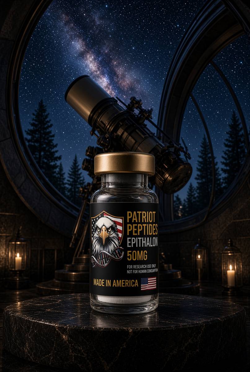
Research-use-only. Not for human consumption / not a medicine. The lyophilised tetrapeptide on the listing is Ala-Glu-Asp-Gly, HPLC-characterised, labelled for in-vitro work and for the animal protocols you already know how to write: a 24-hour melatonin curve in a controlled photoperiod, a Per2 and Bmal1 panel, AANAT protein if the factory is the claim, TRAP and karyotype if you've crossed into the telomere essay. The physiology in the paragraphs above is public, cited, and older than the vial. Use it to design the experiment you have the controls for, with the photoperiod named, the time points written down, and the indoleamine not confused with the four-mer. Read Takahashi, read Klein, read Van Cauter, read the Khavinson curves as a starting library, then weigh the cake. We'll sell you the sequence. We won't tell you it's melatonin, and we won't tell you it replaces a dark night. The 24-hour gene is a transcriptional loop. This reagent is four residues at the edge of that loop, with a chromatogram on the bench beside it.
In short. The vial is a research tetrapeptide for named clock and pineal assays, not a medicine and not the night hormone. The biology is public. Darkness is still free.
- SCN: ~20,000 neurons, CLOCK/BMAL1 and PER/CRY, melanopsin ipRGCs at ~480 nm. Master clock, not a sleep centre.
- Pineal: SCN → PVN → IML → superior cervical ganglion → norepinephrine → AANAT → melatonin. Light in, hormone out.
- Periphery: liver BMAL1 gates gluconeogenesis; β-cell clocks gate insulin; food is a liver zeitgeber. Not just sleep.
- Ageing flattens the nocturnal peak. IARC Group 2A for night-shift work. Van Cauter: one short night already moves glucose.
- Epithalon is AEDG, ~390 Da, Khavinson's tetrapeptide, a starting library at that oscillator. Not melatonin. Not a dark night.
- Assays: 24-hour curve, AANAT, clock genes at two times, photoperiod in lux. The sun is still free.
Questions the essay actually answers
- Is melatonin the same as Epithalon?
- No. Melatonin is N-acetyl-5-methoxytryptamine from pinealocytes, made when AANAT fires. Epithalon is Ala-Glu-Asp-Gly, a synthetic tetrapeptide studied for pineal and TERT effects. Different molecules, overlapping conversation — people mash them because both get mentioned next to night.
- Can morning light replace a peptide?
- Morning light is how you set the SCN, via melanopsin ipRGCs. It's free and it works. Epithalon is the characterised tetrapeptide for the ageing-pineal question in the Khavinson library. Use the light either way.
- Why does this sit next to a glucose essay?
- Hepatic BMAL1 gates gluconeogenic enzymes. Pancreatic clocks gate insulin. A mismatched photoperiod is a glucose insult before it is a sleep complaint. Van Cauter's sleep-restriction work is the next page, and it's ruder than this one.
- What is the SCN?
- The suprachiasmatic nucleus: about twenty thousand neurons above the optic chiasm that keep ~24-hour time and teach the rest of the body. Lesion it and timing falls apart. It's the master clock, not a sleep centre.
- What does AANAT do?
- Arylalkylamine N-acetyltransferase acetylates serotonin to N-acetylserotonin, the rate-limiting night step towards melatonin. Protein rises tens-fold after dark and collapses in minutes if you turn a light on. Klein called it the Timezyme.
- Why is night-shift work a cancer conversation?
- IARC classifies night-shift work as a probable carcinogen (Group 2A), argued in part as circadian disruption: internal desynchrony, light-at-night, flattened melatonin, clock-gated DNA-repair and metabolic transcripts. A tetrapeptide curve in an old rat doesn't unscrew a roster.
- Do peripheral organs really have clocks?
- Yes. Almost every tissue runs a CLOCK/BMAL1 loop. The liver can be reset by meal timing while the SCN still follows light (Damiola, Schibler). β-cell clocks gate insulin secretion (Marcheva, Bass). Sleep is one output of a much larger transcriptional programme.
- Does ageing change melatonin?
- The nocturnal peak often flattens even in the dark: less AANAT competence, a noisier SCN, a pineal that has calcified in many adults. Cause versus marker is still argued. That flattened oscillator is the gerontology the Khavinson papers sat on.
- How is NAD+ involved?
- NAMPT, the salvage bottleneck, is a CLOCK/BMAL1 target, so the NAD+ pool cycles (Ramsey, Bass, 2009). SIRT1 spends NAD+ on the loop. Neighbourhood of cellular time, not identity with melatonin or with AEDG. The cofactor essay is next door.
- What assays actually matter for the tetrapeptide on this page?
- A 24-hour melatonin curve in a controlled photoperiod, AANAT if you can, and a clock-gene panel at two or more times. TRAP and karyotype if you've crossed into the TERT claim. A single morning serum isn't that list.
Hypothetical research reconstitution
How this vial is typically mixed
Hypothetical research reconstitution for the named catalogue vial. Not a protocol, not medical advice, not a use instruction. These amounts sit in published and commonly cited laboratory ranges. The vial is labelled for research use only — not for human or veterinary administration.
Epithalon
50mg
Mix with 5 ml bacteriostatic water → 10 mg/ml
- Hypothetical aliquot
- 5–10 mg
- 0.50–1.00 ml · 50–100 units on a U-100 syringe
- How often
- Once daily, evening, for 10–20 consecutive days
- 10–20 days, two cycles a year in the Khavinson-school notes
Bench steps
- Let the vial sit until it is no longer cold to the touch.
- Wipe the stopper with 70% isopropyl alcohol. Let it dry.
- Draw 5 ml bacteriostatic water (0.9% benzyl alcohol).
- Run the water slowly down the inside glass — do not blast the cake.
- Roll between finger and thumb until the cake is gone. Do not shake.
- Label the date. Store the solution at 2–8 °C. Do not freeze. Use within 30 days unless the note below says otherwise.
Tetrapeptide (AEDG). Short pulses, not a daily-forever molecule in that literature.
Bacteriostatic water and sterile syringes ship with peptide orders over £75. Kit details · 10 ml bacteriostatic water
The molecule in the essay
The same published structure the essay describes — HPLC-characterised.
Read next
49 min · long read · Peptide research
Epithalon, telomeres and the pineal clock
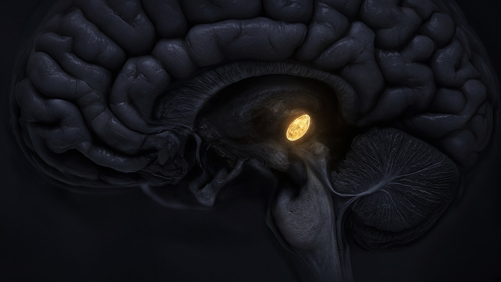
A four-residue peptide from the Khavinson school sits where melatonin, telomerase and aging research overlap. The claim is large. The molecule is tiny. The assays are specified.
46 min · long read · Metabolism
Sleep is a glucose drug you cannot buy
Spiegel, Leproult and Van Cauter restricted healthy people to four hours a night. Next-day glucose tolerance looked like a milder type 2. Cortisol, GH pulses and the hepatic clock are why. The bread did not change.
50 min · long read · Peptide research
Sirtuins, caloric restriction and the NAD+ budget
Yeast SIR2, worm daf-16, mouse SIRT1 — a conserved story about spending NAD+ to deacylate the proteins that decide whether a cell is in famine or feast. The cofactor is the budget. We stock it at 1000mg.
More in this desk
53 min · long read · Peptide research
Recovery, GH pulses and the CJC / ipamorelin pair
Somatotrophs have two 'go' receptors. CJC without DAC is a slightly longer GHRH pulse. Ipamorelin is a selective ghrelin-receptor key that does not yank ACTH. Together they are the pair Bowers already showed is more than additive — not a gym protocol.
53 min · long read · Peptide research
How research peptides are made — and why HPLC actually matters
Solid-phase peptide synthesis builds a chain one residue at a time. HPLC then asks whether the main peak is what you think it is. ≥98% is not a slogan. It is a chromatogram.
52 min · long read · Peptide research
Fatigue, cellular energy and the NAD+ / MOTS-c neighbourhood
NAD+ is the rechargeable chip every cell spends on fuel and DNA repair — and the pool shrinks with age. MOTS-c is a mitochondrial 16-mer sent out under metabolic stress. Two answers to 'I have no energy' that are not coffee.
51 min · long read · Peptide research
Melanocortin circuits: pigment, appetite and PT-141
Melanotan II lights MC1, MC3, MC4 and MC5 — pigment plus the rest of the sheet. PT-141 is the free-acid cousin pointed at MC3 and MC4, the circuitry papers use for desire and energy, not skin colour. Same Arizona family. Different question.
Essays describe published research. They are not medical advice and they do not authorise human use of any catalogue item.