

Peptide research · 53 min · 11,718 words
How research peptides are made — and why HPLC actually matters
Solid-phase peptide synthesis builds a chain one residue at a time. HPLC then asks whether the main peak is what you think it is. ≥98% is not a slogan. It is a chromatogram.
What this essay actually tells you
- Bruce Merrifield's solid-phase synthesis (1963, Nobel 1984) grows a chain on a resin, one residue per cycle of deprotection and coupling. That's still how most of the catalogue is made.
- A missed coupling makes an N-1 deletion peptide that can look almost right on a cheap UV scan and is the wrong molecule in an assay. We've seen that chromatogram. You don't want it.
- ≥98% HPLC means the main peak dominates the chromatogram. Mass spectrometry still has to confirm that peak has the right mass, because UV is not a molecular identity card.
What this actually means
A research peptide is built, not fished out of a gland. Chemists grow the chain on a resin, one amino acid at a time, then cut it off, fold or modify it if the sequence needs it, and freeze-dry the solid. HPLC (high-performance liquid chromatography) separates the wanted chain from deletion sequences, leftover reagents and truncated junk. Mass spectrometry asks whether the main peak has the right mass. A certificate that says ≥98% HPLC means the main peak dominates. That is honesty about the vial, not a clinical claim.
A research peptide is a molecule somebody decided to make, which sounds obvious until you notice how many catalogues treat the vial as a harvest: a name, a milligram number, a photograph of a white cake. The cake is a freeze-dried solid. The name is a published primary structure. Between those two facts sits a factory that isn't a gland and isn't a ribosome. Bruce Merrifield, in 1963, showed that you can park the C-terminus of a growing chain on an insoluble resin and add residues one at a time, washing away the reagents that didn't react. High-performance liquid chromatography then asks whether the main species that came off that resin is the species you named. Mass spectrometry asks whether that species has the mass the sequence predicts. Those three sentences are peptide manufacture as a grown-up activity. Everything else on this page is the chemistry that makes them true, the failure modes that make them necessary, and the certificate that's supposed to prove they happened. The interesting number is never whether a peptide was made. It is what else is in the peak.
In short. A research peptide is built on a bead, one amino acid at a time, then checked by a chromatogram and a mass. The name on the vial isn't that work.
We stock retatrutide because it's the published LY3437943 structure: a unimolecular agonist at the GIP, GLP-1 and glucagon receptors, fatty-acylated so albumin will carry it. Coskun and colleagues drew that chain in Cell Metabolism in 2018. Jastreboff and colleagues reported a Phase 2 weight-loss curve in the New England Journal of Medicine in 2023. The vial on this catalogue is the American-made research solid, HPLC-MS on the certificate, labelled for the bench. It isn't Eli Lilly's pen and it isn't a medicine. The neighbouring essays — what a peptide is, why the cake is freeze-dried, and the triple-agonist receptor story — sit next door. This page is the factory and the chromatogram. You need both if you're going to hold a thirty-nine-residue designed ligand and believe the label. A ribosome would have made the amide bonds too. A ribosome would also have had a message, a proofreading budget, and a cell around it. Solid-phase synthesis has a programme a chemist wrote and a column afterwards. The column isn't optional, and the mass analyser isn't a luxury.
In short. Retatrutide here is the published research chain, checked by HPLC and mass spec. This page is how that chain is made and tested.
Most people meet HPLC as a percentage on a certificate, and the number looks like a grade until you learn it isn't. Greater than or equal to ninety-eight percent. It's the area of the main peak as a fraction of the total integrated peak area at a wavelength where the peptide bond absorbs, usually 214 or 220 nanometres, on a reverse-phase C18 column eluted with an acetonitrile gradient in dilute trifluoroacetic acid. That sentence is long because every clause is load-bearing. Change the wavelength and you change which impurities you can see. Change the column chemistry and you change which species co-elute. Report a number without a chromatogram and you have reported a rumour. Report a chromatogram without a mass and you have reported a clean peak of an unnamed chain. The rest of this page unpacks the factory that produces the mixture the column is asked to judge, and then the column, and then the mass analyser. If you already knew that a ninety-percent peptide is an experiment with an undeclared co-solute, you're in the right room.
In short. HPLC purity is the size of the main peak on a specific chromatogram, not a brand score. Without a mass, even a clean peak can be the wrong chain.
Purity is a chromatogram. Identity is a mass. A name on a vial with neither is a rumour with a stopper.
The bond that is the whole argument
A peptide bond is an amide linkage between the carboxyl carbon of one amino acid and the α-amino nitrogen of the next, and that join is the whole argument. The reaction is a condensation: water leaves, the chain lengthens by one residue, and the new bond is planar because resonance gives the C–N link partial double-bond character. Pauling and Corey had the geometry in the 1950s; every undergraduate who has drawn a trans peptide plane is repeating their argument. The omega torsion sits near 180 degrees. Rotation about that bond is expensive, which is why a backbone has preferred secondary structure even when it's only a dozen residues long. The carbonyl and the NH are a hydrogen-bond pair waiting to happen. An α-helix is that pair, i to i+4, written along the chain. A β-strand is the same pair pointing at a neighbour. None of this is mysticism. It is why a short designed ligand can present a surface a receptor already knows, and why the same ligand can aggregate on a resin bead if the sequence is hydrophobic enough to prefer sheet. The covalent trick is one condensation. The conformational consequences last for the rest of the essay.
In short. A peptide bond joins two amino acids by an amide, held flat by chemistry. That flat backbone is why short chains still fold and why some sequences stick together.
Length is a working classification, not a metaphysical split. Oligopeptides typically sit under twenty residues: GHK is three, KPV is three, BPC-157 is fifteen. Polypeptides run longer. Proteins are the folded machines, often past fifty residues, with tertiary structure worth drawing. Insulin, fifty-one residues, was the first peptide anyone bothered calling a medicine; Banting and Best, 1921, and a century of footnotes. GLP-1 is thirty-one. Retatrutide is a designed chain in that band, with a fatty-acyl handle the native incretin doesn't have. Haemoglobin is a protein. The shared word 'peptide' is an accident of language. What the vial contains is a defined sequence. What the cell contains, if it translated the same sequence, would be that sequence plus whatever the ribosome and the chaperones and the modifying enzymes did to it. Solid-phase chemistry gives you the bonds you asked for and none of the ones you didn't, which is both the point and the limitation. There's no intron. There's no glycosylation unless you put it in. There's no folding guarantee. There's a chromatogram.
In short. Short chains are peptides; folded machines are proteins. Insulin sits on the peptide side. The vial holds a defined sequence, not a cell's finished protein.
The same bond is why HPLC can see the molecule at all. Peptide bonds absorb in the far ultraviolet, a π-to-π* transition centred near 190 nanometres and still useful at 214 and 220, where the solvents and the detectors in a routine laboratory are happier. Aromatic side chains add absorbance at 280. A chain with no tryptophan, tyrosine or phenylalanine is nearly invisible at 280 and perfectly visible at 214. That's why peptide chromatograms are run at 214 or 220 and why a purity number generated at 280 on a tyrosine-poor sequence is a way of not looking. The detector doesn't know the sequence. It knows that something with a peptide bond eluted at that time. A deletion peptide, missing one residue, still has peptide bonds. An incompletely deprotected chain still has peptide bonds. A truncated failure still has peptide bonds. Ultraviolet absorbance counts bonds, not identities. Keep that in your pocket until the mass spectrometry heading. It's the reason the two techniques travel together.
In short. HPLC sees peptide bonds by their ultraviolet absorbance. A wrong chain still has those bonds, so a peak isn't yet a name.
Diagram
- Amino acid~110 DaTwenty side chains. The alphabet.
- Peptide bondamide, planarCarboxyl carbon to the next nitrogen. Resonance holds it flat.
- Oligopeptide< ~20 residuesMost hormones and fragments. GHK is three. KPV is three.
- Polypeptide20–50+Insulin 51. GLP-1 31. Retatrutide is a designed chain in this band.
- Proteinfolded machineHaemoglobin, a GPCR, lysyl oxidase. Tertiary structure worth drawing.
Insulin (Banting and Best, 1921) was the first peptide anyone bothered calling a medicine. A collagen hydrolysate is food. A named sequence with a mass and a chromatogram is a research peptide. The shared word is the accident.
Solution-phase was a career, not a cycle
Before the resin, making a peptide was a solution-phase argument with protecting groups, couplings, and a purification after every residue — a career, not a cycle. Vincent du Vigneaud synthesised oxytocin in 1953 — nine residues, a disulphide, the first polypeptide hormone anyone built from amino acids rather than isolated from a gland — and received the Nobel Prize in Chemistry in 1955. That work was heroic in the ordinary sense: milligrams, months, a group that could lose the product at any step because a failed coupling in solution leaves you with a mixture you must separate by the chemistry of the 1950s. Each intermediate is soluble. Each intermediate must be isolated. Yields compound downward. A fifteen-residue chain done that way is a dissertation. A thirty-nine-residue designed agonist with a fatty-acyl side chain isn't a solution-phase weekend. The field knew how to make amide bonds. It didn't know how to make them as a repeating industrial cycle. That's the problem Merrifield actually solved. He didn't invent the peptide bond. He invented a way to stop purifying after every residue.
In short. Oxytocin was built in solution in 1953, one hard purification after another. That works for a nine-residue hormone. It doesn't scale to a catalogue.
The arithmetic of solution-phase is unforgiving in a way that has nothing to do with incompetence. Suppose each coupling-and-isolation returns eighty-five percent of the last intermediate, which would be a cheerful number in 1955. After ten residues you have about twenty percent of the starting material in the target chain, and the rest is a family of failures you have been carrying forward. After twenty residues you have a few percent. You can improve the couplings. You can't abolish the isolations. Every isolation is a chance to lose material on a column, in a crystallisation, in a flask that went to dryness at the wrong moment. The protecting-group scheme has to survive all of those manipulations. The more residues you add, the more the scheme has to be orthogonal, and orthogonality in the 1950s meant a small menu of acids, bases and hydrogenations that didn't always agree with the sequence. Solution-phase peptide synthesis is still used, especially for short fragments that will later be ligated. It isn't how a catalogue of lyophilised research solids gets made. The factory needed a different geometry.
In short. In solution you purify after every amino acid, and the yield collapses as the chain grows. Short fragments still use that route. A catalogue doesn't.
What the field wanted was a cycle: add a residue, wash away everything that isn't the chain, repeat. The chain has to sit still while the reagents come and go. Insoluble support is the obvious answer once somebody has the nerve to try it. Organic chemists had used polymer supports for other transformations; peptide chemists had good reasons to be suspicious, because a failed coupling on a bead is a failed coupling you can't crystallise away, and because the bead has to survive acid, base, and the solvents that dissolve protected amino acids. Merrifield's contribution was to take that suspicion into the laboratory and show that a chloromethylated polystyrene resin, a C-terminal amino acid anchored as a benzyl ester, and a repeating cycle of deprotection and coupling could produce a tetrapeptide you could cleave off and recognise. The 1963 paper is short. The consequences are the rest of this factory. Every automated synthesiser in a peptide house is that paper, running overnight.
In short. The field needed a cycle that washed leftovers away instead of purifying after every step. That meant parking the chain on a solid bead.
Merrifield, 1963: park the C-terminus
R. B. Merrifield, Journal of the American Chemical Society, 1963: Solid Phase Peptide Synthesis. I. The Synthesis of a Tetrapeptide. He anchored the C-terminal residue to a polystyrene bead and built toward the N-terminus, which is the opposite of how a ribosome writes a protein and the same direction every Fmoc synthesiser still uses. The C-terminus is parked. The N-terminus is the working face. Each cycle has two chemical jobs. First, the temporary protecting group on the α-amine is removed so a free nitrogen is available. Second, an incoming amino acid, itself protected on its nitrogen and on any reactive side chain, is coupled to that nitrogen through an activated carboxyl. Excess reagents are washed away because they are soluble and the growing chain is not. Then you do it again. A four-residue peptide is four cycles. A fifteen-residue peptide is fifteen. A thirty-nine-residue triple agonist is thirty-nine couplings plus whatever extra chemistry the fatty-acyl handle and the side-chain protections demand. The Nobel Prize in Chemistry arrived in 1984, twenty-one years after the tetrapeptide, which is about right for a method that had to become a field before the committee could see it.
In short. Merrifield stuck the first amino acid to a bead and added the rest one by one. The chain stays put; leftover chemicals wash away. He won the Nobel in 1984.
Direction matters because the two ends of a peptide aren't equivalent. The C-terminus is a carboxyl; the N-terminus is an amine. Anchor the carboxyl to the resin and every subsequent coupling is an amine attacking an activated incoming carboxyl, which is the reliable direction for amide-bond formation. Anchor the other way and you're asking a resin-bound carboxyl to be activated in the presence of whatever else is on the bead, which is a racemisation argument you don't want. Nature's ribosome also writes N-to-C, but it writes in the other direction relative to the parked end: the growing chain's C-terminus is the working face, held as a peptidyl-tRNA. Solid-phase chemistry parked the C-terminus because the bead isn't a tRNA and the chemist isn't a peptidyl transferase. The practical consequence is simple. The residue you load first is the C-terminal residue of the finished peptide. The residue you load last is the N-terminus. If the sequence is GEPPPGKPADDAGLV, valine goes on the resin first. Get that wrong and you have made the reverse chain. The mass might even look plausible. The assay will not.
In short. The last amino acid of the finished chain goes on the bead first. Build the rest toward the front. Reverse that and you have made the wrong molecule.
The wash is the whole invention, restated, and it's the bit students underestimate until they watch a bead. In solution, leftover coupling reagent, leftover amino acid, and leftover base are contaminants you must remove by isolation. On a resin, they are solutes. You drain the vessel. You rinse with dimethylformamide, with dichloromethane, with methanol, with whatever the protocol names. The chain stays. The leftovers leave. That's why the cycle can be automated: a synthesiser is a box that adds solutions, waits, and drains, on a clock a chemist wrote. It's also why a failed coupling is a different kind of disaster from a failed solution-phase coupling. The failed residue is still on the bead, sitting as a free amine or as a truncated chain, and unless you cap it you will couple the next residue to a mixture of the intended N-terminus and the leftover N-terminus from the miss. That mixture is a deletion peptide. It will travel with the target through much of the subsequent chemistry. It is why capping with acetic anhydride after a difficult coupling isn't fussiness. It is how you convert a deletion problem into a truncated, acetylated problem the column can usually see more clearly.
In short. Washing the bead replaces purifying every intermediate. If a coupling misses, the next amino acid can skip a position and make a deletion chain.
Automation followed because a cycle that's only adding, waiting and draining is a cycle a machine can run without getting bored. The first Merrifield synthesisers were laboratory constructions; the current ones are boxes with amino-acid racks, solvent lines, and a computer that will happily couple at two in the morning. Microwave heating, which a later generation added, can accelerate difficult couplings and can also accelerate the side reactions you were hoping to avoid, so it's a tool and not a personality. Parallel synthesis — many sequences on many resins in one run — is how a research house fills a catalogue without employing a du Vigneaud group per vial. None of this abolished the chemistry. A synthesiser that couples in the wrong solvent, or with a stale bottle of activator, or on a resin that wasn't swollen, will produce a crude that looks like a crude produced by a tired postdoc in 1972. The machine runs the cycle. The chemist still owns the sequence, the resin choice, the coupling conditions, and the chromatogram that comes afterwards. If you think automated means solved, you haven't watched a hydrophobic 30-mer refuse the next residue.
In short. Machines now run the same add-wait-drain cycle overnight. Automation doesn't forgive a bad sequence or a missed coupling. The chromatogram still decides.
Diagram
1. Resin
C-terminus parked. Merrifield, 1963.
2. Deprotect
Piperidine on Fmoc. The next nitrogen is free.
3. Couple
HATU / HBTU / DIC–Oxyma. One residue, one bet.
4. Repeat
N-terminal growth. Aggregation is the classic failure.
5. Cleave
TFA cocktail. Protecting groups off. Then HPLC.
Miss a coupling and you have a deletion peptide that looks almost right on a cheap UV scan. That is why the interesting number is not ‘did we make a peptide’ but ‘what else is in the peak’.
Boc, then Fmoc, then the chemistry you actually run
Merrifield's original cycle used tert-butyloxycarbonyl, Boc, as the temporary N-protecting group. Boc comes off with acid, typically trifluoroacetic acid in dichloromethane. Side chains were held with benzyl-based groups that needed a much stronger acid, anhydrous hydrogen fluoride, to release them at the end. The scheme is orthogonal in the sense that mild acid takes Boc and only HF takes the side chains and the peptide off the resin. It worked. It also meant that every peptide laboratory had to handle HF, which is a reagent that eats glass and people with equal enthusiasm, and that every cycle exposed the growing chain to acid. Acid-sensitive sequences suffered. Tryptophan alkylated. Some resins didn't love the repeated TFA. The Boc/benzyl era made the field. It isn't the era most research peptides now come from. You will still meet Boc chemistry in older papers, in some industrial fragments, and in chemists who learned it first and never saw a reason to switch. For a catalogue of lyophilised solids in 2026, the working cycle is almost always Fmoc.
In short. Early solid-phase work used Boc protecting groups and a harsh final acid. It worked. Most research peptides are now made with a gentler Fmoc cycle.
Fmoc, 9-fluorenylmethoxycarbonyl, is a base-labile N-protecting group. Carpino and Han introduced it in the 1970s; Atherton and Sheppard, among others, put it onto peptide synthesis in a form a laboratory could live with. Twenty percent piperidine in dimethylformamide takes Fmoc off in minutes, by a β-elimination that releases dibenzofulvene. The side chains, in the now-dominant Fmoc/tBu scheme, are held with tert-butyl-based groups that come off with TFA at the end, along with cleavage from the resin. Base for the temporary group, acid for the permanent groups: orthogonality the other way round from Boc. No HF. No repeated strong-acid cycles on the growing chain. Piperidine isn't a toy, and the dibenzofulvene by-product has to be washed away, but the cycle is compatible with a much wider range of sequences and with automation that doesn't want an HF line. When this essay says deprotection, it means piperidine on Fmoc unless it's telling a Boc story. That's the chemistry the catalogue is made with. It's also the chemistry whose failure modes occupy the next few headings.
In short. Fmoc comes off with a base, piperidine. Side chains come off later with acid. That split's why modern peptide synthesis can run without hydrogen fluoride.
Side-chain protecting groups are the quiet half of the cycle and the reason a crude chromatogram is a forest. Serine, threonine and tyrosine carry tert-butyl ethers. Aspartic and glutamic acids carry tert-butyl esters. Lysine carries Boc on the ε-amine. Arginine carries Pbf, a bulky sulphonyl group that's one of the slower things to leave during cleavage. Histidine, cysteine, asparagine and glutamine often carry trityl. The point of all of that furniture is to stop the side chain from reacting when you wanted only the α-amine and the incoming carboxyl to react. The cost is that every one of those groups has to come off cleanly at the end, and every one of them can fail to. Incomplete deprotection of Pbf-arginine is a classic high-mass impurity. Trityl that stays on histidine is another. A tert-butyl that stays on aspartic acid is a third. Each of those species has a mass you can calculate, a retention time that's usually close to the target, and an ultraviolet absorbance that will put it on the chromatogram as a shoulder if you're lucky and as a co-eluting liar if you're not. Protecting-group chemistry is why the mass spectrum isn't optional.
In short. Each reactive side chain wears a shield during synthesis. If a shield stays on after the final cut, you have a heavier, wrong molecule that can hide near the main peak.
Coupling is a bet, named by the reagent
An amide bond doesn't form because you mix an amine and a carboxylic acid and wait. The carboxyl has to be activated. Classically that meant a carbodiimide — dicyclohexylcarbodiimide in the old literature, diisopropylcarbodiimide now — which converts the acid into a reactive O-acylisourea. That intermediate can couple. It can also rearrange to an unreactive N-acylurea, and it can racemise the incoming residue by forming an oxazolone. Additives were invented to intercept the O-acylisourea as an active ester that couples faster and racemises less. HOBt, hydroxybenzotriazole, was the additive of a generation and is now treated as an explosive when dry, which is why Oxyma, ethyl cyano(hydroxyimino)acetate, has taken much of its place. DIC/Oxyma is a working pair in a modern laboratory: the carbodiimide to activate, the oxime to make the active ester, the resin-bound amine to attack. You don't need to memorise the arrow-pushing to own the physiology of the factory. You do need to know that 'coupling' is a named reaction with named failure modes, not a synonym for 'the next amino acid went on'.
In short. The incoming amino acid must be chemically activated before it will join. Modern labs use named reagent pairs for that job, because an unactivated acid simply won't couple.
Aminium and phosphonium reagents are the other half of the drawer. HBTU and HATU — uronium/aminium salts that generate HOBt or HOAt active esters in situ — are fast, and HATU is the one you reach for when the coupling is difficult: hindered residues, N-methyl amino acids, a sequence that has started to aggregate. They're also expensive, and they can guanidinylate the N-terminus if you let the reagent sit with the amine in the absence of the carboxylic acid, which is a contamination that mass spectrometry will find and the chemist won't enjoy explaining. The practical rule is boring and correct. Activate the acid, then introduce it to the resin. Do not premix HATU with the amine and hope. Double-coupling a difficult residue — drain, add a second portion of activated amino acid — is how you push a stubborn step toward completion. Capping afterwards is how you live with the fraction that still didn't go. None of this is glamour. It is why two houses can synthesise the same published sequence and produce crudes that look like different molecules until both have been through a column.
In short. HATU and HBTU are faster coupling reagents for stubborn steps. Difficult amino acids get coupled twice, and leftovers get capped, so mistakes don't snowball.
Racemisation is the stereochemical tax. Amino acids in the catalogue are L unless the sequence actually calls for D, as some designed analogues do. The α-carbon can lose and regain its hydrogen during activation, especially if the residue is histidine, cysteine, or a residue being coupled as a fragment rather than as a urethane-protected amino acid. Fmoc protection reduces racemisation relative to some older schemes because the urethane is less willing to form the oxazolone. Histidine remains a problem because the imidazole can catalyse its own racemisation; the protecting group on the pi-nitrogen isn't a decorative choice. Cysteine racemises by a different path, and a D-Cys in a disulphide peptide is a conformational mutation. A chromatogram at 214 nanometres won't always separate epimers. A receptor often will. That is one of several reasons a clean ultraviolet peak isn't yet a pharmacological identity. Chiral methods exist. They're not on every certificate. Mass spectrometry won't catch an epimer at all, because D and L have the same mass. The honest certificate is HPLC plus MS plus a synthesis route that had a reason not to racemise. Three legs. Two won't stand.
In short. The wrong mirror-image amino acid can sneak in during coupling, especially at histidine and cysteine. A chromatogram and a mass can both miss that error.
Failure modes have names because they keep happening
A deletion peptide is the product of a missed coupling that wasn't capped, or was capped incompletely. One residue is absent. The mass is the target minus that residue. The hydrophobicity is close. The ultraviolet absorbance is almost the same. On a lazy gradient the two species co-elute, or the deletion sits as a shoulder you could integrate away if you were the sort of person who integrates shoulders away. In an assay the deletion can be inert dirt, a weaker agonist, a competitive antagonist, or a chain with an unexpected activity at a related receptor. You won't know which until the result is already confusing. N-minus-one deletions are the commonest. Double deletions happen on difficult stretches. Insertion peptides, the opposite error, happen if Fmoc comes off during coupling and a second copy of the incoming residue adds. Truncations happen when you cap, or when a chain falls off the resin early. The crude is a family. If you've run a difficult sequence, you've met at least one member of it. The column is how you stop inviting the family into the assay.
In short. If one amino acid fails to add, you get a nearly-right chain with a residue missing. It looks similar on a scan and can ruin an experiment.
Hydrophobic aggregation on resin is the failure mode that makes experienced chemists flinch at certain sequences. As the chain grows, hydrophobic stretches — leucines, isoleucines, valines, phenylalanines, a run of alanine — start to form β-sheet with their neighbours on adjacent beads or on the same bead. The N-terminus hides. The next activated amino acid can't find it. Coupling yields collapse, often suddenly, at a specific residue that everyone in the group will remember by number. Pseudoproline dipeptides, which insert a kink that breaks sheet, are one answer. Better solvents, DMSO or NMP in the DMF, are another. Microwave, heat, a different resin with a longer linker, backbone protection: the toolbox is real and sequence-specific. None of it's guaranteed. A fatty-acylated chain such as retatrutide has a hydrophobic handle as part of the design, which is good for albumin binding and not good for the resin step that has to install it. Difficult sequences are why crude purities of fifty or sixty percent still appear in serious laboratories. The crude isn't the product. The product is what survives the column.
In short. Sticky stretches of the chain can clump on the bead and hide the growing end, so the next amino acid can't add. Some sequences are famous for this.
Aspartimide is a ring that aspartic acid has no business forming and forms anyway. Under basic conditions — and piperidine deprotection is basic — the backbone nitrogen of the next residue can attack the side-chain ester of Asp, especially when that next residue is glycine, asparagine or serine. The five-membered imide then opens two ways: back to Asp, or to a β-aspartyl peptide with a rearranged backbone. Both the imide and the β-peptide are isomers or near-isomers of the target. Mass spectrometry may see the imide as a dehydration, minus eighteen daltons, or may see the opened β-peptide as isobaric with the target. HPLC may or may not resolve them. Asp-Gly is the notorious pair; anyone writing a sequence with that motif has a protecting-group and deprotection-time argument to have before the synthesiser is loaded. Additives, shorter piperidine treatments, backbone protection of the glycine, a different Asp protecting group: all of these exist because the ring keeps winning if you ignore it. A ninety-eight percent peak that's half β-aspartyl is a ninety-eight percent peak of the wrong backbone. The receptor will notice.
In short. At aspartic acid next to glycine, the chain can snap into a small ring and reopen wrongly. The mass can look right. The backbone is not.
Incomplete deprotection is the impurity class that mass spectrometry is almost unfairly good at catching. A Pbf that stayed on arginine adds a known mass. A trityl that stayed on cysteine or histidine adds another. A tert-butyl that stayed on a carboxylic acid or an alcohol adds 56 daltons, which is a peak you learn to look for the way a radiologist learns to look for a fracture. TFA cleavage is supposed to take all of them off. TFA cleavage is also a set of carbocations looking for something to alkylate, which is why scavengers exist and why tryptophan, methionine and cysteine are the residues that teach humility. If the scavenger cocktail is wrong, you get alkylated tryptophan, tert-butylated methionine, and a yellow crude that smells like a mistake. If the cleavage is too short, protecting groups remain. If it's too long, sensitive residues degrade. The window is sequence-dependent. A certificate that reports a mass matching the fully deprotected, fully cleaved target is a certificate that this window was found. A certificate that reports only an HPLC percentage hasn't been asked that question.
In short. If leftover protecting groups stay attached, the chain is heavier than it should be. A mass measurement catches that. A purity percentage alone may not.
Diagram
- DNA replication + MMR10⁻⁹ to 10⁻¹⁰A genome of 6 Gbp (diploid) accumulates a handful of mutations per division.
- Transcription~10⁻⁵RNA is disposable. The cell can afford a wrong letter in a message that lasts hours.
- Translation~10⁻⁴One wrong amino acid per ten thousand. Proteins turn over. DNA does not.
- mtDNA10–100× nuclearNo histones, ROS next door, weaker repair. The second genome ages faster.
The genome is sacred, the message is cheap, the protein is cheaper. Ageing is partly what happens when the sacred copy still drifts — and when mitochondria, which never got the nuclear repair budget, drift faster.
The arithmetic of a missed step
Coupling yields compound. That's the sentence a synthesiser can't repeal. If each cycle installs the intended residue on ninety-nine percent of the chains, then after ten residues the fraction of full-length, correct sequence in the crude is 0.99 to the tenth, about ninety percent, before you even count racemisation, aspartimide, aggregation and leftover protecting groups. After twenty residues it's about eighty-two percent. After thirty, about seventy-four. After thirty-nine, the length of a designed triple agonist, about sixty-seven percent if you were lucky enough to hold ninety-nine percent at every step. Real difficult sequences don't hold ninety-nine percent at every step. A single cycle at ninety percent, which is what aggregation looks like, punches a hole in the arithmetic that no later cycle can fill. This is why crude peptide is a mixture by construction, why preparative HPLC is a factory step rather than a nicety, and why a house that sells crude as if it were finished material is selling an undeclared combinatorial library. The cell, for comparison, translates at about one error in ten thousand amino acids. Solid-phase chemistry is closer to transcription than to replication. Purification is the mismatch repair.
In short. Even a ninety-nine percent success at each step leaves a lot of wrong chains after thirty additions. That's why crude peptide must be purified.
Length is therefore a risk function, not a badge. A tripeptide such as GHK is three cycles and a chromatogram that a competent laboratory can make look boring. A fifteen-residue chain such as BPC-157 is a standard SPPS object, still well inside the range where Fmoc chemistry is routine, still a crude that needs a column. A thirty-nine-residue fatty-acylated agonist is a different proposition: more cycles, a hydrophobic handle, more chances for aspartimide and aggregation, a preparative gradient that has to separate a forest. A hundred-and-ninety-one-residue hormone such as somatropin is not, in any serious factory, an SPPS product. It's a recombinant protein, the ribosomal pipeline hijacked in a tank, because the error arithmetic of thirty-nine cycles is already a negotiation and the error arithmetic of one hundred and ninety-one cycles is a defeat. When a catalogue mixes a 15-mer, a 39-mer and a 191-mer on adjacent pages, it's mixing two factories. The amide bond is the same. The way you ask whether the main species is the named species is not. HPLC-MS on a 15-mer and a peptide map plus intact mass on a recombinant hormone are different sentences.
In short. Short peptides are routine on a bead. A thirty-nine-residue designed chain is harder. A 191-residue hormone is made in cells, not on resin.
The honest comparison with polymerase error isn't that SPPS is DNA chemistry. It is that both are stepwise polymer factories whose mistakes look almost like the product. A DNA polymerase with mismatch repair finishes a genome at about one error in a billion to ten billion bases; the archive is sacred and the cell spends ATP to keep it so. Transcription is sloppier, about one in a hundred thousand, because the message turns over. Translation is sloppier again, about one in ten thousand, because the protein turns over too. Solid-phase peptide synthesis, at a percent-scale miss per cycle, is sloppier than all of them. It can afford to be, because the chemist is allowed to throw the mistakes away on a column afterwards. The cell doesn't have preparative HPLC. It has the proteasome and autophagy, which is a different essay. What the two factories share is the near-miss: a wrong base, a wrong residue, a deletion, still a polymer, still able to fool a careless readout. UV absorbance is a careless readout. Mass is less careless. A receptor assay with a mixture isn't a readout at all. It's a story about a family.
In short. Cells keep DNA almost error-free and allow more mistakes in RNA and protein. Peptide synthesis makes more mistakes still, which is why a column has to remove them.
Cleavage is a reaction, not an afterthought
When the last residue is on, the chain is still on the resin, still wearing its side-chain protecting groups, still not a product. Cleavage is the step that releases it. For Fmoc/tBu chemistry that means a trifluoroacetic acid cocktail, typically ninety-five percent TFA with scavengers making up the rest. The acid cuts the linker to the resin and cuts the tert-butyl, trityl, Boc and Pbf groups off the side chains. Both cuts generate carbocations. Carbocations alkylate tryptophan, methionine, cysteine and tyrosine if you let them. Scavengers — triisopropylsilane, water, 1,2-ethanedithiol, phenol, thioanisole, depending on the sequence and on which Reagent K or Reagent R variant the laboratory inherited — catch those cations before they catch the peptide. A cleavage cocktail is therefore a small, unpleasant piece of organic chemistry, not a tap you open. Get the scavengers wrong and you will see the alkylated masses. Get the time and temperature wrong and you will see either leftover protecting groups or degraded residues. The crude that comes off the resin is a mixture by definition. Freeze-dry that crude and sell it as a finished peptide and you've skipped the factory floor.
In short. Acid cuts the finished chain off the bead and strips its protecting groups. Extra chemicals in the mix catch reactive debris so it doesn't damage the peptide.
Work-up is the unglamorous hour after the acid. The resin is filtered off. The peptide is in TFA. Ether precipitation is the classical next move: pour the cleavage mixture into cold diethyl ether, watch the peptide crash out, wash, dry. What you now have is crude peptide as a solid, still a mixture, often as a TFA salt because the column hasn't happened yet and the acid is the counter-ion sitting on every basic side chain. Some laboratories go straight from cleavage into a preparative HPLC injection, which is cleaner if the crude will dissolve and if the column can take it. Either way, this is the point at which a serious house and a corner-cutting house diverge. The serious house looks at an analytical chromatogram of the crude, estimates how ugly the forest is, and sets a preparative gradient accordingly. The other house weighs the solid, writes a milligram number, and prints a label. Crude yields look generous. They should. Much of the mass isn't the target. A certificate that doesn't distinguish crude from purified hasn't yet entered the argument this essay is about.
In short. After the acid step you have a mixed solid, often still full of wrong chains. A real factory takes that mixture to a purification column next.
Disulphides, if the sequence has cysteines that need them, are a further reaction. Oxytocin's disulphide was part of du Vigneaud's problem in 1953 and is still part of anyone's problem when two thiols must find each other in the right pairing. Air oxidation, DMSO, iodine, a directed protecting-group scheme that lets you open one pair at a time: choose according to how many cysteines you have and how many ways they can mispair. A misfired disulphide is another isobaric impurity. HPLC often sees it; MS may not, because the mass of the wrong pair is the mass of the right pair. Retatrutide isn't a disulphide story. Plenty of research peptides are. The general point is that 'cleavage' is sometimes the start of a second synthetic campaign, not the end of the first. Modifications — fatty acylation, C-terminal amidation, N-terminal acetylation, a PEG, a biotin — each add a step, a failure mode, and a mass increment the certificate has to account for. A published structure that includes a handle includes the obligation to show the handle is on.
In short. If the chain needs a sulphur bridge or a fatty handle, that's extra chemistry after the bead. The mass on the certificate has to include those extras.
Reverse-phase HPLC is a conversation with hydrophobicity
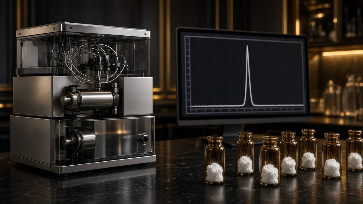
High-performance liquid chromatography, in the form a peptide laboratory actually runs, is almost always reverse-phase chromatography on a silica particle coated with C18 alkyl chains — a conversation with hydrophobicity. The stationary phase is hydrophobic. The mobile phase is water and acetonitrile, almost always with 0.1 percent trifluoroacetic acid in both, because TFA keeps acidic groups protonated, improves peak shape, and ion-pairs with basic residues so the chain behaves. You inject the dissolved crude. Polar junk — leftover salts, very short truncations — comes off first. More hydrophobic species hold onto the C18 longer and come off as the acetonitrile fraction rises. The target peptide has a retention time on that gradient, on that column, at that temperature. A deletion peptide that lost a leucine will usually come off a little earlier. A peptide that kept a tert-butyl will usually come off later. A fatty-acylated chain holds on longer than its unacylated analogue, which is how you know the handle is on before the mass spectrometer confirms it. The method is a hydrophobicity argument. It's a very good hydrophobicity argument. It isn't a sequence argument.
In short. A C18 column holds greasy chains longer and lets polar ones go first. Acetonitrile then peels them off in order. That order isn't yet a name.
Detection, as the peptide-bond heading already flagged, is ultraviolet absorbance at 214 or 220 nanometres. The peptide bond is the chromophore you can count on. Diode-array detectors will also show you 280 if you have aromatics, and a 280-only method will lie about a tyrosine-poor impurity. Fluorescence and evaporative light-scattering exist; they aren't how a routine peptide CoA is generated. What the integrator reports as purity is the area of the main peak divided by the total area of all integrated peaks, at that wavelength, on that method. Species that don't absorb at 214 — salts, some small scavenger leftovers — are invisible to this number. Species that absorb more strongly than the peptide, per gram, are over-counted. Species that co-elute under the main peak are counted as if they were the main peak. This is why method details matter and why a single percentage, unaccompanied by a chromatogram, a wavelength and a column identity, is a number wearing a lab coat. ≥98% HPLC, on a C18 method at 214 nanometres with a stated gradient, is a specification. 'Purity 98%' on a marketing slide is a mood.
In short. The detector watches ultraviolet light at 214 nanometres, where peptide bonds absorb. Purity is the main peak's share of that light, not a weigh-in of the powder.
Shoulders are the honesty test. A symmetric, baseline-resolved main peak with small, named impurities to either side is the adult chromatogram. A fat front, a fat tail, a lump on the side that the integrator was told to ignore, is a mixture wearing the target's clothes. Co-elution is the failure the area-percent number can't confess: two species, one retention time, one peak, a purity that looks excellent and a mass spectrum that, if anyone bothers to take it across the peak, shows two envelopes. Changing the gradient, changing the temperature, changing to a C8 or a phenyl column, adding a second dimension, are how you ask whether the peak is one thing. A laboratory that never re-runs a suspicious peak on a different method is a laboratory that has decided not to know. Preparative fractions should be re-analysed analytically. That sentence is so obvious it's embarrassing, and it's the step that gets skipped when a queue is long. The certificate you want is the analytical re-run of the pooled fractions, not the preparative trace the operator was watching while they cut.
In short. A fat shoulder on the main peak is a mixture. If two chains come off the column together, the purity number will happily pretend they are one.
Area percent is also not weight percent of peptide, and it isn't net peptide content. Those three numbers get collapsed in internet arguments and they are three different measurements. HPLC area percent at 214 asks what fraction of the absorbing, eluting material is the main peak. Net peptide content asks what fraction of the weighed solid is peptide rather than water, counter-ion and residual solvent, usually by elemental analysis or by amino-acid analysis or by a calibrated ultraviolet measurement. A vial can be ≥98% HPLC and 75% peptide by weight because the rest is TFA and moisture. Both numbers can be true. Both numbers should be on a serious certificate if the assay is quantitative. Confusing them is how a bench worker over-makes a stock solution by a third and then writes a paper about an EC50 that belongs to the counter-ion. The lyophilisation essay next door is the moisture half of this. This page is the chromatogram half. Weighing a cake isn't characterisation. It is weighing a cake.
In short. A high HPLC percentage says the main peak dominates the chromatogram. It doesn't say the powder is all peptide; salt and water can still be in the vial.
Diagram
Reverse-phase C18 holds hydrophobic chains longer. A fat shoulder is a mixture wearing a compound’s clothes. ≥98% HPLC means the main peak dominates. Without MS you can still have a clean peak of the wrong chain.
Preparative HPLC is the factory floor
Analytical HPLC is a question. Preparative HPLC is a harvest. The chemistry is the same — C18, acetonitrile, TFA, 214 nanometres — and the columns are larger, the loadings are higher, the fractions are collected rather than merely observed. A crude that looked like a forest on a 4.6 millimetre analytical column is loaded onto a 20 or 50 millimetre preparative column and eluted with a gradient shallow enough that the target can be cut away from its deletions. Each fraction is an analytical re-run waiting to happen. Pool the fractions that are clean enough. Throw the rest. The yield of purified peptide from crude is often a disappointment to anyone who thought the crude mass was the product mass. It's also the step at which ≥98% becomes a possible specification rather than a hope. A house that quotes ≥98% without a preparative step is quoting a wish. A house that quotes ≥98% from a preparative cut that wasn't re-analysed is quoting an operator's memory of a screen. The specification is the analytical chromatogram of the pooled, lyophilised solid you actually put in the vial.
In short. Big columns do the same separation so you can collect the main peak and throw the rest away. The purity on the vial should come from a re-check of what was collected.
Crude versus purified is the distinction catalogues blur when they want the crude yield to look like generosity. Crude peptide is the solid after cleavage, before the column, a mixture whose main component may be the target and whose other components are the family this page has been naming. Purified peptide is what came off the preparative column in the fractions that met the specification. In-process peptide is everything in between. Selling crude as research material is lawful if you say it's crude. Selling crude with a purity number borrowed from a different batch isn't characterisation. It is costume. For a receptor assay, crude is how you discover that a deletion peptide is a partial agonist and spend a month believing the literature is wrong. For a quantitative binding curve, purified and weighed with a net peptide content is the minimum. The specification on this catalogue is the purified solid, HPLC-MS against the published structure. That's a factory claim. It isn't a clinical claim, and it doesn't become one because the percentage is high.
In short. Crude is the mixed solid before the column. Purified is what was collected from the main peak. Those words aren't interchangeable on a label.
After the column, the peptide is in aqueous acetonitrile with TFA. You can't ship that. Lyophilisation freezes the solution and sublimes the ice under vacuum so the chain remains as a porous cake, which is the neighbouring essay and won't be retold here except as a hand-off. Residual moisture, measured by Karl Fischer, predicts whether hydrolysis resumes in the solid state. The cake is usually a TFA salt at this point, because TFA was the ion-pair on the column and nobody has yet exchanged it. Storage is cold, dark, and dry. Reconstitution is mass divided by volume in a solvent the sequence will actually dissolve in, which for a fatty-acylated chain isn't always water. The freeze-dried cake isn't characterisation. It is how you stop the characterised solid from becoming a different molecule in the post. This page stops at the chromatogram and the mass. The reconstitution essay starts at the cake. Read both if you're holding a vial. Skipping either is how a careful synthesis becomes a sloppy stock solution.
In short. The purified peak is freeze-dried into a cake so it can ship without rotting in water. How you then dissolve that cake is a separate essay.
Mass spectrometry is the identity card
Electrospray ionisation, ESI, is the common identity method for a research peptide. The chain is dissolved, sprayed, and picked up as a family of protonated species: [M+H]+, [M+2H]2+, [M+3H]3+, and so on, depending on how many basic sites it has and on the instrument's m/z range. A thirty-nine-residue agonist with a handful of lysines and arginines will typically present as a multiply charged envelope. Deconvoluting that envelope gives the intact mass. MALDI-TOF is the other routine option, a matrix-assisted laser desorption that often gives [M+H]+ with less charge complexity, useful for a quick look and for larger chains. Either method is asking the same question. Does the mass of the main species match the mass calculated from the published sequence, including modifications, within the instrument's error? For a small peptide, that error should be a fraction of a dalton. For a larger chain, a few daltons. 'The peak looks right' isn't an answer to that question. 'The mass matches LY3437943, including the fatty-acyl handle' is an answer.
In short. Mass spectrometry weighs the main chain. The number should match the published sequence, including any fatty handle. A pretty chromatogram can't replace that weigh-in.
The calculated mass isn't a single number until you specify which mass you mean. Monoisotopic mass is the mass of the species containing only the most abundant isotope of each element, 12C, 1H, 14N, 16O, 32S. Average mass is the weighted average of the isotopic distribution. For a small peptide the two are close and a unit-resolution instrument will see a simple isotope pattern. For a thirty-nine-residue chain the isotope envelope is wide, carbon-13 is no longer a rounding error, and reporting the wrong flavour of mass against the wrong flavour of calculation is a classic way to think you're 2 daltons off when you're actually on. Sodium and potassium adducts sit 22 and 38 daltons above the protonated species and fool people who haven't met them. TFA adducts appear. A dehydration, minus 18, is the aspartimide flag. A plus 56 is a leftover tert-butyl. Learning to read an envelope is a morning's work. Refusing to look at one is a style of manufacture. Identity is sequence plus mass plus chromatogram. Drop any of the three and you're guessing.
In short. You must compare the right kind of mass with the right calculation, and watch for salt adducts and leftover groups. Those details are how a match is real.
Purity without mass can still be the wrong chain. That's the sentence this page exists to make boringly obvious. A synthesis programmed with a transposed residue, a D-amino acid where L was specified, a missing handle, a C-terminal acid where the published structure is an amide, can still produce a single beautiful peak. The peak is homogeneous. The homogeneous substance isn't the substance on the label. Ultraviolet can't tell. A receptor sometimes can't tell, and then you publish a curve that belongs to a different molecule and the literature acquires a ghost. Sequence confirmation at the residue level — tandem MS/MS, Edman, a tryptic digest — is the gold standard and isn't on every research CoA. Intact mass plus a synthesis record plus HPLC is the working standard. For retatrutide, the fatty-acyl handle is a mass increment you can see; its absence is a different, lighter chain that might still look like a peptide on C18. Our retatrutide is the published LY3437943 structure, made in the United States, HPLC-MS on the certificate. The mass is part of why that sentence is allowed.
In short. A single clean peak can still be the wrong sequence if nobody weighed it. Mass spec is how you check the chain, not just the chromatogram.
Tandem mass spectrometry, when it's done, is the residue-level argument. Fragment the intact ion, read b- and y-ions, reconstruct the sequence. For a 15-mer this is routine. For a 39-mer it's a longer afternoon and still cheaper than believing a label. Edman sequencing, the older chemistry, chews residues off the N-terminus one at a time and is defeated by an N-terminal modification; a fatty-acylated N-terminus will simply refuse. Peptide mapping — digest with trypsin or another protease, HPLC-MS the fragments — is how recombinant houses confirm a 191-residue hormone and how a thorough peptide house can confirm a difficult synthetic chain. None of these is a potency assay. All of them are identity. A certificate that offers HPLC area percent, an intact mass matching the calculated monoisotopic mass of the published sequence, and a lot number you can ask questions about, is a certificate that has done the working standard. A certificate that offers a name and a smile has done marketing. Keep the first. File the second in the bin you reserve for rumours.
In short. Breaking the chain into pieces and weighing those pieces can confirm the actual order of amino acids. A serious certificate at least has the intact mass and the chromatogram.
- Merrifield SPPS
- 1963
- Nobel Prize in Chemistry
- 1984
- Fmoc deprotection
- piperidine / DMF
- Coupling reagents
- HBTU, HATU, DIC/Oxyma
- Per-cycle yield, 99%
- 0.99^30 ≈ 74%
- Detection wavelength
- 214 / 220 nm
- Specification
- ≥98% HPLC
- Identity
- [M+nH]n+
JACS tetrapeptide. C-terminus on polystyrene. The cycle starts here.
Twenty-one years after the paper, once the method had become a field.
Base-labile N-protection. tBu side chains wait for TFA.
Named activators, not synonyms for 'the residue went on'.
Target sequence in crude after thirty steps, before other failures.
Peptide-bond absorbance. 280 nm will miss a tyrosine-poor impurity.
Main peak area percent on a stated C18 method, then a mass.
ESI or MALDI against the calculated mass of the published sequence.
What ≥98% HPLC actually buys the assay
In a culture dish, a ninety-percent peptide is an experiment with an undeclared co-solute. Ten percent of the weighed solid, if that ten percent is absorbing at 214 and eluting as other peaks, is other chains. Some of those chains are truncations that don'thing. Some are deletions that still bind. Some are protecting-group leftovers that are insoluble and will precipitate on your cells as if the peptide were toxic when the peptide is fine. You will attribute the mess to the literature, to the cell passage number, to the moon. ≥98% HPLC is a specification because that's the level at which the main species is allowed to be the variable. The remaining two percent is still a confession, not a halo. It's a small enough confession that a well-designed assay can live with it, provided the identity of the main peak is the identity on the label. No clinical claim hides in that number. It is laboratory hygiene. If the assay is quantitative, you still want a mass on the same certificate, and you want a net peptide content if you're converting milligrams into molarity.
In short. A ninety percent peptide means one tenth of the vial is something else. Ninety-eight percent is the point where the named chain is allowed to be what you're testing.
Deletion sequences deserve a second sitting because they are the impurities most likely to lie in a receptor assay. A chain missing a residue from a binding face may lose affinity. A chain missing a residue that had been sterically in the way may gain it. A chain missing a residue that conferred selectivity may occupy a related receptor the parent didn't occupy. Inverse agonism, partial agonism, competitive antagonism: all of these have been reported for peptide impurities in the literature of laboratories that bothered to isolate the shoulder and test it. Most laboratories don't bother. They run the mixture, they get a curve, they publish. The next laboratory, using a cleaner lot, can't reproduce the curve and a disagreement is born that was always a manufacturing disagreement. This isn't a reason to demand 99.9% on a 39-mer, which is a specification that throws away most of the batch for a gain the assay can't feel. It's a reason to demand that the main peak dominate, that the mass match, and that the lot be consistent enough that two vials with the same lot number are the same experiment.
In short. A chain with one amino acid missing can still stick to a receptor, sometimes more, sometimes less. That's why leftover deletion peptides confuse assays.
Purity isn't potency, and mixing those two nouns is how a certificate starts pretending to be an assay. HPLC asks what's in the vial. A cell assay, a binding assay, a functional cAMP assay, asks what the contents do at a named receptor. You want both, in that order. Identity first, or the potency number is a story about a mixture. Potency without identity is how a catalogue acquires a folklore EC50 that belongs to a batch nobody can make again. Identity without a functional check is fine if you're using the peptide as a chromatographic standard; it's incomplete if you're using it as a ligand and want to know that this lot occupies the receptor the papers named. We don't print a potency number on a research certificate and pretend it's a medicine. We print a chromatogram and a mass against a published structure. If you then run a dose-response in a system you control, that dose-response is yours, with your cells, your passage numbers, your readout. The vial supplied a characterised ligand. It didn't supply a result. Treat ≥98% as a pharmacological claim and you've mixed the factory floor with the assay plate.
In short. Purity says what sits in the vial. Potency says what that material does in a test. You need the first before the second means anything.
Counter-ions are chemistry too
The peptide that comes off a TFA-containing reverse-phase column is a TFA salt. Arginine, lysine, the N-terminus — every basic site — is ion-paired with trifluoroacetate. That salt is a large fraction of the mass of a basic peptide. It's also a biologically active nuisance in some assays: TFA can be cytotoxic at high concentration, can shift NMR spectra, can inhibit some enzymes, and can make a stock solution more acidic than the researcher who dissolved '1 milligram' expected. Acetate exchange — a second ion-exchange or a reverse-phase step with acetic acid instead of TFA — swaps the counter-ion. Hydrochloride is another option. The peptide is the same sequence. The weighed solid isn't the same object. A certificate that names the salt form is a certificate that knows this. A certificate that doesn't is a certificate that will surprise the first person who runs a TFA-sensitive assay and blames the sequence. For many receptor occupancy experiments the TFA salt is fine if you buffer the stock. For others it's not. The right move is to know which assay you're running, not to treat counter-ion as a footnote the chemist is being fussy about.
In short. Peptides often arrive as trifluoroacetate salts from the column. That salt can affect some tests, which is why acetate-exchanged material exists.
Net peptide content is how you stop the counter-ion from stealing your molarity. If sixty-five percent of the solid is peptide and the rest is TFA and water, a 1 milligram-per-millilitre solution is 0.65 milligrams of peptide per millilitre, and the micromolarity you wrote in the notebook is wrong by the same factor. Amino-acid analysis, nitrogen elemental analysis, or a calibrated quantitative NMR can give you that fraction. Not every research vial carries it. Quantitative work wants it. Qualitative screening of a receptor in a dish can often live without it if the HPLC is clean and the mass is right and you aren't publishing an EC50 to three significant figures. The failure mode is the paper that compares two analogues by weighing cakes of different salt forms and concluding that analogue A is more potent, when analogue A was the acetate and analogue B was the TFA and the peptide contents weren't the same. Counter-ions don't make this page a purchasing guide. They make it a reminder that the solid in the vial is a salt of a sequence, not a sequence in a vacuum. Weigh accordingly.
In short. Salt and leftover water are part of the powder's weight. If you care about exact concentration, you need to know how much of the powder is actually peptide.
Retatrutide as the worked example
Retatrutide is LY3437943, a fatty-acylated unimolecular agonist at GIPR, GLP-1R and GCGR. Coskun, Sloop and colleagues, Cell Metabolism 2018, for the engineering: one chain, three class-B GPCRs, a fatty handle so circulating albumin will carry it past the minutes-scale destruction DPP-4 would otherwise inflict on a native gut peptide. Jastreboff and colleagues, New England Journal of Medicine 2023, Phase 2 in obesity: 24.2 percent mean weight loss at 12 milligrams, 48 weeks. That paper is an investigational-medicine trial. It isn't a manufacturing CoA, and it isn't a licence for a research house to pretend it's Eli Lilly. The published primary structure is a public object. A competent peptide factory can make it the way this page has described: Fmoc SPPS, the hydrophobic handle as a designed difficulty, TFA cleavage, preparative C18, lyophilisation, HPLC-MS against the calculated mass of LY3437943 including the acyl group. The chemistry is the chemistry of every other long research peptide, with a handle that makes the resin step harder and the chromatogram later. Harder isn't impossible. It is why the certificate has to work.
In short. Retatrutide is a published three-receptor chain with a fatty handle. The trial papers describe a medicine in testing. A research vial is the same backbone, made and checked as chemistry.
What the certificate has to show, for this chain specifically, is that the main peak is the intact, fully deprotected, fatty-acylated sequence and not a family of deletions around the handle. The handle is hydrophobic. It shifts retention time. An unacylated analogue, if it's present, will usually come off earlier and should be visible if the gradient is honest. A deletion that lost a hydrophilic residue may sit closer to the target than you would like. Intact mass should match LY3437943, not a nearby analogue somebody's programme auto-filled. Multiply charged ESI envelopes for a peptide of this size are expected; deconvolution is part of the method, not an optional extra. Our material is made in the United States, HPLC-MS on the certificate, the published backbone. It isn't Lilly's pen. It isn't Mounjaro, which is tirzepatide, a different dual agonist. It isn't semaglutide. Three incretin-adjacent chains, three masses, three chromatograms. Mixing them up is a category error the factory is supposed to make impossible and the label is supposed to make obvious. Read the sequence. Then read the mass.
In short. The certificate for retatrutide should show the fatty handle is on and the mass matches LY3437943. It isn't Lilly's pen and it isn't a different incretin drug.
Why this chain is the example, rather than a 15-mer that would have made the factory look easier: because the catalogue's most famous research ligand is also one of its more demanding SPPS objects, and because the internet that discusses it's full of purity numbers detached from chromatograms. A 15-mer can be ≥98% without anyone thinking very hard. A 39-mer with a handle can be ≥98% only if the preparative cut was real and the mass was checked. The receptor story — GIP, GLP-1, glucagon, the Phase 2 curve — lives in the retatrutide essay. This page only needs the manufacturing sentence. Occupancy at those receptors, in a system you control, requires that the ligand in the tube be the ligand in the paper. A purity percentage is part of that requirement. A mass is another part. A lot number that traces to a chromatogram is the third. We synthesise the published structure. We put HPLC-MS on the certificate. We don't put a weight-loss percentage on the certificate, because that number belongs to Jastreboff's trial and not to a research solid. Neighbourhood isn't identity. The factory isn't a clinic.
In short. Retatrutide is a hard chain to make well, which is why it's the example. The trial's weight-loss number belongs to the trial, not to a research certificate.
What a certificate is, and what it is not
A certificate of analysis worth keeping names the sequence, the lot, the HPLC method and the area percent of the main peak, the observed mass against the calculated mass, the appearance of the solid, and preferably the salt form — that's a receipt, not a personality. Some will add water content, residual solvents, endotoxin if the material is destined for a cell assay that cares. The chromatogram itself, as a trace, is better than a percentage without a picture. The mass spectrum, as an envelope, is better than a single number without a charge state. Lot-to-lot comparison is how you know the factory is a factory and not a rumour that got lucky once. None of this is decoration. It's the minimum set of documents that lets a second laboratory repeat the first laboratory's experiment with the same ligand. A PDF that says '99% pure' and a company logo isn't that set. A name on a vial isn't that set. The two numbers that do most of the work remain the ones this page has been circling since the lead: HPLC for purity, MS for identity, against the published structure. That's the receipt.
In short. A useful certificate shows the sequence, the main-peak percentage, the mass, the lot, and preferably the chromatogram itself. A logo and a round number aren't enough.
What the certificate is not: a potency assay, a sterility licence, a clinical authorisation, a promise that the peptide will dissolve in the first solvent you try, or a substitute for reading the papers that named the sequence. It won't tell you whether LY3437943 occupies GCGR in your particular cell line. It won't tell you how to reconstitute the cake; that's arithmetic plus a solvent the sequence accepts, and the lyophilisation essay is next door. It won't tell you whether a 90% lot from a different vendor is 'close enough', because close enough is an experimental question and the answer is usually no. It won't convert a research solid into a pen. Those refusals are the point of a grown-up CoA. The document should be precise about what it measured and silent about what it did not. Silence isn't a defect in a certificate. Silence about the mass, when the mass wasn't measured, is a defect. Learn the difference and the rest of the catalogue gets easier to read.
In short. A certificate doesn't say how strong the peptide is in a cell test, and it isn't permission to use it as a medicine. It records what was measured about that lot.
The neighbouring essays complete the map. What peptides are: the bond, the length, the difference between a ligand and a collagen hydrolysate. Lyophilised peptides and reconstitution: why the solid is a cake, what Karl Fischer moisture predicts, why bacteriostatic water is a laboratory solvent and not a ritual. Retatrutide, the triple agonist: Coskun's chain, Jastreboff's curve, three receptors, and it isn't a brown-fat story. This page is the factory that sits under all of them. Merrifield, 1963, C-terminus on a bead. Fmoc, piperidine, HATU, a TFA cocktail, a C18 column, 214 nanometres, an ESI envelope. ≥98% HPLC is a chromatogram. Identity is a mass. A ninety-percent peptide is an experiment with an undeclared co-solute. Purity without MS can still be the wrong chain. Counter-ion is chemistry. Crude isn't purified. The solids are characterised sequences for a bench. Research-use-only is the legal class of the reagent, not a potency claim hiding in a purity number, and not a medicine. The chromatogram will still be the chromatogram in the morning, whether or not anyone opened a vial.
In short. This page is how the chain is made and checked. Next door: what a peptide is, why it's freeze-dried, and the retatrutide receptor story. The vial is a characterised research solid.
The chain is a sequence. The factory is a cycle. The column is a hydrophobicity argument. The mass is an identity card. Staple a name to a stopper without those four and you have a rumour. Staple them together and you have a reagent.
- Merrifield, 1963; Nobel 1984. C-terminus on resin, N-terminal growth, a wash instead of an isolation after every residue.
- Fmoc/tBu is the working cycle. Piperidine takes Fmoc off. TFA cleavage takes the side-chain groups off and the chain off the bead.
- Coupling is HBTU, HATU, DIC/Oxyma. A miss is a deletion peptide. Aggregation, aspartimide, His/Cys racemisation and leftover protecting groups are the named failures.
- Crude is a mixture by arithmetic: 0.99 to the n, before the other failures. Preparative C18 is a factory step. ≥98% is the analytical re-run of the cut.
- Detect at 214 or 220 nanometres. Area percent is not net peptide content and is not potency. Co-elution is the lie a single percentage cannot confess.
- ESI or MALDI for [M+nH]n+. Identity is sequence plus mass plus chromatogram. Purity without a mass can still be the wrong chain.
- TFA versus acetate is a real assay variable. Weigh a salt, not a vacuum. Retatrutide is published LY3437943, US-made, HPLC-MS on the certificate, not Lilly's pen.
Questions the essay actually answers
- What is solid-phase peptide synthesis?
- Merrifield's 1963 method: the C-terminal amino acid is anchored to an insoluble resin and the chain is built toward the N-terminus in cycles of deprotection and coupling. Excess reagents wash away. Fmoc/tBu chemistry, with piperidine deprotection and TFA cleavage, is what most research peptides are made with now. The Nobel Prize was 1984.
- What does ≥98% HPLC actually mean?
- The area of the main peak as a fraction of the total integrated peak area, usually on reverse-phase C18 at 214 or 220 nanometres, with an acetonitrile/TFA gradient. It's a chromatogram, not a grade and not a potency number. Method details — wavelength, column, gradient — are load-bearing. A percentage without a trace is a rumour.
- Can a peptide be 99% pure and still the wrong sequence?
- Yes, if nobody measured the mass. A transposed residue, a missing fatty-acyl handle, a C-terminal acid instead of an amide, can still give a single beautiful peak. Purity without identity is a clean peak of the wrong thing. HPLC and MS travel together on a certificate worth keeping.
- Is HPLC the same as a potency assay?
- No. HPLC asks what is in the vial. A cell or binding assay asks what that material does at a named receptor. You want both, in that order. Identity first, or the potency number is a story about a mixture. A research certificate carries the chromatogram and the mass, not a clinical EC50.
- What is a deletion peptide?
- A chain missing one or more residues because a coupling failed and the next amino acid added anyway. The mass is the target minus those residues. Hydrophobicity is close. On a lazy gradient it can hide under the main peak. In an assay it can be inert, weaker, stronger, or active at the wrong receptor.
- Why not 100%?
- Because coupling yields compound, because aspartimide and leftover protecting groups exist, and because a preparative cut that throws away every detectable shoulder will throw away the batch. ≥98% on a stated method is a working specification for a research ligand. The remaining fraction is a confession the assay has to be able to live with, provided the main peak is the named chain.
- Why does the counter-ion matter?
- Material off a TFA column is a TFA salt. Trifluoroacetate can be cytotoxic in some assays, can shift NMR, and is a real fraction of the weighed mass. Acetate exchange is a second step some laboratories run on purpose. Net peptide content tells you how much of the solid is peptide rather than salt and water. Sequence is the same. The solid isn't.
- Is the retatrutide in this catalogue Lilly's medicine?
- No. It's the published LY3437943 structure, made in the United States, HPLC-MS on the certificate, labelled for research. Coskun drew the chain; Jastreboff reported a Phase 2 curve. Neither fact is a marketing authorisation. The pen, tirzepatide and semaglutide are different objects. This is the backbone the papers drew.
- Why is somatropin not made this way?
- A 191-residue hormone is past the error arithmetic of stepwise solid-phase chemistry. Even at 99% per cycle, a chain that long is a mixture the column can't honestly rescue. Somatropin is made recombinantly: the ribosomal pipeline in a tank, then a different set of identity tests. Same amide bond. Different factory.
- What should be on a peptide certificate?
- Sequence, lot, HPLC method and main-peak area percent, observed mass against the calculated mass of the published structure, appearance, and preferably salt form and a chromatogram trace. Intact mass plus HPLC is the working standard. A name and a round percentage aren't.
Hypothetical research reconstitution
How this vial is typically mixed
Hypothetical research reconstitution for the named catalogue vial. Not a protocol, not medical advice, not a use instruction. These amounts sit in published and commonly cited laboratory ranges. The vial is labelled for research use only — not for human or veterinary administration.
Retatrutide
30mg
Mix with 3 ml bacteriostatic water → 10 mg/ml
- Hypothetical aliquot
- 1–2 mg to start; published trial arms ran higher by week
- 0.10–0.20 ml · 10–20 units on a U-100 syringe (at 1–2 mg)
- How often
- Once weekly
- The Jastreboff NEJM 2023 arms ran 48 weeks. That is a trial, not a shop protocol.
Bench steps
- Let the vial sit until it is no longer cold to the touch.
- Wipe the stopper with 70% isopropyl alcohol. Let it dry.
- Draw 3 ml bacteriostatic water (0.9% benzyl alcohol).
- Run the water slowly down the inside glass — do not blast the cake.
- Roll between finger and thumb until the cake is gone. Do not shake.
- Label the date. Store the solution at 2–8 °C. Do not freeze. Use within 30 days unless the note below says otherwise.
LY3437943 architecture. Weekly, not daily. Those milligram figures are what the papers used on the investigational medicine — they are not a use instruction for this reagent.
Bacteriostatic water and sterile syringes ship with peptide orders over £75. Kit details · 10 ml bacteriostatic water
The American-made molecule
Identical to Eli Lilly’s LY3437943. Synthesised in the United States. HPLC-characterised.
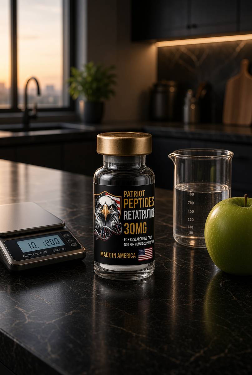 Made in USAOut of stock
Incretin
Retatrutide
US-made retatrutide 30mg — the published structure LY3437943, HPLC-MS verified.
4.6(609)
93 browsing this now · 5 purchased in the last 24 hours
30mg
£120.00
Read next
49 min · long read · Peptide research
What are peptides? Short chains that run the body
Peptides are amino acids joined by peptide bonds — shorter than proteins, more specific than most small-molecule drugs. Insulin was the first. GLP-1 agonists are the current proof. The catalogue here is the same chemistry, labelled for research.
47 min · long read · Peptide research
Why peptides are freeze-dried — and what reconstitution actually is
Water hydrolyses peptide bonds, oxidises methionine and grows microbes. Lyophilisation sublimes the ice so the chain ships as a porous cake. Reconstitution is mass divided by volume — laboratory handling, not a protocol for a body.
46 min · long read · Peptide research
Retatrutide (LY3437943): the published triple-agonist structure
A fatty-acylated unimolecular agonist at GIPR, GLP-1R and GCGR. US-made, HPLC-MS verified, labelled for laboratory research — not a medicine.
More in this desk

53 min · long read · Peptide research
Recovery, GH pulses and the CJC / ipamorelin pair
Somatotrophs have two 'go' receptors. CJC without DAC is a slightly longer GHRH pulse. Ipamorelin is a selective ghrelin-receptor key that does not yank ACTH. Together they are the pair Bowers already showed is more than additive — not a gym protocol.
52 min · long read · Peptide research
Fatigue, cellular energy and the NAD+ / MOTS-c neighbourhood
NAD+ is the rechargeable chip every cell spends on fuel and DNA repair — and the pool shrinks with age. MOTS-c is a mitochondrial 16-mer sent out under metabolic stress. Two answers to 'I have no energy' that are not coffee.
51 min · long read · Peptide research
Melanocortin circuits: pigment, appetite and PT-141
Melanotan II lights MC1, MC3, MC4 and MC5 — pigment plus the rest of the sheet. PT-141 is the free-acid cousin pointed at MC3 and MC4, the circuitry papers use for desire and energy, not skin colour. Same Arizona family. Different question.
51 min · long read · Peptide research
GHRH and ghrelin mimetics: two keys on the same pituitary lock
Somatotrophs have two ‘go’ receptors — GHRHR (Gs–cAMP) and GHSR (Gq). Bowers showed that occupying both releases more GH than the sum of either. That is synergy as a receptor fact, not a smoothie.
Essays describe published research. They are not medical advice and they do not authorise human use of any catalogue item.