

Peptide research · 49 min · 10,746 words
What are peptides? Short chains that run the body
Peptides are amino acids joined by peptide bonds — shorter than proteins, more specific than most small-molecule drugs. Insulin was the first. GLP-1 agonists are the current proof. The catalogue here is the same chemistry, labelled for research.
What this essay actually tells you
- A peptide bond is an amide between one amino acid's carboxyl carbon and the next residue's nitrogen. Oligopeptides are typically under 20 residues. Proteins fold past that.
- Insulin (51 residues, Banting & Best 1921) was the first peptide medicine. GLP-1 agonists are the current industrial proof of the same grammar, which is why the field still exists.
- A research peptide has a defined sequence, a mass and a chromatogram. Hydrolysed collagen in coffee has none of those, and calling it a peptide is how the word got mushy.
What this actually means
A peptide is a short chain of amino acids joined by amide bonds. Proteins are the long ones that fold into actual machines. Peptides are the shorter messages: hormones, fragments, receptor ligands, the things that tell those machines what to do. Insulin is a peptide. GLP-1 is a peptide. BPC-157 is a 15-residue scrap of a gastric protein. Length and job, that's the split. A peptide bond is planar because of resonance. Oligopeptides typically sit under 20 residues; the grey band around 50 is where insulin (51, two chains, disulphides) sits and people argue. Most of the signalling ones occupy GPCRs at nanomolar to picomolar concentrations; insulin occupies a receptor tyrosine kinase. Proteases — DPP-4, neprilysin, the trypsin family — eat native gut peptides in minutes, which is why medicines get a fatty-acyl handle and research solids ship freeze-dried. Collagen powder is hydrolysed food. A research peptide has a defined sequence, a mass and a chromatogram. We stock named sequences as lyophilised research solids, characterised, labelled for the bench. Same chemistry the papers already wrote down.
Peptides are short amino-acid chains. Amino acids are the twenty small building blocks your cells string together; a peptide is a handful of those blocks joined end to end. Insulin is one you already know — fifty-one amino acids, two chains, the hormone that moves glucose into muscle and fat. GLP-1 is another: thirty-one residues, written in the gut after a meal, asking the pancreas for more insulin. BPC-157 is a fifteen-residue fragment of a gastric protein, written GEPPPGKPADDAGLV. Same family, three jobs you can actually hold. The join itself is a peptide bond, which is an amide. Each residue offers a carboxyl carbon and an α-amino nitrogen. A molecule of water leaves. The two amino acids are now one molecule. Repeat the condensation and you have a primary structure: a one-letter code, a calculated mass, a backbone that can hydrogen-bond itself into a helix or a sheet. Proteins are the long versions of the same chemistry, folded into machines with tertiary structure worth drawing. Peptides are shorter. They are the hormones, fragments and receptor ligands that tell those machines what to do. Length and job. That is the split.
In short. A peptide is a short chain of amino acids. Insulin, GLP-1 and BPC-157 are three you can name. Proteins are the long folded versions.
You have probably met the word in two very different places, and it is worth putting both on the table before we draw a bond. Collagen powder is sold as peptides. A thirty-nine-residue triple agonist that occupies the GIP, GLP-1 and glucagon receptors is also a peptide. One of those is hydrolysed food: thousands of overlapping fragments, a gram weight on a tub, no single HPLC peak to point at. The other is a designed ligand with a receptor occupancy curve, a mass, and a chromatogram. Length, sequence and receptor are the facts that actually matter. If you cannot name the residues, you are looking at a mixture, not a research peptide. English hung one syllable on two objects, and the objects did not become the same thing. A scoop and a one-letter code can share a mass band and still be different molecules. We are going to stay with the chemistry from here: the receptors, the proteases, and five sequences on the shelf that can actually be written down. Once you can write the letters, the grocery-aisle use of the word becomes a different, perfectly honest conversation about food.
In short. Collagen powder and a named research ligand both get called peptides. Only the second has a sequence, a mass and a chromatogram.
Your cells already run on this language, which is the bit that is easy to forget when peptides get talked about as a 2020s invention. Pituitary releasing hormones are peptides. The incretins that amplify insulin after a meal are peptides. Defensins, melanocortins, oxytocin, vasopressin, the gastric pentadecapeptide BPC-157, the copper tripeptide GHK: all short sequences with published receptors or transcriptomic signatures. Small-molecule drugs are keys cut to fit a lock they were not born knowing. Peptides often are the lock's original key, or a close enough copy that the receptor does not argue much. That is why a fifteen-residue surface can pick one receptor out of a family at nanomolar or picomolar concentrations, and why a statin never saw the extracellular face of a class-B GPCR. The chemistry was never new. Insulin has been a peptide medicine since 1921. What changed is range, half-life engineering, and a public that noticed when a gut hormone started moving body weight. We will get to that wave. First we need the bond, the fold, and a few sequences you can actually hold.
In short. The body already uses short amino-acid chains as hormones and signals. Insulin was a peptide medicine in 1921. The idea is not new.
So the working definition we use in the laboratory is deliberately plain. One primary structure. A mass. A chromatogram. A paper that named the residues. BPC-157 is GEPPPGKPADDAGLV. GHK is three letters and a copper. KPV is Lys-Pro-Val, the tail of α-MSH. Ipamorelin is Aib-His-D-2-Nal-D-Phe-Lys-NH2, a selective ligand at the ghrelin receptor. Retatrutide is the published LY3437943 backbone, fatty-acylated, a unimolecular agonist at three class-B GPCRs. If you can point at those letters, that mass and that peak, you are holding the object this page is about. Named sequences, lyophilised, characterised against the published structure. Write the one-letter code on the tube and you have already done most of the identity work. What follows is why that object has a backbone dipole, a preferred secondary structure, a receptor, a protease that wants to eat it, and a century of medicine hanging off the same amide. You do not need a third category. You need the letters, the mass, and a little patience with the chemistry.
In short. A research peptide is one named sequence, a measured mass, and a chromatogram. If you can write the letters, you are holding that object.
Insulin in 1921 was the first peptide anyone bothered calling a medicine. Everything since has been a longer argument with the same backbone chemistry.— Banting and Best, plus a century of footnotes
The peptide bond is an amide
A peptide bond is an amide linkage between the carboxyl carbon of one amino acid and the α-amino nitrogen of the next, formed by condensation. Water is the leaving group. The product is a planar unit: the carbon, the oxygen, the nitrogen and the hydrogen sit in one plane because the nitrogen lone pair conjugates into the carbonyl, giving the C–N bond partial double-bond character. Pauling and Corey measured that planarity in the 1930s and 1950s, and it is why a polypeptide backbone has restricted φ and ψ angles — the two rotations at each residue — rather than free spinning at every join. Ramachandran later mapped the allowed combinations. The cis isomer is rare except at proline, which is why proline is a helix-breaker and a turn-maker, and why a proline-rich gastric fragment such as BPC-157 behaves differently in acid than a run of ordinary residues would. Resonance is doing real work here. It is why even a short chain has a backbone dipole and a preferred secondary structure, and why a 15-mer is already a three-dimensional object rather than a floppy string of letters.
In short. A peptide bond is an amide, held flat by resonance. That flat join is why a short chain can already fold into a helix or a turn.
The condensation is thermodynamically a little unhappy in water. Hydrolysis is favoured — the bond would rather come apart than stay made. Kinetics save the day: at cold and dry, the bond lasts. Warmth, leftover water, oxygen and light cancel that truce, which is why research peptides ship as a lyophilised cake rather than a millilitre of solution. Aspartate next to glycine is a known weak point, because the side-chain carboxyl can attack the backbone and make an aspartimide. Asparagine deamidates to a mix of Asp and iso-Asp. Methionine becomes methionine sulphoxide. Cysteine forms the wrong disulphides. Tryptophan goes yellow. These are ordinary organic reactions, and they will run in a vial if you let them. A ribosome lives with the same chemistry in a crowded cytosol, except the cell has chaperones, quality control and a proteasome, and a cake in a fridge has a stopper. There is a neighbouring page on lyophilisation if you want the storage half of that argument. Here we are staying with the bond itself, because once you know it can hydrolyse, the freeze-dried cake starts looking like chemistry rather than a peculiar commercial choice.
In short. Water slowly undoes peptide bonds. That is why research peptides are freeze-dried cakes, not bottles of solution sitting on a shelf.
Diagram
- Amino acid~110 DaTwenty side chains. The alphabet.
- Peptide bondamide, planarCarboxyl carbon to the next nitrogen. Resonance holds it flat.
- Oligopeptide< ~20 residuesMost hormones and fragments. GHK is three. KPV is three.
- Polypeptide20–50+Insulin 51. GLP-1 31. Retatrutide is a designed chain in this band.
- Proteinfolded machineHaemoglobin, a GPCR, lysyl oxidase. Tertiary structure worth drawing.
Insulin (Banting and Best, 1921) was the first peptide anyone bothered calling a medicine. A collagen hydrolysate is food. A named sequence with a mass and a chromatogram is a research peptide. The shared word is the accident.
Twenty proteinogenic amino acids, twenty side chains, an alphabet. Average residue mass is about 110 daltons, which is why a 15-mer such as BPC-157 sits near 1.4 kilodaltons and a 51-residue insulin monomer near 5.8. The side chains do the chemistry a receptor actually reads: charges, hydrogen bonds, a hydrophobic patch, a histidine that can hold copper, a proline that kinks the backbone. The backbone does the geometry. An α-helix has 3.6 residues per turn and a hydrogen bond from the carbonyl of residue i to the amide of residue i+4. A β-strand hydrogen-bonds to its neighbour. A turn is often proline and glycine. Secondary structure is not a protein privilege. Short peptides form helices in water when the sequence wants to, and they form them more readily when a membrane or a receptor pocket pays the entropic bill. That is why a 15-residue surface is already a lot of contacts, and why small-molecule drugs, which have far less surface, rarely match peptide affinities at the large extracellular faces of class-B GPCRs. You can hold the alphabet in one hand. The receptor is reading the side chains with the other.
In short. Twenty amino acids, about 110 daltons each. The backbone sets the shape; the side chains are what a receptor actually touches.
The ribosome makes this bond with RNA catalysis, at five or six residues a second in a mammalian cytosol, directed by a codon. Solid-phase peptide synthesis makes the same bond with an activating reagent on a resin bead, one cycle at a time, directed by a chemist. Same amide. Different factory. Bruce Merrifield published the resin trick in 1963 and collected the chemistry Nobel in 1984. Almost every short chain you will meet on a research certificate is the second factory: Fmoc chemistry, TFA cleavage, HPLC, mass spectrometry, a freeze-dried cake. Somatropin, at 191 residues, is the first factory hijacked — a recombinant tank, not a bead — because a 191-mer on resin is a different proposition from a 15-mer. The neighbouring essay on synthesis and HPLC is the making and the receipt. The point of naming both factories here is only this: the peptide bond does not care who made it. A receptor does not either. Identity is sequence plus mass plus chromatogram, not a romantic origin story about a gland. Once you believe that, the ribosome and the bead can sit on the same bench without an argument.
In short. Cells make peptide bonds on a ribosome. Chemists make the same bond on a resin bead. A receptor reads the sequence, not the factory.
- Amino acid
- ~110 Da, ~0.8 nm
- Peptide bond
- amide, planar
- GHK
- 3 residues
- BPC-157
- 15 residues, ~1.4 kDa
- Insulin
- 51 residues, two chains
- Typical mammalian cell
- 10–30 µm
Twenty proteinogenic side chains. The alphabet.
Carboxyl carbon to the next nitrogen. Resonance holds it flat.
Gly-His-Lys, plus copper. A tripeptide with a transcriptome.
GEPPPGKPADDAGLV. A few nanometres of gastric fragment.
The grey band. First peptide medicine, 1921.
A 15-mer is four orders of magnitude shorter, and a trillion times smaller in volume.
Oligopeptide, polypeptide, protein — length and job
Oligopeptides typically sit under 20 residues. That is a convention, not a law, and it is a useful one. GHK is three. KPV is three. Oxytocin is nine. The gastric fragment BPC-157 is fifteen. Most of the signalling messages in a mammal live in this band: short enough to be made and destroyed quickly, long enough to pick a receptor out of a family. Polypeptides run longer. GLP-1 is thirty-one residues in its active form. Glucagon is twenty-nine. A designed incretin analogue with a fatty-acyl handle sits in the same neighbourhood. Proteins are the folded, functional assemblies, often past 50 residues, with tertiary structure worth drawing: a hydrophobic core, disulphides, domains, a job that is catalysis or structure or transport rather than a message to a receptor. Haemoglobin is a protein. A GPCR is a protein. Lysyl oxidase is a protein. The ligand that occupies the GPCR is often a peptide. Mixing those two objects is how a grocery aisle and a laboratory ended up sharing a word, and how a scoop of hydrolysate started being talked about as if it were a research tool. Keep the lengths in mind and the jobs stay clear.
In short. Under about 20 residues is an oligopeptide. Longer chains are polypeptides. Proteins are the folded machines, usually past 50 residues.
There is a grey band around 50 residues where people argue, and then they get on with the experiment. Insulin sits on that line: 51 residues, two chains, three disulphides, a folded monomer that dimerises and hexamerises around zinc in the granule, and a ligand at a receptor tyrosine kinase. Everyone calls it a peptide hormone. Everyone also knows it has tertiary structure. The argument is taxonomic and does not change the blot. A 40-residue designed agonist is a peptide in every catalogue that stocks it. A 191-residue growth hormone is a protein, made recombinantly, occupying a cytokine receptor. IGF-1 LR3 is 83 residues and lives in the same recombinant neighbourhood. The split that matters in a laboratory is not the dictionary. It is whether you can write a one-letter code, weigh a single mass, and show a main HPLC peak. That test puts BPC-157, GHK, KPV, ipamorelin and retatrutide on one side of the bench, and a tub of collagen hydrolysate on the other. Insulin, grey band and all, passes the test. You can call it a hormone, a peptide, a small protein. The sequence, the mass and the peak do not care.
In short. Around 50 residues people argue about the word. Insulin has 51 and is still called a peptide hormone. The useful test is a single sequence and a single peak.
Diagram
- 0.1 nmHydrogen atomA proton and an electron. Chemistry starts here.
- 0.3 nmWater molecule70% of a cell by mass. The solvent life is.
- 1 nmAmino acidTwenty kinds. Peptide bonds string them.
- 2–4 nmResearch peptideA named chain. BPC-157 is 1.4 kDa, 15 residues.
- 4–10 nmGlobular proteinHaemoglobin, a GPCR’s extracellular face.
- 25 nmRibosomeThe factory that reads mRNA into protein.
- 5 nmMembraneA lipid bilayer. Every compartment starts here.
- 0.5–1 µmMitochondrionA bacterium the cell swallowed and kept.
- 6–10 µmNucleusTwo metres of DNA folded into a sphere.
- 10–30 µmTypical cellA city. 10¹⁰ proteins. One genome.
- 1 mmTissue grainA thousand cells talking across ECM.
- 1.7 mYou~36 trillion human cells. Most of them are red blood cells.
Lengths are characteristic, not exact. A research peptide is closer in size to a water molecule than to the cell that assays it — which is why a 15-mer can occupy a receptor pocket a small-molecule drug also wants.
Put a 15-mer next to a cell and the arithmetic still startles people. An amino acid is about 0.8 nanometres along the backbone. Fifteen residues, fully extended, would be a dozen nanometres; as a compact coil or a short helix it is a few. A typical mammalian somatic cell is 10–30 micrometres across, two to four picolitres, on the order of ten billion protein molecules. The gap is four orders of magnitude in length and about twelve in volume. A research peptide is a ligand that fits one protein on or in that cell. It is not a miniature version of the cell, and it is not a treatment plan for the 36 trillion cells that make a person. Ron Milo and Rob Phillips spent a decade putting these numbers in BioNumbers so the rest of us would stop waving our hands. Sender, Fuchs and Milo counted the cells. The catalogue lives on the first few nanometres of that ruler. Physiology lives on the last few rungs. Miss a rung and you start asking a milligram of lyophilised chain to stand in for a body. The scale diagram is there so we do not.
In short. A 15-residue peptide is a few nanometres long. A cell is ten thousand times longer. The peptide is a key for one lock, not a miniature body.
Secondary structure at this scale is already chemistry, not a rumour of a protein. A short helix has a dipole, positive at the N-terminus, negative at the C-terminus, which is why so many peptide hormones present a charged end to a receptor pocket. Amphipathic helices can sit on a membrane and concentrate next to a GPCR. Disulphides lock a fold that would otherwise breathe apart in water; insulin's three bridges are the famous case, but oxytocin and vasopressin each have one, and a cyclised analogue is often a chemist doing by hand what a cell did with cysteines. Stapled peptides, Verdine's hydrocarbon bridges, are the industrial version of the same idea: lock a helix so a protease cannot find a flexible loop. None of that makes a 15-mer into a protein. It makes a 15-mer into a three-dimensional ligand, which is the only form a receptor ever sees. Primary structure is the letters. The letters fold. The fold is what occupies the pocket. Once you have that picture, a string of one-letter code on a page starts to look like what it always was: a recipe for a shape.
In short. Even a short peptide can form a helix or be locked by disulphides. The receptor sees a shape, not a string of letters on a page.
Insulin, 1921: the first peptide medicine
Frederick Banting and Charles Best, Toronto, 1921. A pancreatic extract that dropped blood sugar in a depancreatised dog, then in a dying boy, Leonard Thompson, in January 1922. Collip cleaned the extract enough to inject. Macleod ran the department and shared the 1923 Nobel with Banting, who shared his prize money with Best. The molecule itself took another thirty years to become a sequence. Sanger worked out the two chains and the disulphides in the 1950s and collected his first chemistry Nobel in 1958 for that, the first protein anyone had sequenced. Fifty-one residues: an A chain of 21, a B chain of 30, three disulphide bridges, two of them interchain, one of them a loop on A. A zinc-coordinated hexamer in the granule, a monomer at the receptor. The first peptide anyone bothered calling a medicine, and still the manufacturing education for the whole class: two chains, three disulphides, a recombinant industry that had to be invented when animal glands ran short. Everything after is a longer argument with the same backbone chemistry. We are still having it.
In short. Banting and Best isolated insulin in 1921. Sanger sequenced its 51 residues in the 1950s. It is still the ancestor of peptide medicine.
The receptor is not a GPCR. Insulin occupies the insulin receptor, a receptor tyrosine kinase, a dimer already waiting in the membrane. Ligand binding rearranges the extracellular α-subunits; the intracellular β-subunits autophosphorylate; IRS proteins dock; PI3K–Akt and Ras–MAPK run. Glucose transporters move to the muscle and adipose membrane. Hepatic gluconeogenesis quiets. That is a different lock from the seven-helix story most of the catalogue occupies, and it is worth keeping the locks named so they do not blur. IGF-1 is the cousin ligand at IGF1R, another RTK, which is why IGF-1 LR3 sits in a different mechanistic essay from ipamorelin. The point of putting insulin at the front of a peptide essay is historical and chemical, not a claim that every research vial is a hormone replacement. Insulin proved that a short amino-acid chain, injected, could occupy a receptor and change a life. The GLP-1 medicines of the last decade proved it again, at a different receptor, with a different half-life trick. The chemistry did not change in between. The lock did, and the half-life did, and that is plenty.
In short. Insulin binds a different kind of surface receptor from most of the catalogue. That binding moves glucose transporters and still defines the class.
Recombinant manufacture is the industrial half of the ancestor story. Animal glands, then semi-synthesis, then Genentech's 1978 insulin in E. coli, approved in 1982 as Humulin: the first recombinant peptide medicine, and the template for every subsequent tank. A 191-residue somatropin followed. A catalogue of research 15-mers did not. Solid-phase synthesis is cheaper and cleaner for chains that short, and it does not ask a bacterium to fold two chains and form the right disulphides. The two factories still share the amide, the HPLC question, and the legal distinction between a licensed pen and a characterised reagent. Insulin remains a medicine. A lyophilised 15-mer with a paper behind it remains a reagent. Those two jobs can share a backbone without sharing a label. The ancestor is allowed to be both a historical fact and a manufacturing lesson without turning every sequence on a shelf into a prescription. We will keep saying medicine when we mean a pen, and reagent when we mean a cake, because the chemistry is related and the law is not.
In short. Recombinant insulin in 1982 taught the industry how to make a peptide at scale. Short research chains are still built on resin, not in a tank.
The body already spoke this language
Walk a hypothalamus and you are walking a peptide catalogue. GHRH, 44 residues in its full form, 29 in the active fragment, occupies GHRHR on the somatotroph and raises cAMP. Somatostatin occupies SSTR2 and SSTR5 and shuts the same cell down. GnRH is ten residues and runs the gonadotroph. CRH runs the corticotroph. TRH is three residues, a pyroglutamate-histidine-proline amide, and it is enough to occupy the thyrotroph. These are not metaphors for signalling. They are the signals. The anterior pituitary is a peptide-reading organ, and the portal blood from the median eminence is a millimetre-scale delivery system designed around half-lives measured in minutes. Local production, a short trip, a receptor, a protease. That architecture is why a native gut peptide also lasts minutes in plasma, and why every therapeutic analogue since has been an argument with dipeptidyl peptidase-4, neprilysin, and the trypsin-family enzymes that treat a peptide bond as food. Nature solved stability with geography. We solve it with chemistry. Same problem, two clever answers.
In short. Hypothalamic releasing hormones are short peptides. They travel a millimetre, occupy a pituitary receptor, and are destroyed in minutes.
The gut is the other original catalogue. Secretin, 27 residues, was Bayliss and Starling's 1902 hormone, the first time anyone used the word, a duodenal peptide that tells the pancreas to secrete bicarbonate. Gastrin, cholecystokinin, GLP-1, GIP, PYY, ghrelin: an intestine and a stomach writing messages to islets, brain, and gallbladder. GLP-1 is cleaved from proglucagon in the L-cell, occupies GLP-1R on the β-cell and in the brainstem, amplifies glucose-stimulated insulin secretion, slows gastric emptying, and reduces intake. Native half-life is about two minutes because DPP-4 clips it at alanine-2. GIP is the sister incretin from the K-cell. Glucagon, from the α-cell, occupies GCGR on the hepatocyte and raises glucose output. Three class-B GPCRs, three native peptides, and now a unimolecular agonist that occupies all three. The incretin decade did not invent peptide signalling. It industrialised a gut language that Bayliss and Starling had already named, and that insulin had already proved could be a medicine, once you kept the chain alive long enough to matter. We noticed when the weight-loss curves arrived. The language was older.
In short. Gut hormones such as secretin, GLP-1 and ghrelin are peptides. The incretin drugs are copies of that intestinal language, built to last longer.
Defensins are peptides too, and they are a reminder that not every short chain is a hormone. They are cationic, amphipathic, disulphide-locked, and they disrupt microbial membranes. Melanocortins are the other reminder: α-MSH is thirteen residues, a cleavage product of POMC, and it occupies MC1R on the melanocyte (pigment), MC4R in the hypothalamus (intake), and a set of anti-inflammatory programmes that KPV, the C-terminal tripeptide, still carries without the tanning. Oxytocin and vasopressin are nine-residue amides with one disulphide each, occupying class-A GPCRs, running milk ejection, water reabsorption, social behaviour, a century of obstetric and endocrine practice. Atrial natriuretic peptide is 28 residues from the heart. Substance P is eleven, a tachykinin, pain and inflammation. The mammal is a peptide-writing animal. A research catalogue is a drawer of the published primary structures from that animal, plus the analogues chemists made when the native chain died too fast or bound too many family members. The drawer is a reading list. It is not the animal. Holding that distinction makes the rest of the shelf easier to love.
In short. Defensins, melanocortins, oxytocin and gut hormones are all short chains. A research catalogue is a drawer of those published sequences, not a body.
Paracrine versus endocrine is geography, not a different chemistry. A gastric fragment that acts on nearby endothelium is a peptide. A pituitary hormone in the bloodstream is a peptide. The bond is the same. The receptor logic is the same. The difference is how far the chain has to travel before a protease finds it, and whether a chemist has hung a fatty acid on it so albumin will carry it past that protease. Native GLP-1 is an endocrine peptide with a paracrine-scale half-life, which is why it is a terrible drug and a beautiful hormone. BPC-157, in the Sikiric literature, is argued as a gastric-endothelial peptide with unusual stability in gastric juice, proline-rich, a 15-mer that does not need a fatty-acyl handle to survive the organ it came from. GHK is a circulating fragment of the extracellular matrix, a copper ligand, a transcriptome in fibroblasts. Three geographies, three half-lives, one backbone. Naming the geography is how you stop treating every vial as a systemic hormone. Some messages are whispered next door. Some are posted in the blood. You need to know which you are holding.
In short. Some peptides act next door; some travel in blood. Same bond, different distance, and a different race against the enzymes that cut them.
Occupancy at a receptor, then a flood
Diagram
Outside
Peptide ligand
Named sequence in the nM–µM pocket. Shape complementarity, not vibes. A 15-mer and a 4-mer do not fit the same hole.
Membrane
7-TM receptor
Helices rearrange. The cytoplasmic face becomes a GEF for a heterotrimeric G protein (Gs, Gi, Gq, G12/13).
Inside
Second messengers
cAMP, IP₃, Ca²⁺, β-arrestin. One occupied receptor can spawn thousands of messenger molecules. That is amplification.
~800 GPCRs in the human genome. Seven transmembrane helices, an extracellular ligand pocket, an intracellular G-protein handshake. Catalogue neighbours: ipamorelin at GHSR, PT-141/MT2 at melanocortin receptors, retatrutide at GLP-1R/GIPR/GCGR.
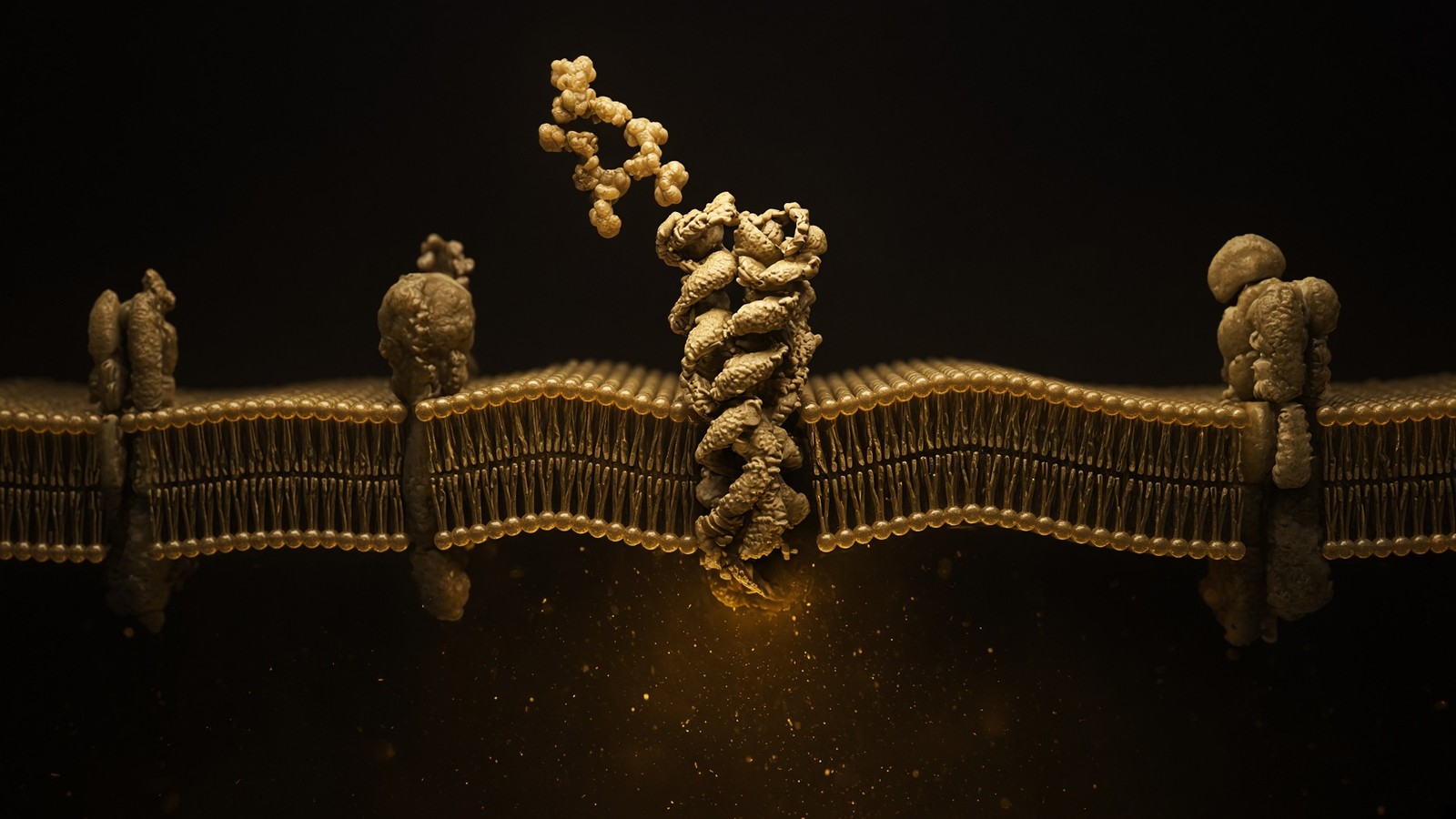
Most bioactive peptides occupy G protein-coupled receptors. About 800 of those in the human genome, seven transmembrane helices, an extracellular face that binds ligand, an intracellular face that acts as a guanine-nucleotide exchange factor for a heterotrimeric G protein. Lefkowitz and Kobilka got the 2012 chemistry Nobel for this family, and they deserved it. Class A, rhodopsin-like, includes GHSR (ghrelin, ipamorelin) and the melanocortin receptors (α-MSH, PT-141, melanotan II, KPV in some assays). Class B, secretin-like, includes GLP-1R, GIPR, GCGR and GHRHR: the incretin and growth-hormone-releasing locks, large extracellular N-termini, peptide ligands that sit across that face rather than in a small-molecule pocket. Occupancy rearranges the helices. The G protein spends GTP. The information that crossed the five-nanometre bilayer is conformation. The peptide, in the boring and correct case, is still outside, or is on its way to a lysosome after the receptor was internalised as a complex. That is how a chain that never enters the cell can run the cell. Shape on the outside. Chemistry on the inside.
In short. Most signalling peptides stick to a receptor on the cell surface. They stay outside. The receptor changes shape, and that is the message.
Receptor tyrosine kinases are the other major lock, and insulin already introduced them. Ligand-induced rearrangement, autophosphorylation, SH2 effectors. IGF-1 LR3 occupies IGF1R. A few peptides occupy ion channels or modulate them, though that is a smaller slice of the catalogue than forum shorthand pretends. A few have intracellular partners: GHK hands copper around, TB-500's LKKTETQ motif binds actin, MOTS-c is a 16-mer a mitochondrion translated from its own 12S rRNA and can go to the nucleus under stress. Those exceptions are named exceptions. They do not smear a tourist story across the whole shelf. If you cannot name the lock, you do not yet have a mechanism. Occupancy at a named protein, then a named second messenger, then a named assay: that is a mechanism you can test. Bioactive peptides do this at nanomolar to picomolar concentrations because a 15-residue surface is a lot of contacts, van der Waals, hydrogen bonds, a salt bridge or two, enough to pick one receptor out of a family that a 400-dalton pill cannot tell apart. Surface is the point. Name it.
In short. Some peptides use other locks: insulin's receptor is a kinase, GHK carries copper, a few bind actin. Name the lock. That is the mechanism.
Affinity is how tightly the ligand binds, Kd. Occupancy is the fraction bound at a given concentration, θ = [L] / ([L] + Kd) for a simple isotherm. Efficacy is what the occupied receptor does: full agonist, partial agonist, biased agonist, antagonist. Tissue is which cell has the receptor, how many copies (typically a thousand to a hundred thousand), and which effectors sit downstream. Spare receptors mean a full response can come from a fraction occupied, which is why EC50 can sit below Kd and why a binding assay and a functional assay are not the same experiment. Nanomolar occupancy at a class-B GPCR is ordinary for a native peptide. Picomolar happens when the contacts are excellent and the receptor is willing. Small molecules rarely match that at these faces because they do not have the surface. That is the pharmacological reason peptides exist as a class, and it is also the reason they are expensive to keep alive: the same amide bonds that make the surface are food for proteases. Specificity and fragility are the same chemistry, read in two directions. We will meet the tax in a moment.
In short. Peptides can bind their receptors at tiny concentrations because a long surface makes many contacts. The same bonds that bind well are easy for enzymes to cut.
Diagram
× 1
Ligand
One peptide in one pocket. nM–µM. Shape, not a mood.
× 10–10²
G proteins
The occupied GPCR is a GEF. Each Gα is a catalyst.
× 10³–10⁴
cAMP / IP₃ / Ca²⁺
Adenylyl cyclase and PLC do not make one molecule. They make a cloud.
× 10⁴–10⁶
PKA / PKC / CaMK
Kinases phosphorylate many substrates per messenger.
× tissue
Secretion, transcription, motility
The organism-level readout. Still not a protocol.
This is the only magic, and it is not magic. A nanomolar ligand can move a micromolar messenger because enzymes sit between them. Desensitisation (GRK, β-arrestin, endocytosis) is how the cell refuses to let ‘more ligand’ mean ‘more signal’ forever.
Amplification is arithmetic. One occupied GPCR can catalyse GDP/GTP exchange on tens to hundreds of G proteins. Each Gαs-GTP can activate adenylyl cyclase to make many cyclic AMP from ATP. Each cAMP-activated protein kinase A can phosphorylate many substrates. A nanomolar occupancy can move a micromolar messenger because the receptor is an enzyme and the cyclase is an enzyme. Gαq takes the other door: phospholipase C, IP3, calcium from stores, protein kinase C. GHSR, the ghrelin receptor that ipamorelin occupies, is mostly this Gq route, which is why Bowers' synergy papers with GHRH (Gs, cAMP) showed super-additive growth-hormone release from a somatotroph: two second-messenger systems, one secretory granule. Gαi lowers cAMP. Gα12/13 talks to Rho. β-arrestin then desensitises, internalises, and sometimes scaffolds MAPK. Two ligands at the same GPCR can prefer G protein versus arrestin, which is biased agonism, an active design axis in the incretin literature and a sentence most catalogue peptides do not yet have a clean published profile for. We should not invent one.
In short. One receptor can load many G proteins, and each enzyme makes many messengers. That is why a tiny amount of peptide can change a whole cell.
Ipamorelin is a worked example of selectivity inside this grammar. Early growth-hormone secretagogues — GHRP-6, GHRP-2, hexarelin — occupied GHSR and also moved ACTH and prolactin. Fine if you were cataloguing side-effects. Poor if you wanted growth hormone as the variable. Raun, Hansen, Thorkildsen and colleagues, European Journal of Endocrinology 1998, designed a pentapeptide, Aib-His-D-2-Nal-D-Phe-Lys-NH2, that kept GHSR occupancy and lost much of that HPA noise. Relative, not absolute. GHSR is also a hunger receptor in hypothalamus and vagus, so ghrelin mimetics still have an appetite conversation. Selectivity versus older GHRPs is the paper, and it is why anyone still bothers with this analogue as a probe. Modified GRF(1-29), often sold as CJC-1295 without DAC, occupies the other pituitary lock, GHRHR, Gs, cAMP, a pulse rather than a multi-day flat line. Two keys, two second messengers, one granule. The GHRH–secretagogue essay next door is that experiment. This page only needs the sentence: a 5-residue analogue can prefer one readout of one receptor, because a peptide surface is specific when you design it to be.
In short. Ipamorelin was built to occupy the ghrelin receptor without dragging cortisol the way older secretagogues did. Selectivity is a published design choice.
Proteolysis is the tax
Peptide bonds are food for proteases. That is not a defect in the design. It is the design. A hormone that lasted for days would still be signalling after the meal, the stress, or the pulse had passed. Dipeptidyl peptidase-4, DPP-4, is the famous incretin tax: a serine protease on endothelial cells and in plasma that cleaves after a proline or alanine at position 2. GLP-1 has His-Ala-Glu at the N-terminus. DPP-4 clips it. Half-life, about two minutes. Sitagliptin and its cousins inhibit that enzyme so native GLP-1 and GIP last a little longer, which is a small-molecule way of doing what a chemist can also do by swapping alanine-2 for a residue DPP-4 does not like, or by hanging a fatty acid so albumin hides the chain. Modified GRF(1-29) puts D-alanine at position 2 for the same reason. Native GHRH dies there too. The enzyme does not care whether the substrate is a gut hormone or a hypothalamic one. It cares about the P1 residue. Sequence is fate, at this end of the argument. Change the letters and you change the clock.
In short. An enzyme in blood cuts many gut and brain peptides near the start, so they last minutes. Medicines are edited or shielded so that cut is slower.
Neprilysin, NEP, CD10, a zinc metalloprotease, is the other famous tax on natriuretic peptides and on a long list of neuropeptides. The trypsin-family serine proteases — trypsin itself in the gut, thrombin, plasmin, a set of kallikreins — cut after lysine or arginine, which is why a research peptide with a dibasic site has a weak point you can read off the one-letter code. Insulin-degrading enzyme takes insulin and amyloid-β. ACE takes angiotensin I to II and also chews bradykinin. The mammal is a protease landscape, and a native peptide is written to be local and brief inside that landscape. Therapeutic analogues are written to cheat it: fatty-acid acylation so albumin carries the chain (liraglutide's C16, semaglutide's C18 diacid, the same handle on tirzepatide and retatrutide), PEGylation so the kidney does not filter it, D-amino acids so a protease's active site does not fit, N-methylation, cyclisation, stapling, C-terminal amidation, which many native peptide hormones already have. Half-life engineering is the industrial core of peptide medicine. It is also why a native 15-mer and a weekly pen are not the same object, even when they rhyme.
In short. The body is full of enzymes that cut peptides. Drug-like analogues add fatty acids, PEG, or D-amino acids so those enzymes work more slowly.
Native gut peptides last minutes, which is why the therapeutic analogues get acylated, PEGylated, or sequence-stabilised. Liraglutide hung a palmitate so albumin would carry it for hours to a day. Semaglutide used a longer C18 diacid spacer and became weekly. Tirzepatide and retatrutide wear the same albumin-handle trick. Oral semaglutide exists because of SNAC, a permeation enhancer that locally raises pH in the stomach and helps a fraction of the dose across; bioavailability is still single-digit percent, which is why the tablet dose is huge next to the injection. That is engineering honesty, not a magic pill. D-amino acids show up in ipamorelin (D-Phe, D-2-Nal) and in a long list of research analogues whose half-life in a dish is the variable someone actually wanted. None of this is glamour. It is why a chain that would last eight minutes in plasma can sit in a fridge as a cake for months, and why a weekly medicine and a lyophilised research solid can share a backbone grammar without sharing a legal class, a formulation, or a device. Two tricks. Two jobs. One amide.
In short. Fatty acids, PEG and D-amino acids are tricks for living longer in blood. A freeze-dried research cake is a different trick: take the water away so the chain waits.
The dish is not a person, and proteases are one reason. Diffusion, unstirred layers, plasma-protein binding, first-pass metabolism, receptor number, spare receptors, desensitisation, and the fact that a human is tens of trillions of cells of many types all sit between a nanomolar well and a physiology. A binding isotherm in a well is a binding isotherm in a well. Fatty-acid acylation is how licensed incretin analogues buy half-life against albumin; a research vial of the published sequence is the ligand, not the pen. DPP-4 in a serum-containing medium will still clip an unmodified incretin analogue while you are watching the cAMP assay, which is why methods sections name the inhibitors and the time points. Trypsin on a poorly handled stock will clip anything with a lysine. These are laboratory facts, the unglamorous kind that save a month. They are also why characterisation — HPLC for purity, mass spectrometry for identity — is the receipt before anyone talks about a receptor curve. A clean peak of the wrong chain is a very tidy way to waste that month. Measure the chain first. Then the receptor.
In short. A dish at nanomolar is not a person. Enzymes, binding proteins and sheer scale sit in between. Measure the chain first, then the receptor.
Five sequences, five jobs
Diagram
| Node | Catalogue | Conversation |
|---|---|---|
| GPCR | Ipamorelin, MT2, PT-141, retatrutide, CJC | Second messengers, secretion, appetite, pigment |
| RTK / IGF1R | IGF-1 LR3 | IRS–PI3K–Akt–mTOR and Shc–ERK |
| Cytokine receptor | Somatropin (HGH) | GHR–JAK2–STAT5b, hepatic IGF-1 |
| Cofactor | NAD+ | Sirtuins, PARPs, CD38, redox |
| Actin buffer | TB-500 / Tβ4 motif | G-actin sequestration, motility |
| Growth-factor-like | BPC-157 | VEGFR2 / FAK / eNOS neighbourhood |
| Copper ligand | GHK-Cu | Transcriptome shift in fibroblasts |
| MC fragment | KPV | NF-κB, PepT1, no pigment |
| Nuclear / pineal | Epithalon (AEDG) | TERT and melatonin literatures |
| mtORF peptide | MOTS-c | AMPK, folate–methionine cycle |
Each row is a different kind of molecular conversation. The catalogue peptides bind at these nodes; they are not interchangeable, and stacking them because a forum did mixes unrelated literatures.
BPC-157 is GEPPPGKPADDAGLV, fifteen residues, a fragment of body protection compound, a gastric protein Sikiric and colleagues isolated and then studied as a pentadecapeptide. Proline-rich, which is part of why it is unusually stable in gastric juice: DPP-4 and many trypsin-family enzymes find less to like. The published literature is endothelial and gastric, with a VEGFR2 and nitric-oxide trail, wound and tendon models, a rat-heavy bibliography that a careful reader will take as a careful reader should. It is a defined 15-mer — not a collagen hydrolysate — with a mass near 1.4 kilodaltons and a chromatogram. We stock it as that object, lyophilised, characterised, because the one-letter code is the claim. The gastric-cytoprotection essay next door is the mechanism in full. This page only needs the sentence that belongs in a definition: a research peptide can be a fragment of a larger protein and still be a ligand with a name, provided someone wrote the residues down and someone else can show they are in the vial. Fifteen letters. A mass. A peak.
In short. BPC-157 is fifteen named residues from a gastric protein. Proline-rich, stable in acid, a single sequence — not a collagen powder.
GHK is glycine-histidine-lysine, three letters, and a copper. Pickart isolated it from human plasma in the 1970s as a factor that made old liver tissue synthesise proteins more like young tissue, and then spent decades on the copper complex and on fibroblast arrays. The histidine holds Cu(II) in a square-planar complex. The transcriptome that follows in culture — collagen, TIMPs, MMPs, a repair-shaped sheet — is the published signature, and it is a copper-chaperone story as much as a peptide-receptor story. Lysyl oxidase, the enzyme that cross-links collagen, is copper-dependent, which is how a tripeptide and a tub of collagen hydrolysate get mentioned in the same breath at dinner, and then someone says 'peptides' as if that settled it. It does not. GHK-Cu is Gly-His-Lys·Cu²⁺, characterised. Collagen hydrolysate is a mixture of many fragments. Different objects, sharing a syllable and a metal. The copper-and-repair-transcriptome essay is the full reading list. Here it is the three-residue end of the length scale, proof that an oligopeptide can still be a ligand. Three letters and a metal. That is enough.
In short. GHK is three amino acids that hold copper. It is a defined ligand with a published fibroblast signature, not a scoop of hydrolysed collagen.
KPV is lysine-proline-valine, the last three residues of α-MSH. Luger's dermatology group and Getting's inflammation group showed that this fragment still calms inflammatory transcription, NF-κB, in keratinocytes and macrophages. Dalmasso, Merlin and colleagues showed that KPV can ride PepT1, the intestinal oligopeptide transporter, which is why DSS-colitis models keep appearing in this literature: the fragment can get into epithelium by a nutrient transporter, not only by a melanocortin receptor. MC1R dependence is incomplete, which is the intellectual reason to use KPV instead of α-MSH or melanotan II when pigment and MC4R appetite circuitry would confound the assay. Three residues, a transporter, an NF-κB literature. Subtract the tan. Keep the quieting. That is a design choice you can only make if the parent hormone was a peptide you could shorten with a pair of scissors and a reason. Small molecules do not always offer that subtraction. Peptides do, because the primary structure is a string you can cut. Hold the parent, cut the tail, ask a cleaner question.
In short. KPV is the last three residues of the tanning hormone, kept for its anti-inflammatory work and stripped of the pigment programme.
Retatrutide is the published LY3437943 structure: a single-chain, fatty-acylated unimolecular agonist at GIPR, GLP-1R and GCGR, three class-B GPCRs, primarily Gs–cAMP. Coskun and colleagues, Cell Metabolism 2018, for the engineering. Jastreboff and colleagues, New England Journal of Medicine 2023, Phase 2 in obesity: 24.2% mean weight loss at 12 mg, 48 weeks, n=338, gastrointestinal events the common adverse events. That paper is an investigational-medicine trial. The research vial is the published backbone, synthesised, HPLC-MS characterised, labelled for the bench. Same architecture on paper. Different legal class, formulation, device, pharmacovigilance. Wilding, NEJM 2021, STEP 1, semaglutide 2.4 mg, 14.9% mean weight loss. Jastreboff, NEJM 2022, SURMOUNT-1, tirzepatide 15 mg, 20.9%. Three papers, three receptor sets, one industrial decade, and the reason a public that had ignored peptides since insulin suddenly cared. The chemistry was never new. The clinical result is what changed. A research sequence of the triple agonist is still a reagent. The therapeutics essay next door is the platform. This page is the definition that makes the platform legible.
In short. Retatrutide is a designed peptide that occupies three gut-hormone receptors. The trial showed large weight loss. A research vial of that chain is still a laboratory ligand.
Ipamorelin, again, because selectivity deserves a second named sentence in a definition essay. Aib-His-D-2-Nal-D-Phe-Lys-NH2. Five residues, two of them D-amino acids, a C-terminal amide, a ghrelin-receptor agonist that Raun et al. built to stop dragging ACTH and prolactin the way GHRP-6 did. GHSR is Gq, calcium, amplification of the GHRH pulse on the somatotroph, and a hunger receptor elsewhere. Using it with modified GRF(1-29) is occupying the two physiological inputs to that cell, which Bowers showed is super-additive, because cAMP and calcium converge on the same granules. It is not a recipe printed on a vial. It is two locks, two ligands, a third receptor (somatostatin) still in the background because physiology did not leave. Five sequences, then, that the catalogue actually holds: a gastric 15-mer, a copper tripeptide, a melanocortin tail, a fatty-acylated triple agonist, a selective pentapeptide. Each can be written in one-letter code, weighed as a single mass, and shown as a main HPLC peak. That is the definition in practice, not in a dictionary. If you can write it down, you can hold it. If you cannot, you are holding something else.
In short. Ipamorelin is a five-residue ghrelin-receptor probe designed for selectivity. With BPC-157, GHK, KPV and retatrutide, it is a named sequence you can actually write down.
Collagen powder is hydrolysed food
Type I collagen is a heterotrimeric (α1)₂α2 triple helix, each chain about a thousand residues, glycine every third position, hydroxyproline to hydrogen-bond the three strands, lysyl-oxidase cross-links to make the cable load-bearing. When a food factory makes 'collagen peptides', it is not purifying that helix. It is digesting it. Endopeptidases cut until the average piece is two to five kilodaltons, so it dissolves in coffee. The product is a population: thousands of overlapping fragments, batch-dependent, no one peak to point at on a chromatogram. After you drink it, Pro-Hyp and related di- and tripeptides do show up in plasma; Iwai, Sato and a trail of nutrition papers documented that, and those fragments are studied as a mixture for fibroblast and bone-cell effects. A mixture can have biology. A mixture still does not have a single sequence, a single mass, or a single receptor curve. Identity tests that mean everything on a research vial — HPLC plus mass spectrometry of one peak — are meaningless as a purity claim on broth. Grams versus a one-letter code. Food versus ligand. Both can be interesting. They are not the same object.
In short. Collagen powder is chopped-up structural protein, a mixture with no single sequence. A research peptide is one chain you can name and weigh.
The muddle stays alive because copper and collagen share a dinner conversation. Lysyl oxidase is copper-dependent. GHK carries copper. Collagen needs lysyl oxidase. BPC-157 is proline-rich and gastric, and collagen is proline-rich and everywhere, so the syllables blur. KPV is three residues and collagen hydrolysate contains three-residue fragments, so a tub and a vial get mentioned in the same breath. Length is not identity. A 15-mer of defined sequence is not a 15-mer-sized piece of a thousand-residue helix of unknown sequence. Ask for the residues. If the answer is a gram weight, it is food. If the answer is a one-letter code and a mass, we can talk. The collagen-versus-research-peptide essay next door is the gory version of that split. This page only has to plant the flag: the word peptide, on a research vial, means a defined primary structure. On a tub in a grocery aisle it means a hydrolysate that happens to be in the same mass band as some hormones. Sharing a mass band is not sharing a molecule. You already knew that, once someone said it out loud.
In short. Copper, proline and the word peptide make collagen powder and research ligands sound related. They are not. Ask for the sequence.
Ask for the sequence. If the answer is a gram weight, it is food. If the answer is a one-letter code and a mass, we can talk.
Merrifield made them. HPLC is the receipt.
Bruce Merrifield, Journal of the American Chemical Society, 1963: Solid Phase Peptide Synthesis. I. The Synthesis of a Tetrapeptide. He anchored a C-terminal residue to an insoluble resin and added the chain, one protected amino acid at a time, washing away excess reagents instead of purifying a soluble intermediate at every step. The Nobel was 1984. The chemistry evolved from Boc/benzyl to Fmoc/tBu, which is what almost everyone uses now: base-labile Fmoc off with piperidine, couple the next residue with HBTU or HATU or DIC/Oxyma, cap any unreacted amines so they do not become deletion sequences, repeat. A 15-residue peptide is fifteen cycles. Each cycle is a chance to fail. Aggregation of hydrophobic stretches, aspartimide at Asp-Gly, racemisation at histidine and cysteine: anyone who has run a difficult sequence has met at least one of those. Cleavage with a TFA cocktail releases the chain and the side-chain protecting groups. What you have then is a crude peptide, and crude is a polite word for a mixture. The interesting number is not whether we made a peptide. It is what else is in the peak.
In short. Merrifield grew peptides on a resin bead, one amino acid per cycle. Miss a cycle and you have a slightly wrong chain that can still look like a peak.
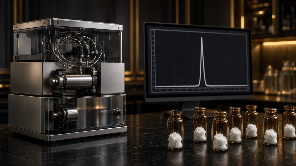
HPLC is a separation. Reverse-phase C18 columns hold hydrophobic chains longer. An acetonitrile/TFA gradient lets them go in order. Detection at 214 or 220 nanometres watches the peptide-bond absorbance. A single sharp peak at the expected retention time is the adult result. A fat shoulder is a mixture wearing a compound's clothes. ≥98% HPLC means the main peak dominates the chromatogram. That is honesty about the vial, not a clinical claim. Mass spectrometry then asks whether the peak's mass matches the calculated monoisotopic mass of the published sequence, including the fatty-acyl handle on a molecule like retatrutide. Purity without identity is a clean peak of the wrong thing. Identity without purity is the right chain in a mess. The two numbers travel together on a certificate worth keeping. The synthesis-and-HPLC essay next door is that receipt in full, with the failure modes and the specification. This page points at it rather than duplicating it. A name on a label does none of that work. The column and the mass analyser do.
In short. A purity test asks whether one peak dominates. A mass test asks whether that peak is the named chain. Keep both numbers.
Lyophilisation is the storage half, and it belongs in one paragraph here so the cake on the bench has a physics. Water hydrolyses peptide bonds, oxidises methionine and grows microbes. Freeze the solution, drop the pressure below the triple point, hold the product colder than its collapse temperature, let the ice sublime, then desorb the bound water. Residual moisture, Karl Fischer, typically wants to sit in the low single-digit percent or the hydrolysis resumes in the solid state. What remains is a porous cake, the ghosts of ice crystals, not kitchen powder. Reconstitution is mass divided by volume, a solvent the sequence actually accepts: polar chains in water or dilute acetic acid, fatty-acylated chains sometimes a co-solvent the paper names. Bacteriostatic water is sterile water plus 0.9% benzyl alcohol so that after you puncture the stopper the next hours are not a culture medium. Laboratory solvent. Arithmetic. The lyophilisation essay is the phase diagram. This page only needs the reason the object is a white plug: the bond is safer dry. Take the water away, and the chain will wait.
In short. Freeze-drying pulls the water off so the chain does not fall apart in storage. Adding a measured volume of solvent is reconstitution: mass over volume.
A gut peptide moved body weight. The chemistry was never new.
Wilding and colleagues, New England Journal of Medicine 2021, STEP 1: semaglutide 2.4 mg, 14.9% mean weight loss. Jastreboff and colleagues, same journal, 2022, SURMOUNT-1: tirzepatide 15 mg, 20.9%. Jastreboff again, 2023: retatrutide 12 mg, 24.2% at 48 weeks, a Phase 2 curve that looks like bariatric surgery drawn with a peptide. Those three numbers, from three papers, are why a public that had ignored peptides since insulin suddenly cared, and why every metabolism group now has a peptide chapter. They are also why a journal that only cheers has not finished the job. Gastrointestinal events, lean-mass questions, and what happens after discontinuation are the adult safety conversation the field is actually having, and it is not settled. More than a hundred peptide drugs are already approved worldwide, depending on how you count analogues and diagnostics. Desmopressin, oxytocin, teriparatide, octreotide, insulin still the ancestor. GLP-1 receptor agonists are the current industrial wave. The wave is not the invention of the idea. The amide was already on the books. The curves made people look.
In short. GLP-1-family medicines produced double-digit weight loss in major trials. That result made peptides famous again. The backbone chemistry dates to insulin.
What the incretin decade actually invented was range and half-life, not the amide. Native GLP-1 was always a peptide. DPP-4 always ate it. Fatty-acid acylation, albumin binding, dual and triple agonists, an oral tablet that exists because of a permeation enhancer: those are industrial answers to a two-minute half-life. Biased agonism at class-B GPCRs, cAMP versus β-arrestin, is an active design axis and still a bit messy in the data. Setmelanotide is daily MC4R agonism for rare obesity syndromes. Bremelanotide is an on-demand melanocortin analogue. Different receptors, same platform problem: keep the chain alive long enough to occupy the target, and no longer than the indication wants. Wang's reviews put the approved count above a hundred. A research catalogue sits underneath that platform as the ligands those programmes were built from or against, not as the pens. The therapeutics essay is the industrial present. This page is the sentence the industrial present stands on: a short amino-acid chain, a receptor, a protease, a chemist who cheated the protease. Insulin already knew. We are still writing footnotes on that knowledge.
In short. The new part was making gut peptides last long enough to be weekly medicines. The amide bond, the receptor and the enzyme tax were already on the books.
At 48 weeks, retatrutide at 12 mg produced a mean body-weight reduction of 24.2%.— Jastreboff AM et al., N Engl J Med. 2023.
A licensed pen is a medicine: formulation, device, pharmacovigilance, a label. A research sequence is the published primary structure on a certificate, used as a ligand in an assay. They can share a backbone and still not share a legal class. Our retatrutide is the published LY3437943 structure, HPLC-MS characterised, not Lilly's product. Insulin in a pharmacy fridge is a medicine. A 15-mer with a Sikiric bibliography is a reagent. The interesting question is no longer whether peptides work. A century of approvals answered that. The interesting question is which receptor set you want to occupy, how long the chain should live, and whether you are holding a medicine or a reagent. On this shelf the last one is always the reagent. The GLP-1 wave proved a gut peptide can move body weight into the range of bariatric surgery. It did not turn a characterised solid into a prescription, and it did not make a collagen tub into a ligand. Chemistry, legal class, and a chromatogram remain three different sentences. Keep them in different pockets and you will be fine.
In short. A medicine pen and a research vial can share a sequence on paper and still be different objects: different law, device, and job.
Why they work when they work
Specificity, mostly. A 15- to 40-residue chain has enough surface to pick one receptor out of a family. That is why ipamorelin can prefer GHSR over the cortisol side-effects that haunted the older GHRPs, and why KPV can occupy melanocortin anti-inflammatory signalling without dragging the whole tanning programme along with it. Small molecules win on oral bioavailability and cost. Antibodies win on specificity at a different scale, a different factory, a different price. Peptides sit in the gap: enough contacts to be picky, small enough to manufacture at insulin-like scale when the sequence cooperates, aimed at the large extracellular faces a statin never saw. The bill comes due as stability. Peptide bonds are food for proteases. Nature solved this with local production and half-lives measured in minutes. Chemists solve it with lyophilisation, fatty-acyl handles, and D-amino acids when the paper actually calls for them. None of that is a personality trait of the molecule. It is physical chemistry. A chain that would last eight minutes in plasma can sit in a fridge as a cake for months because someone took the water away on purpose.
In short. Peptides are specific because they have a large surface. They are fragile because enzymes eat that same surface. Dry storage and chemical edits buy time.
Secondary structure is part of the specificity. A helix that presents the right face to a class-B GPCR's N-terminus is a different ligand from a floppy chain of the same composition. Stapling, lactam bridges, disulphides, a C-terminal amide: chemists lock what the receptor wants to see. The receptor, for its part, is not a hole. It is a conformational machine with an extracellular face, an orthosteric pocket or a peptide-binding groove, allosteric sites, a G-protein interface, an arrestin interface. Two ligands can occupy the same receptor and write different intracellular sentences. That is biased agonism again, and it is why 'this peptide occupies that receptor' is the start of a conversation, not the end of one. Concentration, time, receptor number, the G protein the cell actually expresses, the phosphodiesterases that eat the cAMP, the AKAPs that pin PKA next to one channel and not another: tissue is a variable. A 10 nM dish is a 10 nM dish. A person is not a well. The definition still holds. The extrapolation is the part that wants a different essay, a different n, and a licence.
In short. Shape, not just sequence, decides which receptor a peptide prefers. The cell's own proteins then decide what that binding actually does.
Intracellular partners deserve an honest remaining paragraph so the GPCR story does not swallow the shelf. GHK-Cu is a copper ligand; the metal has an intracellular life, lysyl oxidase among the copper enzymes, a fibroblast array Pickart published. TB-500, the LKKTETQ motif from thymosin β4, binds G-actin and shifts the polymerisation equilibrium; that is a cytoskeleton, not a seven-helix lock. MOTS-c is translated from mitochondrial 12S rRNA in a reading frame nobody asked an rRNA to have; AMPK papers, nuclear translocation under stress, a 16-mer with a different origin story from Merrifield's bead. NAD+ is not a peptide at all; it is a dinucleotide cofactor that happens to sit in the same catalogue because the bench uses lyophilised solids, and because sirtuins and PARPs spend it. Naming the exceptions is how a definition stays honest. Most of the drawer is a GPCR ligand. Some of it is a metal, a motif, a mitochondrial ORF, a cofactor. If the lock is not named, the mechanism is not named. The word peptide does not do that job by itself. We have to.
In short. A few catalogue items do not bind the usual surface receptor: copper carriers, actin motifs, a mitochondrial peptide, a cofactor. Name the lock.
What the vial actually is
Named sequences, lyophilised, characterised against the published structure. That is the object this essay has been defining for several thousand words, and it is a smaller object than the internet wanted. BPC-157 is GEPPPGKPADDAGLV. GHK is three letters and a copper. KPV is the tail of α-MSH. Ipamorelin is a selective GHSR pentapeptide. Retatrutide is the published triple-agonist backbone with a fatty-acyl handle. Each has a mass. Each has a chromatogram. Each has a paper that named the residues. Collagen powder has a gram weight. Insulin has a century, a receptor tyrosine kinase, and a pharmacy fridge. The GLP-1 medicines have weekly pens and three NEJM curves. The research solid shares chemistry with all of those and legal class with none of the medicines. Sister essays take the making (Merrifield, HPLC, mass spectrometry), the storage (lyophilisation, Karl Fischer, reconstitution as arithmetic), and the industrial present (a hundred approvals, a 24% Phase-2 weight-loss curve, SNAC, albumin handles). This page is the molecule they all assume: an amide chain, short, specific, fragile, named.
In short. The vial is a named amino-acid chain, freeze-dried and checked by chromatography and mass. Related to peptide medicines in chemistry, not in law.
A 15-mer is a few nanometres. A cell is ten to thirty micrometres. A person is a census of tens of trillions of cells. Occupancy at a receptor is a conformational change on one protein, then a flood of second messengers inside one cell, then — if you are licensed, randomised, and honest about n — a physiology. Miss a rung and you write a caption the numbers cannot support. The scale diagram, the GPCR diagram, the second-messenger diagram, the peptide-bond diagram, and the map of five sequences are the rungs. Banting and Best opened the century. Sanger wrote the letters. Merrifield made the factory that does not need a gland. Lefkowitz and Kobilka gave the lock a structure. Wilding and Jastreboff showed a gut peptide could move weight into bariatric range. DPP-4, neprilysin and the trypsin family remain the tax. Fatty acids, PEG and D-amino acids remain the cheat. The ribosome and the resin remain two factories for one amide. The word on a grocery tub remains a hydrolysate. The word on this vial remains a primary structure. Hold the rungs and the object stays honest.
In short. A short chain, a receptor, an enzyme tax, a chemist, a chromatogram. That is the whole object, from a few nanometres up to a labelled cake.
The neighbouring essays are the rest of the map. How research peptides are made, and why HPLC actually matters: Merrifield's cycle, deletion sequences, the two numbers on a certificate. Why peptides are freeze-dried, and what reconstitution actually is: the phase diagram, Karl Fischer, mass over volume. Why peptides are the next chapter of medicine: a hundred approvals, three weight-loss numbers, SNAC, the pen and the vial. Collagen powder is not a research peptide: sequence or scoop. How peptides talk to cells: occupancy, amplification, arrestin. From gene to protein: the ribosome as the other factory. Those pages assume this one. A peptide is a short chain of amino acids joined by peptide bonds, shorter than a protein, more specific than most small-molecule drugs, fragile in plasma, stable as a cake, a ligand at a named lock. Insulin was the first. GLP-1 agonists are the current proof. The catalogue here is the same chemistry, labelled for the bench. If you can write the letters, you already know what you are holding.
In short. Read the making, the freeze-drying and the medicines essays next. This one is the definition they all stand on: a short, named, checked amino-acid chain.
- A peptide bond is an amide, planar from resonance, made by condensation. Oligopeptides typically sit under 20 residues; proteins are folded machines, often past 50. Grey band around 50: insulin, 51, two chains, disulphides.
- Bioactive peptides occupy GPCRs, receptor tyrosine kinases, ion channels and intracellular partners at nanomolar to picomolar concentrations, because a 15-residue surface is a lot of contacts.
- Proteolysis is the tax: DPP-4, neprilysin, the trypsin family. Native gut peptides last minutes. Acylation, PEGylation and D-amino acids are the industrial cheat. Lyophilisation is the storage cheat.
- BPC-157 is GEPPPGKPADDAGLV. GHK is three letters and a copper. KPV is the tail of α-MSH. Retatrutide is a fatty-acylated triple agonist. Ipamorelin is a selective GHSR pentapeptide. Collagen powder is hydrolysed food.
- Merrifield, 1963, is the making. HPLC plus mass spectrometry is the receipt. The sister essays take those in full. This page is the molecule.
- Banting and Best, 1921. Wilding 2021, Jastreboff 2022 and 2023: 14.9%, 20.9%, 24.2%. The chemistry was never new. A research vial is still a reagent.
Questions the essay actually answers
- Are peptides the same as proteins?
- Both are amino-acid chains, so the confusion is fair. Peptides are shorter and usually doing signalling. Proteins are longer and folded into machines. Insulin sits on the peptide side of that line, 51 residues, two chains, three disulphides. Haemoglobin does not. There is a grey band around 50 residues where people argue, and then they get on with the experiment.
- What is a peptide bond?
- An amide linkage between the carboxyl carbon of one amino acid and the α-amino nitrogen of the next, formed by condensation — water leaves. The unit is planar because of resonance: the C–N bond has partial double-bond character. That planarity is why a backbone has preferred secondary structure even when the chain is short.
- Are collagen peptides research peptides?
- Hydrolysed collagen is food: a 2–5 kDa mixture of thousands of fragments, no single sequence, no single HPLC peak. A research peptide has a defined sequence, a mass, a chromatogram, and a job in a paper. GHK-Cu is three letters and a metal. BPC-157 is GEPPPGKPADDAGLV. One is a scoop. The other is a chain you can write down.
- How do peptides work in a cell?
- Most stick to a protein on the cell surface, usually a GPCR. Binding rearranges the helices; a G protein spends GTP; enzymes make cyclic AMP or raise calcium; kinases follow. Some occupy receptor tyrosine kinases (insulin, IGF-1). A few have intracellular partners (GHK and copper, TB-500 and actin). Name the lock and you have a mechanism you can test.
- Why do native peptides last only minutes?
- Proteases. DPP-4 clips incretins and GHRH at residue 2. Neprilysin and the trypsin family take others. Nature solved this with local production. Therapeutic analogues cheat with fatty-acid acylation, PEGylation, D-amino acids, cyclisation. A lyophilised cake cheats by removing water so hydrolysis is slow until you add solvent.
- Why do people talk about peptides so much now?
- Because the GLP-1 medicines proved a gut peptide can move body weight into the range of bariatric surgery. STEP 1 semaglutide 14.9%, SURMOUNT-1 tirzepatide 20.9%, retatrutide Phase 2 24.2% at 48 weeks. The chemistry was never new. Insulin has been a peptide since 1921. The clinical result is what changed, and then the internet noticed.
- How is a research peptide made?
- Almost always solid-phase peptide synthesis: Merrifield's resin, 1963, one residue per cycle, Fmoc chemistry, TFA cleavage. HPLC asks whether the main peak dominates. Mass spectrometry asks whether that peak has the right mass. A 191-residue hormone such as somatropin is recombinant, a tank, not a bead. Same amide. Different factory.
- Is a research vial of retatrutide the medicine?
- No. It is the published LY3437943 structure, characterised, labelled for laboratory use. Lilly's medicine is a different product in a different legal frame: formulation, device, pharmacovigilance. Same backbone on paper. Not the same object. The Jastreboff 2023 paper is an investigational-medicine trial, not a protocol for a reagent.
- What does a certificate actually tell you?
- HPLC for purity: the main peak as a percentage of the chromatogram. Mass spectrometry for identity: whether that peak matches the calculated mass of the published sequence. ≥98% HPLC is a specification about the vial, not a clinical claim. A name on a label with neither number is still waiting for those two facts.
- Why ship peptides as a freeze-dried cake?
- Water hydrolyses peptide bonds, oxidises methionine and grows microbes. Lyophilisation sublimes the ice so the chain sits as a porous solid with low residual moisture. Reconstitution is mass divided by volume, in a solvent the sequence accepts. Solutions are for the hours after you add solvent, not for a lorry and a warehouse.
Hypothetical research reconstitution
How this vial is typically mixed
Hypothetical research reconstitution for the named catalogue vial. Not a protocol, not medical advice, not a use instruction. These amounts sit in published and commonly cited laboratory ranges. The vial is labelled for research use only — not for human or veterinary administration.
BPC-157
10mg
Mix with 2 ml bacteriostatic water → 5 mg/ml · 5,000 mcg/ml
- Hypothetical aliquot
- 250 mcg
- 0.05 ml · 5 units on a U-100 syringe
- How often
- Once or twice daily
- 2–4 weeks in the papers that actually run a course
Bench steps
- Let the vial sit until it is no longer cold to the touch.
- Wipe the stopper with 70% isopropyl alcohol. Let it dry.
- Draw 2 ml bacteriostatic water (0.9% benzyl alcohol).
- Run the water slowly down the inside glass — do not blast the cake.
- Roll between finger and thumb until the cake is gone. Do not shake.
- Label the date. Store the solution at 2–8 °C. Do not freeze. Use within 30 days unless the note below says otherwise.
Stable in bacteriostatic water in the fridge. 500 mcg is the upper end of what most bench notes call a daily aliquot; 250 mcg is the usual starting mark.
Bacteriostatic water and sterile syringes ship with peptide orders over £75. Kit details · 10 ml bacteriostatic water
The molecule in the essay
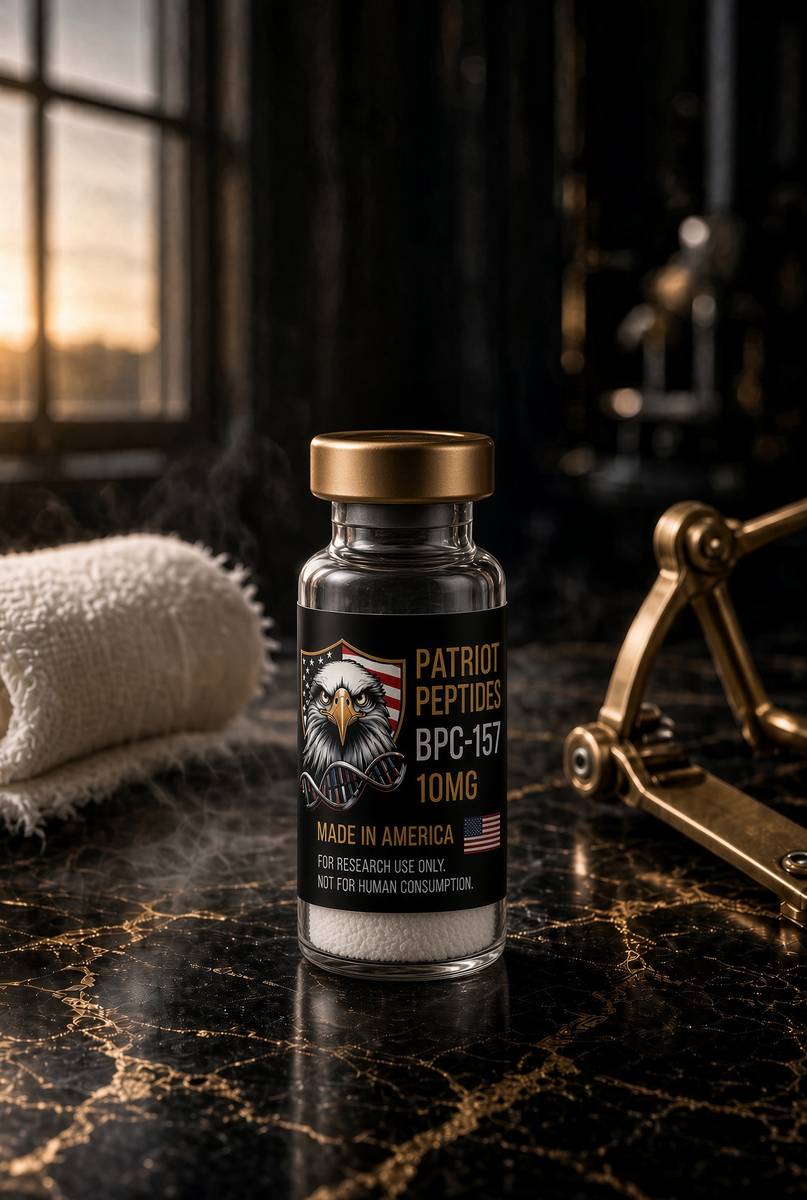
The same published structure the essay describes — HPLC-characterised.
Read next
53 min · long read · Peptide research
How research peptides are made — and why HPLC actually matters
Solid-phase peptide synthesis builds a chain one residue at a time. HPLC then asks whether the main peak is what you think it is. ≥98% is not a slogan. It is a chromatogram.
50 min · long read · Peptide research
Why peptides are the next chapter of medicine
More than a hundred peptide drugs are already approved. Insulin opened the century. GLP-1 agonists moved weight into bariatric territory. Oral tablets, biased agonists and triple ligands are the current argument.
47 min · long read · Peptide research
Why peptides are freeze-dried — and what reconstitution actually is
Water hydrolyses peptide bonds, oxidises methionine and grows microbes. Lyophilisation sublimes the ice so the chain ships as a porous cake. Reconstitution is mass divided by volume — laboratory handling, not a protocol for a body.
More in this desk

53 min · long read · Peptide research
Recovery, GH pulses and the CJC / ipamorelin pair
Somatotrophs have two 'go' receptors. CJC without DAC is a slightly longer GHRH pulse. Ipamorelin is a selective ghrelin-receptor key that does not yank ACTH. Together they are the pair Bowers already showed is more than additive — not a gym protocol.
52 min · long read · Peptide research
Fatigue, cellular energy and the NAD+ / MOTS-c neighbourhood
NAD+ is the rechargeable chip every cell spends on fuel and DNA repair — and the pool shrinks with age. MOTS-c is a mitochondrial 16-mer sent out under metabolic stress. Two answers to 'I have no energy' that are not coffee.
51 min · long read · Peptide research
Melanocortin circuits: pigment, appetite and PT-141
Melanotan II lights MC1, MC3, MC4 and MC5 — pigment plus the rest of the sheet. PT-141 is the free-acid cousin pointed at MC3 and MC4, the circuitry papers use for desire and energy, not skin colour. Same Arizona family. Different question.
51 min · long read · Peptide research
GHRH and ghrelin mimetics: two keys on the same pituitary lock
Somatotrophs have two ‘go’ receptors — GHRHR (Gs–cAMP) and GHSR (Gq). Bowers showed that occupying both releases more GH than the sum of either. That is synergy as a receptor fact, not a smoothie.
Essays describe published research. They are not medical advice and they do not authorise human use of any catalogue item.