

Peptide research · 48 min · 10,588 words
IGF-1 LR3: taking the binding proteins out of the fight
Insulin-like growth factor-1 is the tyrosine-kinase half of the GH conversation. LR3 is an analogue designed to ignore IGFBPs — so the receptor on a cultured cell actually sees it.
What this essay actually tells you
- IGF-1 LR3 is 83 residues: an arginine at position 3 and a 13-residue N-terminal extension that collapse affinity for IGF-binding proteins. That's the engineering. That's why it exists.
- Native IGF-1 spends much of its life tied up by IGFBPs. LR3 was built so the tyrosine kinase (IGF1R → PI3K–Akt–mTOR / ERK) sees the ligand. Occupancy, on purpose.
- IGF1R is related to the insulin receptor. Cross-talk with glucose handling is why this analogue is a precise, and not casual, research tool. Watch the glucose if you watch the kinase.
What this actually means
Growth hormone tells the liver (and some other tissues) to make IGF-1. IGF-1 then tells muscle, bone and many other cells to grow, via a receptor that looks a lot like the insulin receptor. In blood, most IGF-1 is locked up by binding proteins, which is a safety system. Long R3 IGF-1 is a laboratory analogue with an arginine swap and a 13-residue N-terminal extension that collapse that lock, so the peptide can actually reach the receptor in a dish. That's why it exists, and why we stock the 83-residue ligand rather than pretending native IGF-1 will survive a serum-containing medium.
Insulin-like growth factor-1 — IGF-1 — is a seventy-residue peptide held by three disulphide bonds, and it sits so plainly in the insulin superfamily that the two receptors form hybrids. Most of the growing we credit to growth hormone is this ligand doing the tyrosine-kinase work. The liver writes the circulating pool after a growth-hormone pulse occupies a class-I cytokine receptor and STAT5b transcribes IGF1, IGFBP3 and the acid-labile subunit. Tissues write local IGF-1 as well, including a mechanically gated splice in loaded muscle that isn't a vial. In plasma, IGF-binding proteins 1 through 6, together with ALS, sequester more than ninety per cent of the circulating ligand in ternary and binary complexes. That buffer is a safety system: free IGF-1 is short-lived and insulin-like; bound IGF-1 is a reservoir with a half-life of hours. Long Arg3 IGF-1 is an eighty-three-residue laboratory analogue designed to ignore that buffer. An arginine at position 3 and a thirteen-residue N-terminal extension collapse IGFBP affinity by roughly two orders of magnitude while IGF1R agonism remains. A cultured myoblast or hepatocyte actually sees the receptor occupied. That's a design decision about the buffer, not a potency contest against somatropin.
In short. IGF-1 is the growth message the liver writes after growth hormone speaks. Binding proteins lock most of it. LR3 is a lab version those proteins cannot hide.
We stock the eighty-three-residue chain because the papers name it. Catalogue IGF-1 LR3 is lyophilised Long R3, one thousand micrograms, at or above ninety-eight per cent by HPLC, molecular weight 9111.5, CAS 143045-27-6, the Arg3 analogue with the porcine-growth-hormone N-terminal extension. Neighbouring vials on the same axis are modified GRF(1–29) without DAC, ipamorelin, and recombinant 191-residue somatropin. Those four objects occupy four different receptors, so if you mix them you've already asked the wrong question. The neighbouring essay is the whole growth-hormone axis: hypothalamus, stomach, pituitary, liver, tissue. This piece is one microphone, the tyrosine kinase, written at the length the analogue actually needs. We'll walk the seventy-residue native ligand, the heterotetrameric receptor, the two signalling lamps, the six binding proteins, the ternary complex, the arginine swap, the dish that finally sees the kinase, and the insulin-receptor cross-talk that puts glucose in the protocol. It isn't a dosing plan. Papers that already ran IGF-1 analogues in myoblasts and in animals are in PubMed, with concentrations and glucose notes. Stay if you wanted the receptor explained, slowly, with the numbers attached.
In short. The vial is the eighty-three-amino-acid analogue, freeze-dried and checked. It isn't growth hormone, and it isn't the other three axis reagents.
A peptide catalogue with a journal attached has a duty to say which molecule is in the cake, and which receptor that molecule asks. Native IGF-1 is seventy residues. The analogue is eighty-three. The receptor is IGF1R, a receptor tyrosine kinase — a surface protein that tags itself with phosphate when ligand docks — not a GPCR and not a cytokine receptor. Downstream chemistry is IRS–PI3K–Akt–mTOR and Shc–ERK, the same logic insulin uses at the insulin receptor, which is why hybrids exist and why a glucometer belongs next to any protocol that occupies this kinase at high tone. Binding proteins 1 through 6 aren't decoration; they're why native ligand disappears into serum-containing medium and why Long R3 was built. Acid-labile subunit is the liver-specific third partner of the circulating ternary complex. Mechano-growth factor is an autocrine splice in a loaded fibre and isn't this vial. Mecasermin is recombinant native IGF-1, licensed for a named paediatric deficiency, and isn't this vial either. What follows is those objects, in order, with the papers you'd want on the bench before you thaw the cake.
In short. We're walking the receptor, the binding proteins, and the analogue built to skip them. It's a map of the biology, not a plan for a person.
Mature IGF-1 is a 70-residue insulin-family peptide. IGFBPs 1–6, with ALS, sequester more than ninety per cent of the circulating ligand. Long Arg3 IGF-1 is the analogue that takes those binding proteins out of the fight.— The IGF-1 / IGFBP literature, then Francis, Ballard and the GroPep analogue this vial actually is.
Seventy residues, three disulfides
Mature IGF-1 is seventy amino acids, three disulphide bonds, molecular weight a little under 7.7 kilodaltons. The bonds pair Cys6 with Cys48, Cys18 with Cys61, and Cys47 with Cys52, a cysteine pattern inherited from proinsulin and from IGF-2. The fold is two domains that insulin people still call B and A, with a C-region that IGF-1 keeps and insulin discards when C-peptide is clipped. Rinderknecht and Humbel sequenced the human peptide in 1978 and put the homology on paper; every ribbon of IGF-1 drawn next to insulin since then is still in that sentence. The N-terminus is the face IGF-binding proteins read most greedily. That's why the first three residues matter, why des(1–3)IGF-1 exists as a naturally truncated form with reduced IGFBP affinity, and why chemists later put an arginine at position 3 and then built a longer N-terminus still. The receptor-binding surface is a different face. You can wreck the buffer interaction without wrecking agonism. That separation is the whole analogue. Three disulphides hold the shape that both faces need. Reduce them and you've got a seventy-mer that is no longer IGF-1.
In short. Native IGF-1 is seventy amino acids, held by three sulphur bridges, and shaped like insulin. The front end is where binding proteins grab it.
The older name was somatomedin-C. Salmon and Daughaday, 1957, showed that growth hormone made cartilage take up sulphate only if a serum factor sat in between; they called the factor sulfation factor, then somatomedin, and the field spent two decades arguing about how many somatomedins there were. IGF-1 and IGF-2 were the ones that survived purification. Zapf, Froesch, Humbel, Rinderknecht, the Zurich and St Louis papers: a peptide that looks like insulin, circulates at nanomolar rather than picomolar, and doesn't come from a beta cell. The somatomedin hypothesis said GH acts on liver, liver writes the peptide, the peptide does the growing. That hypothesis is still the backbone of a serum IGF-1 assay. It's also incomplete, which Yakar, Le Roith and colleagues demonstrated with a liver-specific IGF-1 knockout: circulating IGF-1 can fall hard while local IGF-1 keeps a surprising amount of postnatal growth going. Endocrine IGF-1 is the serum number and the liver's job. Autocrine and paracrine IGF-1 are the tissue's job. Long R3 is a circulating-style analogue of the endocrine ligand. It isn't a somatomedin hypothesis in a vial, and it isn't the splice a loaded fibre writes.
In short. Doctors first met IGF-1 as a blood factor that carried growth hormone's message to bone. Liver makes the circulating pool. Tissues also make their own.
The human IGF1 gene sits on chromosome 12, six exons, two promoters, a set of splice isoforms that muscle physiologists will not let you forget. Class 1 transcripts use exon 1; class 2 use exon 2; both can join to exon 3 and 4, which encode the mature seventy-mer, and then to alternative E-peptides at the C-terminus. Mechano-growth factor, Goldspink's name for an IGF-1Ec splice with a different E-peptide, is transcribed after stretch and damage. The literature is real and also argued-over in the usual way of splice-variant fields. That transcript is an autocrine event in a fibre that was loaded. The mature seventy-mer can still be processed from several of those transcripts; the E-peptide is a different conversation. Hepatic IGF-1 is mostly a class 2, GH-dependent transcript driven by STAT5b at a defined promoter. Extra-hepatic IGF-1 is often class 1 and isn't optional in the knockout animals. If you add Long R3 to a myotube and write that you've replaced MGF, you've mixed those jobs. The receptor is shared. The question isn't. This analogue is a defined eighty-three-mer of the mature endocrine ligand with the buffer collapsed. It isn't a splice.
In short. The IGF-1 gene can be spliced in more than one way. Muscle writes a local splice when it is loaded. That local message is not the lab analogue.
The axis, from the receptor end
Growth hormone occupies GHR, a class-I cytokine receptor that is already a preformed dimer. Ligand-induced rotation recruits JAK2. JAK2 phosphorylates STAT5b, and STAT5b is the principal transcriptional driver of hepatic IGF1, of IGFBP3, and of the acid-labile subunit. Waters and Brooks put the geometry on paper; Waxman's rat-liver work showed that the pattern of STAT5b pulses writes a different cytochrome-P450 programme than a flattened tone. The three hepatic products assemble in plasma as a ternary complex that carries the large majority of circulating IGF-1 and stretches its half-life from minutes, free, to hours, bound. Acid-labile subunit is liver-specific in a way IGF-1 is not. A GH pulse therefore writes both the ligand and its carrier, which is elegant physiology and a nuisance if you wanted free ligand in a dish. MAPK and PI3K also leave GHR; they matter for the metabolic sentence. Acute GH is lipolytic and insulin-antagonistic. IGF-1, downstream, is insulin-like at IGF1R and at hybrid receptors. A physiological pulse separates those two windows. Flatten the pulse and you stretch the first hour across the day. That's one reading of why continuous GH looks more insulin-antagonistic than pulsatile GH for the same IGF-1 increment.
In short. Growth hormone tells liver cells to write IGF-1 and the proteins that carry it in blood. A burst of GH and a flat drip are not the same message.
Three objects share a receptor and don't share a question. Endocrine IGF-1 is the liver's product, buffered by IGFBP-3 and ALS, measured in a clinic as an integral over hours, blind to whether the night was three large GH bursts or a flattened plateau. Autocrine and paracrine IGF-1 are written in the tissue that will use them; loaded muscle is the example we keep naming because Goldspink's MGF papers sit on every training reading list and because a reconstituted eighty-three-mer isn't that splice. Long R3 is a laboratory analogue of the endocrine ligand with IGFBP affinity collapsed, so a cultured cell sees the tyrosine kinase occupied without a binding-protein sink. Hepatic transcription is STAT5b after GHR. The local splice is mechanical. The analogue is a design decision about the buffer. Treat those three as one 'IGF' and you haven't yet started. The neighbouring axis essay keeps the four microphones in order; this one keeps the three IGF objects in order. Shared receptor, three questions. Write which one you meant, because a blot of phospho-Akt can't tell them apart if you never named the ligand.
In short. Blood IGF-1, local muscle IGF-1, and this lab analogue all talk to the same receptor. They are still three different questions.
The genetic proofs keep the cascade in order, which is why they belong in an analogue essay and not only in a textbook. Laron syndrome is a GHR defect: high GH, very low IGF-1, a receptor that will not rotate and will not recruit JAK2. A GHRH analogue can't rescue Laron. A GH analogue can't rescue a missing STAT5b. Mecasermin, recombinant native IGF-1, is the licensed ligand when the cascade is broken below GHR, in a named paediatric indication. Catalogue IGF-1 LR3 isn't mecasermin, and Laron isn't a research protocol. The little mouse is a Ghrhr missense, one floor up. Human isolated GH deficiency type IB includes GHRHR mutations. Yakar, Liu, Le Roith, PNAS 1999, liver-specific IGF-1 knockout: circulating IGF-1 falls, GH rises, postnatal growth is partly preserved by local IGF-1. That paper is why 'GH makes you grow via IGF-1' is true and incomplete. STAT5b mutations in humans produce a GH-insensitivity picture with immune features, because STAT5b has jobs beyond IGF1. Each rescue is floor-specific. Occupying IGF1R with Long R3 is a precise question. It's a useless question if the kinase, or IRS-1, or mTOR, is the floor that actually failed.
In short. Mutations prove the cascade is ordered: GH receptor, then IGF-1, then the IGF-1 receptor. A ligand cannot rescue a protein it never meets.
Diagram
| Node | Catalogue | Conversation |
|---|---|---|
| GPCR | Ipamorelin, MT2, PT-141, retatrutide, CJC | Second messengers, secretion, appetite, pigment |
| RTK / IGF1R | IGF-1 LR3 | IRS–PI3K–Akt–mTOR and Shc–ERK |
| Cytokine receptor | Somatropin (HGH) | GHR–JAK2–STAT5b, hepatic IGF-1 |
| Cofactor | NAD+ | Sirtuins, PARPs, CD38, redox |
| Actin buffer | TB-500 / Tβ4 motif | G-actin sequestration, motility |
| Growth-factor-like | BPC-157 | VEGFR2 / FAK / eNOS neighbourhood |
| Copper ligand | GHK-Cu | Transcriptome shift in fibroblasts |
| MC fragment | KPV | NF-κB, PepT1, no pigment |
| Nuclear / pineal | Epithalon (AEDG) | TERT and melatonin literatures |
| mtORF peptide | MOTS-c | AMPK, folate–methionine cycle |
Each row is a different kind of molecular conversation. The catalogue peptides bind at these nodes; they are not interchangeable, and stacking them because a forum did mixes unrelated literatures.
IGF1R is a receptor tyrosine kinase
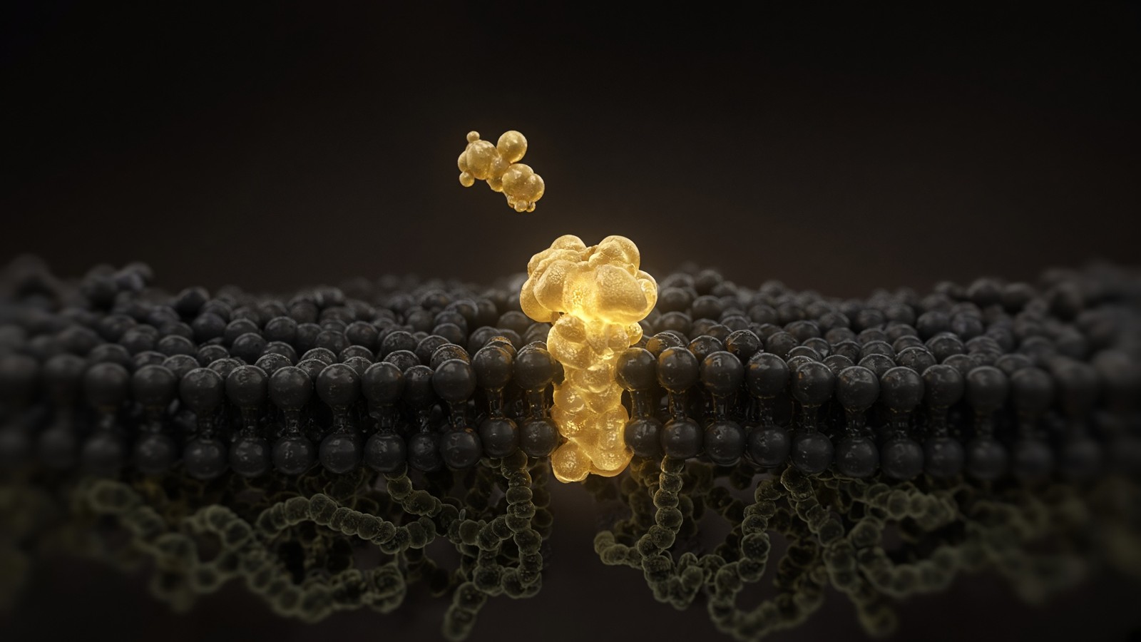
IGF1R is a heterotetramer, two alpha chains and two beta chains, a receptor tyrosine kinase in the same structural family as the insulin receptor. Each alpha-beta pair is cut from a single precursor; disulphides hold the tetramer together. Ligand binding at the alpha chains brings the beta-subunit kinase domains together. They autophosphorylate, principally on tyrosines in the activation loop — 1131, 1135, 1136 in the usual numbering — and on additional tyrosines that recruit adapters. Ullrich, Schlessinger and colleagues cloned the receptor in the mid-1980s; De Meyts, Lemmon, Kavran and the later ectodomain structures showed how one IGF-1 molecule rearranges an already assembled tetramer rather than simply gluing two monomers. That architecture is why IGF1R isn't a GPCR and isn't a cytokine receptor, and why a GHRH analogue or a pentapeptide secretagogue can't be redescribed as a short IGF. Two locks, two ligand lengths, two intracellular languages. The ligand stays outside. Information crosses the membrane as phosphorylation. Downstream chemistry does the rest. If you can't yet say alpha-two-beta-two, you haven't yet described the receptor. You've described a hope about growth.
In short. The IGF-1 receptor is a four-chain tyrosine kinase on the cell surface. The peptide stays outside. Phosphate on the inside is the message.
Phosphotyrosines on the beta subunit are docking sites. IRS-1 and IRS-2 bind through PTB domains to NPXY motifs; Shc binds a different phosphotyrosine neighbourhood. Those adapters are how one occupied receptor opens two lamps. IRS proteins, once phosphorylated, recruit the p85 regulatory subunit of PI3K; PI3K converts PIP2 to PIP3; PIP3 brings PDK1 and Akt to the membrane; Akt is phosphorylated by PDK1 and by mTORC2 and then phosphorylates a census of clients. Shc recruits Grb2-Sos, Ras loads GTP, Raf phosphorylates MEK, MEK phosphorylates ERK. That's the same logic insulin uses at IR, which is why the two receptors form hybrids and why glucose belongs in any protocol that occupies IGF1R at high tone. We'll keep coming back to the glucometer, because the chemistry will. Hybrid IR/IGF1R receptors are common in some tissues. An analogue that ignores IGF-binding proteins will, at sufficient concentration, look a little like insulin. Hypoglycaemia in animals is the organism-level version of that sentence. Occupancy is occupancy. Downstream is two lamps, not one mood.
In short. Once the receptor is on, two inside paths light up: one builds protein and handles fuel, the other tells the cell to divide. Insulin uses the same logic.
The insulin receptor comes in two splice isoforms, IR-A and IR-B, and both can heterodimerise with IGF1R. Hybrid receptors aren't a footnote in muscle, in endothelium, or in some tumours; they're a measurable fraction of the surface census. IGF-1 occupies IGF1R with high affinity and IR with lower affinity. Insulin occupies IR with high affinity and IGF1R poorly. Hybrids sit in between, and their ligand preference is a function of which IR isoform is in the pair. At nanomolar free IGF-1, you're mostly asking IGF1R and IGF1R-containing hybrids. At the concentrations a binding-protein-blind analogue can reach in a well, you can start to occupy IR as well. That isn't a scandal. It's a dose. Native IGF-1 in plasma rarely has that problem because IGFBPs keep free ligand in the sub-nanomolar range. Long R3 was built to defeat those binding proteins. Defeat them and you inherit the insulin-like edge of the dose-response. Siddle's hybrid-receptor papers, the Nakae and Accili genetics of IR versus IGF1R in mice, and the hypoglycaemia warnings on licensed IGF-1 are all the same sentence. Write the concentration. Write whether you measured glucose. Write which receptor you think you occupied.
In short. The IGF-1 receptor and the insulin receptor can pair up. At high dose the analogue can look a little like insulin, so glucose has to be watched.
IRS–PI3K–Akt–mTOR
IRS–PI3K–Akt–mTOR is the growth and survival lamp, and it's the lamp a hypertrophy assay is usually trying to read. PI3K, class I, p110 with p85, makes PIP3. Akt isoforms then phosphorylate TSC2, which relieves inhibition of Rheb, which lets mTORC1 assemble at the lysosome when amino acids are also present. mTORC1 phosphorylates 4E-BP1 and S6K1. 4E-BP1 releases eIF4E; S6K1 phosphorylates ribosomal protein S6 and other translation factors; protein synthesis rises. That's the anabolic sentence. Akt also phosphorylates FOXO transcription factors, which then leave the nucleus, so the catabolic and atrophy programme FOXO was writing goes quiet. GSK3 is another Akt client; so is AS160/TBC1D4, which is how GLUT4 vesicles get a chance to fuse. The pathway is therefore growth, survival, and glucose uptake in one neighbourhood. Rapamycin will close mTORC1 and tell you how much of your readout was that node. Wortmannin or LY294002 will close PI3K and take the whole lamp down, with the usual specificity caveats. Phospho-Akt(Ser473) and phospho-S6 are the blots you'd ask a friend to show you. A heavier myotube without those blots is a photograph, not a mechanism.
In short. One path from the receptor turns on protein building and glucose uptake through Akt and mTOR. That is the growth lamp inside the cell.
Protein synthesis is initiation factors, elongation, amino acids, and an mTORC1 that will not fire if leucine is missing even when Akt is on. A myotube given Long R3 in amino-acid-poor medium is a different experiment from a myotube given Long R3 after a feed. The dish has to say which. GLUT4 translocation is the metabolic half of the same lamp: IGF1R occupancy, in muscle cells, can move glucose transporters to the surface by the AS160 route insulin also uses. That's why an analogue that ignores IGFBPs will, at sufficient concentration, drop medium glucose and, in an animal, drop plasma glucose. FOXO exclusion is the transcriptional half: MuRF1 and atrogin-1, the atrophy ubiquitin ligases, are FOXO targets; Akt that keeps FOXO out of the nucleus is one reason IGF-1 neighbourhoods look anti-atrophy in denervation and unloading papers. Those papers used native IGF-1, transgenes, or various analogues; they didn't all use Long R3. Mechanism is the shared lamp. Ligand identity is still a line you write down. If you claim hypertrophy, show protein synthesis or a defined myofibre-size measurement, show phospho-Akt, and show that PI3K or mTOR blockade took the phenotype down.
In short. Building protein needs the growth lamp and also amino acids. The same lamp can pull sugar into the cell, which is why glucose belongs in the notes.
Classic L6 and C2C12 work is why this analogue sits on a muscle page at all. Myoblasts in culture can proliferate or they can differentiate, fuse, and write a myotube programme. IGF-1 sits on both decisions, which is the awkwardness Florini, Ewton, Coolican and colleagues spent a career making precise. PI3K-dependent signalling supports the differentiation and hypertrophy programme: MyoD and myogenin stay in the game, fusion happens, myosin heavy chain appears, myotubes get wider. At higher tone, particularly when the ERK lamp is also bright, cells stay in the cell cycle and fusion is delayed or reduced. That proliferation-over-fusion tradeoff is the whole point of using a defined analogue rather than a crude serum spike. Serum is a soup of IGF-1, IGFBPs, insulin, and mitogens you didn't name. Long R3 lets you occupy IGF1R at a known concentration without the soup. People have been surprised by the fusion result. They shouldn't be. Occupancy is occupancy. More ligand isn't a monotonic good in a differentiation assay. Titrate. Count nuclei per myotube. Count myosin-positive cells. Then argue.
In short. In cultured muscle cells, IGF-1 can either help fibres grow or keep cells dividing instead of fusing. Dose decides which, so you have to titrate.
Shc–ERK is the other lamp
Shc–ERK is the other lamp, and it's the proliferative one. Phospho-Shc recruits Grb2 and Sos. Sos is a guanine-nucleotide exchange factor for Ras. GTP-Ras recruits Raf. Raf phosphorylates MEK1/2. MEK phosphorylates ERK1/2 on the TEY motif. Phospho-ERK goes to the nucleus and to cytoplasmic clients; ELK1, FOS, cyclin D, a G1 programme. MEK inhibitors — U0126, PD98059, the later trametinib — close this lamp and are how Coolican and Florini separated mitogenic from myogenic IGF actions in the 1997 Journal of Biological Chemistry paper you can still cite. The two lamps aren't a switch you flip once. They run together. Their ratio, the duration of each, and the differentiation state of the cell decide whether you get a myotube or a pile of dividing myoblasts. IGF1R internalisation, phosphatase tone, and dual-specificity phosphatases that chew phospho-ERK all shape the duration. A single twenty-minute phospho-ERK blot is a snapshot. A time course is an argument. If your claim is proliferation, show BrdU or EdU or a cyclin, show phospho-ERK, and show that a MEK inhibitor took the DNA synthesis down. If your claim is fusion, show that the same inhibitor didn't, or did the opposite.
In short. The second path from the receptor tells the cell to divide, through Ras and ERK. Blocking that path is how you tell division from fibre growth.
The proliferation-over-fusion tradeoff isn't a curiosity of one cell line. Satellite cells in an adult fibre, C2C12 myoblasts, L6 myoblasts, primary human myoblasts, all have to choose, on a timescale of hours to days, whether to stay in cycle or to fuse. IGF1R occupancy feeds both choices because both lamps light. High free ligand, high ERK, high cyclin D, delayed p21, delayed myogenin: a proliferative neighbourhood. Moderate ligand, PI3K on, ERK not saturating, amino acids present: a fusion and hypertrophy neighbourhood. Serum concentration in the medium is a hidden IGFBP and mitogen dose. Transferrin, insulin in the ITS supplement, and FGF2 in the growth medium are further hidden ligands. If you write 'IGF-1 LR3, ten nanomolar' and don't write the rest of the medium, you haven't described the occupancy. The analogue takes IGFBPs out of the fight. It doesn't take FGF receptors, insulin receptors, or the cell-cycle machinery out of the fight. Controls are serum-free or low-serum comparisons, an IGF1R kinase inhibitor if you have one that is clean enough, and the MEK and PI3K tools already named. Then the tradeoff is a result. Before that it's a rumour about wider myotubes.
In short. Division versus fusion depends on dose, timing and what else is in the dish. The analogue removes binding proteins. It does not remove the rest of biology.
More IGF isn't a monotonic good, and that sentence is the reason to use a defined analogue rather than to keep turning the tap. In a proliferation assay, a saturating Long R3 dose may be exactly what you wanted. In a differentiation assay, the same dose may be the reason fusion failed and the reason a colleague can't reproduce your myosin blot. In a glucose-uptake assay, the same dose may occupy hybrid receptors and IR, and you'll have measured an insulin-like effect you attributed to IGF1R. Titration isn't timid. It's how receptor biology is done. A three-point or five-point curve, a vehicle, a native-IGF-1 arm if the question is the buffer, and a time point that matches the lamp you claim — phospho-Akt in minutes, myogenin in days — are the minimum. EC50 at IGF1R is nanomolar for native ligand; Long R3 is at least as potent at the receptor and far more available once IGFBPs are in the room. Available isn't the same as specific. Specificity is a function of concentration, of hybrid-receptor census, and of how long you left the analogue on. Write those.
In short. More analogue is not always more of the result you wanted. A dose curve tells you whether you are growing fibres, dividing cells, or acting like insulin.
Diagram
× 1
Ligand
One peptide in one pocket. nM–µM. Shape, not a mood.
× 10–10²
G proteins
The occupied GPCR is a GEF. Each Gα is a catalyst.
× 10³–10⁴
cAMP / IP₃ / Ca²⁺
Adenylyl cyclase and PLC do not make one molecule. They make a cloud.
× 10⁴–10⁶
PKA / PKC / CaMK
Kinases phosphorylate many substrates per messenger.
× tissue
Secretion, transcription, motility
The organism-level readout. Still not a protocol.
This is the only magic, and it is not magic. A nanomolar ligand can move a micromolar messenger because enzymes sit between them. Desensitisation (GRK, β-arrestin, endocytosis) is how the cell refuses to let ‘more ligand’ mean ‘more signal’ forever.
Binding proteins 1 through 6, and ALS
IGF-binding proteins 1 through 6 are a family, not a single sponge. They share a high-affinity IGF-binding domain and they don't share tissue distribution, regulation, or what they do after they bind. IGFBP-3 is the main circulating partner, GH-dependent via STAT5b, abundant in plasma, and the one that forms the ternary complex with ALS. IGFBP-1 is insulin-suppressed, rises in fasting and in hepatic insulin resistance, and is a way the axis knows the metabolic state; a high IGFBP-1 is a low-free-IGF neighbourhood even if total IGF-1 looks ordinary. IGFBP-2 is a developmental and a tumour-biology object, and a cerebrospinal-fluid one. IGFBP-4 is often inhibitory and is cleaved by PAPP-A in tissues, a local-release mechanism. IGFBP-5 sits in extracellular matrix and in bone neighbourhoods. IGFBP-6 prefers IGF-2. Jones and Clemmons, Firth and Baxter, Hwa, Oh and Rosenfeld: those reviews are the census. Treating 'IGFBP' as one protein is how you miss that serum in a dish is mostly IGFBP-3 from foetal bovine serum, and that a hepatocyte's own IGFBP-1 will change when you change glucose and insulin. Name the family member if you blot one. Name serum percentage if you didn't.
In short. Six binding proteins lock IGF-1 with different jobs in different tissues. The main one in blood is IGFBP-3. They are not one sponge.
The ternary complex is IGF-1, IGFBP-3 and the acid-labile subunit, and it's how most circulating IGF-1 actually travels. ALS is a leucine-rich glycoprotein, written almost exclusively by hepatocytes under STAT5b, encoded by IGFALS. Binary IGF-1–IGFBP-3 can leave the circulation; the ternary complex, much larger, is retained, and that retention is the half-life trick. Free IGF-1's half-life is minutes. Ternary IGF-1's half-life is hours, often quoted in the fifteen-to-twenty-hour range in humans. Boisclair, Baxter, and the ALS-deficiency cases in paediatrics are the papers: without ALS, IGF-1 and IGFBP-3 are low because they are cleared, and growth is impaired. More than ninety per cent of circulating IGF-1 sits in ternary and binary complexes; the free fraction is the piece that can occupy IGF1R on endothelium and on tissues the binary complex can still reach. Proteases clip IGFBPs in tissues and raise local free ligand without raising the serum total. That's physiology. In a dish it's a nuisance: foetal bovine serum brings a binding-protein sink, native IGF-1 added to that medium is rapidly bound, and the receptor on a myoblast sees a fraction of what you pipetted. That nuisance is why Long R3 exists.
In short. Most IGF-1 in blood rides as a trio with IGFBP-3 and a liver protein called ALS. That trio lasts hours. Free IGF-1 lasts minutes.
Serum-containing media are an IGFBP experiment whether you wanted one or not. Foetal bovine serum carries bovine IGF-1, bovine IGFBPs, and a protease background. Ten per cent serum isn't a blank. It's a ligand plus a buffer plus a set of mitogens, and it will bind native human IGF-1 you add on top. Charcoal-stripped or IGF-depleted sera exist because people got tired of that confound. Serum-free medium with Long R3 as the only IGF1R ligand is the cleaner design if the question is this receptor. Long R3's collapsed IGFBP affinity means that even in serum it remains largely free, which is the practical reason myoblast protocols adopted it in the 1990s and haven't stopped. Collapsed affinity isn't zero affinity, and it isn't zero cross-talk at IR. It's two orders of magnitude, roughly, which at nanomolar analogue and micromolar IGFBP still leaves most of the analogue unbound. Do the arithmetic for the concentration you actually use. If you're comparing native IGF-1 with Long R3 in the same serum, you're asking a buffer question. If you're not comparing, say so, and don't write that you studied 'IGF-1'.
In short. Serum in a dish already contains binding proteins and some IGF-1. Native ligand gets hidden. LR3 was built so the cell still sees it.
IGFBP-1 is the family member a metabolic protocol actually has to name. Insulin suppresses IGFBP-1 transcription in hepatocytes, so a fed, insulin-high animal has low IGFBP-1 and a higher free-IGF fraction; a fasted or insulin-deficient animal has high IGFBP-1 and a tighter buffer. That's one of the ways the GH–IGF axis listens to fuel. It's also why a hepatocyte dish, an in-vivo analogue experiment, and a myoblast in ten per cent serum are three different IGFBP landscapes. IGFBP-3 in plasma is slower, GH-dependent, a stock rather than a minute-to-minute valve. PAPP-A and other IGFBP proteases are the local valve in tissues: pregnancy-associated plasma protein-A cleaves IGFBP-4 and IGFBP-5, releases ligand, and has a bone and a plaque literature of its own. Adding Long R3 bypasses all of those valves at once. That's the point in culture. In an organism it's a different point, because the valves were physiology and you've just walked around them. Write which landscape you're in. A free-IGF measurement, an IGFBP-1 blot, and a glucose number will tell a colleague more than a total-IGF kit on a homogenate.
In short. IGFBP-1 rises when insulin is low, so fasting tightens the buffer. The analogue walks around that valve, which is useful in a dish and a different choice in an animal.
- Mature IGF-1
- 70 residues
- IGF-1 LR3
- 83 residues
- IGF1R
- α2β2 RTK
- IGFBPs
- 1–6
- Ternary complex
- IGF-1 · IGFBP-3 · ALS
- IGFBP affinity, LR3
- ~100-fold down
- Catalogue cake
- 1000 mcg
- Licensed neighbour
- mecasermin
Three disulfides. Insulin superfamily. Rinderknecht and Humbel, 1978.
Arg3 plus a 13-aa N-terminal extension. 9111.5 g·mol⁻¹. CAS 143045-27-6.
Autophosphorylation, IRS–PI3K–Akt–mTOR and Shc–ERK. Ullrich, 1986.
Family, not a sponge. IGFBP-3 is the main circulating partner.
Sequesters >90% of circulating IGF-1. Half-life hours, not minutes.
Two orders of magnitude. IGF1R agonism remains. Free ligand in serum.
≥98% HPLC. Lyophilised 83-mer. Reagent, not a serving.
Recombinant native 70-mer. Severe primary IGF-1 deficiency. Not this vial.
Long R3: arginine at three
Long Arg3 IGF-1 is an eighty-three-residue analogue. Francis, McDougall, Bagley, Ballard and the GroPep group made it by substituting glutamate at position 3 with arginine and adding a thirteen-residue N-terminal extension derived from porcine growth hormone, MFPAMPLSSLFVN in the usual one-letter string. King, Wells, Krieg and colleagues expressed the recombinant analogue in Escherichia coli and characterised receptor binding and IGFBP interaction in the early 1990s. IGFBP affinity collapses by roughly two orders of magnitude. IGF1R agonism remains. The N-terminal extension and the Arg3 substitution both contribute; des(1–3)IGF-1, which simply lacks the first three residues, already shows reduced IGFBP binding, and R3-IGF-1 without the long extension is an intermediate object. Long R3 is the version that culture adopted. In serum-containing medium the analogue is the ligand the myoblast actually sees; native IGF-1 is the ligand the binding proteins see. That sentence is the entire product. It isn't a claim that eighty-three is better than seventy in an organism. It's a claim that eighty-three is the tool if the dish has a buffer you don't want.
In short. LR3 adds thirteen amino acids at the front and swaps an arginine into position 3. Binding proteins then barely hold it, but the receptor still does.
Des(1–3)IGF-1 is the natural neighbour, and it belongs in this heading so Long R3 doesn't look like a unique magic. Brain, milk and some tissue extracts contain IGF-1 that has lost the N-terminal tripeptide; the truncation is proteolytic. The truncated peptide binds IGFBPs poorly and occupies IGF1R well, which is the same design Long R3 later exaggerated with an extension and an arginine. Tomas, Ballard, Francis and colleagues used des(1–3) and Long R3 in rats to ask nitrogen-retention and muscle-protein-synthesis questions that native IGF-1, buffered, answered more weakly. Those animal papers are real. They're also animal papers, with animal doses, animal glucose, and animal IGFBP profiles. They aren't a protocol, and they aren't a licence to treat Long R3 as a more convenient mecasermin. The licensed ligand is the native seventy-mer, on purpose, because the buffer is a safety system in plasma. The research analogue is the eighty-three-mer, on purpose, because the buffer is a nuisance in medium. Two purposes. Two legal classes. One receptor family. Elide that gap and you're selling a neighbourhood as an address.
In short. A shorter natural form of IGF-1 already dodges binding proteins a little. LR3 is the lab version of that idea, built for culture, not a licensed medicine.
Collapsed affinity is a number, not a metaphor. Native IGF-1 binds IGFBP-3 with nanomolar-to-subnanomolar affinity; Long R3 binds the same proteins roughly a hundred-fold more weakly, sometimes quoted near a thousand-fold depending on the binding protein and the assay. At the IGF-1 receptor the two ligands are comparable agonists. The experimental consequence is constitutive receptor occupancy in culture without a binding-protein sink: add ten nanomolar Long R3 to a well that contains serum IGFBPs, and most of the analogue is still free; add ten nanomolar native IGF-1, and most of it isn't. Two orders of magnitude is why a concentration that looks similar on a pipette isn't a similar free fraction. It's also why you can't convert a native-IGF-1 paper into a Long R3 paper by copying the nanomolar figure. Do a curve. The molecular weight of the analogue is 9111.5; a one-thousand-microgram cake is about 110 nanomoles; reconstitution in one millilitre gives a stock you can dilute into wells at nanomolar free ligand without pretending the arithmetic is mysterious. Mass spectrometry should confirm the mass. HPLC should show a main peak. A certificate that can't show those isn't yet a ligand.
In short. LR3 sticks to binding proteins about a hundred times more weakly than native IGF-1, so more of it stays free in the dish at the same pipetted dose.
The listing is lyophilised Long R3 IGF-1, one thousand micrograms, at or above ninety-eight per cent by HPLC, eighty-three residues, molecular weight 9111.5 grams per mole, CAS 143045-27-6. A white to off-white cake, a certificate of analysis, a reconstitution kit. It's the same backbone the GroPep-era papers characterised. It's a laboratory reagent. Purity is a chromatogram, not a feeling. Water, freeze–thaw and adsorption to plastic are the enemies of a nanomolar peptide in a tube; the lyophilised solid is how you store it, and a carrier protein in the dilute working stock is how some laboratories stop it vanishing onto the wall of the pipette tip. None of those sentences is a dose for an organism. None of them is a route. None of them is a schedule. If you can't say how many nanomoles went into a well, you haven't yet started. The cake exists so a defined eighty-three-mer can be weighed into the assays this essay describes. One thousand micrograms is a mass on a certificate. It isn't a serving.
In short. The vial is a freeze-dried milligram-scale cake of the eighty-three-amino-acid analogue, for weighing into experiments, not a dose for a person.
What the analogue buys you on the bench
What the analogue buys you on the bench is a question you can actually ask. What does IGF1R occupancy do to proliferation, to differentiation, to glucose uptake, or to hypertrophy markers when the binding proteins are taken off the board. Classic myoblast work in L6 and C2C12 shows PI3K-dependent hypertrophy programmes and, at higher tone, a proliferation-over-fusion tradeoff. Hepatocyte work shows a metabolic neighbourhood that overlaps insulin's, which is expected from hybrids and from IRS-2. Chondrocyte and osteoblast work sits in the somatomedin tradition and still uses IGF1R ligands to ask matrix-synthesis questions. None of those assays is a training programme. All of them are receptor assays if you measure the receptor. Phospho-IGF1R, phospho-Akt, phospho-ERK, a PI3K tool, a MEK tool, glucose in the medium, and a named differentiation readout are how you stop a growth-factor essay becoming a mood. The analogue is the ligand those assays need when serum would otherwise hide native IGF-1. It isn't a substitute for the MGF splice, for a GH pulse, or for a licensed recombinant IGF-1 in a child who cannot make one.
In short. On the bench, LR3 lets you ask what the IGF-1 receptor does when binding proteins are not hiding the ligand. Muscle cell lines are the usual test.
Native IGF-1 in serum-containing media is rapidly bound. That isn't a controversy. It's the reason the analogue was invented, and it's still the reason that adding 'IGF-1, ten nanomolar' to ten per cent foetal bovine serum hasn't described free ligand. You can strip the serum, you can use serum-free medium, you can add IGFBP antibodies, or you can use Long R3. The last of those is the practical choice in most myoblast papers of the last thirty years. Practical isn't the same as identical to native. The analogue's N-terminus is different; some IGFBP-independent IGFBP actions — the ones where the binding protein is a ligand in its own right, at LRP receptors or in the nucleus in contested papers — will not see Long R3 the way they see native IGF-1. If your question is an IGFBP as a signalling molecule, this analogue is the wrong tool, and native ligand plus a defined IGFBP is the right one. If your question is IGF1R, Long R3 is the tool that keeps the question from being an IGFBP question in disguise. Choose on purpose.
In short. Native IGF-1 vanishes into serum proteins in a dish. LR3 does not, which is why culture uses it when the question is the receptor itself.
A defined analogue isn't a crude serum spike, and the difference is the whole point of paying for a chromatogram. Serum is IGF-1, IGF-2, insulin, FGF2, PDGF, lipids, and a binding-protein cloud, at concentrations that vary by batch. A spike of that soup into a starved myoblast will move phospho-Akt, phospho-ERK, and a fusion readout, and you won't know which ligand did the work. Long R3 at a stated nanomolar concentration, in a stated medium, with a stated serum percentage or none, is a ligand you can attribute. Attribution is the job. Batch-to-batch serum variation is why differentiation protocols that 'always worked' stop working when the bottle changes; IGFBP content is one of the hidden variables. If you must use serum, use the analogue, write the lot, and write the percentage. If you can avoid serum for the occupancy window, avoid it. Then, if the phenotype survives an IGF1R kinase inhibitor and dies with a PI3K inhibitor, you were looking at this receptor and this lamp. If it dies with the kinase inhibitor, you have a receptor paper. If it survives both, you have a soup, and you should stop calling it IGF-1 LR3.
In short. Serum is a mixed soup of growth factors. A named analogue at a named concentration is how you know which receptor you actually asked.
Insulin-receptor cross-talk
Cross-talk with the insulin receptor isn't a footnote. IGF1R and IR form hybrids. An analogue that ignores IGFBPs will also, at sufficient concentration, look a little like insulin. Hypoglycaemia in animals is the organism-level version of that sentence. Licensed recombinant IGF-1 carries a labelled hypoglycaemia risk even with the binding-protein buffer still in play; Long R3, by design, has less of that buffer. Moving the analogue from a C2C12 well into a mouse without a glucose plan has skipped the hybrid-receptor paragraph. Design the experiment with glucose in the protocol, or don't be surprised. Medium glucose in a dish is the same warning at smaller scale: a high-tone occupancy will drop the sugar, the cells will see a metabolic shift you didn't attribute, and a 'hypertrophy' readout may partly be a fuel-handling readout. Measure glucose. Measure phospho-Akt in parallel with a GLUT4 or 2-deoxyglucose assay if the claim is metabolic. Measure it even if the claim is myogenic, because the lamp doesn't know which paper you're writing. Skip this and the chemistry will still happen; you just won't have named it.
In short. At high concentration the analogue can act a little like insulin and lower glucose. Put glucose in the protocol, in a dish or in an animal.
Glucose belongs in the protocol as a named measurement, not as a feeling about feeding. In culture, record the starting glucose of the medium, the time since feeding, and a medium glucose at harvest if the occupancy was long enough to matter. In animals, a glucometer at defined time points after analogue administration is the minimum; a proper insulin-tolerance or glucose-tolerance test is what a metabolic claim requires. Hybrid receptors and IR occupancy are concentration-dependent, so a dose that didn't drop glucose in one strain at one time point may drop it in another. Fasted animals are more vulnerable; fed animals have insulin and IGFBP-1 in a different place. Write the feeding state. Hypoglycaemia isn't a badge of potency. It's a confound, a welfare issue in animals, and a reason licensed IGF-1 is a specialist medicine rather than a general anabolic. The analogue is a precise probe at IGF1R. Precision isn't the same as safety at organism scale. Native IGF-1 in a ternary complex with IGFBP-3 and ALS is the physiological circulating form. LR3 is the form that ignores the complex. Choose on purpose, and write the choice, including how glucose was watched.
In short. Write down the sugar: starting medium, feeding state, and a measurement after the analogue. A dropped glucose is a result, not a proof of strength.
Long R3 isn't insulin, and it isn't a diet. Insulin is a fifty-one-residue beta-cell peptide occupying IR, with IGF1R cross-talk at the high end of its own dose-response. Long R3 is an eighty-three-residue IGF-1 analogue occupying IGF1R, with IR cross-talk at the high end of its dose-response. The overlap is real and isn't identity. A glucose-uptake assay in myotubes can be run with either ligand; the blot that distinguishes them is phospho-IGF1R versus phospho-IR, with the usual antibody-specificity caveats, and a receptor knockdown or a kinase inhibitor if you have one. An experiment that uses Long R3 and then talks about insulin sensitisation without those blots has described a metabolic phenotype and attributed it to the wrong receptor. An experiment that uses Long R3 and then talks about body composition in a healthy animal has left the dish, left the buffer, and entered a literature that licensed IGF-1 and GH already occupy with labelled risks. We'll sell the named eighty-three-mer for the dish and the receptor assays. We won't redesign the experiment as a meal plan. Fuel handling is a readout of this kinase. It isn't a product claim.
In short. LR3 is not insulin and it is not food. Both can move glucose at the high end. Name the receptor you occupied and blot it if you can.
Diagram
Closed chromatin (H3K27me3, DNA methylation) hides the promoter. Pioneer factors and histone acetyltransferases open it.
PIC: TFIID, TFIIH, Mediator, Pol II. Ser5 phosphorylation of the CTD lets the polymerase leave the promoter.
Elongation ~20–40 nt/s. Capping, splicing, cleavage and polyadenylation happen on the still-growing RNA.
Human genes are islands in 3.1 billion base pairs of mostly noncoding sequence. Promoter, enhancers, chromatin state and the Mediator complex decide whether Pol II is allowed to fire. Epithalon’s literature sits on TERT and pineal clocks — two of the rare promoters anyone bothers to name in a peptide essay.
Four microphones, this one the kinase
CJC without DAC asks what a slightly prolonged GHRH pulse does to somatotrophs when troughs are still allowed. Ipamorelin asks what selective GHS-R1a agonism does to the same granule without dragging ACTH, cortisol and prolactin into the table. Recombinant somatropin asks the GH receptor itself, skipping hypothalamus and pituitary, and the answer will depend on whether you dose it as a pulse or as a drip. IGF-1 LR3 bypasses the pituitary and the liver and asks the tyrosine kinase on a myotube or a hepatocyte, with IGFBPs taken out of the fight; glucose belongs in that protocol. Four ligands, four receptors, four questions. Confuse them and you're assembling a stack. A stack is a purchasing habit. It isn't an experiment. The papers that combined GHRH and a secretagogue in pituitary slices named both concentrations and both receptors. That's the standard. A paper that combines somatropin and Long R3 and then reports a waist hasn't isolated a floor. Walk GH to IGF-1 to a tissue readout, in that order, or occupy IGF1R directly and say that you skipped the walk. Skipping is allowed. Pretending you didn't skip isn't.
In short. Four lab analogues sit on four steps of the growth-hormone axis. LR3 is the last step, the receptor on the tissue, not a stand-in for the others.
The analogue can't substitute for a nocturnal GH pulse, and it can't substitute for the MGF splice a loaded fibre writes. A GH pulse writes hepatic IGF-1, ALS and IGFBP-3, and it writes a direct lipolytic programme that IGF-1 does not copy. Long R3 skips all of that and occupies IGF1R with the buffer collapsed; you won't see a STAT5b phosphorylation in the liver from this ligand, and you shouldn't claim a GH-axis reconstitution. Mechano-growth factor is an autocrine transcript after load; adding an eighty-three-mer to an unloaded myotube doesn't reproduce stretch. Goldspink's papers belong on a muscle-physiology reading list. They don't belong on a certificate of analysis for LR3. Somatropin, dosed daily, already flattens pulsatility relative to Veldhuis bursts; using Long R3 on top of that flattened signal is two flattening decisions, not one clever stack. If the question needs a pulse, the pulse-length GHRH analogue is the microphone. If the question needs GHR, somatropin is the microphone. If the question needs IGF1R without IGFBPs, this vial is the microphone. One question per vial unless you're prepared to unpick two concentrations.
In short. LR3 does not recreate a night-time growth-hormone burst, and it does not recreate the IGF-1 splice a loaded muscle writes. It asks the receptor directly.
Attribution is the job, and sequence identity is the only honest link between a catalogue vial and a paper. Long R3 is a defined eighty-three-mer. Modified GRF(1–29) without DAC is a defined tetrasubstituted twenty-nine-mer. Ipamorelin is a defined pentapeptide. Somatropin is a defined 191-mer. HPLC and mass spectrometry are how we know which of those four chains is in the cake. A certificate that can't show a main peak and a mass isn't yet a microphone; it's a rumour about a white powder. The kit in the box is bacteriostatic water and syringes so reconstitution is a documented step rather than an argument. None of that is a protocol for combining them. It's the minimum a research reagent owes a write-up that will have to put the catalogue number next to a concentration. Same sequences as the papers name, characterised, sitting on four shelves because they are four ligands. Reconstitute two of them into one syringe and call it the axis, and you've stopped being able to attribute the peak. The axis essay exists so that sentence is harder to dodge. This essay exists so the kinase isn't dodged either.
In short. Each vial is a named chain, checked by HPLC and mass. That is how you know which receptor you actually occupied.
Mecasermin is the licensed neighbour
Mecasermin is recombinant native IGF-1, Increlex in the clinic, licensed for severe primary IGF-1 deficiency — a paediatric endocrine object with hypoglycaemia as a labelled risk and with a specialist's hand on the prescription. The ligand is the seventy-mer, on purpose. The buffer in plasma is still there, and the dose still has to be timed around meals because the insulin-like edge is real. That's the clinical existence proof that IGF1R is drug-able, in a named indication, under a marketing authorisation. It isn't Long R3. Long R3 was built for culture, where binding proteins in serum make native IGF-1 a poor tool. Taking the analogue into an organism reintroduces the insulin-like risk without the binding-protein buffer that made native IGF-1 safer in plasma. That's why the dish and the animal are different experiments even at the same receptor. Catalogue Long R3 is HPLC-characterised research material, labelled for in-vitro work. Indication, authorisation, device and pharmacovigilance sit on the mecasermin side of the sentence. Sequence, certificate of analysis and a reconstitution kit sit on this side. Same receptor family. Different object, different job, different fridge.
In short. Licensed IGF-1 is the native seventy-amino-acid hormone for a rare childhood deficiency. LR3 is the research analogue for dishes. Same receptor family, different object.
Mecasermin rinfabate, IGF-1 bound to IGFBP-3, was a brief clinical neighbour under the iPLEX name, an attempt to give the ligand with its buffer rather than against it. The development history is a reminder that plasma IGF-1 without IGFBP-3 is a different pharmacokinetic object from the ternary complex the liver normally ships. Long R3 is the opposite design: ligand against the buffer, not with it. In a dish that opposite is a feature. In plasma it would be a different feature, closer to a free-IGF spike, and the glucose warning would get louder rather than quieter. We'll keep naming the neighbour so that a research vial isn't mistaken for a paediatric medicine, and so that an analogue built to ignore IGFBPs isn't mistaken for a complex built to include them. A catalogue that stocks an eighty-three-mer next to a receptor that already has a licensed seventy-mer has a duty to keep those objects labelled as two objects. We will. The physiology above doesn't depend on the label. The label does depend on not mixing Increlex with a culture analogue.
In short. One clinical product tried giving IGF-1 together with its binding protein. LR3 does the opposite, on purpose, so a dish can see free ligand.
The receptor is taken inside
Occupied IGF1R doesn't sit on the surface forever. Ligand binding, phosphorylation, and recruitment of ubiquitin ligases — Cbl family members, Nedd4, others depending on the paper — tag the receptor. Endocytosis follows, clathrin-dependent in the textbook version, with a fraction recycling and a fraction heading for the lysosome. Down-regulation is why more ligand isn't more signal forever, the same arithmetic Lefkowitz spent a career on at GPCRs, applied here to a tyrosine kinase. A myoblast that sees continuous Long R3 will, over hours, present fewer surface receptors and a smaller phospho-Akt increment per molecule of analogue. That's ordinary receptor biology. It's also why a twenty-four-hour occupancy and a twenty-minute occupancy are different experiments, even at the same nanomolar figure. Surface biotinylation, an IGF1R ELISA on a lysate, and a phospho-IGF1R blot at two time points will show the difference. A single late blot of phospho-Akt may show a pathway that is already adapting. Write the time. Write whether the analogue was left on or washed off. Chronic occupancy is a downregulation experiment. Acute occupancy is a signalling experiment. They share a ligand. They don't share a question.
In short. After the receptor is switched on it is pulled inside and some of it is destroyed. A long treatment and a short treatment are different experiments.
Ubiquitin is the tag that sentences a receptor, and it's why this analogue essay owns a proteostasis diagram rather than borrowing a GPCR cartoon. Hershko, Ciechanover and Rose, Nobel 2004: E1 activates, E2 conjugates, E3 selects the victim. K48-linked chains send proteins to the 26S proteasome; K63-linked chains are more often a trafficking signal. IGF1R can receive both flavours depending on the ligase and the paper. Internalised receptor that is ubiquitylated and not recycled is lysosomal more than proteasomal, which is ordinary for a transmembrane kinase; IRS-1, by contrast, is a proteasomal substrate when chronic insulin or IGF tone keeps it phosphorylated, a negative-feedback that mTORC1 and SOCS proteins both feed. Chronic high ligand therefore spends the adapter as well as the receptor. That's one reading of IGF1R 'desensitisation' that isn't about the kinase domain at all. MG132 will pile up ubiquitylated IRS-1 if the proteasome was the sink; a lysosomal inhibitor will pile up receptor if that was the sink. Use the tools as tools. A growth-factor paper that never mentions ubiquitin is allowed. A chronic-occupancy paper that never mentions it is missing the off-switch.
In short. A small protein tag called ubiquitin marks the receptor and its helpers for removal. That off-switch is why a constant high dose fades.
Diagram
A growing mammalian cell turns over a few percent of its proteome per hour. 10 billion proteins is a warehouse with a shredder on the floor, not a museum. Ageing is partly what happens when the shredder slows and the warehouse fills with unfolded inventory.
Hershko, Ciechanover, Rose — Nobel 2004. E1 activates, E2 conjugates, E3 selects the victim. K48-linked chains go to the 26S proteasome; K63 is more often a signal. Autophagy handles the organelles the proteasome cannot swallow. mTOR vs AMPK is the appetite switch.
How to design an honest assay
Decide what you're measuring before you thaw the cake. Phospho-IGF1R is occupancy at this kinase. Phospho-Akt(Ser473) and phospho-S6 are the PI3K–mTOR lamp. Phospho-ERK(Thr202/Tyr204) is the Shc lamp. A myogenin blot, a myosin heavy-chain blot, and nuclei per myotube are differentiation. EdU or BrdU is proliferation. Medium or plasma glucose is the insulin-like edge. None of those is a substitute for another. A heavier protein pellet without phospho-Akt isn't a PI3K paper. A dropped glucose without phospho-IGF1R isn't an IGF1R paper; it might be IR. An IGF1R kinase inhibitor, a PI3K inhibitor, and a MEK inhibitor are how you name the lamp. Antibodies against the analogue's N-terminus will not see native IGF-1; antibodies against mature IGF-1 may not see Long R3 equally. Mass spectrometry on the reconstituted cake is how you know which chain you diluted. A plate-reader viability dye is a scout, not a hypertrophy assay. Pick the measurement that matches the sentence you want to write, and write the time point, because phospho-Akt is minutes and myosin is days.
In short. Say which readout you mean: the receptor's own phosphate, the growth lamp, the division lamp, fusion, or glucose. They are not interchangeable.
Medium composition is a control, not a convenience. Serum percentage is an IGFBP dose. Insulin in an ITS supplement is an IR occupancy. Glucose concentration is both a fuel and, as it falls, a confound. Amino acids gate mTORC1 independently of Akt; starve them and Long R3 can't give you the hypertrophy blot you expected. A wash-off versus a continuous presence is the downregulation variable of the previous heading. Carrier protein — BSA at a low percentage — stops nanomolar peptide disappearing onto plastic; without it your 'ten nanomolar' may be a wish. Acid-washed tubes, low-binding tips, and a fresh dilution beat a freeze-thawed working stock that has already adsorbed and oxidised. Disulphides on a seventy-mer or an eighty-three-mer aren't immortal in a warm, aerated well. If this sounds fussy, that's because a defined analogue is only as defined as the minute you put it on the cells. Native IGF-1 as a parallel arm, where the question is the buffer, is the cleanest specificity check Long R3 can have: same receptor, different free fraction. If the native arm is silent in serum and the Long R3 arm isn't, you've just shown why the analogue exists.
In short. Write the medium: serum, insulin, sugar, amino acids, and whether the analogue stayed on. Plastic and freeze–thaw can steal a nanomolar peptide.
Cell-type choice is a control, not a convenience. C2C12 and L6 are myoblast lines with a published IGF1R literature; they aren't adult human satellite cells, and they aren't a fibre in a trained muscle. Primary myoblasts vary with donor age and passage. Hepatocytes have a different IRS-2 weighting and write IGFBP-1 themselves. Chondrocytes sit in the somatomedin tradition. Cancer cell lines often over-express IGF1R or IR-A hybrids and will give you a proliferative answer whether or not that was the question. HEK293 will tell you whether a construct phosphorylates; it won't tell you about fusion. A dish at ten nanomolar isn't a person: diffusion, proteases, plasma binding and first-pass sit between the two, and Long R3's collapsed IGFBP affinity is a different feature in plasma than in medium. If the claim is hypertrophy, the cell has to be able to fuse or to be a myotube already. If the claim is glucose uptake, the cell has to have GLUT4 or an equivalent you measure. If the claim is 'growth', say of what, in which cell, at which lamp. Design the assay as if someone who wasn't in the room has to believe it.
In short. Pick the cell that has the job you are claiming. A kidney cell line is not a muscle fibre, and a dish is not a person.
- Name the ligand: Long R3, 83 residues, not native IGF-1, not mecasermin, not MGF.
- Name the receptor: IGF1R, a heterotetrameric RTK. Hybrids with IR at high tone.
- Name the lamp: phospho-Akt / mTOR for growth, phospho-ERK for proliferation, glucose for the insulin-like edge.
- Name the buffer: serum percentage, IGFBPs if you blot them, why the analogue rather than native ligand.
- Write the medium, the time point, and the glucose. Phospho-Akt is minutes. Myosin is days.
- Do not call CJC, ipamorelin or somatropin a substitute. Four microphones. Four questions.
Close: a defined ligand, a named receptor
The topology is short enough to hold in one paragraph, and it's the only thing you need to leave with. Mature IGF-1 is seventy residues, three disulphides, an insulin-family peptide. IGF1R is a heterotetrameric receptor tyrosine kinase. Occupancy autophosphorylates the beta subunit, recruits IRS-1/2 and Shc, and opens PI3K–Akt–mTOR and Ras–Raf–MEK–ERK. IGFBPs 1 through 6, with ALS, sequester more than ninety per cent of circulating IGF-1 in ternary complexes, controlling half-life and tissue availability. Long R3 substitutes Glu3 for arginine and adds a thirteen-residue N-terminal extension, collapsing IGFBP affinity by roughly two orders of magnitude while retaining IGF1R agonism. The experimental consequence is receptor occupancy in culture without a binding-protein sink. Hybrid insulin/IGF signalling is why glucose belongs in the protocol. CJC without DAC, ipamorelin and somatropin are different microphones on the same axis; this analogue is the kinase, not the pulse and not the cytokine receptor. Mecasermin is the licensed seventy-mer. Local MGF is a splice. Three objects, one receptor family, one catalogue eighty-three-mer. That's the map. The figure of trained muscle is a late readout. The receptor was early.
In short. Leave with the map: seventy-amino-acid hormone, six binding proteins, an eighty-three-amino-acid analogue that skips them, and a receptor that can also look like insulin.
The public papers are the reading list, and they're short enough to actually read. Rinderknecht and Humbel, Journal of Biological Chemistry 1978, the sequence and the insulin homology. Ullrich, EMBO Journal 1986, the receptor clone. Jones and Clemmons, and Firth and Baxter, the IGFBP reviews you'd still walk. Boisclair on ALS. Yakar, Le Roith, PNAS 1999, liver-specific IGF-1 knockout, why endocrine IGF-1 isn't the whole of growth. Coolican, Florini, Journal of Biological Chemistry 1997, distinct mitogenic and myogenic lamps, MEK versus PI3K. King, Francis, Ballard, the recombinant Long R3 characterisation and the rat nitrogen-retention neighbourhood. De Meyts and the later ectodomain structures, so the tetramer is a picture rather than a cartoon. Siddle on hybrids. Nakae and Accili on IR versus IGF1R genetics. The Increlex label, so the licensed neighbour stays named. Veldhuis, so pulsatility isn't forgotten one floor up. Goldspink, so MGF isn't written on this certificate. That's a fortnight of evenings, not a guru. Come back to the axis after those, and the four receptors will look like four receptors.
In short. A short stack of named papers covers the sequence, the receptor, the binding proteins, the two inside lamps, the analogue and the licensed neighbour. Read those first.
Leave with a topology, not a shopping list. IGF-1 is the tyrosine-kinase half of the GH conversation. Binding proteins 1 through 6, with ALS, are the safety buffer that makes native ligand a poor tool in serum. Long R3 is the eighty-three-residue analogue that takes those binding proteins out of the fight so a cultured cell actually sees the receptor. Downstream is IRS–PI3K–Akt–mTOR and Shc–ERK, two lamps, a proliferation-over-fusion tradeoff, and an insulin-like edge that puts glucose in the notes. The one-thousand-microgram cake is lyophilised Long R3 for the assays that topology demands. Somatropin, modified GRF(1–29) and ipamorelin are different objects on the same campus. Mecasermin is a different legal object. If your experiment needs the analogue, weigh it, name the free fraction, and name the lamp. If it needs a pulse, the pulse-length GHRH analogue is the microphone. If it needs a medicine, this catalogue doesn't sell one. The receptor will still be on the myotube tonight, whether or not anyone opened a vial, because the axis wasn't waiting for a certificate of analysis. The analogue is how you ask it without the buffer answering first.
In short. Leave with the receptor, the buffer, and the analogue that skips the buffer. The cake in the vial is for that question, not for a stack.
Research-use-only. Not for human consumption / not a medicine. The lyophilised Long R3 IGF-1 on this listing is a laboratory reagent, HPLC-characterised at or above ninety-eight per cent, labelled for in-vitro work: a myoblast well, a phospho-Akt time course, a glucose-uptake assay, a defined comparison against native IGF-1 in serum. The physiology in the paragraphs above is public, cited, and older than the vial. Use it to design the experiment you have the controls for, with the receptor named, the lamp named, the medium written down, and the glucose watched. Read Rinderknecht, read Coolican, read Francis, then weigh the cake. We'll sell you the analogue. We won't tell you it's a substitute for a GH pulse, for a loaded fibre's own splice, or for a licensed seventy-mer in a child who cannot make one. IGF1R is a receptor tyrosine kinase. Occupancy is a concentration. The buffer was the problem the analogue was built to walk around. Walk around it in a dish, with a chromatogram on the bench beside it, and keep the claim the size of that chromatogram.
In short. The vial is a research chemical for experiments, not a medicine and not food. The biology is public. Weigh it, name the receptor, and keep the claim small.
LR3 lets you ask what IGF1R occupancy does when the binding proteins are taken off the board. Occupancy is occupancy. Glucose belongs in the protocol.
- Mature IGF-1: 70 residues, three disulfides, insulin superfamily.
- IGF1R: α2β2 RTK. IRS–PI3K–Akt–mTOR and Shc–ERK. Hybrids with IR.
- IGFBP1–6 plus ALS: ternary complex, >90% of circulating IGF-1, half-life hours.
- Long R3: 83 residues, Arg3, 13-aa N-extension from porcine GH. IGFBP affinity collapsed.
- C2C12 / L6: PI3K-dependent hypertrophy, proliferation-over-fusion tradeoff at higher tone.
- Glucose belongs in the protocol. Hypoglycaemia is the organism-level version of hybrid occupancy.
- Mecasermin is the licensed 70-mer. MGF is a splice. Somatropin is a different receptor.
- Catalogue material: 1000 mcg, ≥98% HPLC, the 83-mer the dish actually sees.
Questions the essay actually answers
- How is IGF-1 LR3 different from IGF-1?
- LR3 has an arginine at position 3 and a 13-amino-acid N-terminal extension. Those changes sharply reduce binding to IGF-binding proteins, so more of the peptide is free to occupy IGF1R in a dish. That is the entire point of the analogue.
- Is this the same as growth hormone?
- No. GH is a 191-amino-acid cytokine-receptor ligand. IGF-1 is the downstream RTK ligand. LR3 bypasses the pituitary and the liver and asks the receptor itself. Four different receptors on one axis, four different questions.
- Are CJC, ipamorelin, HGH and IGF-1 LR3 interchangeable?
- No. CJC asks GHRHR, a class-B Gs GPCR. Ipamorelin asks GHS-R1a, a class-A Gq/Gi GPCR. Somatropin is the 191-residue ligand at the GH receptor. IGF-1 LR3 asks the IGF-1 tyrosine kinase with IGF-binding proteins taken out of the fight.
- What is an IGF-binding protein?
- IGFBPs 1–6 are high-affinity partners for IGF-1 and IGF-2. With the acid-labile subunit, IGFBP-3 sequesters more than ninety per cent of circulating IGF-1 in a ternary complex, stretching half-life from minutes to hours. In a dish, serum IGFBPs hide native ligand. Long R3 was built to ignore that hide.
- Why does glucose belong in the protocol?
- IGF1R and the insulin receptor form hybrids. An analogue that ignores IGFBPs will, at sufficient concentration, look a little like insulin. Hypoglycaemia in animals is the organism-level version. Measure glucose. Medium glucose in a dish is the same warning at smaller scale.
- What does IGF1R actually do inside the cell?
- Ligand binding drives autophosphorylation of the β-subunit kinase domain, recruitment of IRS-1/2 and Shc, then PI3K–Akt–mTOR and Ras–Raf–MEK–ERK. Two lamps: growth and survival on one side, proliferation on the other. Coolican and Florini separated them in myoblasts in 1997.
- Is LR3 the same as mecasermin?
- No. Mecasermin is recombinant native IGF-1, licensed for severe primary IGF-1 deficiency, a paediatric medicine with labelled hypoglycaemia risk. Catalogue Long R3 is the 83-residue research analogue built for culture. Same receptor family. Different backbone, different legal class, different fridge.
- Why not just add native IGF-1 to the dish?
- Native IGF-1 in serum-containing media is rapidly bound by IGFBPs, so the receptor sees a fraction of what you pipetted. Long R3 collapses that affinity by roughly two orders of magnitude. If your question is an IGFBP as a signalling molecule, native ligand is the right tool. If your question is IGF1R, Long R3 keeps the question from being a buffer question in disguise.
- What is the proliferation-over-fusion tradeoff?
- In L6 and C2C12 myoblasts, PI3K-dependent IGF signalling supports differentiation and hypertrophy, while high ERK tone keeps cells cycling and delays fusion. That tradeoff is why a defined analogue beats a crude serum spike, and why more ligand is not always more of the result you wanted in a differentiation assay.
- Is this a medicine?
- No. Mecasermin is a medicine in a named paediatric indication. Recombinant somatropin is a medicine in diagnosed GH deficiency. The listing is HPLC-characterised Long R3 for in-vitro work, labelled as such: same backbone the culture papers name, a reconstitution kit, and a certificate of analysis rather than a marketing authorisation.
Hypothetical research reconstitution
How these vials are typically mixed
Hypothetical research reconstitution for the named catalogue vial. Not a protocol, not medical advice, not a use instruction. These amounts sit in published and commonly cited laboratory ranges. The vial is labelled for research use only — not for human or veterinary administration.
IGF-1 LR3
1000mcg
Mix with 1 ml bacteriostatic water → 1,000 mcg/ml
- Hypothetical aliquot
- 20–50 mcg
- 0.02–0.05 ml · 2–5 units on a U-100 syringe
- How often
- Once daily
- 4–6 weeks, then a pause
Bench steps
- Let the vial sit until it is no longer cold to the touch.
- Wipe the stopper with 70% isopropyl alcohol. Let it dry.
- Draw 1 ml bacteriostatic water (0.9% benzyl alcohol).
- Run the water slowly down the inside glass — do not blast the cake.
- Roll between finger and thumb until the cake is gone. Do not shake.
- Label the date. Store the solution at 2–8 °C. Do not freeze. Use within 30 days unless the note below says otherwise.
A thousand micrograms, not milligrams. 50 mcg is 5 units. Over-mixing the cake with a large water volume makes the marks unreadable — 1 ml is the point.
HGH
24 IU
Mix with 2 ml bacteriostatic water → 12 IU/ml
- Hypothetical aliquot
- 1–2 IU
- 0.08–0.17 ml · 8–17 units on a U-100 syringe
- How often
- Once daily, usually an evening aliquot in the somatropin notes
- 8–12 weeks, then a pause
Bench steps
- Let the vial sit until it is no longer cold to the touch.
- Wipe the stopper with 70% isopropyl alcohol. Let it dry.
- Draw 2 ml bacteriostatic water (0.9% benzyl alcohol).
- Run the water slowly down the inside glass — do not blast the cake.
- Roll between finger and thumb until the cake is gone. Do not shake.
- Label the date. Store the solution at 2–8 °C. Do not freeze. Use within 30 days unless the note below says otherwise.
24 IU in 2 ml. Two IU is about 17 units on the syringe. Gentle roll only — somatropin denatures if you beat it.
CJC-1295 (no DAC)
10mg
Mix with 2 ml bacteriostatic water → 5 mg/ml · 5,000 mcg/ml
- Hypothetical aliquot
- 100–300 mcg
- 0.02–0.06 ml · 2–6 units on a U-100 syringe
- How often
- Once daily, often with ipamorelin in the same window
- 8–12 weeks
Bench steps
- Let the vial sit until it is no longer cold to the touch.
- Wipe the stopper with 70% isopropyl alcohol. Let it dry.
- Draw 2 ml bacteriostatic water (0.9% benzyl alcohol).
- Run the water slowly down the inside glass — do not blast the cake.
- Roll between finger and thumb until the cake is gone. Do not shake.
- Label the date. Store the solution at 2–8 °C. Do not freeze. Use within 30 days unless the note below says otherwise.
No DAC — the pulse, not the drip. This is not CJC with DAC. Fridge. Often paired with the ipamorelin listing or the 10/10 blend.
Ipamorelin
10mg
Mix with 2 ml bacteriostatic water → 5 mg/ml · 5,000 mcg/ml
- Hypothetical aliquot
- 200–300 mcg
- 0.04–0.06 ml · 4–6 units on a U-100 syringe
- How often
- Once or twice daily (morning and/or evening)
- 8–12 weeks
Bench steps
- Let the vial sit until it is no longer cold to the touch.
- Wipe the stopper with 70% isopropyl alcohol. Let it dry.
- Draw 2 ml bacteriostatic water (0.9% benzyl alcohol).
- Run the water slowly down the inside glass — do not blast the cake.
- Roll between finger and thumb until the cake is gone. Do not shake.
- Label the date. Store the solution at 2–8 °C. Do not freeze. Use within 30 days unless the note below says otherwise.
GHS-R1a hexapeptide. The 200 mcg mark is the usual starting aliquot. Stacks with CJC-1295 no DAC in the papers that run both.
Bacteriostatic water and sterile syringes ship with peptide orders over £75. Kit details · 10 ml bacteriostatic water
The vials this essay sits on
Named sequences the essay maps — IGF-1 LR3, HGH, CJC without DAC, Ipamorelin. Hypothetical research neighbourhood, not a protocol, not a medicine. One press puts every in-stock vial in the bag.
 Research only
 Research only
 Research only
Growth axis
CJC without DAC
10 mg CJC without DAC — a GHRH pulse, not a weekly drip.
4.6(620)
63 browsing this now · 4 purchased in the last 24 hours
10mg · In stock
£30.00
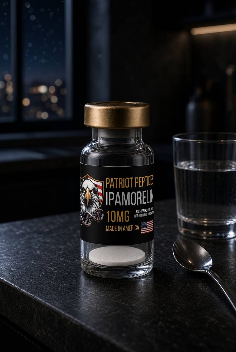 Research only
Growth axis
Ipamorelin
10 mg ipamorelin. The clean ghrelin-receptor pentapeptide.
4.7(457)
90 browsing this now · 4 purchased in the last 24 hours
10mg · In stock
£30.00
Research use only. Not a combined-use instruction.
Read next
44 min · long read · Peptide research
The growth-hormone axis, properly explained
GHRH, ghrelin, somatotropin and IGF-1 are one conversation. CJC without DAC, ipamorelin, HGH and IGF-1 LR3 are four different microphones.
48 min · long read · Peptide research
Somatropin: the 191-residue ligand
Recombinant human growth hormone is one of the most studied proteins in endocrinology. Pulsatility, GHR–JAK2–STAT5b, lipolysis, IGF-1 generation — the map is public.
More in this desk

53 min · long read · Peptide research
Recovery, GH pulses and the CJC / ipamorelin pair
Somatotrophs have two 'go' receptors. CJC without DAC is a slightly longer GHRH pulse. Ipamorelin is a selective ghrelin-receptor key that does not yank ACTH. Together they are the pair Bowers already showed is more than additive — not a gym protocol.
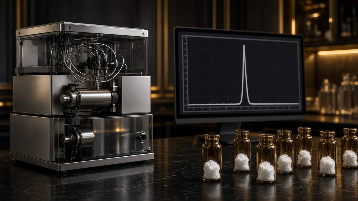
53 min · long read · Peptide research
How research peptides are made — and why HPLC actually matters
Solid-phase peptide synthesis builds a chain one residue at a time. HPLC then asks whether the main peak is what you think it is. ≥98% is not a slogan. It is a chromatogram.
52 min · long read · Peptide research
Fatigue, cellular energy and the NAD+ / MOTS-c neighbourhood
NAD+ is the rechargeable chip every cell spends on fuel and DNA repair — and the pool shrinks with age. MOTS-c is a mitochondrial 16-mer sent out under metabolic stress. Two answers to 'I have no energy' that are not coffee.
51 min · long read · Peptide research
Melanocortin circuits: pigment, appetite and PT-141
Melanotan II lights MC1, MC3, MC4 and MC5 — pigment plus the rest of the sheet. PT-141 is the free-acid cousin pointed at MC3 and MC4, the circuitry papers use for desire and energy, not skin colour. Same Arizona family. Different question.
Essays describe published research. They are not medical advice and they do not authorise human use of any catalogue item.