

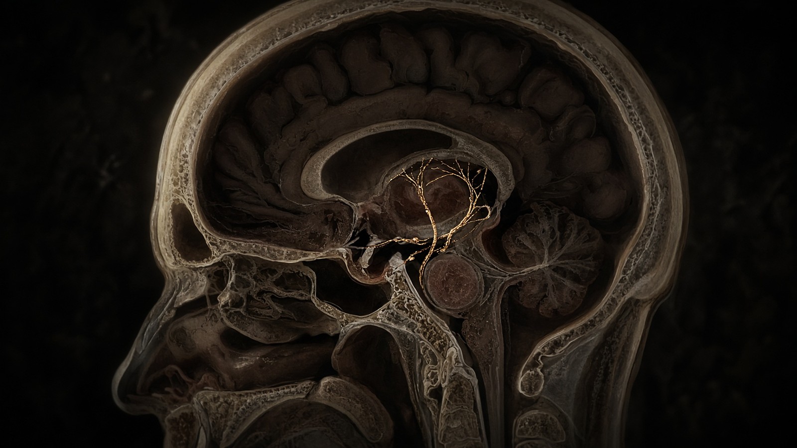
Peptide research · 44 min · 9,769 words
The growth-hormone axis, properly explained
GHRH, ghrelin, somatotropin and IGF-1 are one conversation. CJC without DAC, ipamorelin, HGH and IGF-1 LR3 are four different microphones.
What this essay actually tells you
- Pituitary GH is a pulse, mostly at night. Veldhuis showed that flattening the pulse changes the hepatic message: more insulin antagonism, less of the IGF pattern people think they want.
- GHRH occupies GHRHR (Gs). Ghrelin/ipamorelin occupy GHSR (Gq). Somatropin occupies the GH receptor. IGF-1 LR3 occupies IGF1R and ignores IGFBPs. Four doors. Don't mix the keys.
- CJC without DAC is DPP-IV-resistant GRF(1-29) that still pulses. The DAC version hangs on albumin and turns a pulse into a flat line. We stock the pulse.
What this actually means
Your pituitary releases growth hormone in pulses, mostly at night, when the hypothalamus says go (GHRH) and ghrelin turns up the volume, and somatostatin says stop. The liver then makes IGF-1, which does much of the growing. Laboratory analogues exist at every step. They're not interchangeable, which is why we stock all four as four named sequences rather than one mix.
The growth-hormone axis is a conversation with four named speakers and a clock. The hypothalamus writes two peptides into pituitary portal blood: growth-hormone-releasing hormone, GHRH, which says go, and somatostatin, which says stop. Ghrelin, written in the stomach and in a handful of arcuate neurons, occupies the secretagogue receptor GHS-R1a — that's the ghrelin receptor — and turns the volume up. The anterior pituitary, hearing that ratio, releases growth hormone in pulses, mostly at night, as a 191-residue four-helix bundle. The liver, hearing those pulses at a class-I cytokine receptor, transcribes IGF-1, the acid-labile subunit, and IGFBP3. Much of the growing people attribute to growth hormone is IGF-1 occupying a receptor tyrosine kinase. Four laboratory analogues sit on those four microphones: modified GRF(1–29) without DAC, ipamorelin, recombinant somatropin, and IGF-1 LR3. They're four different questions. We'll walk the axis, the receptors, the pulses, the hepatic programme, and why the Drug Affinity Complex is a design decision rather than a convenience.
In short. Your hypothalamus says go or stop, your stomach turns the volume up, your pituitary pulses growth hormone, and your liver makes IGF-1. Four lab tools sit on four steps.
We stock those four sequences because the papers name them, each as a distinct ligand. CJC without DAC is DPP-IV-resistant GRF(1–29) that still pulses at the GHRH receptor. Ipamorelin is a selective GHS-R1a pentapeptide with minimal ACTH and prolactin at growth-hormone-effective doses, which is why Raun 1998 is still the paper we'd hand you first. Somatropin is the 191-residue ligand itself. IGF-1 LR3 is an 83-residue analogue with collapsed IGF-binding-protein affinity, so a cultured myotube or hepatocyte actually sees the tyrosine kinase occupied. Mix those four objects in your head and you've already designed the wrong experiment. The catalogue keeps them as named sequences, HPLC-characterised, with the kit in the box. Neighbouring essays take each microphone in turn. This piece is the wiring diagram they share, written at the length the axis actually needs. If a protocol needs one receptor, it needs one vial; if it needs two, it names both concentrations and unpicks the peak.
In short. The four analogues ask four receptors. Mix them into one protocol and you stop knowing which step actually did the work.
Let's put names on the anatomy before we put analogues on it. GHRH is a 44-residue hypothalamic peptide; the first twenty-nine residues retain full agonism at a class-B GPCR — a seven-helix receptor that catches a hormone on the outside. Ghrelin is a 28-residue, serine-3-octanoylated stomach hormone whose receptor was cloned as an orphan before the ligand was found. Growth hormone is a 22-kilodalton cytokine. IGF-1 is a 70-residue insulin-family peptide. Those four sentences are the map. The receptors are a second map: class-B Gs at GHRHR, class-A Gq/Gi at GHS-R1a, class-I cytokine JAK2–STAT5b at GHR, receptor tyrosine kinase at IGF1R. Second messengers and transcription factors do the rest. Treat the axis as a single 'growth' slider and you're asking a somatotroph, a hepatocyte and a myoblast to answer as if they were one cell. They aren't. The rest of this piece is those four floors, in order, with the papers you'd want on the bench beside you.
In short. Four ligands, four receptor classes. The axis is a map of named proteins, not a single growth slider.
If you only read one paper on this pentapeptide, make it Raun 1998. The dose–response tables are the whole argument.— Raun K, Hansen BS, Nielsen KL, Heinig CH, Andersen PH, Thøgersen H, Ankersen M, Madsen K. Eur J Endocrinol. 1998; 139: 552–561.
Two hypothalamic peptides, and a stomach one
The hypothalamus is the first floor. Magnocellular and parvocellular neurons write peptides into the portal capillaries that bathe the anterior pituitary, a millimetre or two away. GHRH neurons sit mainly in the arcuate nucleus. Their axons end on the portal plexus. The peptide they release is forty-four residues in the human, processed from a larger precursor, with an amidated C-terminus. Guillemin, Schally, Rivier and colleagues isolated it from tumour and hypothalamic extracts in the early 1980s after a decade of false starts; the clinical and physiological literature that followed is why a GHRH analogue is a serious research object. The first twenty-nine residues, GRF(1–29)-NH2, occupy the receptor with full efficacy. That truncation is the backbone of every laboratory GHRH analogue that matters, including the tetrasubstituted peptide sold as CJC-1295 without DAC. Length isn't decoration. The receptor's extracellular domain reads a defined N-terminal face, and chemists have known that since the isolation papers.
In short. GHRH is a 44-residue hypothalamic go peptide. The first 29 residues are enough to occupy the pituitary receptor fully.
Somatostatin is the stop. Brazeau, Vale, Guillemin, 1973: a fourteen-residue cyclic peptide from hypothalamic extracts that inhibited growth-hormone release in pituitary cells, named for that job before its wider life as a gut and pancreatic hormone was mapped. A twenty-eight-residue N-terminally extended form exists, and in some tissues it isn't a footnote. On the somatotroph the relevant receptors are mainly SSTR2 and SSTR5, class-A GPCRs coupled to Gi — the inhibitory G protein. Adenylyl cyclase falls. Cyclic AMP falls. The L-type calcium current that GHRH had opened is opposed. Granule fusion slows. Somatostatin tone is high between pulses and drops as a pulse is allowed through; the 'go' is therefore a double event, GHRH up and somatostatin down, arriving in portal blood as a ratio rather than as two independent knobs. Add a GHRH analogue and ignore the stop peptide, and you've described half of what the hypothalamus actually said.
In short. Somatostatin is the hypothalamic stop. On the pituitary it cuts cAMP and calcium, so GH granules stay put until the ratio flips.
Portal blood is the medium, and it isn't a peripheral plasma sample. Concentrations in the long portal vessels are high, pulsatile, and largely cleared before they reach a cubital vein. That's why a GHRH measurement in your arm is a poor proxy for what the somatotroph just heard, and why the clinical tests that still use GHRH — often with arginine to suppress somatostatin — are provocations, not photographs of the portal stream. The arcuate GHRH neuron is itself gated: sleep, particularly slow-wave sleep, fasting, sex steroids, thyroid hormone, and a dense inhibitory innervation from somatostatin neurons in the periventricular nucleus. A pulse is a hypothalamic computation before it's a pituitary secretion. Flattening that computation from the periphery, with a long-acting analogue or with continuous recombinant GH, is a different experiment from asking what a pulse-length ligand does to a somatotroph that still has a clock. The analogue occupies one receptor. The village is still there when it arrives.
In short. Portal blood carries high, pulsatile GHRH and somatostatin. A vein in the arm is not that stream, and a long-acting analogue is not a pulse.
The genetic proofs sit on two floors of the same cascade, and they're the reason we can be sure of the order. The little mouse carries a missense mutation in Ghrhr; it's GH-deficient and small, and it's why Mayo's clone was more than a sequence on a gel. Human isolated GH deficiency type IB includes GHRHR mutations, particularly in consanguineous cohorts that paediatric endocrinology still cites. Laron syndrome is the next floor down: GHR mutations, high GH, very low IGF-1, a receptor that will not rotate and will not recruit JAK2. A GHRH analogue can't rescue Laron. A GH analogue can't rescue a missing STAT5b. Mecasermin, recombinant native IGF-1, is the licensed ligand when the cascade is broken below GHR, in a named paediatric indication. Catalogue IGF-1 LR3 isn't mecasermin, and Laron isn't a research protocol. The genetics are here so the four microphones stay in order. Each rescue is floor-specific. Occupy the wrong receptor in a broken cascade and a beautiful ligand becomes an uninformative blot.
In short. Mutations prove the cascade is ordered: GHRHR, then GHR, then IGF-1. A ligand cannot rescue a receptor it never meets.
Diagram
Hypothalamus
GHRH · somatostatin
Go and stop. Class-B GPCRs on the somatotroph.
Stomach / arcuate
ghrelin → GHS-R1a
Volume knob. Synergises with GHRH. Ipamorelin sits here.
Pituitary
GH pulses
191 residues. Night-time bursts. Veldhuis spent a career on the pattern.
Liver
IGF-1 + IGFBP3 + ALS
JAK2–STAT5b at the GH receptor. Much of the growing is this hormone.
Tissue
IGF1R
RTK. IRS–PI3K–Akt–mTOR. IGF-1 LR3 asks this microphone with IGFBPs taken out.
CJC without DAC is DPP-IV-resistant GRF(1–29) that still pulses. Ipamorelin is selective GHS-R1a. Somatropin is the 191-residue ligand. IGF-1 LR3 bypasses the pituitary. Confuse the four and the methods section is already wrong.
Ghrelin is the third speaker sitting next to the hypothalamus, and most of its circulating mass isn't hypothalamic in origin. Kojima, Hosoda, Date, Nakazato, Matsuo and Kangawa, Nature 1999: a 28-residue peptide from the stomach, uniquely octanoylated on serine 3 by ghrelin O-acyltransferase, occupying the receptor that Howard, Smith and colleagues had cloned three years earlier as an orphan growth-hormone secretagogue receptor. The acylation is obligatory for GHS-R1a agonism. Des-acyl ghrelin circulates in larger amounts and is a different conversation. At the pituitary, GHS-R1a is expressed on somatotrophs and synergises with GHRH. In the arcuate, the same receptor sits on NPY/AgRP neurons and is the reason ghrelin is an orexigenic signal — a hunger signal — as well as a secretagogue. Vagal afferents carry a third copy of the story. One ligand, several addresses, one G protein family. A catalogue pentapeptide that occupies GHS-R1a will inherit that geography. Hunger comes with the receptor, even if GH was the only thing you meant to sample.
In short. Ghrelin is a 28-residue stomach hormone with an octanoyl group. It occupies GHS-R1a, raises GH, and also talks to hunger circuits.
- Native GHRH
- 44 residues
- Somatostatin-14
- 14 residues, cyclic
- Ghrelin
- 28 aa, Ser3-octanoylated
- Ipamorelin
- 5 residues
- Mature GH (somatropin)
- 191 aa, ~22 kDa
- IGF-1
- 70 residues
- IGF-1 LR3
- 83 residues
- mod GRF(1–29), no DAC
- 29 residues, tetrasubstituted
Hypothalamic go peptide. GRF(1–29)-NH2 retains full agonism at GHRHR.
The stop. SSTR2 and SSTR5 on the somatotroph, Gi-coupled.
Stomach and arcuate. GHS-R1a, Gq/Gi. Kojima, Nature 1999.
Aib-His-D-2-Nal-D-Phe-Lys-NH2. Selective GHS-R1a. Raun 1998.
Four-helix bundle. The somatotroph's product and the recombinant ligand.
Hepatic, STAT5b-driven. Ternary complex with IGFBP3 and ALS in plasma.
Arg3 plus a 13-aa N-terminal extension. Collapsed IGFBP affinity.
CJC without DAC. DPP-IV-resistant pulse, tens of minutes, not days.
The GHRH receptor is a class-B GPCR
GHRHR is a class-B secretin-family GPCR, the same structural neighbourhood as GLP-1R, GIPR, GCGR and the secretin receptor itself. Seven transmembrane helices, a large N-terminal extracellular domain that captures the C-terminal half of the peptide, and a transmembrane pocket that reads the N-terminus. Occupancy rearranges the helices. The intracellular face becomes a guanine-nucleotide exchange factor for Gs — the stimulatory G protein that loads adenylyl cyclase. Mayo cloned the receptor in the early 1990s; inactivating mutations cause a rare isolated growth-hormone deficiency, which is the genetic proof that this protein isn't optional in the axis. Class B is a different extracellular architecture from the rhodopsin-like class A that GHS-R1a belongs to. If you're designing a GHRH analogue, you're designing against a hormone-binding ectodomain, not against a small-molecule cleft. That's why the first twenty-nine residues still matter, and why a five-residue ghrelin-receptor agonist can't be redescribed as a short GHRH. Two locks. Two ligand lengths. One granule downstream, if both locks turn.
In short. The GHRH receptor is a class-B seven-helix protein. Occupancy loads Gs. It is a different lock from the ghrelin receptor.
Gs is the first amplifier. GTP-bound Gαs occupies adenylyl cyclase. Cyclic AMP rises, in somatotrophs, by enough to occupy protein kinase A. PKA phosphorylates CREB, which is the transcriptional half of a longer GHRH exposure, and phosphorylates the machinery that opens L-type voltage-gated calcium channels. Calcium enters. Local calcium at the granule is the proximate trigger for exocytosis of stored growth hormone. The granule is a dense-core vesicle packed with GH, already synthesised, waiting. A pulse, on the seconds-to-minutes clock, is mostly that fusion event. Hours of GHRH tone will also write more GH gene transcription, more granules, a larger readily-releasable pool. Two clocks, one receptor. A short-acting analogue asks the first clock. An albumin-conjugated analogue asks both, and then asks what the liver does with a flattened GH tone, which is a third question. Cyclic AMP is hydrolysed by phosphodiesterases; somatostatin, via Gi, opposes the cyclase. The pulse is therefore a contest at the cyclase as well as a contest at the portal.
In short. GHRH raises cAMP, PKA opens calcium channels, and GH granules fuse. A short analogue asks that pulse. A week-long analogue asks a different clock.
Diagram
Outside
Peptide ligand
Named sequence in the nM–µM pocket. Shape complementarity, not vibes. A 15-mer and a 4-mer do not fit the same hole.
Membrane
7-TM receptor
Helices rearrange. The cytoplasmic face becomes a GEF for a heterotrimeric G protein (Gs, Gi, Gq, G12/13).
Inside
Second messengers
cAMP, IP₃, Ca²⁺, β-arrestin. One occupied receptor can spawn thousands of messenger molecules. That is amplification.
~800 GPCRs in the human genome. Seven transmembrane helices, an extracellular ligand pocket, an intracellular G-protein handshake. Catalogue neighbours: ipamorelin at GHSR, PT-141/MT2 at melanocortin receptors, retatrutide at GLP-1R/GIPR/GCGR.
L-type calcium current is the sentence that's easy to skip and shouldn't be. Somatotrophs are electrically excitable. They fire action potentials. The depolarisation that GHRH and ghrelin both encourage, by different G proteins, opens Cav1 channels. The calcium that enters is the fusion signal. Block those channels with a dihydropyridine in a pituitary slice and the GH pulse collapses, which is old pharmacology and still a useful control. GHRH gets there through cAMP and PKA. Ghrelin gets there through Gq, phospholipase C, IP3, and a rise in intracellular calcium that also depolarises. The two paths converge on the same granule. That convergence is why co-application of a GHRH analogue and a GHS-R1a agonist releases more GH than either ligand alone, in slices and in animals, and why textbooks draw the large pulses as a combination. Synergy is a G-protein fact, not a stacking tip. Isolate each ligand and you'll know which receptor did the work. Name both if you apply both.
In short. Both GHRH and ghrelin raise calcium in the pituitary cell, by different G proteins, and GH granules fuse. Together they make the large pulses.
Diagram
× 1
Ligand
One peptide in one pocket. nM–µM. Shape, not a mood.
× 10–10²
G proteins
The occupied GPCR is a GEF. Each Gα is a catalyst.
× 10³–10⁴
cAMP / IP₃ / Ca²⁺
Adenylyl cyclase and PLC do not make one molecule. They make a cloud.
× 10⁴–10⁶
PKA / PKC / CaMK
Kinases phosphorylate many substrates per messenger.
× tissue
Secretion, transcription, motility
The organism-level readout. Still not a protocol.
This is the only magic, and it is not magic. A nanomolar ligand can move a micromolar messenger because enzymes sit between them. Desensitisation (GRK, β-arrestin, endocytosis) is how the cell refuses to let ‘more ligand’ mean ‘more signal’ forever.
Desensitisation is why more ligand isn't more signal forever. Occupied GHRHR is phosphorylated by GRKs, binds β-arrestin, and is internalised. Recycle or degrade, depending on the duration and the cell. A somatotroph that sees continuous GHRH tone will, within hours, present fewer receptors and a smaller cAMP increment per molecule of ligand. That's ordinary GPCR biology, the same arithmetic Lefkowitz spent a career on, applied to a class-B receptor whose ligand in life is a pulse. It's one of the reasons a Drug Affinity Complex on a GHRH analogue is a design choice with a cost: you buy duration, you spend the pulse and some of the receptor census. Tesamorelin, dosed once daily as a licensed medicine, lives with that trade because its indication asked for a sustained IGF-1 rise and a visceral-fat readout, not a copy of nocturnal physiology. A research analogue without DAC was built for the other trade. You get the minutes. You keep the trough.
In short. If GHRH tone stays high, the receptor is pulled inside and the cell hears less. A long-acting analogue buys time and spends some of that hearing.
Ghrelin turns the volume up
GHS-R1a is a class-A GPCR, rhodopsin-like, with a high constitutive activity that the field has argued about since the clone. The dominant coupling is Gq/11: phospholipase C, PIP2 cleavage, IP3, a calcium rise from stores and from the plasma membrane. A Gi component is real, and it's part of why the electrophysiology of a somatotroph seeing ghrelin isn't a carbon copy of one seeing GHRH. β-arrestin is recruited; biased ligands exist in the medicinal-chemistry literature and they aren't this catalogue. The receptor is unusual in requiring the octanoyl on native ghrelin, a post-translational lipid that GOAT installs in the stomach. Synthetic growth-hormone secretagogues — the peptide and non-peptide ligands that occupied this receptor before ghrelin was found — don't need that lipid because they were built against the orphan. Ipamorelin is one of those synthetic peptides. It isn't ghrelin. It's a pentapeptide agonist at the same receptor, and the distinction is the whole product. Same lock. Different key. Different molecule in the cake.
In short. The ghrelin receptor is a class-A GPCR that raises calcium through Gq. Ipamorelin occupies that receptor. It is not the stomach hormone itself.
Bowers spent the 1980s making smaller, stronger peptides that released GH from pituitary cells without being GHRH. GHRP-6, His-D-Trp-Ala-Trp-D-Phe-Lys-NH2, worked. It also moved ACTH, cortisol and prolactin, which is a messy experiment if the question was isolated growth-hormone release. Hexarelin, a related hexapeptide, did the same circus with more potency. When Smith's group cloned GHS-R, those ligands had a named target; they still had the off-axis baggage. The secretagogue field then split, as fields do, into cleaner tools and louder ones. A louder one isn't a better reagent. It's a reagent plus a confound. Cortisol will change the hepatic programme you thought you were reading as an IGF-1 effect. Prolactin will confuse a reproductive readout. If you wanted GHS-R1a, you wanted a ligand that occupies GHS-R1a and leaves the corticotroph and lactotroph as nearly silent as the tables allow. That's a design brief, not a personality contest between peptides.
In short. Early ghrelin-receptor peptides released GH and also shoved ACTH and prolactin around. That extra noise is a confound, not a bonus.
Synergy with GHRH is the physiological point of GHS-R1a on the somatotroph. In hypothalamic–pituitary clamp experiments, a GHRH pulse in the presence of a secretagogue produces a much larger GH burst than GHRH alone; a secretagogue in the absence of GHRH produces a smaller one. The large nocturnal pulses that Veldhuis deconvolved are, on this picture, a GHRH event amplified by ghrelin tone and permitted by a somatostatin trough. Fasting raises ghrelin and amplifies GH, which is why a secretagogue assay in a fed animal and a fasted animal aren't the same assay. Hyperglycaemia suppresses GH and will blunt a secretagogue challenge. Those gates are why a clean GHS-R1a ligand still isn't a push-button. Two receptors, two G proteins, one granule. Name both ligands if you apply both. Name the metabolic state of the animal. Then measure. That's how you keep the large pulse as a result you can attribute, rather than as a peak you can't unpick.
In short. Ghrelin makes a GHRH pulse larger. Fasting turns ghrelin up; high sugar turns GH down. The secretagogue is not a button that ignores those gates.
Ipamorelin is the clean pentapeptide
Ipamorelin is Aib-His-D-2-Nal-D-Phe-Lys-NH2, five residues, a C-terminal amide, two D-amino acids, an aminoisobutyric acid at the N-terminus that resists aminopeptidase. Novo Nordisk published it. Raun, Hansen, Nielsen, Heinig, Andersen, Thøgersen, Ankersen and Madsen, European Journal of Endocrinology, 1998, 139: 552–561. In vitro, on rat pituitary cells, it released GH with a potency and efficacy that sat in the secretagogue family. In vivo, in swine and in rats, plasma GH rose. ACTH, cortisol and prolactin did not, at doses that were effective for GH, in contrast to GHRP-6 and to hexarelin run in the same protocols. That last clause is the argument. The dose–response tables are assigned reading because they're how you tell a selective tool from a noisy one. The analogue doesn't meaningfully bind the motilin receptor, a related GPCR that some ghrelin-family ligands graze. Oral bioavailability is poor, as you'd expect for a pentapeptide with those side chains; research use is parenteral in animal protocols or in vitro.
In short. Ipamorelin is a five-amino-acid ghrelin-receptor agonist. Raun 1998 showed GH goes up while ACTH, cortisol and prolactin mostly do not.
Selectivity is the entire product. A GHS-R1a agonist that also occupies the corticotroph's secretagogue-sensitive machinery writes a stress-axis sentence on top of the GH sentence. Downstream, that cortisol will antagonise some of the IGF-1 pattern, mobilise glucose, and alter immune readouts. If you wanted isolated GHS-R1a agonism, you've then measured a mixture. Raun's tables are why ipamorelin is still the cleanest peptide secretagogue to put on a bench a quarter of a century later. Non-peptide secretagogues — the mk-series, anamorelin in the cachexia literature — are a different chemical class with their own selectivities and their own clinical programmes; they aren't this vial. A later Novo Nordisk and Helsinn programme asked whether ipamorelin would recover postoperative ileus. It didn't meet its primary endpoint. That's a useful reminder that a clean GH secretagogue isn't a universal gut drug, even though GHS-R1a is expressed in the enteric nervous system. Receptor geography isn't a licence to generalise the indication.
In short. Clean GHS-R1a agonism is the point of ipamorelin. A later gut-recovery trial failed, which is information: a GH secretagogue is not a general gut medicine.
What a selective pentapeptide lets you ask, then, is precise. What does isolated GHS-R1a agonism do to somatotroph calcium and GH release when GHRH is present, and when it is absent. What does it do to gastric emptying and to arcuate NPY neurons without a cortisol confound. What does it do in combination with a DPP-IV-resistant GHRH analogue: two receptors, two G proteins, one pulse, each ligand named. Those are experiments. Combining them as a purchasing habit isn't. The ipamorelin we stock is that pentapeptide, ten milligrams, HPLC at or above 98 per cent, the sequence Raun published. The GHRH analogue sits on the next shelf as a different sequence. Reconstitute both into one syringe and call it the axis, and you've stopped being able to attribute the GH peak. Attribution is the job. We're walking the wiring so that sentence is harder to dodge when you're holding two vials.
In short. Ipamorelin asks the ghrelin receptor on its own. Combine it with a GHRH analogue only if you name both ligands and unpick which one did what.
Pulses are the point
Johannes Veldhuis spent a career showing that growth hormone is a digital signal. Deconvolution of frequent-sampled plasma GH, in humans and in animals, yields discrete bursts superimposed on a low interpulse baseline. Most of the mass is in the bursts. Most of the bursts, in a healthy adult, cluster after sleep onset, in slow-wave sleep, with a smaller daytime population. Amplitude falls with age, with abdominal adiposity, with hyperglycaemia. Amplitude rises with fasting, with oestradiol in some protocols, with a GHRH or secretagogue challenge. The pattern is the message. A single morning clinic GH is almost uninterpretable without an IGF-1, and even IGF-1 is an integral, not a pulse photograph. The papers are in Journal of Clinical Endocrinology and Metabolism, in Endocrine Reviews, in a run of American Journal of Physiology pieces that a generation of fellows were handed. Report 'GH' as a continuous infusion or as a daily subcutaneous bolus and then talk as if you'd reproduced physiology, and you haven't yet read Veldhuis on those bursts.
In short. GH is released in bursts, mostly at night. Veldhuis showed the pattern is the message. A flattened dose is a different signal from a pulse.
Sleep is the largest physiological gate. Slow-wave sleep and the first nocturnal GHRH-permissive window coincide; deprive the slow waves and the GH peak shrinks. That's why night-time sampling exists, and why a daytime secretagogue challenge is a different probe from a nocturnal endogenous pulse. Thyroid hormone is permissive for GH synthesis; a hypothyroid somatotroph writes fewer granules. Glucocorticoids, in excess, suppress the axis at hypothalamus and pituitary; in replacement they're allowed. Sex steroids shape amplitude and frequency, which is the next heading. Free fatty acids suppress GH release, one of the reasons obesity is a low-GH state. Ghrelin and fasting push the other way. The somatotroph isn't an isolated oscillator. It's a cell reading a hypothalamic ratio, a metabolic plasma, and a sleep clock. A laboratory analogue occupies one receptor in that village. The village is still there when the analogue arrives, which is both the constraint and the interest of the experiment.
In short. Sleep, thyroid, sugar, fat, and fasting all gate GH pulses. A lab analogue occupies one receptor in that village; the rest of the village remains.
Sampling is the unglamorous half of pulsatility, and it's where a lot of GH numbers quietly die. A single clinic GH, drawn at nine in the morning, reports a trough more often than a peak and can't distinguish deficiency from a nadir between bursts. Frequent sampling, every ten or twenty minutes overnight, is how Veldhuis deconvolved mass and frequency; it's also how you prove a pulse if that's the claim. IGF-1 is the integral: a hepatic average over hours, useful, and blind to whether the night was three large bursts or a flattened plateau. IGFBP-3 and ALS are slower integrals still. Occupy GHRHR with a thirty-minute analogue and then draw IGF-1 at a week, and you've measured the liver's running mean, not the somatotroph's burst. Draw GH at twenty-minute intervals around the dose if the question is the pulse. Draw IGF-1 if the question is the hepatic programme. Name which integral you meant, because the two numbers don't substitute for each other.
In short. One morning GH blood test is usually a trough. Frequent night samples show pulses. IGF-1 averages hours of liver output and hides the bursts.
Continuous growth hormone isn't pulsatile growth hormone at the liver. That sentence is the design constraint for every GHRH analogue, and it's why this catalogue stocks CJC without the Drug Affinity Complex. Hepatocytes reading a pulsatile STAT5b phosphorylation write IGF1, ALS and IGFBP3, the ternary-complex programme that circulating IGF-1 actually travels in. Hepatocytes reading a flattened GH tone write a different programme: more insulin antagonism, a different lipid-gene set, less of the IGF pattern many protocols thought they wanted. Receptor down-regulation contributes. SOCS2, a STAT5b target, feeds back and shuts the receptor down when occupancy is unrelieved. Jørgensen, the older rodent infusion studies, and the clinical experience with continuous subcutaneous GH pumps all point the same way. In the rat, a three-hour GH infusion and a pair of pulses matched for area-under-the-curve don't write the same cytochrome P450 genes. Pattern is information. Flatten it and you've changed the ligand even if the amino-acid sequence is identical.
In short. The liver reads GH pulses as one programme and a continuous GH drip as another. Flatten the pulse and you change the message.
GH is a digital signal: night-time bursts, suppression by hyperglycaemia, amplification by fasting and ghrelin. Flatten the pulse and the liver writes a different programme.— The Veldhuis programme, in the language this desk uses. See Veldhuis JD, Bowers CY. Endocr Rev and the JCEM deconvolution series.
Sexual dimorphism is not a footnote
Male rodents fire large, regular nocturnal GH bursts against a near-silent baseline. Female rodents run a more nearly continuous, lower-amplitude tone. The liver reads those two patterns as two programmes. A classic molecular signature: male-pattern GH, in the rat, induces some cytochrome P450 genes (CYP2C11) and suppresses others (CYP2C12); female-pattern GH does the reverse. STAT5b pulsing versus STAT5b occupancy that never quite returns to baseline is a large part of the mechanism. Knock out STAT5b and the male-specific liver programme collapses toward female. That literature, from Waxman, Norstedt, Mode and others, is why a pharmacologist can't treat 'GH exposure' as area-under-the-curve and be done. Humans are less cartoonishly dimorphic than rats, which is a species sentence we should keep making, but the principle survives: women have more frequent, smaller pulses and a higher interpulse baseline; men have larger, less frequent bursts. Oral oestrogen, in particular, antagonises hepatic GHR signalling and lowers IGF-1 even as it may raise GH secretion — a first-pass effect that transdermal oestrogen doesn't copy.
In short. Males and females fire GH in different patterns, and the liver reads those patterns as different gene programmes. Area-under-the-curve is not the whole story.
The practical consequence for a reagent is blunt, and it's why two similarly named vials are not two strengths of one thing. A pulse-length GHRH analogue, dosed so that the somatotroph still sees peaks and troughs, is asking a question that can in principle look like physiology, including its sexual dimorphism. An albumin-conjugated GHRH analogue, or a daily subcutaneous bolus of recombinant GH that never returns to baseline, is asking what a flattened tone does. Both questions are lawful. They aren't the same question. They won't give the same hepatic IGF-1, the same insulin sensitivity, or the same cytochrome pattern in a rodent liver. Treat CJC with DAC and CJC without DAC as two strengths of one reagent and you've already failed the dimorphism heading, the Veldhuis heading, and the STAT5b heading in a single table footnote. We stock the pulse-length ligand because the physiology we're trying to look like is a pulse. The other molecule exists. It's a different object.
In short. A short GHRH analogue can still pulse. A week-long one flattens the tone. Those are two experiments, including at the level of male versus female liver genes.
The 191-residue ligand and its receptor
Mature human growth hormone is 191 amino acids, about 22 kilodaltons, a four-helix bundle in the class-I cytokine family, the same structural neighbourhood as prolactin, erythropoietin and the interleukins that use similar receptors. A 20-kilodalton splice variant lacks residues 32–46 and is a minority of circulating GH; assays that don't see it will under-count. Disulfide bonds hold the bundle. The protein is synthesised as a precursor, cleaved, stored in dense-core granules in somatotrophs, which are half or more of the anterior pituitary by cell number in a young adult. Recombinant somatotropin — somatropin in INN language — is that 191-residue sequence, made in bacteria or in mammalian cells depending on the manufacturer, folded, characterised. The 24 IU vial in this catalogue is that backbone, lyophilised, HPLC-characterised. It's the ligand the receptor evolved to bind. Occupying GHR directly is a different experiment from occupying GHRHR or GHS-R1a, because you've skipped the somatotroph's pulse generator and its somatostatin gate. You're now the pulse, or the drip, depending on how you dose.
In short. Growth hormone is a 191-amino-acid protein. Recombinant somatropin is that same chain. Giving it skips the pituitary and talks to the GH receptor directly.
The 20-kilodalton splice isn't a contaminant. Alternative splicing of GH1 skips 45 bases in exon 3 and deletes residues 32–46; the variant is a fraction of pituitary output, binds GHR with a different affinity profile, and is missed by some monoclonal assays. If you report GH in nanograms per millilitre, say which epitope the antibody sees. Placental GH, from GH2, is a different gene, a different pregnancy physiology, and not this vial. The 191-residue recombinant protein is the 22-kilodalton species, the majority ligand, the one the adult-GHD trials used. Isoform literacy is how you stop arguing with an assay. It's also why HGH as a three-letter nickname is a worse name than somatropin: the nickname doesn't tell you which chain, which splice, or which gene. Pegvisomant, a GHR antagonist used in acromegaly, is the inverse probe: occupy the receptor so that STAT5b does not write. A licensed antagonist and a licensed agonist on the same receptor are further proof that occupancy at GHR is a variable, not a mood called growth.
In short. Most GH is the 191-residue, 22-kilodalton form. A shorter splice exists and some assays miss it. Pegvisomant blocks the same receptor from the other direction.
GHR is a class-I cytokine receptor, a single-pass transmembrane protein that sits as a preformed dimer. Ligand doesn't recruit a second chain so much as rotate the dimer, which is the picture Waters, Brooks and colleagues established after an earlier sequential-dimerisation model. Rotation brings the intracellular boxes into a geometry that JAK2, already loosely associated, can use. JAK2 phosphorylates itself and phosphorylates tyrosines on the receptor tail. STAT5b SH2 domains dock, STAT5b is phosphorylated, dimerises, and goes to the nucleus. That's the principal transcriptional driver of hepatic IGF1, ALS and IGFBP3. MAPK and PI3K arms leave the same receptor and account for some of the non-transcriptional and some of the extra-hepatic actions. SOCS2 is transcribed and comes back to shut the receptor down. The whole assembly is a cytokine-receptor logic, not a steroid-receptor logic, which is why talking about GH as if it were a stronger testosterone is a category error. Different kingdom. Different transcription factor. Different circulating messenger.
In short. The GH receptor is a cytokine receptor. Ligand twists a ready-made pair, JAK2 fires, STAT5b goes to the nucleus and writes IGF-1. It is not a steroid.
Direct metabolic actions of GH, independent of IGF-1, are real, and they're part of why the axis isn't a pure growth slider. Hormone-sensitive lipase is activated in adipocytes; free fatty acids rise. Insulin signalling is antagonised in muscle and in liver: more glucose output, less glucose uptake, a shift toward lipid oxidation. In the growing skeleton, GH and IGF-1 both talk to epiphyseal chondrocytes, which is why childhood GH deficiency is a stature diagnosis and why acromegaly, the experiment of nature for excess, grows acral soft tissue and has a cardiomyopathy conversation. Adult GH-deficiency replacement, in the Jørgensen and Bengtsson literature, shifts body composition — less fat, more lean mass — with a more modest effect on strength and a real discussion of glucose tolerance. Those are labelled-medicine trials in diagnosed deficiency. They're the clinical existence proof that GHR occupancy does something to composition. They aren't a licence to treat a research 191-mer as a training protocol. Acromegaly remains the cautionary physiology: chronic excess is a disease, not a goal.
In short. GH also burns fat and opposes insulin, aside from making IGF-1. Replacement in true deficiency changes body composition. Chronic excess is acromegaly, a disease.
STAT5b writes the hepatic programme
The hepatocyte is where much of the circulating IGF-1 is written. GHR occupancy, JAK2, STAT5b, then IGF1 transcription, ALS transcription, IGFBP3 transcription. The three products assemble in plasma as a ternary complex that carries the large majority of circulating IGF-1, extending its half-life from minutes (free) to hours (bound). Acid-labile subunit is liver-specific in a way IGF-1 is not; extra-hepatic IGF-1, especially the mechanically gated splice in loaded muscle, is a different conversation and isn't a vial. Yakar, Le Roith and colleagues, with the liver-specific IGF-1 knockout, showed that circulating IGF-1 can fall hard while local IGF-1 keeps a surprising amount of postnatal growth going. That paper is why 'GH makes you grow via IGF-1' is true and incomplete. Endocrine IGF-1 is the serum assay and the liver's job. Autocrine and paracrine IGF-1 are the tissue's job. IGF-1 LR3, coming two headings from here, is a circulating-style analogue engineered to skip the binding-protein buffer. It is neither hepatic transcription nor a mechano-growth splice.
In short. The liver writes most of the IGF-1 in blood, together with two partners that carry it. Tissues also make local IGF-1. A lab analogue is neither of those jobs.
MAPK and PI3K leaving GHR are the other arms, and they matter for the metabolic sentence. ERK signalling contributes to some of the proliferative and to some of the gene-expression programme. PI3K–Akt is the metabolic neighbourhood GH shares, awkwardly, with insulin and with IGF-1: awkward because GH is insulin-antagonistic at the whole-body level even as its receptor can light a PI3K lamp. Tissue and timing sort that apparent contradiction. Acute GH is lipolytic and insulin-antagonistic. IGF-1, downstream, is insulin-like at IGF1R and at hybrid receptors. The net of a physiological pulse is therefore a lipolytic first hour and an IGF-1-mediated later window. Flatten the pulse and you stretch the first hour across the day, which is one reading of why continuous GH looks more insulin-antagonistic than pulsatile GH for the same IGF-1 increment. The papers aren't unanimous in every protocol, which is honest. The direction of the argument is stable enough to make DAC a scientific decision rather than a convenience of dosing.
In short. GH both opposes insulin and, later, raises IGF-1, which is insulin-like. A pulse separates those two windows. A drip blurs them.
SOCS2 is the negative-feedback protein the axis deserves a named sentence for. STAT5b transcribes it. SOCS2 binds the phosphorylated GHR tail and dampens JAK2. Animals lacking SOCS2 are large, which is the genetic proof. Continuous GHR occupancy writes more SOCS2 and the receptor quiets; troughs in a physiological pulse let the receptor recover. That's ordinary cytokine-receptor hygiene, the same logic interferon and EPO use, applied to a hormone whose information is a burst. It's also why a weekly albumin-conjugated GHRH analogue and a pulse-length analogue will not occupy GHR in the same temporal pattern even if mean IGF-1 looks similar on a sparse blood draw. Mean IGF-1 is an integral. The receptor's day is a series of STAT5b phosphorylations. Integrals hide series. Veldhuis taught that about GH in plasma. It's equally true about STAT5b in a hepatocyte nucleus. If you only draw the mean, you've asked the liver to summarise a day it experienced as peaks.
In short. SOCS2 is the off-switch STAT5b writes. Pulses let the GH receptor recover. A continuous tone keeps the off-switch planted.
IGF-1 does much of the growing
Mature IGF-1 is seventy residues, three disulfide bonds, a member of the insulin superfamily so obviously that the two receptors form hybrids. IGF1R is a heterotetramer, two α and two β chains, a receptor tyrosine kinase. Ligand binding at the α chains brings the β-subunit kinase domains together; they autophosphorylate; phosphotyrosines recruit IRS-1 and IRS-2 and Shc. IRS proteins open PI3K, PIP3, Akt, then mTOR, the growth and survival lamp. Shc opens Ras, Raf, MEK, ERK, the proliferative lamp. That's the same logic insulin uses at IR, which is why glucose belongs in any protocol that occupies IGF1R at high tone, and why we'd be uneasy about a dish or an animal that forgot the glucometer. Hybrid IR/IGF1R receptors are common in some tissues. An analogue that ignores IGF-binding proteins will, at sufficient concentration, look a little like insulin. Hypoglycaemia in animals is the organism-level version of that sentence. The receptor is a cousin of insulin's. Treat it that way and the glucose plan writes itself.
In short. IGF-1 occupies a tyrosine-kinase receptor related to the insulin receptor. The cell then grows through Akt and mTOR, and can also drop its glucose.
IGF-binding proteins 1 through 6, with ALS, sequester more than ninety per cent of circulating IGF-1 in ternary and binary complexes. That buffer is a safety system: free IGF-1 is short-lived and insulin-like; bound IGF-1 is a reservoir with a half-life of hours. IGFBP-3 is the main circulating partner and is itself GH-dependent via STAT5b, which is why a GH pulse writes both the ligand and its carrier. IGFBP-1 is insulin-suppressed and rises in fasting, which is one of the ways the axis knows the metabolic state. Proteases clip IGFBPs in tissues and release ligand locally. The experimental nuisance, in a dish, is that serum-containing media present a binding-protein sink. Native IGF-1 added to such a medium is rapidly bound and the receptor on a myoblast sees a fraction of what you pipetted. That nuisance is why Long Arg3 IGF-1 exists. It's a design decision about the buffer, not a potency contest against somatropin, which is a different receptor. Same superfamily. Different lock. Different question.
In short. Most IGF-1 in blood is locked to binding proteins as a safety buffer. In a dish those proteins steal the ligand, which is why the LR3 analogue exists.
Local IGF-1, especially in loaded muscle, is the sentence a vial can't honestly buy. Mechano-growth factor, an IGF-1 splice with a different E-peptide, is transcribed after stretch and damage; the literature is real and also argued-over in the usual way of splice-variant fields. That transcript is an autocrine event in a fibre that was loaded. Hepatic IGF-1 is an endocrine event downstream of GHR. IGF-1 LR3 is a laboratory analogue of the endocrine ligand with the buffer collapsed. Three objects. Treating a reconstituted 83-mer as a substitute for the splice the fibre already writes is mixing jobs. The receptor is shared. The question isn't. We'll keep saying that, because the axis is one conversation and the microphones still aren't interchangeable. Goldspink's MGF papers belong on a muscle-physiology reading list. They don't belong on a certificate of analysis for LR3. If the fibre already wrote the splice, the analogue is answering a different question, in a different compartment, with a different buffer.
In short. Muscle can make its own IGF-1 splice when it is loaded. That local message is not the same object as a lab analogue added to a dish.
Mecasermin is recombinant native IGF-1, licensed for severe primary IGF-1 deficiency, a paediatric endocrine object with hypoglycaemia as a labelled risk. Increlex isn't LR3. LR3 was built for culture, where binding proteins in serum make native IGF-1 a poor tool. Taking that analogue into an organism reintroduces the insulin-like risk without the binding-protein buffer that made native IGF-1 safer in plasma. That's why the dish and the animal are different experiments even at the same receptor, and why moving LR3 from C2C12 into a mouse without a glucose plan skips the hybrid-receptor sentence. The 83-mer is a precise probe. Precision isn't the same as safety at organism scale. Native IGF-1 in a ternary complex with IGFBP3 and ALS is the physiological circulating form. LR3 is the form that ignores the complex. Choose on purpose, and write the choice down, including how glucose was watched. The kinase doesn't care which experiment you thought you were running. Glucose will tell you anyway.
In short. Licensed IGF-1 is the native 70-mer, buffered by binding proteins. LR3 ignores that buffer so a dish can see it. Moving it into an animal needs a glucose plan.
CJC without DAC still pulses
Native GHRH dies in minutes in plasma. Dipeptidyl peptidase-IV cleaves the Ala2–Asp3 bond, a truncation that abolishes agonism at GHRHR. The same enzyme is why GLP-1 is a poor medicine as a native peptide and why sitagliptin exists. Chemists therefore tetrasubstituted GRF(1–29): typically D-Ala2 to resist DPP-IV, Gln8, Ala15, Leu27 to resist oxidation and to tidy pharmacokinetics, while preserving receptor efficacy. The result, modified GRF(1–29), often sold as CJC-1295 without DAC, has a half-life on the order of tens of minutes rather than minutes, and rather than days. Tens of minutes is still a pulse on a somatotroph's clock. It's long enough to be a useful experimental ligand and short enough that troughs can still happen. That window is the entire point of the substitutions. A longer window is a different molecule, obtained by a different trick, and it asks a different question of the liver. Four amino acids buy you the minutes. They don't buy you the week.
In short. Blood enzymes chew native GHRH in minutes. Four amino-acid swaps make a version that lasts tens of minutes, which is still a pulse.
DAC is Drug Affinity Complex, ConjuChem's maleimide linker, typically an N-ε-maleimidopropionyl-lysine on a similar backbone, that Michael-adds to cysteine 34 of circulating albumin. The peptide then rides albumin for days. Teichman and colleagues, 2006, showed multi-day GH and IGF-1 elevation after a single dose of CJC-1295 with DAC: continuous rather than pulsatile tone, IGF-1 that stays up. Albumin conjugation is a beautiful trick for a weekly incretin. It's the wrong trick if the message you want is a somatotroph pulse. The with-DAC molecule is a real reagent for a real question — what does flattened GHRH tone do to IGF-1 and to body composition. It's the wrong reagent if you're trying to look like physiology. We picked the pulse-length ligand for that reason. The name in the trade, CJC-1295 without DAC, is a historical accident of how the albumin version was branded. The chemistry is modified GRF(1–29). The decision is the absence of the maleimide.
In short. DAC glues the peptide to albumin for days, flattening GH tone. Without DAC, modified GRF(1–29) can still pulse. That is the version on the shelf.
DPP-IV resistance without albumin conjugation is a general peptide-design move, and it's worth separating from half-life folklore. Sitagliptin inhibits the enzyme so native incretins last longer. Semaglutide and tirzepatide go further and add a fatty-acid handle so albumin carries them through the week. Modified GRF(1–29) stops at the first move: resist the enzyme, keep the pulse. That's a smaller, more physiological intervention than fatty acylation or maleimide conjugation, and it's why this analogue and those incretin analogues shouldn't be described as the same trick. They share an enemy, DPP-IV, and they don't share a design goal. GHRH wants a peak and a trough. GLP-1R agonism in a medicine wants a week of tone. Copying the incretin half-life trick onto GHRH is how you accidentally run the Teichman experiment when you thought you were running a pulse. We label the vial without DAC so that accident is harder to have. The maleimide is a decision, not a missing ingredient.
In short. Stopping DPP-IV is not the same as gluing a peptide to albumin. GHRH wants peaks and troughs. Weekly incretins want the opposite.
Tesamorelin is the licensed neighbour
Tesamorelin is a trans-3-hexenoyl GRF(1–44), Egrifta in the United States, licensed for reduction of excess abdominal fat in HIV-associated lipodystrophy. Falutz and colleagues ran the trials; IGF-1 rose, visceral adipose tissue fell, glucose had to be watched. That's the clinical existence proof that GHRHR is drug-able, in a named indication, under a marketing authorisation, as a daily subcutaneous medicine. It is a GHRH analogue. It isn't modified GRF(1–29) without DAC, and it isn't in this fridge. The hexenoyl cap is a different DPP-IV-resistance trick from D-Ala2; the backbone is the full forty-four rather than twenty-nine. Daily dosing of a still-relatively-short analogue is closer to a pulse series than a weekly albumin conjugate is, which is why tesamorelin and CJC-with-DAC shouldn't be collapsed into one sentence either. The licensed molecule is the neighbour we're obliged to name so that a research vial isn't mistaken for it. Same receptor family. Different backbone, different legal class, different job.
In short. Tesamorelin is a licensed GHRH analogue for HIV lipodystrophy. It proves the receptor can be a medicine. It is not the research vial in this catalogue.
We'll keep naming the neighbour, because licensed analogue and research analogue are easy to mix up and expensive to confuse. Retatrutide's neighbour is tirzepatide and semaglutide; the research vial is the published LY3437943 structure, not Lilly's pen. Somatropin's neighbour is every recombinant GH medicine on an endocrine ward; the research 191-mer is the same amino-acid sequence, labelled for a different job. Tesamorelin is the GHRH-analogue neighbour. Modified GRF(1–29) without DAC is the pulse-length ligand one step upstream of that, HPLC-characterised, for the bench. Indication, authorisation, device, and pharmacovigilance sit on one side of that sentence. Sequence, certificate of analysis, and a reconstitution kit sit on the other. Collapse the gap and you're selling a neighbourhood as an address. The axis is interesting enough, and the four microphones are distinct enough, that the elision is unnecessary as well as unlawful as a claim. Two objects. Two labels. One receptor family they happen to share.
In short. Licensed GHRH analogue on one side, research modified GRF(1–29) on the other. Same receptor family, different object, different job.
Four probes, four questions
CJC without DAC asks: what does a slightly prolonged GHRH pulse do to somatotrophs, to GH burst mass, and to the hepatic STAT5b programme when troughs are still allowed. Ipamorelin asks: what does selective GHS-R1a agonism do to the same granule, and to arcuate and gastric addresses, without dragging ACTH, cortisol and prolactin into the table. Recombinant somatropin asks the GH receptor itself, skipping the hypothalamus and the pituitary, and the answer will depend on whether you dose it as a pulse or as a drip. IGF-1 LR3 bypasses the pituitary and the liver and asks the tyrosine kinase on a myotube or a hepatocyte, with IGFBPs taken out of the fight; glucose belongs in that protocol. Four ligands, four receptors, four questions. Confuse them and you're assembling a mix you can no longer attribute. The papers that combined GHRH and a secretagogue in pituitary slices named both concentrations and both receptors. That's the standard, and it's a kind one: you get the synergy, and you still know which microphone sang.
In short. Each analogue asks one receptor one question. Treating the four as a mix is a shopping list, not an experiment.
Diagram
| Node | Catalogue | Conversation |
|---|---|---|
| GPCR | Ipamorelin, MT2, PT-141, retatrutide, CJC | Second messengers, secretion, appetite, pigment |
| RTK / IGF1R | IGF-1 LR3 | IRS–PI3K–Akt–mTOR and Shc–ERK |
| Cytokine receptor | Somatropin (HGH) | GHR–JAK2–STAT5b, hepatic IGF-1 |
| Cofactor | NAD+ | Sirtuins, PARPs, CD38, redox |
| Actin buffer | TB-500 / Tβ4 motif | G-actin sequestration, motility |
| Growth-factor-like | BPC-157 | VEGFR2 / FAK / eNOS neighbourhood |
| Copper ligand | GHK-Cu | Transcriptome shift in fibroblasts |
| MC fragment | KPV | NF-κB, PepT1, no pigment |
| Nuclear / pineal | Epithalon (AEDG) | TERT and melatonin literatures |
| mtORF peptide | MOTS-c | AMPK, folate–methionine cycle |
Each row is a different kind of molecular conversation. The catalogue peptides bind at these nodes; they are not interchangeable, and stacking them because a forum did mixes unrelated literatures.
IGF-1 LR3 is the 83-residue object, and it deserves the same precision the secretagogue got. Francis, McDougall, Bagley, Ballard and colleagues made Long Arg3 IGF-1 by substituting Glu3 for arginine and adding a thirteen-residue N-terminal extension derived from porcine GH. IGFBP affinity collapses by roughly two orders of magnitude. IGF1R agonism remains. In serum-containing culture the analogue is the ligand the myoblast actually sees; native IGF-1 is the ligand the binding proteins see. Classic L6 and C2C12 work shows PI3K-dependent hypertrophy programmes and, at higher tone, a proliferation-over-fusion tradeoff. That tradeoff is a reason to use a defined analogue rather than a crude serum spike, and a reason not to treat 'more IGF' as a monotonic good in a differentiation assay. Cross-talk with the insulin receptor isn't a footnote. Design the experiment with glucose in the protocol. The 1000 microgram listing is that 83-mer, HPLC-characterised. It isn't somatropin, it isn't GHRH, and it isn't a substitute for the MGF splice a loaded fibre writes.
In short. IGF-1 LR3 is an 83-amino-acid analogue that ignores the binding proteins, so a cultured cell actually sees it. Watch glucose. It is not growth hormone.
Somatropin as a probe has a flattening problem of its own, even without DAC. A daily subcutaneous bolus of recombinant GH doesn't reproduce nocturnal pulsatility. It produces a broad plasma peak and a long tail, a pattern closer to a drip than to a Veldhuis burst, and the liver notices. Children with GH deficiency still grow on that regimen, which is one of the cleaner endocrine success stories of the late twentieth century, because a growing epiphysis will accept a flattened IGF-1 rise. Adults with true deficiency shift composition on it. Healthy adults given the same flattened signal are a different population, and acromegaly remains the experiment of nature for what chronic excess does to soft tissue, insulin sensitivity and myocardium. The research 191-mer is the ligand those programmes used. Dosing it as if it were a nocturnal pulse is a claim the pharmacokinetics won't support. If the question needs a pulse, the pulse-length GHRH analogue is the microphone, not a larger IU count of somatropin.
In short. Daily recombinant GH is a broad peak, not a night burst. It grows deficient children and flattens physiology. A pulse-length GHRH analogue is the other tool.
A dish isn't a somatotroph in a sleeping human, and a somatotroph in a sleeping human isn't a hepatocyte, and a hepatocyte isn't a myotube. Species is a variable: rodent sexual dimorphism of GH pattern is sharper than human; GHS-R1a constitutive activity and tissue distribution aren't identical across mammals; IGFBP profiles differ. Concentration is a variable: nanomolar in a well isn't a plasma free-fraction after proteases, first-pass and binding proteins have had their turn. DPP-IV will still try to eat an unmodified GHRH even in a tube if plasma is present. The four probes are how you isolate floors of the axis. They aren't how you skip floors. Pathophysiology, in the genome-to-function essay on this desk, is receptor then cell then tissue then organism. Apply a GHRH analogue and measure GH, then IGF-1, then a body-composition endpoint, and you've walked the floors. Apply four analogues at once and measure a waist, and you haven't. One microphone at a time is how the wiring stays readable.
In short. A dish, a rat, and a person are three systems. Use one analogue to isolate one floor. Using all four at once hides which floor moved.
Sequence identity is the only honest link between a catalogue vial and a paper. Modified GRF(1–29) without DAC is a defined tetrasubstituted 29-mer. Ipamorelin is a defined pentapeptide. Somatropin is a defined 191-mer. IGF-1 LR3 is a defined 83-mer. HPLC and mass spectrometry are how we know which of those four chains is in the cake. A certificate that can't show a main peak and a mass isn't yet a microphone; it's a rumour about a white powder. The kit in the box is bacteriostatic water and syringes so reconstitution is a documented step rather than an argument. None of that is a protocol for combining them. It's the minimum a research reagent owes an experiment that will have to write the catalogue number next to a concentration. Same sequences as the papers name, characterised, sitting on four shelves because they are four ligands. That's the whole product, restated as chemistry rather than as a mix.
In short. Each vial is a named chain, checked by HPLC and mass. That is how you know which microphone you reconstituted.
- GHRH occupies GHRHR, a class-B Gs GPCR. cAMP, PKA, L-type Ca2+, GH granule fusion. Somatostatin opposes via Gi.
- Ghrelin, or ipamorelin, occupies GHS-R1a, a class-A Gq/Gi GPCR. Calcium, synergy with GHRH, orexigenic addresses as well.
- GH occupies GHR, a class-I cytokine receptor. JAK2, STAT5b, hepatic IGF1/ALS/IGFBP3, plus MAPK and PI3K. Pattern is information.
- IGF-1 occupies IGF1R, a receptor tyrosine kinase. IRS–PI3K–Akt–mTOR and Shc–ERK. IGFBPs buffer the circulating ligand.
- Four laboratory analogues sit on those four occupancies. Name the microphone.
The axis, named, then left alone
The axis is a hypothalamic ratio, a stomach volume knob, a pituitary pulse generator, a hepatic transcription programme, and a tyrosine kinase on bone, muscle and most other tissues that still divide. GHRH receptor: class-B GPCR, Gs–cAMP–PKA, L-type Ca2+, GH exocytosis. GHS-R1a: Gq/Gi, Ca2+, synergistic with GHRH. GH receptor: class-I cytokine receptor, JAK2–STAT5b, hepatic IGF1/ALS/IGFBP3, plus MAPK/PI3K. IGF1R: RTK, IRS–PI3K–Akt–mTOR and Shc–ERK. CJC without DAC is DPP-IV-resistant GRF(1–29) that still pulses. Ipamorelin is a selective GHS-R1a pentapeptide with minimal ACTH and prolactin. Somatropin is the 191-residue ligand. IGF-1 LR3 is an 83-residue analogue with collapsed IGFBP affinity. Pulsatility is the design constraint: flatten the pulse and you change the message, which is the whole reason the DAC decision exists. Those sentences are the molecular picture, written out at the length they need. The four ligands isolate those floors; they don't skip them. Walk GHRH to GH to IGF-1 to a tissue readout, in that order, and the axis is an experiment.
In short. Four receptors, four lab analogues, and a pulse that the liver reads as information. Flatten the pulse and the message changes.
The neighbouring essays take each microphone. CJC-1295 without DAC: why the maleimide is absent, and why tesamorelin is the licensed neighbour rather than the vial. Ipamorelin: Raun's tables, the pentapeptide sequence, the failed ileus programme as a reminder that selectivity isn't a universal drug. Somatropin: the 191, JAK2–STAT5b, adult-GHD composition trials, acromegaly as caution. IGF-1 LR3: the 83-mer, the Arg3 substitution, the dish that actually sees the kinase, glucose in the protocol. How peptides talk to cells: occupancy, amplification, arrestin, the class-B and class-A distinction this axis uses twice. Membranes: why the ligand doesn't need to enter. Pathophysiology: genome to function, floors you don't skip. Read those if you're holding one vial and want that vial's paper. Stay here if you wanted the wiring. The athlete in the figure is an organism-level readout. The readout is late. The receptors are early. That's the order we kept, because it's the order the cell keeps.
In short. Separate essays cover each analogue. This one is the shared wiring: hypothalamus to IGF-1 receptor, with the pulse as the design constraint.
Protocols, doses and combinations live in the papers, not here. The studies that already ran GHRH analogues, secretagogues, recombinant GH and IGF-1 analogues in animals and in dishes are in PubMed, with concentrations, sampling intervals and exclusion criteria. Go there if you're running an experiment. Stay here if you wanted the axis explained at this depth. The four sequences are stocked as the molecules the papers name, same backbones, kit included. Tesamorelin remains the licensed GHRH analogue in its indication. Recombinant somatropin remains a medicine in diagnosed deficiency. This catalogue remains a research house. The axis will still pulse tonight, whether or not anyone opened a vial, because the hypothalamus and the stomach and the somatotroph weren't waiting for a certificate of analysis. The athlete in the opening figure is a late readout of that pulse, of hepatic IGF-1, and of an IGF1R on bone and muscle. The receptors were early. The map was the point.
In short. This is the map, not a dosing plan. The four named sequences are research reagents. The axis pulses whether or not a vial is opened.
Four probes, four questions. Confuse those four and you are not writing a paper, you are assembling a stack. Attribution is the job.
- Hypothalamus: GHRH (go), somatostatin (stop). Portal blood, not a cubital vein.
- GHRHR: class-B GPCR, Gs–cAMP–PKA, L-type Ca2+, GH exocytosis. DPP-IV eats native GHRH in minutes.
- GHS-R1a: class-A, Gq/Gi, synergistic with GHRH. Ipamorelin is the selective pentapeptide; Raun, Eur J Endocrinol 1998.
- GH pulses, mostly night. Veldhuis. Sexual dimorphism of pattern. Continuous GH is a different hepatic programme.
- GHR: class-I cytokine receptor, JAK2–STAT5b, IGF1/ALS/IGFBP3, plus MAPK/PI3K. SOCS2 is the off-switch.
- IGF1R: RTK, IRS–PI3K–Akt–mTOR and Shc–ERK. IGF-1 LR3 collapses IGFBP affinity. Glucose belongs in the protocol.
- CJC without DAC is the pulse-length ligand. DAC is albumin conjugation, a weekly drip. Tesamorelin is the licensed neighbour.
- Four microphones. Four questions. The catalogue stocks all four as named sequences.
Questions the essay actually answers
- What is the growth-hormone axis?
- The hypothalamus releases GHRH; ghrelin turns the volume up; somatostatin says stop; the pituitary pulses GH; the liver makes IGF-1. Four laboratory analogues sit on four different microphones of that conversation: modified GRF(1–29) without DAC, ipamorelin, somatropin, and IGF-1 LR3.
- Why stock CJC without DAC instead of the long-acting version?
- Without the Drug Affinity Complex, modified GRF(1–29) still pulses. DAC is a maleimide that glues the peptide to albumin for days. Teichman 2006 showed multi-day IGF-1 elevation with the conjugated analogue — continuous tone, a different hepatic question. The pulse is the design constraint.
- Are CJC, ipamorelin, HGH and IGF-1 LR3 interchangeable?
- No. CJC asks GHRHR, a class-B Gs GPCR. Ipamorelin asks GHS-R1a, a class-A Gq/Gi GPCR. Somatropin is the 191-residue ligand at the GH receptor. IGF-1 LR3 asks the IGF-1 tyrosine kinase with IGF-binding proteins taken out of the fight. Four questions, four receptors.
- What did Raun 1998 actually show?
- Ipamorelin, Aib-His-D-2-Nal-D-Phe-Lys-NH2, released GH in vitro and in vivo with no meaningful ACTH, cortisol or prolactin rise at GH-effective doses, unlike GHRP-6 and hexarelin in the same protocols. European Journal of Endocrinology, 139: 552–561. The dose–response tables are the argument for selectivity.
- Why does pulsatility matter at the liver?
- Hepatocytes reading pulsatile STAT5b write IGF-1, ALS and IGFBP3. Hepatocytes reading a flattened GH tone write more insulin antagonism and less of that IGF pattern. Sexual dimorphism of pulse shape is part of the same story. Veldhuis spent a career on the plasma side; Waxman's rat-liver P450 work is the transcriptional side.
- Is tesamorelin the same as CJC without DAC?
- No. Tesamorelin is a trans-3-hexenoyl GRF(1–44), licensed as Egrifta for HIV-associated lipodystrophy. Catalogue CJC is tetrasubstituted GRF(1–29) without a Drug Affinity Complex, the pulse-length ligand. Same receptor family. Different backbone, different legal class, different fridge.
- What is IGF-1 LR3 for, if somatropin already raises IGF-1?
- Somatropin occupies GHR and asks the liver to transcribe native IGF-1, which then sits in IGFBP complexes. LR3 is an 83-residue analogue (Arg3 plus a 13-aa extension) that collapses IGFBP affinity so a cultured cell sees the tyrosine kinase occupied. Different floor, different question. Watch glucose.
- Does ghrelin only release growth hormone?
- No. GHS-R1a sits on somatotrophs, on arcuate NPY/AgRP neurons, and on vagal afferents. Native ghrelin is orexigenic as well as a secretagogue, and it needs an octanoyl on serine 3. Ipamorelin occupies the same receptor as a selective pentapeptide; it inherits that geography even if the protocol only sampled GH.
- How is a GHRH analogue different from recombinant GH?
- A GHRH analogue occupies GHRHR on the somatotroph and still has to live with somatostatin, sleep, glucose and the readily-releasable granule pool. Recombinant somatropin occupies GHR directly and skips that gate. Daily subcutaneous GH also flattens the native pulse. If the question needs a pulse, the pulse-length GHRH analogue is the microphone.
- Is this a medicine?
- Tesamorelin is a medicine in a named indication. Recombinant somatropin is a medicine in diagnosed GH deficiency. The four catalogue sequences are HPLC-characterised research reagents, labelled as such: same backbones the papers name, a reconstitution kit, and a certificate of analysis rather than a marketing authorisation.
Hypothetical research reconstitution
How these vials are typically mixed
Hypothetical research reconstitution for the named catalogue vial. Not a protocol, not medical advice, not a use instruction. These amounts sit in published and commonly cited laboratory ranges. The vial is labelled for research use only — not for human or veterinary administration.
CJC-1295 (no DAC)
10mg
Mix with 2 ml bacteriostatic water → 5 mg/ml · 5,000 mcg/ml
- Hypothetical aliquot
- 100–300 mcg
- 0.02–0.06 ml · 2–6 units on a U-100 syringe
- How often
- Once daily, often with ipamorelin in the same window
- 8–12 weeks
Bench steps
- Let the vial sit until it is no longer cold to the touch.
- Wipe the stopper with 70% isopropyl alcohol. Let it dry.
- Draw 2 ml bacteriostatic water (0.9% benzyl alcohol).
- Run the water slowly down the inside glass — do not blast the cake.
- Roll between finger and thumb until the cake is gone. Do not shake.
- Label the date. Store the solution at 2–8 °C. Do not freeze. Use within 30 days unless the note below says otherwise.
No DAC — the pulse, not the drip. This is not CJC with DAC. Fridge. Often paired with the ipamorelin listing or the 10/10 blend.
Ipamorelin
10mg
Mix with 2 ml bacteriostatic water → 5 mg/ml · 5,000 mcg/ml
- Hypothetical aliquot
- 200–300 mcg
- 0.04–0.06 ml · 4–6 units on a U-100 syringe
- How often
- Once or twice daily (morning and/or evening)
- 8–12 weeks
Bench steps
- Let the vial sit until it is no longer cold to the touch.
- Wipe the stopper with 70% isopropyl alcohol. Let it dry.
- Draw 2 ml bacteriostatic water (0.9% benzyl alcohol).
- Run the water slowly down the inside glass — do not blast the cake.
- Roll between finger and thumb until the cake is gone. Do not shake.
- Label the date. Store the solution at 2–8 °C. Do not freeze. Use within 30 days unless the note below says otherwise.
GHS-R1a hexapeptide. The 200 mcg mark is the usual starting aliquot. Stacks with CJC-1295 no DAC in the papers that run both.
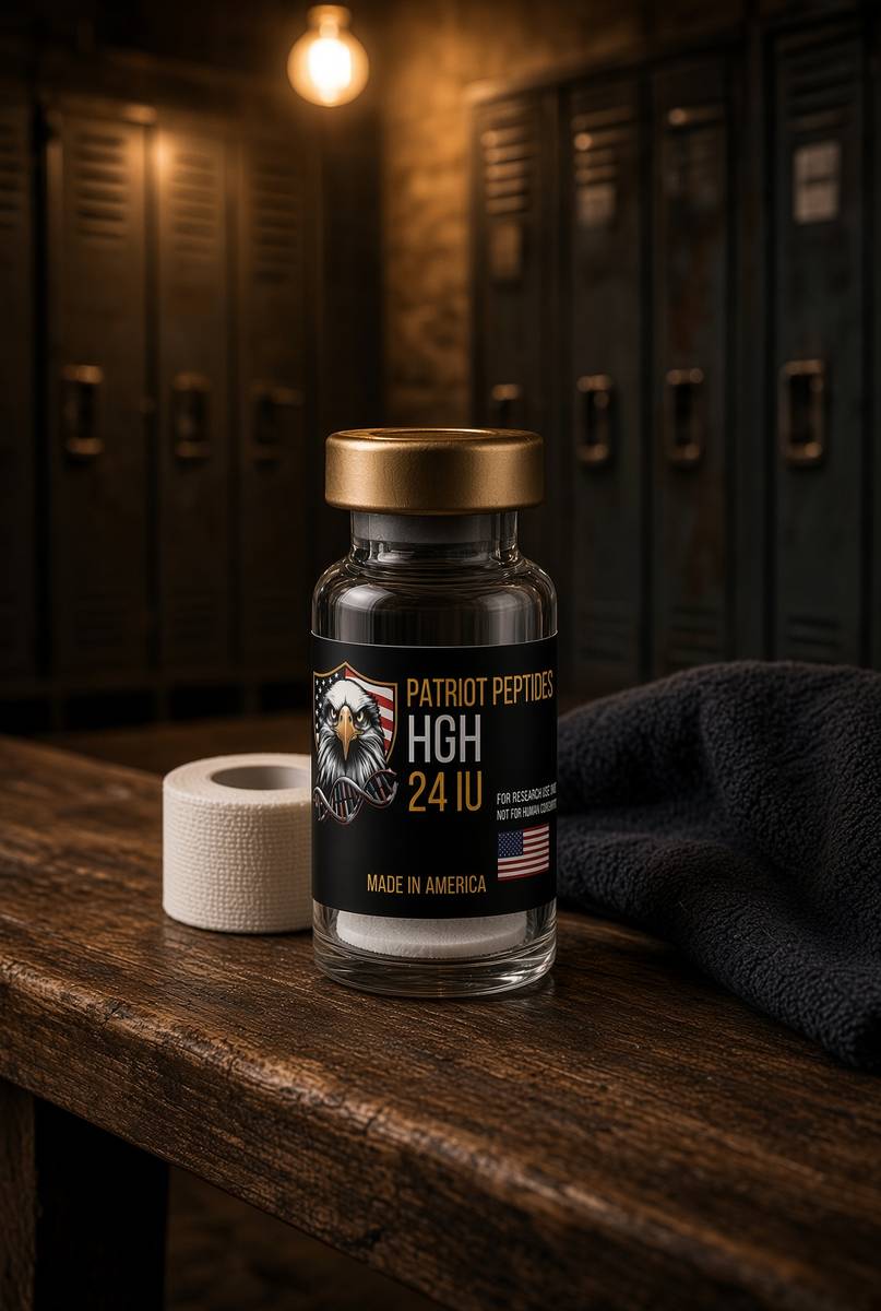
HGH
24 IU
Mix with 2 ml bacteriostatic water → 12 IU/ml
- Hypothetical aliquot
- 1–2 IU
- 0.08–0.17 ml · 8–17 units on a U-100 syringe
- How often
- Once daily, usually an evening aliquot in the somatropin notes
- 8–12 weeks, then a pause
Bench steps
- Let the vial sit until it is no longer cold to the touch.
- Wipe the stopper with 70% isopropyl alcohol. Let it dry.
- Draw 2 ml bacteriostatic water (0.9% benzyl alcohol).
- Run the water slowly down the inside glass — do not blast the cake.
- Roll between finger and thumb until the cake is gone. Do not shake.
- Label the date. Store the solution at 2–8 °C. Do not freeze. Use within 30 days unless the note below says otherwise.
24 IU in 2 ml. Two IU is about 17 units on the syringe. Gentle roll only — somatropin denatures if you beat it.
IGF-1 LR3
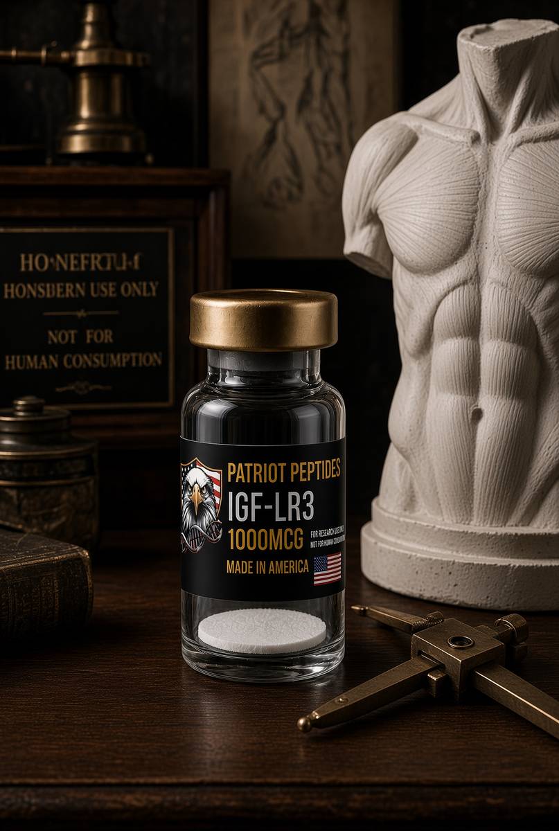
1000mcg
Mix with 1 ml bacteriostatic water → 1,000 mcg/ml
- Hypothetical aliquot
- 20–50 mcg
- 0.02–0.05 ml · 2–5 units on a U-100 syringe
- How often
- Once daily
- 4–6 weeks, then a pause
Bench steps
- Let the vial sit until it is no longer cold to the touch.
- Wipe the stopper with 70% isopropyl alcohol. Let it dry.
- Draw 1 ml bacteriostatic water (0.9% benzyl alcohol).
- Run the water slowly down the inside glass — do not blast the cake.
- Roll between finger and thumb until the cake is gone. Do not shake.
- Label the date. Store the solution at 2–8 °C. Do not freeze. Use within 30 days unless the note below says otherwise.
A thousand micrograms, not milligrams. 50 mcg is 5 units. Over-mixing the cake with a large water volume makes the marks unreadable — 1 ml is the point.
Bacteriostatic water and sterile syringes ship with peptide orders over £75. Kit details · 10 ml bacteriostatic water
The vials this essay sits on
Named sequences the essay maps — CJC without DAC, Ipamorelin, HGH, IGF-1 LR3. Hypothetical research neighbourhood, not a protocol, not a medicine. One press puts every in-stock vial in the bag.
 Research only
Growth axis
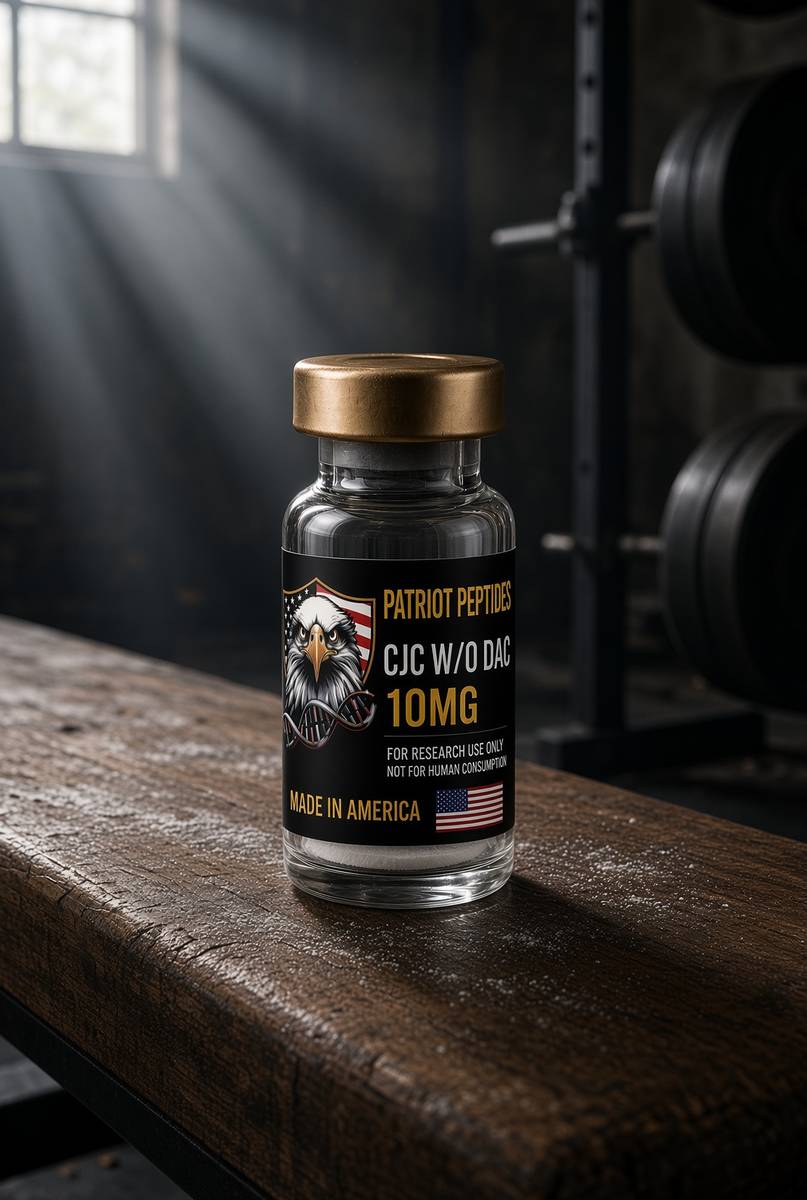
CJC without DAC
10 mg CJC without DAC — a GHRH pulse, not a weekly drip.
4.6(620)
58 browsing this now · 4 purchased in the last 24 hours
10mg · In stock
£30.00
 Research only
Growth axis
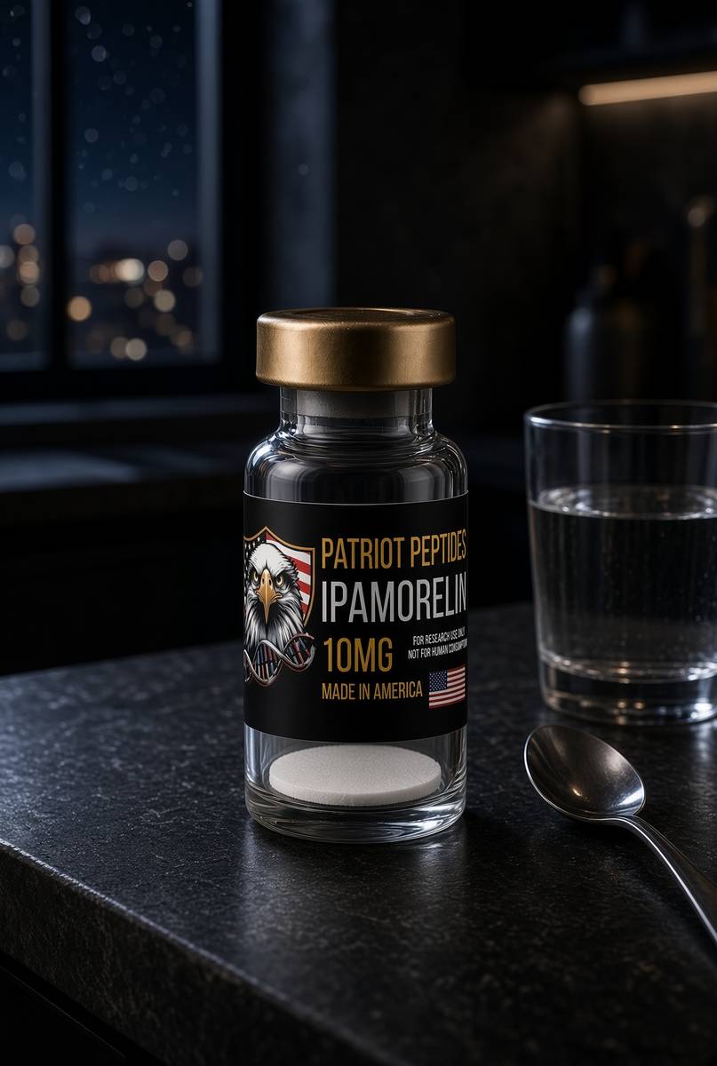
Ipamorelin
10 mg ipamorelin. The clean ghrelin-receptor pentapeptide.
4.7(457)
42 browsing this now · 4 purchased in the last 24 hours
10mg · In stock
£30.00
 Research only
 Research only
Research use only. Not a combined-use instruction.
Read next
41 min · long read · Peptide research
CJC-1295 without DAC: a GHRH analogue that still pulses
Modified GRF(1–29) with four substitutions that dodge DPP-IV, and deliberately without the Drug Affinity Complex that would glue it to albumin for a week. The design constraint is physiology.
51 min · long read · Peptide research
Ipamorelin: the ghrelin receptor without the circus
A pentapeptide GHS-R1a agonist that raises GH with minimal ACTH, cortisol or prolactin. Raun 1998 is still the paper. Selectivity is the entire product.
48 min · long read · Peptide research
Somatropin: the 191-residue ligand
Recombinant human growth hormone is one of the most studied proteins in endocrinology. Pulsatility, GHR–JAK2–STAT5b, lipolysis, IGF-1 generation — the map is public.
48 min · long read · Peptide research
IGF-1 LR3: taking the binding proteins out of the fight
Insulin-like growth factor-1 is the tyrosine-kinase half of the GH conversation. LR3 is an analogue designed to ignore IGFBPs — so the receptor on a cultured cell actually sees it.
More in this desk

53 min · long read · Peptide research
Recovery, GH pulses and the CJC / ipamorelin pair
Somatotrophs have two 'go' receptors. CJC without DAC is a slightly longer GHRH pulse. Ipamorelin is a selective ghrelin-receptor key that does not yank ACTH. Together they are the pair Bowers already showed is more than additive — not a gym protocol.
53 min · long read · Peptide research
How research peptides are made — and why HPLC actually matters
Solid-phase peptide synthesis builds a chain one residue at a time. HPLC then asks whether the main peak is what you think it is. ≥98% is not a slogan. It is a chromatogram.
52 min · long read · Peptide research
Fatigue, cellular energy and the NAD+ / MOTS-c neighbourhood
NAD+ is the rechargeable chip every cell spends on fuel and DNA repair — and the pool shrinks with age. MOTS-c is a mitochondrial 16-mer sent out under metabolic stress. Two answers to 'I have no energy' that are not coffee.
51 min · long read · Peptide research
Melanocortin circuits: pigment, appetite and PT-141
Melanotan II lights MC1, MC3, MC4 and MC5 — pigment plus the rest of the sheet. PT-141 is the free-acid cousin pointed at MC3 and MC4, the circuitry papers use for desire and energy, not skin colour. Same Arizona family. Different question.
Essays describe published research. They are not medical advice and they do not authorise human use of any catalogue item.