


Peptide research · 48 min · 10,499 words
KPV: three residues, an NF-κB off-switch
The C-terminal tripeptide of α-MSH keeps much of the anti-inflammatory pharmacology and almost none of the tanning. Gut epithelium, PepT1, and a very small molecule.
What this essay actually tells you
- KPV is Lys-Pro-Val, the C-terminal tripeptide of α-MSH. It suppresses NF-κB in epithelium and macrophages. Three residues. That's the fragment.
- DSS-colitis models and keratinocyte work are the right sandbox. There is evidence for PepT1 uptake and MC1R-independent activity, which is why it can calm without tanning.
- If your question is pigment, this is the wrong melanocortin fragment. That job belongs to MC1R pan-agonists such as MT2. File KPV under inflammatory tone.
What this actually means
α-MSH is a tanning hormone that also calms inflammation. Most of the pigment lives in the middle of the sequence. Most of the anti-inflammatory signal survives in the last three amino acids: lysine–proline–valine, or KPV. In gut and skin models that tripeptide reduces NF-κB activity, the master switch for a lot of inflammatory transcription, and it can hitch a ride into epithelial cells on the peptide transporter PepT1. That's a precise, small, interesting reagent. It is not a pan-melanocortin agonist and it will not tan a dish, which is why we reach for it when pigment would just get in the way.
KPV is lysine-proline-valine. Three residues, a mass of 342.4 grams per mole, CAS 67724-34-9, the C-terminal tripeptide of α-melanocyte-stimulating hormone. α-MSH itself is thirteen residues, acetylated at the amino terminus, amidated at the carboxyl terminus, sequence Ac-SYSMEHFRWGKPV-NH2, a product of pro-opiomelanocortin once the convertases have finished with ACTH. The last three letters of that sequence are the molecule we’re talking about. Medicinal chemistry is often addition: a D-amino acid here, a lactam bridge there, a pan-agonist that occupies four receptors because the chemist refused to pick one. KPV is subtraction. Keep the tail. Lose most of the pigment, most of the MC4R appetite agonism, most of the autonomic extras a cyclic analogue brings into a first-in-human room. What remains is small enough to be a substrate of the intestinal oligopeptide transporter PepT1, and stubborn enough, in the papers that bother to measure it, to suppress NF-κB in epithelium and in macrophages. That’s a precise, small, interesting reagent. It isn’t a weaker α-MSH. It’s a different question, asked with three letters instead of thirteen.
In short. KPV is the last three amino acids of the tanning hormone α-MSH. Labs keep the anti-inflammatory tail and drop most of the pigment.
The parent hormone is a tanning signal that also calms inflammatory transcription, which is how a pigment peptide walked into a gastroenterology journal and a dermatology review in the same decade. Most of the pigment lives in the middle of the sequence, in the His-Phe-Arg-Trp tetrapeptide that melanocortin receptors actually want in the pocket. Most of the anti-inflammatory signal, on the Luger and Getting reading, survives in the last three amino acids. In gut and skin models that tripeptide reduces NF-κB activity — NF-κB is the master switch for a lot of inflammatory transcription — and it can hitch a ride into epithelial cells on PepT1. Several groups have shown residual activity when MC1R is missing or blocked. That incomplete dependence is the intellectual reason to reach for the fragment instead of α-MSH or Melanotan II when a tan, an appetite crash or a melanocortin extras would confound the assay. File it under inflammatory tone in a barrier tissue. File pigment under a different ligand. The tail does a job the core was never asked to do quietly.
In short. The middle of α-MSH turns pigment receptors on. The last three letters quiet inflammatory genes, and they can enter gut lining on a food-peptide transporter.
We stock lyophilised Lys-Pro-Val, 10 mg, ≥98% by HPLC, because that’s the tripeptide the NF-κB, PepT1 and DSS-colitis papers actually dissolve into a well. The neighbouring melanocortin essay is the family portrait: one precursor, five receptors, a pan-agonist, a licensed MC4R agonist, and this tail. The irritable-bowel essay is the organ portrait: a Rome-criteria cluster, a one-cell lining, BPC-157 on vessels and nitric oxide, KPV on IκB and a nutrient transporter. Neighbourhood isn’t identity. A gastric 15-mer and a melanocortin tripeptide share an organ on a reading list and don’t share a lock. Melanotan II shares a precursor and occupies almost every receptor in the family on purpose. This listing is the fragment designed in the other direction. The 10 mg cake isn’t a gastroenterology appointment and isn’t a tanning protocol. The rest of this piece is the biochemistry you’d want before designing the assay you actually have the controls for: the precursor, the tail, the transcription factor, the gut door, and the mouse models that sit under the word colitis without becoming a diagnosis.
In short. The named three-letter chain is what the inflammation papers dissolve. A tanning analogue and a stomach 15-mer sit nearby. They are different experiments.
Three residues is below the length at which a chain folds into a globular domain. KPV is a ligand and a transporter cargo, not a tiny enzyme. It doesn’t have an active site of its own. Its published jobs are to be recognised, in some assays, at a melanocortin receptor, and, more interestingly, to enter a cell on PepT1 and leave IκB standing so NF-κB p65 stays out of the nucleus. Those are different jobs, and the literature has spent thirty years mixing them. If you can’t say which job you came to measure, you haven’t yet chosen an experiment. A 342-dalton tripeptide isn’t a licensed inflammatory-bowel medicine; the label is the legal class of the object. Replication quality varies by lab, as it does in all peptide inflammation work. The mechanistic core — NF-κB down, PepT1 in, MC1R not required — has been seen more than once, which is the most we ask of a three-residue literature before we put the chain on a shelf. Small isn’t simple. Small is a reason to name the door and the switch, because there’s so little chain to hide behind.
In short. Three amino acids is too short to be a folded machine. The useful claims are a quieter inflammatory switch and a named door into gut cells.
Thirteen residues, then three
The POMC gene sits on the short arm of chromosome 2, at 2p23.3. The primary translation product is a polyprotein with paired basic residues marking the cut sites. Prohormone convertase 1/3 in a corticotroph releases ACTH and β-lipotropin. Prohormone convertase 2, abundant in melanotrophs and in hypothalamic POMC neurons, takes ACTH further; peptidylglycine α-amidating monooxygenase amidates the short piece, and an N-acetyltransferase acetylates it. The product of those two modifications is α-MSH: Ac-SYSMEHFRWGKPV-NH2. Thirteen residues is a small hormone, and almost every position has been mutated by someone with a reason. The N-terminal acetyl and the C-terminal amide aren’t decorations. They slow exopeptidases and they change potency at the receptors. γ-MSH comes from the N-terminal fragment. β-endorphin comes from β-lipotropin. One gene, a handful of convertases, a tissue-specific peptide cocktail. A corticotroph isn’t a melanotroph wearing a different hat. Write POMC peptides as if they were interchangeable and you haven’t yet chosen a ligand. KPV is the last three letters of one of those products, after the convertases and the amidating enzyme have done their work.
In short. One precursor protein is cut into ACTH in the pituitary and into the thirteen-residue tanning hormone elsewhere. KPV is the last three letters of that hormone.
The core pharmacophore of α-MSH is His-Phe-Arg-Trp, residues six to nine, the HFRW tetrapeptide that melanocortin receptors actually want in the pocket. That’s the tanning pharmacophore, the appetite pharmacophore, the sebum pharmacophore: one motif, five receptors, different tissues. Melanotan II was built around a cyclic version of that core, with a D-phenylalanine that proteases dislike and a lactam that holds the ring shut. Setmelanotide was built around the same ancestral pocket, pointed at MC4R. Afamelanotide is [Nle4, D-Phe7]-α-MSH, the linear photoprotective analogue. All of those molecules are commentaries on HFRW. KPV is what remains when you throw the core away. Lysine, proline, valine. A charged amino terminus, a stiff ring in the middle, a hydrophobic carboxyl end. Often C-terminally amidated in the research solid, matching the parent hormone’s amide. The fragment isn’t a weaker key for the same lock. It’s a short chain that can still quiet a transcription factor after the lock you first thought of has been taken off the door. That’s the design, and it’s why three letters earned their own vial.
In short. The middle four amino acids of α-MSH fit the pigment and appetite receptors. KPV is what you have when you throw that core away.
Write the three letters in three-letter code and the chemistry is already visible. Lysine carries a primary amine on a four-carbon tether, solvent-exposed, a place for a charge and, in some older binding papers, a handle people argued about at MC1R. Proline is the residue that does most of the telling at this length. The side chain loops back onto the backbone nitrogen, so proline is both an amino acid and a fold-breaker; it starves several proteases of a comfortable cut site and it kinks a tripeptide into a shape a transporter can notice. Valine is a branched hydrophobic, the carboxyl end of the parent hormone before amidation. Theoretical monoisotopic mass for the free tripeptide is 342.4 daltons; the molecular formula is C16H30N4O4 if you write the uncharged chain. The lyophilised cake on a certificate will also report trifluoroacetate or acetate counter-ions depending on the cleavage and the salt exchange, and a conscientious write-up says which. Purity ≥98% HPLC is a peak, not a physiology. A dipeptide missing the valine can hide under a cheap ultraviolet trace and still wreck an uptake assay, which is why the interesting chromatogram is the one with a mass on it.
In short. Lysine, proline, valine: a charge, a stiff kink, a hydrophobic end. Mass about 342 daltons. A clean peak with that mass is identity.
Five G-protein-coupled receptors, MC1R through MC5R, read the parent family. They’re primarily Gs-coupled, so occupancy raises cyclic AMP. What the cell does with that rise depends on which receptor you occupied and where it lives. MC1R on melanocytes pushes tyrosinase toward eumelanin; on macrophages and keratinocytes it’s also the receptor the inflammation literature first reached for. MC2R is the ACTH receptor on the adrenal cortex and wants a longer ligand. MC3R and MC4R live in hypothalamus, with MC4R the appetite brake Farooqi and O’Rahilly put into the obesity clinic and setmelanotide later occupied on an FDA label. MC5R lives on sebaceous epithelium. Treating the family as a sunbed story is how you miss four of those five sentences. Treating every fragment of α-MSH as a Gs ligand is how you miss the point of KPV. The fragment can still bind MC1R in some assays. The reason it exists as a research object is that a useful fraction of the anti-inflammatory work doesn’t require that binding. The family essay next door is the receptor sheet. This essay is the tail that was kept when pigment would just get in the way.
In short. Five related receptors read the parent hormone: pigment, adrenal, appetite, oil glands. The tripeptide was kept because a lot of its quieting work does not need the pigment receptor.
A circulating hormone of thirteen residues is already small. A fragment of three is a different pharmacokinetic object, and that’s chemistry rather than a shorthand. Di- and tripeptides are the native cargo of PepT1 and PepT2, proton-coupled oligopeptide transporters that exist to harvest dietary protein after brush-border peptidases have finished. A thirteen-residue amidated hormone isn’t that cargo. A cyclic heptapeptide with a lactam isn’t that cargo. Lys-Pro-Val is exactly that cargo: two peptide bonds, a free amino terminus, a size the binding site of SLC15A1 was built for. Dalmasso, Merlin and colleagues made that the sentence a gut paper has to cite. Oral delivery in a DSS mouse is coherent for this chain in a way it isn’t coherent for Melanotan II. Coherence isn’t a capsule claim. Coherence is why the colitis models keep turning up, and why adding KPV to a medium and adding α-MSH to a medium aren’t the same experiment even when the downstream blot is p65. Size decided the door. The door decided the organ. The organ isn’t a licence. It’s a reason the fragment and the parent hormone ask different questions in the same tissue.
In short. Food protein is cut to two- and three-letter pieces that a gut transporter is built to carry. KPV fits that hole. The full tanning hormone does not.
Diagram
MC1R
melanocyte
Eumelanin vs pheomelanin. Red-hair alleles. Afamelanotide’s receptor.
MC3R
hypothalamus
Energy balance. Occupied by MT2 because MT2 occupies almost everything.
MC4R
hypothalamus
Appetite brake. Loss-of-function obesity. Setmelanotide (FDA 2020).
MC5R
sebaceous
Sebum. A first-class output people still treat as a footnote.
KPV
tail of α-MSH
NF-κB off-switch. PepT1 uptake. Designed not to tan.
MT2
pan-agonist
Ac-Nle-cyclo[Asp-His-D-Phe-Arg-Trp-Lys]-NH2. Pigment, appetite, flushing, arousal.
α-MSH is Ac-SYSMEHFRWGKPV-NH2. The last three letters are KPV. Melanotan II is what a pan-agonist looks like. KPV is the design move in the other direction. Match the ligand to the question.
NF-κB, IκB, and the papers that named the off-switch
Nuclear factor kappa-light-chain-enhancer of activated B cells is a transcription-factor family, not a mood. In the canonical pathway the heterodimer that matters is p65/RelA with p50. In a resting cell that dimer sits in the cytosol, bound to IκBα, the inhibitor whose name is the whole mechanism: inhibitor of κB. A receptor signal — TNF receptor 1, IL-1 receptor, a Toll-like receptor, a dozen others — converges on the IκB kinase complex, IKKα/IKKβ/NEMO. IKK phosphorylates IκBα on serines 32 and 36. The phosphodegron is recognised by a β-TrCP ubiquitin ligase, IκBα is proteasomally destroyed, and p65 goes to the nucleus. On the DNA it writes TNF, IL-6, IL-1β, IL-8, COX-2, a cassette of adhesion molecules and chemokines, the inflammatory page a barrier tissue turns to when the lining is under attack. Sen and Baltimore named the factor in B cells in 1986. Karin, Maniatis, Hoffmann, Baltimore again: the kinase and the degron followed. If a peptide paper says anti-inflammatory and can’t name IκB, it’s described a feeling. The KPV literature that earned its keep named the inhibitor and asked whether it was still there. That’s the switch we’re going to keep naming.
In short. NF-κB is a gene switch for inflammatory proteins. A partner called IκB holds it in the cytosol. Destroy IκB and the switch enters the nucleus and writes cytokines.
Preservation of IκB is the mechanistic sentence Luger’s dermatology group and Getting’s inflammation group kept returning to. α-MSH, and then the C-terminal tripeptide, reduced IκB degradation after TNF, IL-1β or lipopolysaccharide, so p65 nuclear translocation fell, so NF-κB reporter activity fell, so the cytokine transcripts fell. Brzoska, Luger, Böhm, Haycock, Moustafa: keratinocytes, melanocytes, melanoma lines, macrophages, a set of cells that already had a reason to express MC1R and a set that made the MC1R-independence question worth asking. The 2003 Annals of the New York Academy of Sciences review from Luger’s group, the 2008 Endocrine Reviews from Brzoska, Luger, Maaser, Abels and Böhm, Getting’s pharmacological targeting papers out of the William Harvey: those are the papers to put on the bench. Cyclic AMP and PKA are the default melanocortin second messengers, and in some of these assays they still matter. In others the fragment quieted NF-κB without a cAMP rise you could measure, which is already a clue that the tail isn’t just a short α-MSH. A second-messenger diagram still earns a place later, so we don’t borrow amplification language for a chain that often works by leaving a cytosolic inhibitor standing.
In short. Luger's skin group and Getting's inflammation group showed the tripeptide can keep IκB from being chewed up, so NF-κB stays out of the nucleus.
Name the cytokines, because they are easy to skip. TNF, tumour necrosis factor, is both an NF-κB target and an NF-κB inducer: a feed-forward loop a barrier tissue can trap itself in. IL-6 is the acute-phase writer the liver is listening to. IL-1β is the inflammasome product that IL-1 receptor then hands back to IKK. IL-8, CXCL8, is the neutrophil chemokine a monolayer secretes into the medium when you want a number rather than a blot. Dalmasso measured IL-8 in Caco-2 monolayers. Keratinocyte papers measured IL-1β and TNF transcripts, and sometimes an NF-κB luciferase. Macrophage papers measured the same cassette after LPS. Antimicrobial-adjacent effects have been described for α-MSH fragments, as they have for other cationic peptides of this size, and they’re a different assay: killing curves, not reporter genes. Replication quality on the antimicrobial side is thinner. The mechanistic core — NF-κB down, IκB preserved, p65 less nuclear — has been seen in more than one cell type, which is why it’s the sentence we’ll keep. A cytokine ELISA without an IκB blot is a phenotype. An IκB blot without a PepT1 or MC1R control is still a phenotype, just a more fashionable one.
In short. The genes this switch writes include TNF, IL-6, IL-1β and IL-8. Papers that name those, and show IκB still present, are the ones worth keeping.
Keratinocytes are the skin half of the sandbox, and they aren’t melanocytes. A keratinocyte is the barrier cell of epidermis; it expresses MC1R at low level in several reports, it runs a great deal of NF-κB when TNF or IL-1 hits it, and it’s the cell Haycock and Moustafa used when they asked whether α-MSH and KPV could stop p65 after TNF. HaCaT, the spontaneously immortalised human keratinocyte line, is the usual workhorse, with the usual caveats of a line that isn’t primary and isn’t a reconstructed epidermis. Primary human keratinocytes are the better object and the worse cell-culture week. Contact-hypersensitivity models in mice, from Luger’s group, put the tripeptide into a living ear or a living back and asked about sensitisation and elicitation, which is a different experiment from a luciferase well. Topical and systemic both appeared in those papers. A reconstructed epidermis or a living ear isn’t a cream. It’s a barrier assay with a named hapten. The catalogue vial is a laboratory solid for those assays, not a moisturiser. The interesting part was always the named hapten, the named cell, and whether p65 still went to the nucleus.
In short. Skin lining cells are one sandbox: after an inflammatory push, the tripeptide can keep NF-κB down. That is a dish or a mouse-ear assay, not a cream.
Macrophages and other myeloid cells are the immune half. LPS through TLR4, IKK, IκB, p65, TNF out: the canonical innate loop. α-MSH has been a macrophage peptide since the 1980s, which is why Lipton and Catania’s fever and inflammation reviews sit in the same library as Luger’s dermatology. KPV inherited a fraction of that pharmacology. Getting’s group spent a career putting melanocortin peptides into gouty, ischaemic and inflamed tissues and asking which receptor did the work; MC3R, not MC1R, is the receptor a lot of that macrophage work actually wants, and ACTH fragments rather than KPV are often the ligands. That distinction is how you stop a family review becoming a single-molecule cartoon. For the tripeptide, the myeloid sentence is: NF-κB reporter activity falls in macrophages in several papers, IL-1β and TNF follow, and MC1R dependence is incomplete. Dalmasso later showed PepT1 on immune cells as well as on epithelium, which is a second door into the same cell. Two doors, one transcription factor, a set of cytokines. Name which door you closed if you’re about to claim a mechanism. The family is allowed to be larger than this fragment. This fragment still has to name its own door.
In short. Immune cells are the other sandbox. After bacterial-wall molecules, the same inflammatory switch can fall. Which door the tripeptide used still has to be named.
MAP kinases sit next to NF-κB in the Dalmasso figures, and they deserve a paragraph so they don’t become a rumour. ERK, JNK and p38 are three phosphorylation cascades a cytokine receptor or a Toll-like receptor can light at the same time as IKK. Dalmasso reported that nanomolar KPV reduced those phosphorylations in Caco-2 cells after IL-1β, alongside the NF-κB result. Later keratinocyte papers, including work on particulate-matter injury, have put p38 and ERK back on the blot. A peptide that quiets both IKK and a MAP-kinase cassette still isn’t a pan-kinase inhibitor; it’s a ligand or a cargo whose downstream look includes more than one node. The clean experiment is still IκB, because IκB is the named off-switch in the title of this piece and because a p38 blot without a PepT1 knockdown is a phosphorylation anecdote. Run both if you have the wells. Write which one you’re hanging the mechanism on. The off-switch metaphor is allowed once. After that it is IκB preservation, p65 localisation, a reporter, a cytokine. Metaphor is how you enter the heading. Named proteins are how you leave it, and they’re how a three-residue paper stays a three-residue paper.
In short. Some papers also show quieter MAP-kinase signals. The named off-switch remains IκB: keep that protein, and NF-κB stays out of the nucleus.
Most of the anti-inflammatory activities of α-MSH can be attributed to its C-terminal tripeptide KPV. Pigment is the obstacle that made the fragment interesting. IκB is the protein that made it mechanistic.— Brzoska T, Luger TA, Maaser C, Abels C, Böhm M. α-Melanocyte-stimulating hormone and related tripeptides. Endocr Rev. 2008; 29: 581–602. Reading against Haycock and Moustafa on p65 in keratinocytes.
PepT1 is a nutrient transporter, not a melanocortin receptor
PepT1 is SLC15A1, a proton-coupled oligopeptide transporter of the major facilitator superfamily, twelve transmembrane helices, an extracellular face that binds di- and tripeptides, a proton that rides in with the cargo. Daniel, Rubio-Aliaga, Adibi, the physiological reviews: this is how a small-bowel enterocyte harvests the bulk of dietary amino acids after brush-border peptidases have cut protein to two- and three-residue pieces. The driving force is the proton electrochemical gradient the apical sodium-proton exchanger helps to keep. Glycyl-sarcosine, Gly-Sar, is the non-hydrolysable dipeptide you’d use as a tracer and as a competitor. The transporter sits on the apical membrane of small-bowel epithelium at high abundance in health. Colon epithelium in health expresses little of it. Inflamed colon, in human inflammatory-bowel biopsies and in several rodent models, induces it. That induction is the pharmacokinetic gift the KPV gut papers live on: a ligand small enough to be cargo, a transporter that appears where the inflammation is. A GPCR on the surface is occupancy without entry. PepT1 is entry. Name which you closed. The door and the receptor are not interchangeable sentences, even when they sit on the same cell.
In short. PepT1 is a gut door for two- and three-amino-acid food pieces. Inflamed colon makes more of it. KPV is the right size to walk through.
Dalmasso, Charrier-Hisamuddin, Nguyen, Yan, Sitaraman and Merlin, Gastroenterology 2008, is the paper to put on the bench. Caco-2-BBE monolayers, apical KPV, rapid intracellular acidification consistent with proton-coupled uptake, competition with Gly-Sar, nanomolar KPV reducing NF-κB and MAP-kinase activation after IL-1β, less IL-8 secreted. siRNA against PepT1 abolished the anti-inflammatory effect in the dish. Immune cells carrying PepT1, not only enterocytes. Then the mouse: KPV in the drinking water, dextran-sodium-sulphate colitis and TNBS colitis, histology and cytokine mRNA down. PepT1-null animals didn’t get the benefit, which is the specificity check a transporter paper actually owes. The discussion section of that paper wondered about inflammatory-bowel therapy. Discussion sections are allowed to wonder. A research tripeptide doesn’t inherit a wonder as a claim. The experimental object that survived is uptake via SLC15A1, IκB/NF-κB quieter, two chemical-colitis models improved on oral fragment. That object is why oral KPV is a coherent design in a colitis assay, and why the DSS models keep turning up. Coherence isn’t a human capsule. It’s a mouse and a filter that named the door.
In short. A 2008 gut paper showed KPV enters lining cells on PepT1, quiets NF-κB, and eases two mouse colitis models when PepT1 is present.
Caco-2 is a human colon-adenocarcinoma line that polarises on a filter and pretends, usefully, to be small-bowel epithelium. It expresses PepT1. It’s the monolayer Dalmasso used, and it’s the monolayer a new bench will use if it wants to repeat the uptake half without a mouse. Transepithelial electrical resistance, 14C-Gly-Sar flux, intracellular pH dyes, an NF-κB luciferase or a p65 antibody, IL-8 ELISA: those are the machines. A filter that was never tight isn’t a barrier paper. A Gly-Sar competition that was never run isn’t a PepT1 paper. An siRNA without a rescue, or a knockout mouse without a littermate, is a suggestion. The 2008 figures already contain the design. Copy them. HT-29, T84, primary human colonoids, murine enteroids: lawful next objects, each with a different PepT1 abundance and a different tight-junction baseline. A HEK293 well transfected with SLC15A1 is how you ask whether the transporter is sufficient for uptake. It isn’t how you ask what a colonocyte does with IκB. Sufficiency and physiology are two experiments. Write which one you ran. The filter is allowed to be boring. Boring is how you know the door was this one.
In short. Polarised gut-cell filters, a competitor dipeptide, and a PepT1 knockdown are how you show the door was this transporter. A mixed dish is not that proof.
Induction in inflammation is the other half of the transporter story, and why a healthy colon and an inflamed colon aren’t the same pharmacokinetic neighbourhood. Merlin’s earlier papers, and the human biopsy literature that followed, put PepT1 protein on inflamed colonic epithelium where textbooks said it should be almost absent. Bacterial peptides, muramyl dipeptide, formyl-Met-Leu-Phe, can ride the same door; some of those cargoes are inflammatory, the darker twin of the KPV design. A transporter that can import an anti-inflammatory tripeptide can also import a bacterial peptide that lights NOD2 or a formyl-peptide receptor. The door is a hole with a substrate preference, not a virtue. KPV was chosen because it’s cargo and because, once inside, it quiets NF-κB rather than lighting those sensors. That’s a design, not a guarantee that every tripeptide on PepT1 will be anti-inflammatory. Gly-Sar is a tracer and a competitor, not a cytokine suppressor. Identity of the cargo still matters once the hole has been named. Block PepT1 and lose the KPV phenotype, and you’ve named the door. Never ask about NOD2, and you haven’t yet asked whether the well was clean of bacterial peptides.
In short. Inflamed colon grows more of this transporter, which is why the fragment can reach the injured sheet. The same door can also carry bacterial peptides, so name the cargo.
PepT2, SLC15A2, is the higher-affinity cousin, abundant in kidney proximal tubule and in glia, a clearance and a brain-border story rather than a gut-harvest story. Some KPV papers mention it. Most of the colitis work doesn’t need it. Peptide transporters are also in the lysosomal membrane (PHT1/SLC15A4, PHT2/SLC15A3), which matters for NOD2 sensing of imported muramyl peptides and is a different invoice from apical PepT1 on an enterocyte. If your claim is oral fragment in a DSS colon, PepT1 is the named door. If your claim is a macrophage in a dish, ask whether that macrophage’s PepT1, or an MC1R, or both, carried the result. Dalmasso put the transporter on immune cells and on epithelium; that sentence is a fork, not a merger. Forks are how a three-residue literature stays honest. Merging them is how a transporter paper becomes a receptor paper becomes a blur. The melanocortin receptors are still GPCRs: occupancy outside, cAMP inside, no requirement for the ligand to enter. PepT1 is the exception that proves the rule for this fragment. Name the lock. Then ask whether cargo is even the point.
In short. A related kidney transporter and some immune-cell doors exist. For a gut-lining experiment, PepT1 is the door to name. Receptor occupancy is a different kind of lock.
DSS-colitis is a model, not a diagnosis
Dextran sodium sulphate is a sulphated polysaccharide that chews the colonic mucus gel and then the sheet underneath. Johansson, Hansson and colleagues at Gothenburg spent a decade showing that colon mucus is two layers: an inner MUC2 gel that is normally sterile, and an outer layer that is looser and colonised. DSS attacks the polymer first. Bacteria reach the epithelium. Neutrophils follow. Myeloperoxidase in the tissue becomes a number. Disease-activity index — weight, stool blood, stool form — becomes another number. Histology, a crypt-damage score, becomes a third. That’s a mucus-injury model before it is an immune model, and it looks a bit like ulcerative colitis on a good slide and looks nothing like Tuesday bloating in a person who meets Rome IV and has a normal calprotectin. TNBS, trinitrobenzene sulphonic acid in ethanol, is a different chemical colitis, more T-cell, more Crohn’s-adjacent on a slide, still not a person. Dalmasso ran both. Kannengiesser, Maaser, Luger and colleagues, Inflammatory Bowel Diseases 2008, put KPV into murine colitis in the same year from the dermatology-and-gut side. Two 2008 mouse neighbourhoods. One fragment. Chemical injury, not a clinic. Useful, named, and still a mouse.
In short. DSS is a chemical that chews colon mucus and then the lining. It is a useful mouse injury. It is not irritable bowel and not a human diagnosis.
What those papers reported is the positive finding worth keeping. Disease activity down. Myeloperoxidase down. Inflammatory transcripts down, including after oral delivery, consistent with PepT1 uptake in inflamed gut. Histology less ugly. Cytokine mRNA — TNF, IL-1β, IL-6, the usual cassette — quieter. Kannengiesser’s group, working from the Brzoska and Luger library, saw anti-inflammatory potential in murine IBD models and said so in the title; potential is the right word and should have been left in the title when the PDF started travelling. Replication quality varies by lab, as it does in all peptide inflammation work. Strain, DSS lot, microbiota, the week the animal house had a problem, whether the drinking-water peptide was still the peptide on Friday: those are the covariates a colitis write-up actually lives on. The mechanistic core, NF-κB down and PepT1 in, has been seen more than once. That’s the most we ask of a three-residue literature before we stop calling it a rumour. It also isn’t a licence to caption a vial with a syndrome. A reduced DAI in a DSS mouse is a reduced DAI in a DSS mouse. Hold the finding at the size of the finding, and it stays interesting.
In short. Mouse papers report less disease score, less neutrophil enzyme and quieter cytokine genes, including when the fragment is drunk. That is a model result, not a treatment.
Inflammatory bowel disease is a different drawer, and the two searches keep landing on the same afternoon. Crohn’s disease can involve any stretch of gut, often ileum and colon, transmural, discontinuous, granulomas on a good day. Ulcerative colitis is continuous mucosal inflammation from the rectum upward. Both have ulcers, bleeding, strictures, dysplasia risk, and an endoscopic score a trial can move. Both have licensed medicines: corticosteroids and 5-aminosalicylates as the old layer; thiopurines; anti-TNF antibodies; vedolizumab against α4β7; ustekinumab and the IL-23 p19 antibodies; Janus-kinase inhibitors; sphingosine-1-phosphate receptor modulators. Infliximab is a 150-kilodalton chimeric antibody against TNF. Vedolizumab stops α4β7-positive lymphocytes homing to MAdCAM-1. Those are occupancy stories with Phase 3 programmes and risk registers. Lys-Pro-Val is a named sequence with animal and cell papers. Dalmasso’s discussion wondered whether the fragment might become a therapeutic agent for IBD. A wonder in a discussion isn’t a marketing authorisation, and it isn’t this listing. Human inflammatory-bowel medicine exists. It isn’t this chain. Adjacent organ is a lawful reason to put two papers on the same reading list. It isn’t a reason to merge the drawers.
In short. Crohn's and ulcerative colitis have ulcers and licensed drugs — antibodies, gut-selective integrins, kinase inhibitors. Those medicines are not this three-letter chain.
Irritable bowel syndrome is a third drawer, and the search that lands people on the neighbouring essay. Rome IV, Drossman, 2016: recurrent abdominal pain on average at least one day a week in the last three months, related to defecation or to a change in stool frequency or form, onset at least six months before. A checklist, with subtypes by Bristol stool form, and no receptor, claudin or peptide attached. Functional, in this vocabulary, means no currently demonstrable structural lesion on ordinary endoscopy and histology. It doesn’t mean imaginary, and it doesn’t mean the enteric nervous system is uninvolved. It means the lesion that would re-file the case as Crohn’s or ulcerative colitis hasn’t been found. DSS-colitis isn’t IBS. NSAID gastric lesions, the usual BPC-157 sandbox, aren’t IBS. Adjacent organ is a lawful reason to put two papers on the same reading list, not a reason to caption a vial with a syndrome. The IBS essay unbundles the cluster from the two sequences. This essay stays with the tripeptide, the transporter, and the transcription factor. A Rome object doesn’t become a PepT1 object because they share a search. Pain-and-stool is a checklist. This chain is three letters and a blot.
In short. IBS is a pain-and-stool checklist without a single lesion. It does not name this peptide. A mouse colitis model is a different object again.
Ussing chambers and FITC-dextran are how a lining becomes a number, and they belong here so a colitis score isn’t the only figure. Transepithelial electrical resistance is the ohmic summary of the paracellular seal plus the transcellular path; a tight colon epithelium sits in the hundreds to thousands of ohm·cm², a broken DSS sheet much lower. Flux of 4 kDa FITC-dextran reports a different size window. Claudin-2, the cation pore, rises in human IBD and in several rodent colites; myosin light-chain kinase can open a leak pathway for larger solutes. None of those proteins is a receptor for KPV. KPV’s published door is PepT1, on the apical face, a transporter. A junction blot can still move in a peptide paper if the sheet is less injured. Moving a blot isn’t occupancy of the protein on the blot. Keep the causal verb honest. If you claim barrier rescue, show TEER, show a sized tracer, show a histology, and then show that PepT1 was required. If you only have a DAI, you have a sick-mouse score. Sick-mouse scores are allowed. They aren’t a tight junction.
In short. Labs can measure the lining as electrical resistance and as leak of a labelled dye. A healthier score in a sick mouse is not the same as a named seal protein.
Diagram
Mucus
MUC2 gel
The first argument a microbe has to win.
Epithelium
enterocyte · goblet · Paneth
One cell thick. The wall is the cell, not a fascia.
Tight junctions
claudin · occludin · ZO-1
The seal. ‘Leaky gut’ as a brand is not this protein list.
PepT1
SLC15A1
Oligopeptide transporter. Inflamed gut induces it. KPV can ride it.
Lamina propria
immune cells
Where NF-κB decisions become cytokines.
BPC-157 neighbourhood
NO · VEGFR2 · FAK
Cytoprotection, blood flow, how a damaged lining organises.
IBS is a Rome-criteria cluster. It does not name a receptor. The preclinical literature that named molecules for barrier tissue keeps coming back to BPC-157 and KPV — two ligands, one organ on a reading list, neither a gastroenterology appointment.
- KPV
- Lys-Pro-Val, 342.4 Da
- α-MSH
- 13 residues
- PepT1
- SLC15A1
- Dalmasso
- Gastroenterology 2008
- NF-κB
- p65 / IκBα
- MC1R dependence
- incomplete
- Catalogue cake
- 10 mg, ≥98% HPLC
- GLOW neighbour
- 10 mg in an 80 mg blend
C-terminus of α-MSH. CAS 67724-34-9. Often amidated.
Ac-SYSMEHFRWGKPV-NH2. HFRW is the core. KPV is the tail.
H+-coupled di/tripeptide transporter. Inflamed colon induces it.
Uptake, NF-κB, DSS and TNBS. PepT1-null loses the benefit.
Luger, Getting, Brzoska, Haycock. Preservation of IκB is the sentence.
Residual activity in null or blocked systems is the point of the fragment.
Lyophilised tripeptide. Reagent. Not an IBD medicine.
With GHK-Cu, BPC-157, TB-500. Cap-count. Not a mechanism.
MC1R-independent activity is the point
If KPV were only a short α-MSH, you wouldn’t need it. You’d use α-MSH, or you’d use Melanotan II, and you’d accept the tan, the cAMP, the MC4R appetite tone and the extras a pan-agonist writes into an autonomic nervous system. The fragment exists because a useful fraction of the anti-inflammatory pharmacology survives when the tanning receptor is gone. Residual activity in MC1R-null cells, in antagonist-blocked systems, and in assays where cAMP does not rise, is the experimental object. Luger, Getting, Haycock, and the groups that bothered to knock the receptor down or block it, are the citations. Incomplete dependence isn’t a failure of the parent hormone. Incomplete dependence is the design. Pigment wants HFRW in the MC1R pocket and a MITF programme in a melanocyte. Inflammatory tone in a barrier tissue wants IκB preserved in a keratinocyte or an enterocyte, and it can get there, in the Dalmasso design, through a nutrient transporter that has never heard of melanocortin. That’s why a lab reaches for this chain when pigment would just get in the way. The tail is the tool you pick when the core would flood the readout.
In short. If the pigment receptor explained everything, you would use the full hormone or a tanning analogue. The fragment is used because a lot of the quieting survives without that receptor.
Melanotan II is the comparison that makes the design visible, and it’s a different listing. Victor Hruby and Mac Hadley at the University of Arizona built a cyclic lactam, Ac-Nle-cyclo[Asp-His-D-Phe-Arg-Trp-Lys]-NH2, as a sunless-tanning analogue in a photoprotection programme. It occupies MC1, MC3, MC4 and MC5 because they built it to. The 1990s clinic noticed appetite falling, faces flushing, arousal rising. Those are MC4R, MC3R and autonomic consequences of occupying the whole family at once. Palatin later took a close cousin, bremelanotide, PT-141, to an FDA label for hypoactive sexual desire. Rhythm took setmelanotide to an FDA label for rare MC4R-pathway obesity. Afamelanotide, Melanotan I, is licensed as Scenesse for erythropoietic protoporphyria. The family left the tanning booth years ago, and the pan-agonist remains the research tool you use when the question is the whole sheet. KPV is the tool you use when the question is NF-κB and the sheet would be noise. Same precursor. Opposite design. Two certificates. Two masses. Two jobs. Pick the one that matches the measurement, and the extras stay out of the well.
In short. Melanotan II occupies almost every receptor in the family on purpose: pigment, appetite, flushing. KPV was kept so those extras do not flood the inflammatory readout.
cAMP is still allowed to be in the picture, which is why the second-messenger diagram is in this essay rather than being banned from it. Occupancy at MC1R rearranges seven helices, Gs runs, adenylyl cyclase makes cyclic AMP, PKA phosphorylates CREB, and in a melanocyte that writes MITF and tyrosinase. In a macrophage, cAMP can also lean on inflammatory transcription, a real and older pharmacology. Some KPV assays still live in that neighbourhood: a little binding, a little cAMP, a little IκB. Some do not. Haycock’s keratinocyte work reported calcium transients and NF-κB inhibition without a cAMP rise you could be proud of. Dalmasso’s PepT1 work is intracellular cargo, not a Gs occupancy. The picture is a Venn diagram, not a single arrow. A fraction of the pharmacology is still a melanocortin receptor. A fraction is a transporter and a cytosolic transcription-factor neighbourhood. The reason to pick the tripeptide is the second fraction, and the reason to keep MC1R controls in the well is the first. Dropping the receptor control because the title said independent is how an independent-activity paper becomes uninterpretable. Keep both controls. Then you’ll know which circle you were in.
In short. Some of the effect can still run through the pigment receptor and cyclic AMP. A lot of the interesting effect does not. Keep both controls.
Setmelanotide is the licensed reminder that this family isn’t a cosmetic, and it’s also the reminder that KPV isn’t an appetite drug. Imcivree, FDA 2020, an MC4R agonist for obesity caused by POMC, PCSK1 or leptin-receptor deficiency, later Bardet-Biedl. Farooqi and O’Rahilly, New England Journal of Medicine 2003, put heterozygous MC4R loss-of-function in the clinic as the commonest monogenic severe obesity. The neuron is an appetite brake; break the receptor and the brake fails. α-MSH occupies that receptor because HFRW fits. Melanotan II occupies it because a pan-agonist occupies it. KPV, on the published pharmacology, doesn’t run that brake in any way that would make it an MC4R probe. Body-weight work wants a different ligand. Pigment work wants a different ligand. Sexual-function work wants PT-141 or the Arizona parent, and those are different listings again. Inflammatory tone in epithelium and macrophages is the job this chain was isolated for. Use it as one. A pan-agonist will also tan, suppress appetite and surprise the autonomic nervous system, which is a lot of noise if what you wanted was IκB. Licensed neighbours are how you keep the fragment’s job small and interesting.
In short. A licensed appetite drug occupies a brain receptor in this family. KPV is not that drug. Pigment and body-weight questions want other ligands.
What a pan-agonist confounds, in practice, is the dish you thought you were running. A melanocyte contaminant in a keratinocyte culture will tan the readout. An MC4R-positive neuron in a mixed hypothalamic prep will change firing when you wanted cytokines. A mouse given a cyclic heptapeptide will eat less, flush, and, in the old Arizona rooms, report arousal, while you’re trying to score a colon. None of those extras is a reason to moralise about Melanotan II. They’re a reason to pick the fragment when the extras would drown the signal. They’re also a reason not to stack the fragment with the pan-agonist and call the stack a protocol. Two ligands, two questions. Occupying the whole sheet asks whether the family is involved. Offering the tail asks about inflammatory transcription without the tan. Both words contain melanocortin. Biochemistry didn’t glue them. The neighbouring family essay is the portrait in which both appear. This essay is the tail, at catalogue length, so the pan-agonist can’t wander back into the well by habit. Match the ligand to the measurement, and the extras stay where they belong: on the other listing.
In short. A tanning analogue in the wrong dish adds pigment, appetite and autonomic noise. Pick the tail when those would drown the inflammatory measurement.
Diagram
× 1
Ligand
One peptide in one pocket. nM–µM. Shape, not a mood.
× 10–10²
G proteins
The occupied GPCR is a GEF. Each Gα is a catalyst.
× 10³–10⁴
cAMP / IP₃ / Ca²⁺
Adenylyl cyclase and PLC do not make one molecule. They make a cloud.
× 10⁴–10⁶
PKA / PKC / CaMK
Kinases phosphorylate many substrates per messenger.
× tissue
Secretion, transcription, motility
The organism-level readout. Still not a protocol.
This is the only magic, and it is not magic. A nanomolar ligand can move a micromolar messenger because enzymes sit between them. Desensitisation (GRK, β-arrestin, endocytosis) is how the cell refuses to let ‘more ligand’ mean ‘more signal’ forever.
Neighbours that share a shelf, not a mechanism
BPC-157 is GEPPPGKPADDAGLV, a proline-rich fifteen-residue fragment of a gastric-juice protein, mass about 1419 daltons, acid-stable enough to gavage. The Zagreb corpus put it into NSAID and alcohol gastric lesions, then into endothelium, tendon, a nitric-oxide argument. The cleaner independent papers live at VEGFR2, FAK–paxillin and eNOS. That’s a vessel-and-cytoprotection neighbourhood. It isn’t IκB. It isn’t PepT1. It isn’t a melanocortin tail. The two sequences keep turning up next to the same search because a symptom cluster got bundled with two animal literatures that mention stomach and bowel. Unbundling is the job of the irritable-bowel essay. This paragraph is the unmix for a bench that has both vials on it. Adjacent organ is lawful. A shared receptor is a claim, and the claim hasn’t been made. There’s no heterodimer. There’s no paper in which a defined molar ratio of Lys-Pro-Val and GEPPPGKPADDAGLV was the independent variable and a named kinase the dependent one. Phenotype rhyme — both look like less injury in a rodent gut — is the weakest reason to co-administer two ligands, and the most common. Same organ in the library. Different lock. Different assay.
In short. BPC-157 is a fifteen-amino-acid stomach fragment with vessel and nitric-oxide papers. Same organ in the library as KPV. Different lock. Different assay.
GHK-Cu is glycine-histidine-lysine holding Cu²⁺ in a square-planar complex, Pickart’s plasma tripeptide, a copper escort for lysyl oxidase and SOD1, a microarray claim about a repair transcriptome. Three residues, like KPV, which is an accident of length and not a kinship. One holds a metal. One is a melanocortin tail. One talks to fibroblasts and collagen cross-links. One talks to IκB and PepT1. Calling both copper-free tripeptides a class is how a grocery word eats a ligand. GLOW, on the shelf, is those ideas lyophilised together with BPC-157 and TB-500: 50 mg GHK-Cu, 10 mg of the gastric 15-mer, 10 mg of the actin-adjacent thymosin fragment, 10 mg of KPV, an 80 mg cake so a bench that wants four named sequences doesn’t open four caps. That’s a logistics decision. It isn’t a claim that copper delivery, VEGFR2 traffic, actin sequestration and a PepT1 cargo share a pathway. A blend is a convenience. A stack is a protocol. We’ll sell the convenience to a laboratory that can name the four chains. Four sequences. Four jobs. One stopper if you wanted the cap-count, and still four questions when you thaw it.
In short. Another three-letter peptide carries copper into repair enzymes. A mixed cake puts both next to two more chains as a cap-count. Four mechanisms, not one juice.
TB-500 lives in a different building again, and the map is how you keep from walking into it by accident. Full-length thymosin β4 is a forty-three-residue G-actin sequestering peptide; research TB-500 orbits the LKKTETQ actin-binding motif. That analogue got glued to BPC-157, then, when GLOW appeared, to KPV as well, because all of them showed up in injury models and all of them were short enough to lyophilise. Biochemistry didn’t glue them. One parks actin monomers so a cell can push a lamellipodium. One talks to a receptor tyrosine kinase. One preserves IκB after a nutrient-transporter ride. There’s no shared receptor across that sentence. There’s a shared customer: a laboratory that reads barrier and repair papers and would rather open one cap. Customer isn’t mechanism. The neighbouring BPC-and-TB essay takes that pair apart at catalogue length. The GHK-Cu essay is the metal. The melanocortin essay is the family. The IBS essay is the organ. This piece is Lys-Pro-Val alone, so the blend can’t speak for it. Shared injury stories aren’t a shared receptor. Stay with the melanocortin tail and the other vials stay other vials.
In short. An actin-binding fragment sits in the same blend. Shared injury stories are not a shared receptor. This essay stays with the melanocortin tail.
Retatrutide, if it is on your bench, is occupancy at GLP-1R, GIPR and GCGR, a triple agonist, organism-level fuel demand, a different floor of the building. NAD+ is a dinucleotide the sirtuins and PARP spend. Neither is an NF-κB fragment. A catalogue that holds an incretin, a cofactor, a copper tripeptide, a gastric 15-mer and a melanocortin tail is a catalogue of named objects, not a protocol generator. Neighbourhood, in this journal, is a courtesy on a reading list. It isn’t a combination claim. Match the ligand to the question: inflammatory tone in epithelium or macrophages, without pigment, with a PepT1 story if the tissue is gut. That sentence doesn’t mention weekly incretins, hydride transfer, or lysyl oxidase. If your question is those, other essays exist. If your question is pigment, Melanotan II is the pan-agonist and afamelanotide is the licensed photoprotective analogue. If your question is a Rome-criteria bowel, a clinic is the object, and a peptide isn’t. The map that follows is four objects that get filed together by search bars. A write-up has to take them apart again. Gut-hormone agonists and a cellular cofactor occupy different things. A reading list can sit them together. An experiment can’t.
In short. Gut-hormone agonists and a cellular cofactor live nearby in energy biology. They occupy different things. A reading list can sit them together. An experiment cannot.
Diagram
| Node | Catalogue | Conversation |
|---|---|---|
| GPCR | Ipamorelin, MT2, PT-141, retatrutide, CJC | Second messengers, secretion, appetite, pigment |
| RTK / IGF1R | IGF-1 LR3 | IRS–PI3K–Akt–mTOR and Shc–ERK |
| Cytokine receptor | Somatropin (HGH) | GHR–JAK2–STAT5b, hepatic IGF-1 |
| Cofactor | NAD+ | Sirtuins, PARPs, CD38, redox |
| Actin buffer | TB-500 / Tβ4 motif | G-actin sequestration, motility |
| Growth-factor-like | BPC-157 | VEGFR2 / FAK / eNOS neighbourhood |
| Copper ligand | GHK-Cu | Transcriptome shift in fibroblasts |
| MC fragment | KPV | NF-κB, PepT1, no pigment |
| Nuclear / pineal | Epithalon (AEDG) | TERT and melatonin literatures |
| mtORF peptide | MOTS-c | AMPK, folate–methionine cycle |
Each row is a different kind of molecular conversation. The catalogue peptides bind at these nodes; they are not interchangeable, and stacking them because a forum did mixes unrelated literatures.
How to design an honest assay
Identity first, then the blot. HPLC-MS is how you know which chain you reconstituted. A three-letter abbreviation is how you pretend you already know. Mass 342.4 for the free tripeptide; an amide on the carboxyl if your solid is KPV-NH2, which the parent hormone would suggest and which the certificate should say; a trifluoroacetate adduct if the cleavage left TFA in the cake. Purity ≥98% at 214 nanometres is necessary and not sufficient. Co-eluting dipeptides, truncated sequences, and the occasional wrong chain from a sloppy synthesis will sit under a cheap ultraviolet peak and still give you a phenotype, which is then a phenotype of the wrong molecule. The certificate should carry the sequence, the mass, the HPLC trace, and the counter-ion. A conscientious bench repeats the mass on its own instrument before the first well is seeded. That’s the difference between a ligand and a rumour in a vial. We synthesise the named tripeptide. We put the sequence and the mass on the certificate. We don’t put a percentage-calmed-colon on it, because a percentage isn’t a property of three amino acids. It’s a property of a model, a concentration, a clock, and a control group.
In short. The certificate should show Lys-Pro-Val, the mass and a clean peak. Repeat the mass before the first dish. A calmer-colon percentage is not a property of three letters.
Decide what you’re measuring before you thaw the cake. IκBα protein by Western, with a phospho-IκBα blot if you want the degron. p65 nuclear translocation by fractionation or by microscopy. An NF-κB luciferase or a κB-site EMSA if you’re old-fashioned. TNF, IL-6, IL-1β, IL-8 at transcript and at protein. MAP-kinase phosphorylations if you’re repeating Dalmasso. Those are the inflammatory-transcription set. PepT1 protein or mRNA, Gly-Sar competition, SLC15A1 siRNA or a knockout, intracellular pH if you want the proton-coupled signature. Those are the door set. MC1R protein, an antagonist, a null cell, a cAMP assay. Those are the receptor set. Run the set that matches the claim. A cytokine ELISA without a door and without a receptor is a phenotype, and phenotypes are allowed as scouts. They aren’t allowed as the only figure in a mechanism title. Quench, time point, and the stimulus — TNF, IL-1β, LPS, IL-1, a hapten, DSS in the animal — written down, because a peptide added twenty-four hours after p65 has already gone to the nucleus is a different experiment from a peptide added an hour before. Say which test you’re running. Those are different tests.
In short. Say whether you are measuring the inhibitor protein, the transcription factor in the nucleus, a cytokine, the gut transporter, or the pigment receptor. Those are different tests.
Pharmacological and genetic controls are how you name the door. Gly-Sar in excess if you claim PepT1. A PepT1 inhibitor from the literature, used with the usual humility about off-targets. SLC15A1 knockdown, knockout, or a non-expressing line transfected back. For the receptor: agouti signalling protein or a small-molecule MC1R antagonist if you have one you trust; better, an MC1R-null cell. If the phenotype dies with Gly-Sar and survives an MC1R block, you have a transporter story. If it dies with the receptor block and survives Gly-Sar, you have a melanocortin story, and you might have wanted α-MSH. If it dies with both, you have two doors and a paper that has to say so. If it dies with neither, you have a contamination, a different receptor, or a result that will not replicate. FK866 and olaparib are the wrong tools; they belong in the NAD+ essay. SU5416 is the wrong tool; it belongs in the BPC-157 essay. Borrowing another molecule’s inhibitor because it’s in the freezer is how neighbourhoods collapse. The right tools for this chain are a PepT1 competition, an MC1R control, and an IκB blot.
In short. Block or delete the gut transporter, and block or delete the pigment receptor, so you can tell which door the result used.
Cell-type choice is a control, not a convenience. Caco-2-BBE on a filter is Dalmasso’s object: polarised, PepT1-positive, IL-8 as a number. HaCaT is the keratinocyte workhorse for p65 after TNF. Primary macrophages or a named myeloid line for the LPS cassette. HEK293 will tell you whether transfected PepT1 takes the peptide up; it won’t tell you what an enterocyte does with IκB. A melanocyte will tan the interpretation if you’re not careful. Aged mouse colon isn’t a late-passage dish, and a late-passage dish isn’t colitis. If the claim is DSS, the animal has to drink DSS, the strain and the lot have to be named, and the PepT1 genotype, if you can afford it, has to be in the figure, because Dalmasso already showed the null loses the benefit. If the claim is keratinocyte NF-κB, a reconstructed epidermis is stricter than HaCaT and harder to run. Pick the cell that has the job you’re claiming. A kidney-cell line isn’t a colonocyte, and a mixed old colon is partly immune cells that may be the actual PepT1. Cell sort, if you can.
In short. Pick the cell that has the job you are claiming. A gut-filter line is not a skin cell, and a mixed inflamed colon is partly immune cells carrying the same transporter.
In the animal, oral fragment in drinking water is the Dalmasso design and the reason PepT1 induction matters. Gavage is a different curve. Injection skips the apical transporter and asks a different question, lawful, not the origin story of the gut papers. DSS lot, duration, and whether you start the peptide before the injury or after are the variables that decide whether you have a prevention assay or a treatment assay in a mouse, both still mice. Score DAI blind. Score histology blind. Weigh the animals. Measure colon length. Run MPO. Run the cytokine mRNAs. Then, if you’re making a mechanism claim, run the PepT1-null or a knock-down, and run an IκB or p65 readout in the tissue, not only a stool score. A paper that reports a prettier colon without a door and without a transcription factor is a phenotype paper. Phenotype papers are how a fragment gets on a shelf. Mechanism papers are how it stays there after the first replication fails to match the DAI. Copy Dalmasso for the door. Copy Luger and Haycock for the transcription factor. Read Kannengiesser as the second mouse neighbourhood, not as a substitute for either node.
In short. In mice, say whether the fragment was drunk or injected, and score the colon blind. A prettier bowel without the transporter and without IκB is only a first look.
Dose in a dish is a concentration, not a milligram rumour. Dalmasso used nanomolar KPV in Caco-2, which is the range a PepT1 paper can be proud of; micromolar is where a lot of peptide folklore lives and where off-target becomes the default hypothesis. Serum in the medium has to be written down, because serum is cytokines, peptidases, and a hundred other reasons an NF-κB reporter will move. A zero-peptide well in the same stimulus is the control. A scrambled tripeptide is the one a reviewer should demand and often doesn’t get; Gly-Sar itself is the transporter control that shouldn’t quiet IκB if the cargo identity matters. In the animal, drinking-water concentration, stability over the week, and whether the bottle was changed, are methods, not fussiness. A scrambled peptide in vivo is expensive and still worth it if you’re about to hang a career on a DAI. This literature has more phenotype than scrambled controls, which is normal for a short inflammatory peptide and still a reason to be picky about which figure you photocopy onto a grant. Photocopy Dalmasso. Photocopy Brzoska. Read the rest as a surrounding library. Write the concentration in molar. Nanomolar first. Then argue.
In short. Write the concentration. Nanomolar in a gut-cell filter is the published range. A scrambled three-letter chain is the control most papers skipped and a reviewer should ask for.
- Name the chain: Lys-Pro-Val, mass 342.4, amide or not, HPLC-MS on your own instrument.
- Name the stimulus: TNF, IL-1β, LPS, a hapten, DSS or TNBS. Write the time point.
- Name the transcription readout: IκBα, nuclear p65, a reporter, a named cytokine.
- Name the door: PepT1 competition or deletion, and an MC1R block or null. Both, if the claim is independent.
- Name the tissue: polarised epithelium, keratinocyte, macrophage, a scored colon. Not a blended soup.
- Write the concentration in molar, not in milligrams of folklore. Nanomolar first.
Close: three residues, public papers, laboratory reagent
The node is small, which is the only reason a tanning hormone, a nutrient transporter and a transcription-factor inhibitor can sit in one essay without being a collage. α-MSH is Ac-SYSMEHFRWGKPV-NH2. The last three letters are KPV. Luger and Getting put the fragment on IκB and p65 in keratinocytes and macrophages. Dalmasso put it on PepT1 and into two chemical colites, orally, with the transporter required. MC1R-independent activity is the point of using it instead of a pan-agonist. Melanotan II occupies the whole sheet on purpose. Setmelanotide occupies MC4R as a licensed medicine in rare genetic obesity. Infliximab occupies TNF as a licensed medicine in inflammatory bowel disease. This chain occupies a well, a filter, a scored mouse colon. Conservation of the parent family from fish pigment to human appetite isn’t a licence to treat a DSS figure as a human protocol. It’s a licence to take the biochemistry seriously enough to measure it, in the cell you have, with the door named. Three residues are easy to remember. The work got honest for the same reason: there’s less to hide behind. From a tanning hormone’s tail to a gut transporter and an inflammatory gene switch: that’s the map.
In short. From a tanning hormone's tail to a gut transporter and an inflammatory gene switch: that is the map. A mouse colitis result is not automatically a human plan.
The public papers are the reading list, and they’re short enough to actually read. Luger, Scholzen, Brzoska, Böhm, Annals of the New York Academy of Sciences 2003, α-MSH and related peptides in the immune system. Brzoska, Luger, Maaser, Abels, Böhm, Endocrine Reviews 2008, the tripeptide review that still earns its keep. Haycock and Moustafa on p65 in keratinocytes. Getting on melanocortin peptides as anti-inflammatory pharmacology, with the receptor caveats a myeloid paper needs. Dalmasso, Merlin, Gastroenterology 2008, PepT1, DSS, TNBS. Kannengiesser, Maaser, Luger, Inflammatory Bowel Diseases 2008, the second mouse neighbourhood. Farooqi and O’Rahilly, 2003, so MC4R stays in the family and off this ligand. The melanocortin essay on this desk for the five receptors and the pan-agonist. The IBS essay for Rome, claudins, and why a 15-mer and a tripeptide aren’t a syndrome. That’s a fortnight of evenings, not a guru. Sit with IκB and PepT1 on the same afternoon and the three letters look the size of the papers. Read the named papers. The switch and the door are in print.
In short. A short stack of named papers covers IκB, the gut transporter, two mouse colites, and why pigment and appetite are different ligands. Read those first.
What you should leave with is a topology, not a shopping list. KPV is Lys-Pro-Val, 342 daltons, the C-terminus of α-MSH. IκB preservation and quieter NF-κB in epithelium and macrophages are the transcription half. PepT1, SLC15A1, induced in inflamed gut, is the door half, and the reason oral fragment in DSS is a coherent design. MC1R-independent activity is why the fragment isn’t a short tanning hormone. Melanotan II is the pan-agonist comparison. BPC-157 is a gastric 15-mer on a different lock in the same organ. GLOW is a cap-count. Licensed IBD drugs and licensed melanocortin medicines exist; this listing isn’t among them. Mice, given KPV in water, show quieter chemical colitis in named papers. Humans, given a research tripeptide, aren’t a protocol this catalogue will write. The 10 mg cake is lyophilised Lys-Pro-Val for the assays that topology demands. If your experiment needs the fragment, weigh it, name the door, and show IκB. If it needs a pan-agonist, that listing is next door. If it needs a medicine, this catalogue doesn’t sell one. Three letters, IκB, PepT1, not the pigment receptor, not a bowel medicine. The milligrams are for the assay that map requires.
In short. Leave with the map: three letters, IκB, PepT1, not the pigment receptor, not a bowel medicine. The milligrams are for the assay that map requires.
We won’t dose from this page. We won’t tell you how to quiet a gut, how to calm a keratinocyte in a person, how to combine this chain with a gastric 15-mer or a copper tripeptide, or what a person should expect from a DSS mouse. We won’t launder an IL-8 ELISA into a clinic. We won’t pretend that a discussion-section sentence about IBD therapy is a Phase 3, or that a Rome-criteria cluster is a PepT1 deficiency. The papers that already ran the models wrote their methods: nanomolar in a filter, drinking water in a mouse, a hapten on an ear. Go there if you’re running an experiment. Stay here if you wanted the fragment distinguished from the parent hormone, from the pan-agonist, and from the rumour. Dosing, stacking the tail with the 15-mer, and filing a lyophilised cake under gastroenterology or dermatology as a treatment are category errors this desk won’t make. The receptors will still be Gs-coupled in the morning, and IκB will still be the inhibitor, whether or not anyone opened a vial. This is the biology of the fragment, not instructions for using it. The papers already contain the methods.
In short. This is the biology of the fragment, not instructions for using it. The papers already contain the methods. The chain is a laboratory sequence with a name and a mass.
Research-use-only. Not for human consumption / not a medicine. The lyophilised KPV on this listing is a laboratory reagent, HPLC-characterised at ≥98 percent, labelled for in-vitro work: a Caco-2 filter, a keratinocyte p65 assay, a macrophage LPS cassette, a PepT1 competition, a scored DSS colon in a laboratory animal whose ethics section you actually wrote. The physiology in the paragraphs above is public, cited, and older than the vial. Use it to design the experiment you have the controls for, with the door named, the transcription factor named, and the time point written down. Read Luger, read Dalmasso, read Getting, then weigh the cake. We’ll sell you the tripeptide. We won’t tell you it’s a gastroenterology appointment or a tan you can file under a different word. Inflammatory transcription is a set of rates. This rate you can measure, in a well, with a chromatogram on the bench beside it, and with IκB still standing if the fragment did the job the papers said it does.
In short. The solid is a research chemical for experiments, not a medicine and not food. The biology is public. Weigh it, name the door, and keep the claim the size of the blot.
Questions the essay actually answers
- What is KPV?
- KPV is Lys-Pro-Val, the C-terminal tripeptide of α-MSH (Ac-SYSMEHFRWGKPV-NH2), mass 342.4 Da, CAS 67724-34-9. It retains anti-inflammatory activity in epithelial and myeloid models — IκB preservation, quieter NF-κB — with evidence for PepT1 uptake. Three residues. That is the fragment.
- Will KPV tan cells?
- That is not its job. The tanning pharmacophore of α-MSH is the His-Phe-Arg-Trp core, upstream of KPV. The tripeptide is used precisely because it is a weak pigment signal. If your question is pigment, Melanotan II is the pan-agonist; file KPV under inflammatory tone.
- How does it get into gut cells?
- PepT1 (SLC15A1) transports di- and tripeptides, proton-coupled, on enterocytes, and is induced in inflamed colon. Dalmasso et al., Gastroenterology 2008, showed KPV rides it; siRNA or PepT1-null mice lose the anti-inflammatory effect. Thirteen-residue α-MSH and cyclic Melanotan II do not fit that hole.
- What did Dalmasso 2008 actually show?
- Nanomolar KPV reduced NF-κB and MAP-kinase activation and IL-8 in Caco-2 cells via PepT1. Oral KPV reduced DSS- and TNBS-colitis injury in mice; PepT1-null animals lost the benefit. The discussion wondered about IBD therapy. The experimental object that survived is uptake, transcription, two chemical colites.
- Is KPV an IBD medicine?
- No. Crohn's and ulcerative colitis have licensed antibodies, gut-selective integrins, IL-23 blockers and JAK inhibitors. DSS-colitis is a mucus-injury mouse model. This listing is a characterised laboratory tripeptide. A sentence in a discussion is not a marketing authorisation.
- How is KPV different from Melanotan II?
- Melanotan II is a cyclic pan-agonist at MC1, MC3, MC4 and MC5: pigment, appetite, flushing, arousal, because Hadley built it to occupy the sheet. KPV is the anti-inflammatory tail, with MC1R-independent activity and PepT1 uptake. Same precursor family. Opposite design. Two listings.
- What does IκB have to do with it?
- IκBα holds NF-κB p65 in the cytosol. Inflammatory stimuli destroy IκB and p65 goes to the nucleus to write TNF, IL-6, IL-1β, IL-8. Luger, Brzoska, Haycock and Getting showed α-MSH and KPV can preserve IκB, so the switch stays off. That is the named off-switch in the title.
- How is this different from BPC-157 or GLOW?
- BPC-157 is a gastric 15-mer (GEPPPGKPADDAGLV) with VEGFR2 and nitric-oxide papers. GLOW is 50 mg GHK-Cu plus 10 mg each of BPC-157, TB-500 and KPV — a cap-count, not a shared pathway. KPV is the melanocortin tail. Adjacent organ on a reading list is not a mechanism.
- Does KPV need MC1R?
- Not completely, and that is the point. Residual activity in MC1R-null or antagonist-blocked systems is why a lab reaches for the fragment when pigment, appetite or autonomic extras would confound the assay. Keep an MC1R control anyway; a fraction of the pharmacology can still run through the receptor.
- Is this a supplement or a medicine?
- Neither. The listing is lyophilised Lys-Pro-Val, 10 mg, ≥98% HPLC, for in-vitro and laboratory-animal assays: a PepT1 competition, an IκB blot, a scored DSS colon. Licensed melanocortin medicines and licensed IBD drugs are different objects, different labels, different evidence.
Hypothetical research reconstitution
How these vials are typically mixed
Hypothetical research reconstitution for the named catalogue vial. Not a protocol, not medical advice, not a use instruction. These amounts sit in published and commonly cited laboratory ranges. The vial is labelled for research use only — not for human or veterinary administration.
KPV
10mg
Mix with 2 ml bacteriostatic water → 5 mg/ml · 5,000 mcg/ml
- Hypothetical aliquot
- 250–500 mcg
- 0.05–0.10 ml · 5–10 units on a U-100 syringe
- How often
- Once or twice daily
- 2–4 weeks
Bench steps
- Let the vial sit until it is no longer cold to the touch.
- Wipe the stopper with 70% isopropyl alcohol. Let it dry.
- Draw 2 ml bacteriostatic water (0.9% benzyl alcohol).
- Run the water slowly down the inside glass — do not blast the cake.
- Roll between finger and thumb until the cake is gone. Do not shake.
- Label the date. Store the solution at 2–8 °C. Do not freeze. Use within 30 days unless the note below says otherwise.
α-MSH tripeptide. Same reconstitution arithmetic as BPC-157. Some barrier-tissue papers also dissolve it for well work rather than a drawn aliquot.
MT-2
10mg
Mix with 2 ml bacteriostatic water → 5 mg/ml · 5,000 mcg/ml
- Hypothetical aliquot
- 100–250 mcg
- 0.02–0.05 ml · 2–5 units on a U-100 syringe
- How often
- Every other day, or 3× weekly
- Until the pigment question in the assay is answered; then a hold
Bench steps
- Let the vial sit until it is no longer cold to the touch.
- Wipe the stopper with 70% isopropyl alcohol. Let it dry.
- Draw 2 ml bacteriostatic water (0.9% benzyl alcohol).
- Run the water slowly down the inside glass — do not blast the cake.
- Roll between finger and thumb until the cake is gone. Do not shake.
- Label the date. Store the solution at 2–8 °C. Do not freeze. Use within 30 days unless the note below says otherwise.
Start at 100 mcg in the notes that bother to titrate. Nausea is the classic first-hour finding in the melanocortin literature. Protect from light.
Bacteriostatic water and sterile syringes ship with peptide orders over £75. Kit details · 10 ml bacteriostatic water
The vials this essay sits on
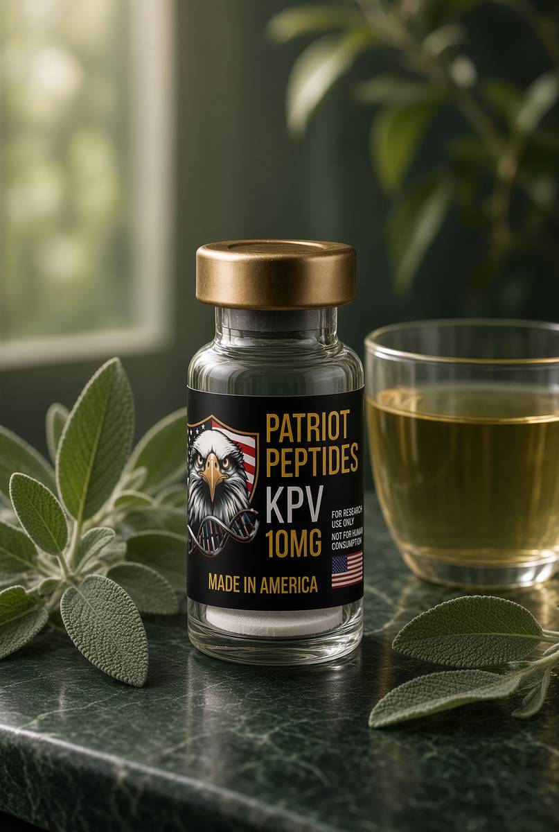
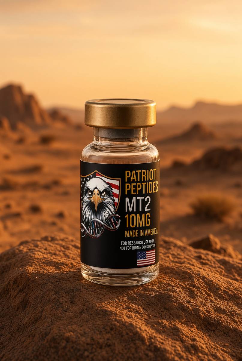
Named sequences the essay maps — KPV, MT2. Hypothetical research neighbourhood, not a protocol, not a medicine. One press puts every in-stock vial in the bag.
 Research only
 Research onlyOut of stock
Melanocortin
MT2
10 mg Melanotan II. Cyclic pan-melanocortin super-agonist.
4.6(720)
50 browsing this now · 3 purchased in the last 24 hours
10mg
£20.00
Research use only. Not a combined-use instruction.
Read next
46 min · long read · Peptide research
Melanocortins: from tanning receptors to inflammatory off-switches
MC1R makes pigment. MC4R runs appetite — and its loss is a monogenic obesity syndrome. KPV keeps the anti-inflammatory tail of α-MSH. MT2 is the pan-agonist that does not know subtle.
48 min · long read · Peptide research
IBS, gut lining and the two research peptides that sit on that literature
Irritable bowel is a symptom cluster, not a receptor. The papers that name molecules for barrier tissue keep coming back to a gastric 15-mer and a three-residue tail of α-MSH. That map — not a gastroenterology appointment.
More in this desk

53 min · long read · Peptide research
Recovery, GH pulses and the CJC / ipamorelin pair
Somatotrophs have two 'go' receptors. CJC without DAC is a slightly longer GHRH pulse. Ipamorelin is a selective ghrelin-receptor key that does not yank ACTH. Together they are the pair Bowers already showed is more than additive — not a gym protocol.
53 min · long read · Peptide research
How research peptides are made — and why HPLC actually matters
Solid-phase peptide synthesis builds a chain one residue at a time. HPLC then asks whether the main peak is what you think it is. ≥98% is not a slogan. It is a chromatogram.
52 min · long read · Peptide research
Fatigue, cellular energy and the NAD+ / MOTS-c neighbourhood
NAD+ is the rechargeable chip every cell spends on fuel and DNA repair — and the pool shrinks with age. MOTS-c is a mitochondrial 16-mer sent out under metabolic stress. Two answers to 'I have no energy' that are not coffee.
51 min · long read · Peptide research
Melanocortin circuits: pigment, appetite and PT-141
Melanotan II lights MC1, MC3, MC4 and MC5 — pigment plus the rest of the sheet. PT-141 is the free-acid cousin pointed at MC3 and MC4, the circuitry papers use for desire and energy, not skin colour. Same Arizona family. Different question.
Essays describe published research. They are not medical advice and they do not authorise human use of any catalogue item.