

Peptide research · 48 min · 10,524 words
IBS, gut lining and the two research peptides that sit on that literature
Irritable bowel is a symptom cluster, not a receptor. The papers that name molecules for barrier tissue keep coming back to a gastric 15-mer and a three-residue tail of α-MSH. That map — not a gastroenterology appointment.
What this essay actually tells you
- IBS is a Rome-criteria symptom cluster. It does not name a receptor. The peptides people file next to it are not IBS drugs.
- BPC-157 is GEPPPGKPADDAGLV: acid-stable gastric 15-mer, NO tone and cytoprotection papers. KPV is Lys-Pro-Val: NF-κB down, PepT1 in, DSS-colitis sandbox.
- Two mechanisms, one organ on a reading list. Research use only. Not a gastroenterology appointment.
What this actually means
IBS is a clinic word for pain, bloating, diarrhoea or constipation without a single lesion you can point at. Rome IV is the checklist. Inflammatory bowel disease is a different drawer: ulcers, licensed antibodies and JAK inhibitors. The lining itself is mucus, a one-cell epithelium, named tight-junction proteins (claudin, occludin, ZO-1), transporters, and a blood supply. Leaky gut as a wellness brand isn't that protein list. The research peptides people keep lining up next to the syndrome aren't IBS drugs. BPC-157 is a 15-amino-acid piece of a stomach protein that survived acid; the animal papers sit on NSAID and alcohol lesions, intestinal blood flow, eNOS/NO tone, VEGFR2 and FAK–paxillin in endothelium. KPV is three letters from the tail of α-MSH; it turns down NF-κB p65 nuclear translocation and can ride PepT1, the oligopeptide transporter inflamed intestine already over-expresses. Two mechanisms. One organ in the papers. Neither vial is a medicine, a dose, or a plan for a human bowel.
Irritable bowel syndrome is a diagnosis of pattern, not of a hole you can photograph. The Rome IV criteria, published in Gastroenterology in 2016 under Drossman's chairmanship, ask for recurrent abdominal pain on average at least one day a week in the last three months, associated with two or more of: relation to defecation, a change in stool frequency, a change in stool form. Symptom onset at least six months before the diagnosis is made. That's a checklist a clinic can apply after it has ruled out a lesion that would reclassify the complaint as inflammatory bowel disease, coeliac disease, colorectal cancer, bile-acid diarrhoea, or microscopic colitis. The checklist doesn't name a receptor. It doesn't name claudin-2. It doesn't name a peptide. Two research sequences keep turning up next to that clinic word because their animal papers mention stomach and bowel. We'll take them apart as chemistry and as a clinic object, because sharing an organ in a paper isn't sharing a mechanism.
In short. IBS is a checklist of pain and stool change without a single lesion. It doesn't name a receptor or a peptide. We'll keep those two objects apart.
The two sequences that keep turning up next to that search are a fifteen-residue gastric fragment and a three-residue tail of a pituitary hormone, and they're worth meeting as chemistry before anyone files them under a syndrome. BPC-157 is GEPPPGKPADDAGLV — a proline-rich piece of a gastric-juice protein that survives pepsin and pH 1.5, which is why a 15-mer can be studied in a stomach at all. KPV is Lys-Pro-Val, the C-terminus of α-melanocyte-stimulating hormone, α-MSH, small enough to ride an oligopeptide transporter the inflamed intestine already over-expresses. One literature sits on NSAID and alcohol gastric lesions, intestinal blood flow, and a nitric-oxide set-point. The other sits on NF-κB p65 nuclear translocation, PepT1, and dextran-sodium-sulphate colitis. Adjacent organ. Different ligand. Different assay. We stock both because a bench that reads barrier papers will meet both names, not because biochemistry wrote them as a pair. That's physiology with a paper trail. It isn't a gastroenterology appointment, and it isn't a combined plan for a person.
In short. BPC-157 is a 15-mer from stomach protein. KPV is three letters from α-MSH. Same organ in the papers. Different jobs. Not a pair for a person.
A 15-mer that survives the stomach is interesting because most 15-mers do not. The rest of the literature is almost inevitable once endothelium notices it.— Reading of the BPC-157 corpus against the KPV inflammation papers. Adjacent organ. Different ligand.
Rome is a checklist, not a receptor
Manning, Kruis, and then the Rome process spent decades turning a complaint into something a trial could enrol. Rome I in 1989, Rome II in 1999, Rome III in 2006, Rome IV in 2016: each revision tightened the pain criterion and tried to keep the syndrome from swallowing every unhappy bowel in the waiting room. Rome IV moved from 'discomfort' to pain, raised the frequency to one day a week, and kept the three-month window with a six-month lead-in. Subtypes are Bristol stool form, not personality: IBS with constipation (IBS-C), IBS with diarrhoea (IBS-D), mixed (IBS-M), unsubtyped (IBS-U). The Bristol scale itself is a seven-point picture of stool that a person can point at. That's how a functional gastrointestinal disorder is built: operational criteria, exclusion of organic disease, a language a trial can use. It's honest work. It's also why 'IBS receptor' is a phrase that has never earned a gene symbol. Syndromes of this kind can still have mechanisms — visceral hypersensitivity, altered motility, bile-acid handling, a post-infectious scar on the mucosal immune conversation. Mechanism is allowed. A single ligand as the cause is a different claim, and the field hasn't made it.
In short. Rome IV is a pain-and-stool checklist with subtypes by stool form. Useful for clinics and trials. It doesn't name one molecule as the cause.
What the clinic actually does, before anyone writes IBS on a letter, is look for the diseases that would make the letter a mistake. Coeliac serology. Faecal calprotectin if inflammation is on the table. Age-appropriate colorectal imaging. Thyroid. Medicines that already explain the stool. Bile-acid sequestrant trials in selected diarrhoea. The negative result is part of the diagnosis. That's why irritable bowel is sometimes called a diagnosis of exclusion as well as a positive symptom cluster, and why a person who hasn't had that work-up isn't yet a Rome object. Functional is a technical word here: a disorder of function without a currently demonstrable structural lesion on ordinary endoscopy and histology. It doesn't mean imaginary. It doesn't mean the enteric nervous system is uninvolved. It means the lesion that would re-file the case as Crohn's disease or ulcerative colitis hasn't been found. A peptide paper that starts from 'IBS' and never says which of those sentences it means hasn't started from the clinic object.
In short. Before IBS is written, the clinic rules out lesions and other diseases. Functional means no ordinary structural finding, not that the bowel is imagining it.
Prevalence numbers are large and a little soft, as syndrome numbers always are. Depending on the Rome version and the country, somewhere between five and fifteen percent of adults meet criteria at a given moment; women are over-represented in most Western clinic series; a substantial fraction never see a gastroenterologist. Quality of life scores can match organic inflammatory disease even when the mucosa looks ordinary, which is one reason the field stopped treating 'functional' as a polite way of saying mild. Work, diet, sleep, and a hypervigilant attention to gut sensation all sit in the phenotype. Psychological comorbidity is common and isn't a verdict that the pain is invented: the dorsal horn and the insular cortex are part of how a viscus is felt. Camilleri's reviews in Gastroenterology and Nature Reviews Gastroenterology & Hepatology are the papers I'd put in your hand before you file a peptide under this word. The word is a cluster. The cluster has biology. The biology is several neighbourhoods, not one occupancy at one lock.
In short. IBS is common, often untreated in clinic, and can wreck a week without a lesion on the camera. Several biologies, not one receptor.
Crohn's and colitis have lesions. Licensed drugs live there.
Inflammatory bowel disease is a different drawer, and the internet keeps shoving it into the same search as bloating. Crohn's disease can involve any stretch of gut, often ileum and colon, in a discontinuous, transmural pattern, with granulomas on a good histology day. Ulcerative colitis is continuous mucosal inflammation from the rectum upward. Both have ulcers, bleeding, strictures, fistulae, dysplasia risk, and an endoscopic score a trial can move. Both have licensed medicines: corticosteroids and 5-aminosalicylates as the old layer; thiopurines; anti-TNF antibodies (infliximab, adalimumab, golimumab, certolizumab); anti-integrin vedolizumab against α4β7; ustekinumab against the IL-12/23 p40 subunit and the newer IL-23 p19 antibodies (risankizumab, mirikizumab, guselkumab); Janus-kinase inhibitors (tofacitinib, upadacitinib, filgotinib); sphingosine-1-phosphate receptor modulators (ozanimod, etrasimod). That list is a reminder, not a prescription. Human inflammatory-bowel medicine exists. It is monoclonal, small-molecule, and sitting in a formulary with a risk register. A lyophilised 15-mer and a tripeptide aren't in that paragraph, and pretending they are is how a research catalogue becomes a rumour.
In short. Crohn's and ulcerative colitis have ulcers and licensed drugs — antibodies, JAK inhibitors, gut-selective integrins. Those medicines are not these sequences.
The reason the two drawers get confused isn't mysterious. Both involve bowel. Both involve, in some people, a conversation about barrier, immune tone, and pain. DSS-colitis, the usual KPV sandbox, is an acute chemical injury that looks a bit like ulcerative colitis on a histology slide and looks nothing like Tuesday bloating in a person who meets Rome IV and has a normal calprotectin. NSAID gastric lesions, the usual BPC-157 sandbox, are ulcers in a rat stomach after indomethacin or diclofenac, which is a gastroenterology object, and still not IBS. Adjacent organ is a lawful reason to put two papers on the same reading list. Adjacent organ isn't a reason to caption a vial with a syndrome. We'll keep naming the licensed world so the research solids can't be mistaken for it. Infliximab is a 150-kilodalton chimeric antibody against TNF. Vedolizumab stops α4β7-positive lymphocytes homing to MAdCAM-1 on gut endothelium. Those are occupancy stories with Phase 3 programmes and black-triangle histories. GEPPPGKPADDAGLV and Lys-Pro-Val are named sequences with animal and cell papers. Keep them apart.
In short. DSS-colitis and NSAID ulcers are animal models of injury. They are not IBS. Licensed IBD drugs are a different legal and clinical class from these peptides.
A lining is a seal, a factory and a blood supply
The intestinal epithelium is one cell thick. That sentence should sit in the front of your mind whenever anyone says gut barrier, because the whole argument lives in that sheet. On the luminal side, a mucus gel: MUC2 mucin, polymerised, a sieve that a commensal has to negotiate before it even reaches a microvillus. Goblet cells write the gel. Paneth cells, in the small-bowel crypt, write antimicrobial peptides — defensins, lysozyme, phospholipase A2 — so the stem-cell neighbourhood isn't a bacterial culture. Enterocytes do the absorptive job: SGLT1 and GLUT2 for glucose, PepT1 for di- and tripeptides, a forest of other SLC proteins, and a brush border of peptidases that destroy most dietary protein before it is a signal. Enteroendocrine cells sample the lumen and talk to the pancreas and the brainstem with GLP-1, PYY, CCK, serotonin. Tuft cells taste parasites. The sheet sits on a basement membrane. Under that, lamina propria: fibroblasts, plasma cells, macrophages, a vascular plexus, lacteals. Under that, muscularis mucosae, then submucosa, then the circular and longitudinal muscle the enteric nervous system actually drives. Barrier is the whole stack, not a catchphrase about leak.
In short. The gut lining is one cell thick, with mucus, specialised cells, and blood underneath. Barrier means that whole stack, not a catchphrase.
Surface area is the other number people skip. Small-bowel mucosa, unfolded, is on the order of a tennis court or a studio flat depending on which textbook you trust and whether you count microvilli; Helander and Fändriks re-measured it downward in 2014 and still left tens of square metres. Colon is less, and wetter, and more microbial. Transit time is hours in small bowel, longer in colon, highly variable, itself a clinical variable in IBS-C and IBS-D. Blood flow to the splanchnic bed is a quarter of cardiac output after a meal. The lining is therefore a metabolic organ as much as a wall: it spends ATP on transport, on mucus, on turnover. Epithelial cells last a few days; the crypt-villus (or crypt-surface) conveyor is one of the fastest in the body, which is why radiation and chemotherapy hit it, and why a repair literature found the stomach and the intestine in the first place. A peptide that talks to endothelium or to inflammatory transcription in this sheet is talking to a tissue that already rebuilds itself every week. That's the organ. Keep the organ in view when a model reports a change in lesion area.
In short. The lining is huge, fast-turning, and hungry for blood after a meal. It already rebuilds itself every few days. That is the tissue the papers sit on.
Mucus is two layers in colon, and treating it as fog is how a model gets misread. Johansson, Hansson and colleagues at Gothenburg spent a decade showing an inner MUC2 gel that is normally sterile, densely polymerised, anchored to goblet cells, and an outer layer that is looser, colonised, and the place a commensal actually lives. Small-bowel mucus is thinner and more discontinuous, which is why the small bowel can absorb and the colon can ferment. DSS, the KPV sandbox, attacks this gel first: the polymer fails, bacteria reach the sheet, neutrophils follow. That's a mucus-injury model before it is an immune model. IBS, in most people, doesn't look like a failed inner mucus layer on a fluorescent MUC2 stain. Some post-infectious and some diarrhoea-predominant cohorts show goblet and mucus changes; the finding is modest. A peptide paper that reports a DSS score has measured a chewed gel and a chewed sheet. It hasn't measured Tuesday bloating. Keep MUC2 on the diagram because it is the first argument a microbe has to win. Keep it off the vial caption, because a 15-mer and a tripeptide are not mucins.
In short. Colon mucus has a sterile inner gel and a looser outer one. DSS chews that gel. Most IBS does not look like that injury on a stain.
Diagram
Mucus
MUC2 gel
The first argument a microbe has to win.
Epithelium
enterocyte · goblet · Paneth
One cell thick. The wall is the cell, not a fascia.
Tight junctions
claudin · occludin · ZO-1
The seal. ‘Leaky gut’ as a brand is not this protein list.
PepT1
SLC15A1
Oligopeptide transporter. Inflamed gut induces it. KPV can ride it.
Lamina propria
immune cells
Where NF-κB decisions become cytokines.
BPC-157 neighbourhood
NO · VEGFR2 · FAK
Cytoprotection, blood flow, how a damaged lining organises.
IBS is a Rome-criteria cluster. It does not name a receptor. The preclinical literature that named molecules for barrier tissue keeps coming back to BPC-157 and KPV — two ligands, one organ on a reading list, neither a gastroenterology appointment.
The protein list: claudin, occludin, ZO-1
A tight junction is a belt, not glue. Adjacent enterocytes stitch their lateral membranes together near the apex with a set of transmembrane proteins, backed by a cytoplasmic plaque that ties the belt to the actin cytoskeleton. Claudins are the seal and the pore. Occludin is the regulator everyone measured first because the antibody existed. Tricellulin (MARVELD2) sits at the three-cell corner, where a bicellular strand would leave a hole. Junctional adhesion molecules, especially JAM-A (F11R), live in the neighbourhood and matter for polarity and for immune-cell traffic. The plaque is zonula occludens-1, ZO-2 and ZO-3 (TJP1, TJP2, TJP3), plus cingulin: PDZ-domain proteins that bind the tails of claudins and occludin and bind actin. Farquhar and Palade photographed the junctional complex in 1963. Furuse, Tsukita and colleagues cloned occludin in 1993 and claudin-1 and claudin-2 in 1998, at which point 'the barrier' became a gene list. If a brochure says leaky gut and can't name three of those proteins, it hasn't described a junction. It has described a feeling.
In short. Tight junctions are named proteins: claudins, occludin, tricellulin, JAMs, and ZO-1 tying them to actin. Leaky gut as a brand is not that list.
Claudins do two jobs, and mixing them is how a field gets a catchphrase instead of a measurement. Barrier claudins (claudin-1, claudin-3, claudin-4, claudin-5 in endothelium, claudin-7, claudin-8) tighten the paracellular cleft. Pore-forming claudins, especially claudin-2, make a cation-selective pore that lets sodium and water through; claudin-15 does related work in small bowel. Claudin-2 is induced in human inflammatory bowel disease and in several rodent colitis models, which is a real, blot-able finding: more pore, more leak of a particular kind. Turner, Shen, Buschmann and colleagues spent two decades showing that myosin light-chain kinase can pull on the perijunctional actomyosin ring and open a leak pathway distinct from the pore pathway — a size-selective, inflammation-gated route for larger solutes, including some bacterial products. That's permeability as a set of numbered, inhibitable proteins and kinases. It's also why 'the gut is leaky' is a measurement waiting to happen, not a diagnosis. Name the claudin. Name the tracer. Name the resistance. Then talk.
In short. Some claudins seal. Claudin-2 makes a salt-and-water pore and rises in inflamed bowel. Permeability is a measurement of named proteins, not a mood.
Occludin remains the protein a barrier paper reaches for when it wants a Western blot that looks like a junction. Knock it out in a mouse and the junction mostly still holds, which was a surprise and a lesson: redundancy is the design. Phosphorylation state, endocytosis, and pairing with ZO-1 are how occludin actually behaves in an inflamed sheet. ZO-1 is closer to essential; it is the scaffold. JAM-A-deficient mice have a permeability phenotype and a susceptibility to colitis that several groups have used as a sandbox. None of these proteins is a receptor for BPC-157. None of them is a receptor for KPV. KPV's published door into the epithelium is PepT1, a transporter, which is a different protein on a different face of the cell. BPC-157's published neighbourhood is endothelium, VEGFR2, FAK–paxillin and nitric-oxide tone, which is the blood-supply side of the same organ. A junction blot can still move in a peptide paper. Moving a blot isn't occupancy of the protein on the blot. Keep the causal verb honest.
In short. Occludin and ZO-1 are how labs blot a barrier. Neither is a receptor for these peptides. KPV rides PepT1. BPC-157's papers sit on vessels and nitric oxide.
The machine that made barrier a number is the Ussing chamber: a piece of mucosa, or a monolayer on a filter, clamped between two buffers, a voltage and a current you can read. Transepithelial electrical resistance is the ohmic summary of the paracellular seal plus the transcellular path; a tight colon epithelium sits in the hundreds to thousands of ohm·cm², a leaky small-bowel epithelium lower, a broken DSS sheet much lower. Flux of a labelled probe — 4 kDa FITC-dextran, 51Cr-EDTA, Lucifer yellow — reports a different size window. In a person, the lactulose/mannitol test is the poor cousin: two sugars, different absorption, a urine ratio, renal clearance as a confound. TEER can fall because claudin-2 opened a cation pore, because MLCK opened a leak pathway, because cells died and left holes, or because the filter was never tight. Papers that report a peptide effect on barrier without saying which of those, and without a paracellular marker the size of the claim, are photographing a mood. A characterised ligand in a well-sealed monolayer is an experiment. A caption on a shop page is a different object.
In short. Labs measure the seal as electrical resistance and as leak of a labelled sugar or dye. Say which measurement. A drop in resistance has several causes.
- Rome IV pain
- ≥1 day / week
- Epithelium
- one cell thick
- Tight-junction pore
- Ångströms to nanometres
- BPC-157
- 15 aa, 1419 Da
- KPV
- 3 aa
- PepT1 (SLC15A1)
- H⁺-coupled
- Infliximab
- ~150 kDa
- DSS colitis
- chemical injury
Last 3 months, onset ≥6 months. A checklist. Not a receptor.
Enterocyte, goblet, Paneth, enteroendocrine. The wall is the cell.
Claudin-2 cations; leak pathway for larger solutes when MLCK pulls.
GEPPPGKPADDAGLV. Pro-rich, acid-stable. Gastric origin.
Lys-Pro-Val. C-terminus of α-MSH. PepT1 substrate.
Di- and tripeptides. Induced in inflamed intestine. Dalmasso 2008.
Licensed anti-TNF. The human IBD world these vials are not.
KPV's usual sandbox. Looks like colitis. Does not look like Rome IV.
Leaky gut as a brand is not a Western blot
The phrase leaky gut escaped the laboratory and became a wellness product. In the laboratory, intestinal permeability is a set of assays: transepithelial electrical resistance on a monolayer; flux of FITC-dextran, 51Cr-EDTA, or lactulose/mannitol in a person or an animal; confocal localisation of claudin and ZO-1; Ussing-chamber conductance on a biopsy. Each assay has a size selectivity, a confound (mucus, blood flow, unstirred layers, renal clearance of the sugar probes), and a dynamic range. A lactulose/mannitol ratio that rises in a post-infectious cohort is a finding. A shop that sells a powder under the same words is a different object. Fasano's zonulin work on coeliac disease is real chemistry around a proposed regulator of the junction; the leap from that literature to a general theory of everything is how a paper becomes a brand. We'll keep using the protein names. Claudin-2, occludin, ZO-1, myosin light-chain kinase. If a sentence can't survive that substitution, it wasn't a barrier sentence.
In short. Labs measure permeability with sugars, dyes and resistance. Leaky gut as a shop phrase is not that measurement. Name the protein and the tracer.
Some people who meet Rome criteria do have measurable permeability changes. The finding isn't universal, isn't stable from Tuesday to Thursday, and isn't a licence to relabel the syndrome as a tight-junction disease. Dunlop, Spiller, and others showed increased small-bowel permeability in subsets of post-infectious IBS, particularly after Campylobacter, with low-grade increases in enterochromaffin cells and T-lymphocytes that can last months. Barbara, De Giorgio, Stanghellini and colleagues in Bologna spent a career on mast cells adjacent to colonic nerves, histamine and tryptase, a pain-fibre conversation you can photograph. Camilleri has reviewed the permeability subset without letting it swallow the rest of the pie. A subset is a subset. Treating every bloated afternoon as a claudin event is how a measurement becomes a personality. Treating none of them as a barrier event is how you ignore a blot. The adult position is the subset, the assay, and the refusal to caption a peptide with the brand.
In short. Some IBS patients, especially after infection, show more gut leak on sugar tests. A subset is not the whole syndrome, and not a peptide indication.
Post-infectious IBS is the cleanest natural experiment the syndrome has. A documented gastroenteritis — Campylobacter, Salmonella, Giardia, norovirus, sometimes C. difficile — is followed, in a minority, by Rome-positive symptoms that outlast the pathogen. Spiller's Nottingham series and the Walkerton outbreak follow-up (Marshall and colleagues, after a municipal E. coli and Campylobacter contamination in Ontario) are the papers to start with. Risk rises with the severity of the acute illness, with female sex, and with anxiety at the time of infection, which already tells you the phenotype is gut plus brain. Histology can show a modest rise in intraepithelial lymphocytes and mast cells, a serotonin-cell hyperplasia, a change in bile-acid handling in some diarrhoea-predominant cases. That's low-grade. It isn't Crohn's. Calprotectin is usually not in the IBD range. The animal models that dump DSS into drinking water to chew the mucus and the sheet are modelling a different injury: acute, chemical, bloody, weight-losing. Useful for NF-κB questions. A poor portrait of a post-Campylobacter year.
In short. After a bad gut infection, a minority keep IBS symptoms. That is a real, modest inflammatory scar. DSS-colitis in a mouse is a harsher, different injury.
The rest of the syndrome is not a lining
Visceral hypersensitivity is the finding that most consistently survives across IBS cohorts: a balloon in the rectum or colon that a control subject rates as pressure, the IBS subject rates as pain, at a smaller volume. Dorsal-horn sensitisation, descending inhibitory control from brainstem, insular and anterior-cingulate attention to gut signals, a pain-fibre that has been talked to by mast-cell histamine — pick the paper, pick the floor of the stack. Mayer, Tillisch and Labus put the brain–gut axis on imaging; the maps are real and they aren't a peptide target in this catalogue. Motility is the other half: accelerated transit in a subset of IBS-D, delayed in IBS-C, a meal-related gastrocolic reflex that is louder than the person wanted. Serotonin is the enterochromaffin cell's broadcast; 5-HT3 antagonists (alosetron, ondansetron in some diarrhoea protocols) and 5-HT4 agonists (prucalopride, and the withdrawn tegaserod) are licensed or once-licensed occupancy at those receptors. Bile-acid diarrhoea, measurable with SeHCAT in the UK, sits under a chunk of what was labelled IBS-D and answers, sometimes, to a sequestrant. Several neighbourhoods. One syndrome word. No gastric 15-mer in that paragraph.
In short. IBS also means a sensitive gut-pain system, odd motility, serotonin, and sometimes bile acids. Those are separate neighbourhoods from a lining peptide.
The microbiome literature is large, noisy, and not yet a ligand. Diversity scores move a little, some genera are over- or under-represented depending on the cohort and the method, faecal microbiota transplantation has been tried with mixed IBS results that would not license a product, and the low-FODMAP diet — which starves fermentable carbohydrates the microbiota would otherwise eat — remains one of the more reproducible clinical tools. Rifaximin, a poorly absorbed antibiotic, has a licensed IBS-D indication in some countries after target-phase trials; that's a medicine, with a number needed to treat, not a peptide. Mast-cell stabilisers and histamine antagonists have wandered through small trials. Psychological therapies, gut-directed hypnotherapy and cognitive behavioural protocols, move symptoms in ways a balloon study can also see, which is information about the stack, not an insult to the mucosa. A research peptide doesn't inherit any of this by proximity. File the microbiome under the microbiome. File KPV under NF-κB and PepT1. File BPC-157 under gastric lesions and endothelial assays. The filing is the honesty.
In short. Bugs, fermentable carbs, a gut antibiotic, and talking therapies all have IBS papers. They do not make BPC-157 or KPV into those tools.
Medicines a gastroenterologist can already name
IBS itself has a formulary, modest and real. Antispasmodics (mebeverine, hyoscine, peppermint oil in some guidelines). Loperamide for diarrhoea, carefully, because pain can worsen if you freeze a sensitive colon. Osmotic laxatives, then linaclotide or lubiprostone or plecanatide — guanylate-cyclase-C or chloride-channel occupancy that increases small-bowel fluid and can ease pain as well as stool form in IBS-C. Prucalopride as a 5-HT4 agonist for constipation. Eluxadoline as a mixed opioid-receptor ligand for IBS-D, with a sphincter-of-Oddi warning. Low-dose tricyclic antidepressants as neuromodulators, which is a dorsal-horn and central-attention story at doses that would not treat depression. SSRIs in selected anxiety-forward phenotypes. Rifaximin as above. That's occupancy, licensed or guideline-backed, with numbers. A person holding this page who has a clinic question has a clinic. The catalogue isn't that room. Naming the room is how the vials stay in their legal class when a search term is a syndrome.
In short. IBS already has licensed or guideline medicines: antispasmodics, gut-fluid drugs, a poorly absorbed antibiotic, low-dose tricyclics. Those live in clinic.
Inflammatory bowel disease, to say it again because the search traffic will not, is further on. Anti-TNF changed the natural history of Crohn's fistulae and ulcerative-colitis admissions. Vedolizumab is gut-selective on purpose, an integrin answer to the question 'can you stop the lymphocytes without stopping the rest of the person'. JAK inhibitors are oral and fast and carry herpes-zoster and thrombosis conversations. IL-23 antibodies are the current wave. Surgery remains an honest sentence: ileocaecal resection, colectomy, an ostomy that gives a life back. None of this is a research peptide. The reason to spend a paragraph on it in a page that will then discuss BPC-157 and KPV is so a reader who arrived from a forum can see the scale difference. Infliximab is given in milligrams per kilogram with an induction schedule and a tuberculosis screen. A 10 milligram cake of a 15-mer is a characterised solid for a tube. Adjacent reading list. Different object, different law, different claim.
In short. IBD medicines include antibodies, JAK inhibitors and operations. They are a different scale and a different law from a research 15-mer or tripeptide.
BPC-157: GEPPPGKPADDAGLV, the 15-mer that survived acid
Body Protection Compound was isolated as a protective protein in human gastric juice. The fragment that survived the subsequent chemistry, and that the literature actually uses, is fifteen amino acids: Gly-Glu-Pro-Pro-Pro-Gly-Lys-Pro-Ala-Asp-Asp-Ala-Gly-Leu-Val. Mass 1419.5 daltons. Four prolines in fifteen residues is a lot of proline. Proline kinks a chain, resists many peptidases, and is why a 15-mer can be given by oral gavage in a rat and still be a story rather than lunch. Most research peptides of this length wouldn't survive pepsin and pH 1.5 long enough to be an experiment in a stomach. That's the origin story, and it's a chemical story before it is a vascular one. The catalogue name BPC-157 is that sequence, lyophilised, HPLC-characterised. It isn't a gastric protein. It isn't a food supplement. It isn't a licensed cytoprotective drug. Misoprostol, proton-pump inhibitors and sucralfate are the clinic's gastric-protection drawer. This 15-mer is the named ligand those Zagreb and later papers put in the drinking water, the gavage, or the dish.
In short. BPC-157 is fifteen amino acids, proline-rich, stable in stomach acid. That chemistry is why gut papers exist. It is the named sequence, not a stomach drug.
Predrag Sikiric's group at the University of Zagreb built the largest single-laboratory corpus on this peptide, over three decades, across gastric, intestinal, tendon, muscle, vessel and, more controversially, central-nervous-system models. The recurring claim is cytoprotection in Robert's sense: an ability to limit lesion area after an insult that would otherwise ulcerate, with a particular interest in the nitric-oxide system. Sikiric has argued, in reviews you can actually cite, that the peptide doesn't simply raise or lower nitric oxide but restores a physiological set-point after NSAID, ethanol, or surgical disruption — a claim that is tested, in those papers, by co-administration of L-NAME (an NOS inhibitor) and L-arginine, and by watching which combination abolishes or restores the protection. Single-laboratory programmes of this size need outside replication. That isn't an insult. It's how a literature is read. The gastric-lesion and endothelial-migration pieces have more company from independent groups than the more extravagant extensions. Read the independent ones first. Keep the assays you can name.
In short. A Zagreb group built a huge BPC-157 file, especially on ulcers and nitric oxide. Independent labs have copied some vessel and tendon pieces more cleanly than the rest.
NSAID gastropathy is the sandbox that matches the origin story most cleanly. Indomethacin, diclofenac, aspirin: cyclo-oxygenase inhibition, a drop in protective prostaglandins, a rise in gastric acid injury, lesions you can photograph and planimeter. Robert's original cytoprotection work used prostaglandins against ethanol and boiling water; the BPC papers sit in that tradition with a different ligand. Reported endpoints are lesion area, microscopic damage scores, and sometimes gastric blood flow by hydrogen clearance or laser Doppler. Alcohol, as absolute ethanol gavage, is the other classical insult: a chemical burn of the mucosa, vascular stasis, a necrotic band. Cysteamine duodenal ulcers, acetic-acid gastric ulcers, anastomosis models, colitis models the Zagreb group also ran — the list is long, which is itself a reading problem. A long list from one laboratory isn't the same as a long list from many. For a gut-lining question, the assays that earn their keep are lesion area after NSAID or ethanol, a blood-flow measurement, and an eNOS or nitrite readout. Name those. Leave the rest in the review until someone else has run them.
In short. The cleanest BPC-157 gut papers are NSAID and alcohol ulcers in animals, with lesion size and blood flow as the readouts. Those are the assays to keep.
André Robert, in the 1970s, showed that prostaglandins could protect gastric mucosa against insults — boiling water, absolute ethanol, acid — at doses that didn't even inhibit acid secretion. He called it cytoprotection, and the word stuck, for better and worse. Misoprostol is the licensed prostaglandin analogue that came out of that physiology, used against NSAID ulcers with a diarrhoea and pregnancy warning. Sucralfate, bismuth, proton-pump inhibitors, H2 antagonists: the clinic's drawer is already full. BPC-157 papers sit in Robert's tradition in the sense that they measure lesion area after an insult and talk about protection of the sheet and its blood flow, not about acid as the only variable. PGE2 acts at EP receptors, Gs and Gq, a named occupancy. GEPPPGKPADDAGLV doesn't become misoprostol because both sentences contain the word stomach. Reading the 15-mer as a cytoprotection reagent in a rat NSAID model is lawful. Reading it as a replacement for a proton-pump inhibitor or a prostaglandin analogue in a person is a category error the legal class of a research solid already forbids, and we won't make it.
In short. Cytoprotection started as prostaglandins saving a stomach from a chemical insult. Misoprostol is the licensed cousin. BPC-157 papers sit in that tradition, not in that formulary.
Intestinal blood flow is the sentence that takes the 15-mer out of the mucus and into the vessel. A damaged lining doesn't heal as a sheet floating in medium. It heals as a tissue with a capillary plexus that has to deliver oxygen and take heat and lactate away, the same physics a tendon repair bed already taught the musculoskeletal papers. BPC-157 has been reported to preserve or restore mesenteric and gastric mucosal flow after insults that would otherwise leave a pale, stasis-heavy mucosa. Nitric oxide is the obvious mediator to reach for: eNOS in endothelium, a tonic vasodilator, a platelet quietener, an angiogenic partner of VEGF. The 'tone reset' language in the Sikiric reviews is an attempt to explain why the peptide can look protective both when NO is too low and when an NO donor would be expected to overshoot. Whether that's a true set-point mechanism or a set of model-dependent effects is a live reading question. It's still a more adult sentence than 'boosts nitric oxide'. Independent endothelial work, coming in the next heading, is cleaner on migration and VEGFR2 than on the philosophy of a set-point.
In short. BPC-157 papers often report better gut blood flow after injury, with nitric-oxide enzymes in the story. Reset of tone is the claim. Migration assays are cleaner.
The causal tool in many Zagreb nitric-oxide papers is a pair of compounds a vascular physiologist already has on the shelf. L-NAME (Nω-nitro-L-arginine methyl ester) inhibits nitric-oxide synthases; L-arginine is the substrate those enzymes want. Give L-NAME and many NO-dependent protections collapse. Give L-arginine and some of them return. Give both, in a factorial design, and you can ask whether a peptide still works when the NO system is clamped high or low. That's a better experiment than a nitrite smear. eNOS (NOS3) is the endothelial isoform; iNOS (NOS2) is the inducible, inflammation-associated one; nNOS (NOS1) lives in nerves, including the enteric nervous system. Which isoform a 15-mer is supposed to talk to isn't always cleanly shown. A blot for eNOS phosphorylation at Ser1177 is a start. An isoform-selective inhibitor is better. A knockout is better still and is scarce in this file. Independent endothelial papers that use VEGFR2 blockers are, again, cleaner than a philosophy of set-points. Keep L-NAME as a tool. Leave 'boosts nitric oxide' on the forum where it belongs.
In short. Zagreb papers often block or feed the nitric-oxide pathway with L-NAME and L-arginine. That is a real design. Which enzyme isoform does the work is less often shown.
Endothelium, VEGFR2, FAK–paxillin
Once a peptide that endothelium notices has been named, scratch assays are almost inevitable. Human umbilical-vein endothelial cells, or a microvascular line, grown to a monolayer, a pipette scratch, closure over hours, a VEGF-pathway inhibitor as the control that makes the closure go away. Several groups outside Zagreb have reported that BPC-157 accelerates that closure and that VEGFR2 is involved: internalisation, phosphorylation, a downstream Akt and eNOS phosphorylation that a blot can show. Hsieh, Chang and colleagues are the names a paper actually reaches for on the VEGFR2 piece. Angiogenesis in a sponge or a lesion bed follows if the in-vitro migration is real and if a vessel can be recruited in vivo. For a stomach or a colon, a denser bed of small vessels in a repair zone is a cytoprotection-adjacent finding: blood is how a sheet lives. It's also how a tumour lives, which is why VEGF occupancy in oncology is a careful, licensed, opposite-direction story (bevacizumab, ramucirumab). A research 15-mer in a migration assay isn't that antibody. Direction, species, dose, and legal class all differ. The neighbourhood is still VEGFR2.
In short. Independent cell work finds BPC-157 helps endothelial cells crawl, with the VEGF receptor in the pathway. That is a vessel story, not an IBS story.
Focal adhesion kinase and paxillin are how a crawling cell holds the floor. Integrins bind extracellular matrix; FAK autophosphorylates at Tyr397; paxillin is recruited; the adhesion talks to actin; the cell pulls. Endothelial cells and tenocytes both live on this machinery. BPC-157 papers have reported FAK–paxillin activation in those cells, which is a lawful molecular sentence about migration and isn't a sentence about thymosin β4. Thymosin β4, and the research analogue TB-500, sequester G-actin so a cell has a monomer pool for the next lamellipodium — an actin-buffer story, a different catalogue entry, a different essay. Forums bundle BPC-157 and TB-500 as a healing stack. Biochemistry doesn't. If this page shows the actin-treadmill diagram, it is to keep the jobs apart: the gastric 15-mer talks to VEGFR2 and to focal adhesions; the actin buffer holds monomers. A fibroblast or an endothelial cell needs both kinds of conversation to crawl, which is why the literatures sit near each other on a bench. Near isn't identical. IBS still hasn't entered this paragraph, and shouldn't.
In short. FAK and paxillin are how crawling cells grip. BPC-157 papers sit there. That is not the actin-buffer peptide TB-500, and it is not an IBS mechanism.
Diagram
BPC-157: Pro-rich, acid-stable, Sikiric corpus. VEGFR2 internalisation, FAK–paxillin, eNOS-dependent NO tone. A cytoprotection story that escaped the stomach.
TB-500: cytoskeletal buffer. Injury releases Tβ4 extracellularly; VEGF, MMPs and keratinocyte migration follow. SDKP is a separate N-terminal anti-fibrotic pharmacophore. Two literatures, two jobs.
Thymosin β4 is the principal G-actin sequestering peptide. TB-500 is built around the LKKTETQ motif. BPC-157 is a gastric 15-mer (GEPPPGKPADDAGLV) that talks to VEGFR2 and focal adhesions. Related in folklore. Unrelated in mechanism.
Tenocyte outgrowth belongs in one paragraph so it doesn't hijack a gut essay. Chang's 2011 Journal of Applied Physiology paper reported faster tendon-cell outgrowth, better survival under oxidative stress, and migration in a BPC-157 dish, with FAK and paxillin in the blots. Rodent tendon-to-bone defects have been used as the in-vivo partner: collagen organisation, vessel density in a hypovascular tissue. That's why gym culture met a gastric peptide. The chemistry didn't change when the tissue did. GEPPPGKPADDAGLV is still a proline-rich 15-mer with an endothelial and focal-adhesion neighbourhood. A rotator cuff isn't a colon. A colon isn't a rotator cuff. The shared object is a repair bed that needs small vessels and cells that can crawl. This page stays with the lining because the search term was IBS. The tendon essay on this desk is next door for anyone who followed the 15-mer out of the stomach and into a collagen dense enough to be a problem. Two tissues. One sequence. Several assays. No protocol in either room.
In short. Some BPC-157 papers are about tendon cells crawling and collagen lining up. Same 15-mer as the stomach work. Different tissue. Still not a treatment plan.
How to read a single-laboratory literature is a skill. Count the models. Count the independent groups. Ask whether the endpoint was pre-specified or a family of scores. Ask whether a VEGFR2 inhibitor, an NOS inhibitor, or a FAK construct was used as a causal tool, or whether the peptide was sprinkled and a phenotype photographed. Prefer a scratch assay with a characterised ligand and a blot over a claim that wanders into behaviour, heart rhythm, or a central-nervous-system rescue without a concentration at the site. The Zagreb corpus is larger than most peptide files and more uneven than a tidy review admits. Independent endothelial migration and some tendon findings are the pieces we'll keep. Gastric lesion area after NSAID or ethanol is the piece a gut-lining page is allowed to keep. Extravagant extensions can wait for company. That reading is why the 15-mer is on a shelf as a named sequence and not on a label as a medicine. Characterisation first. Then the assay the paper actually ran.
In short. Read BPC-157 by naming the assay and who copied it. Vessel crawling and NSAID ulcers have more support than the far-reaching claims. Still a research ligand.
If a peptide makes endothelial cells migrate and a gastric lesion shrinks on a photograph, you do not need a legend. You need the blot, the inhibitor, and a second laboratory.— Working rule for the BPC-157 file. Assays you can name. Replication you can cite.
KPV: three residues from a thirteen-residue hormone
α-Melanocyte-stimulating hormone is thirteen amino acids, acetylated at the N-terminus, amidated at the C-terminus: Ac-SYSMEHFRWGKPV-NH2. It's a cleavage product of pro-opiomelanocortin, the same precursor that yields ACTH and β-endorphin, chopped in the pituitary and in skin. The core His-Phe-Arg-Trp is the melanocortin-receptor pharmacophore, the tetrapeptide that occupies MC1R on melanocytes and writes eumelanin, and that occupies MC3R and MC4R in hypothalamus and writes appetite and a set of autonomic events. The last three letters are lysine, proline, valine. Medicinal chemistry is often addition. KPV is subtraction. Keep the tail. Lose most of the pigment, most of the MC4R appetite agonism, most of the sexual-function circuitry that made Melanotan II famous for the wrong reason. What remains is small enough to be a PepT1 substrate and stubborn enough, in several epithelial and myeloid assays, to suppress NF-κB. That's a design move. It's also why this tripeptide sits on a gut-lining reading list instead of a tanning one.
In short. α-MSH is thirteen letters. The last three are KPV. Keep the anti-inflammatory tail and drop most of the tanning and appetite chemistry.
Diagram
MC1R
melanocyte
Eumelanin vs pheomelanin. Red-hair alleles. Afamelanotide’s receptor.
MC3R
hypothalamus
Energy balance. Occupied by MT2 because MT2 occupies almost everything.
MC4R
hypothalamus
Appetite brake. Loss-of-function obesity. Setmelanotide (FDA 2020).
MC5R
sebaceous
Sebum. A first-class output people still treat as a footnote.
KPV
tail of α-MSH
NF-κB off-switch. PepT1 uptake. Designed not to tan.
MT2
pan-agonist
Ac-Nle-cyclo[Asp-His-D-Phe-Arg-Trp-Lys]-NH2. Pigment, appetite, flushing, arousal.
α-MSH is Ac-SYSMEHFRWGKPV-NH2. The last three letters are KPV. Melanotan II is what a pan-agonist looks like. KPV is the design move in the other direction. Match the ligand to the question.
NF-κB is a transcription-factor family, not a mood. In the canonical pathway, a stimulus — TNF, IL-1, a Toll-like-receptor ligand from a microbe — activates the IκB kinase complex; IκBα is phosphorylated and degraded; p65 (RelA) and p50 move into the nucleus; inflammatory genes are written. The assay is a reporter, an electromobility shift, a nuclear Western for p65, a set of transcripts (TNF, IL-6, IL-1β, CXCL8), sometimes a p65-DNA binding ELISA. KPV has been reported to reduce IκB degradation and p65 nuclear translocation in keratinocytes and in macrophages, and to reduce those transcripts. Brzoska, Luger, Böhm and colleagues at Münster built the melanocortin-inflammation library this sentence sits on; the 2008 Endocrine Reviews article is the map. Residual activity when MC1R is blocked or absent is the intellectually important finding: some of the quieting doesn't require the pigment receptor. That's the reason to use KPV instead of α-MSH or Melanotan II when pigment, appetite, or a pan-agonist circus would confound the dish. Three residues. A nuclear translocation. A reporter going down.
In short. KPV turns down NF-κB: the p65 subunit stays out of the nucleus more. Some of that still works when the pigment receptor is blocked.
PepT1, gene symbol SLC15A1, is a proton-coupled oligopeptide transporter on the apical membrane of small-bowel enterocytes. Its physiological job is dietary: di- and tripeptides from the brush-border digest, a major fraction of amino-acid nitrogen. Its pharmacological job is accidental and useful: valacyclovir, some β-lactams, other peptide-like drugs ride it. Inflammation induces it, including in colon, where baseline expression is lower — a pharmacokinetic gift if your ligand is a tripeptide you want inside an inflamed epithelium. Dalmasso, Merlin, Sitaraman and colleagues, Gastroenterology 2008, showed that KPV uptake into colonic cells was PepT1-dependent, that the tripeptide reduced NF-κB and inflammatory transcripts, and that oral KPV reduced colitis scores in mice. That paper is why this page exists as a pairing rather than as two separate peptide pages. A transporter the diseased sheet already over-expresses is a door. A door isn't a medicine. It is a reason a three-residue ligand can be a coherent colitis reagent when a 13-residue hormone would be a mess of pigment and appetite.
In short. PepT1 is the gut's door for tiny food peptides. Inflamed bowel makes more of it. KPV can ride that door, which is why oral colitis models keep using it.
PepT1 is an electrogenic cotransporter: one proton, one di- or tripeptide, inward. The driving force is the acidic microclimate at the brush border, itself a product of sodium–proton exchange, so a sheet that can't make a pH gradient can't run the door well. Substrate preference is broad and slightly promiscuous — that's how valacyclovir became an oral antiviral — and it's also how some bacterial formyl peptides such as fMLP get in and then talk to immune cells. Inflammation-induced PepT1 in colon is therefore a double sentence: a door for a therapeutic-adjacent tripeptide, and a door for microbial peptides the healthy colon would rather keep out. Dalmasso's KPV work sits on the first sentence. Papers that treat PepT1 only as a nutrient convenience haven't read the second. PepT2 (SLC15A2) is the kidney and brain-adjacent homologue, higher affinity, lower capacity, not the gut-lining story. If a paper claims KPV uptake and doesn't mention SLC15A1, pH, or a competition with a known PepT1 substrate (Gly-Sar is the classic), the door hasn't been shown. It has been assumed.
In short. PepT1 needs an acid gradient to carry di- and tripeptides in. Inflamed colon makes more of it, which can let KPV in and can also let bacterial peptides in.
Dextran sodium sulphate in drinking water is a chemical attack on mucus and epithelium. Over a few days the sheet fails, neutrophils arrive, weight falls, the colon shortens, bleeding is scored, myeloperoxidase is measured as a neutrophil enzyme, and a histologist writes a damage index. It's reproducible, brutal, and only loosely related to human ulcerative colitis, let alone to IBS. Kannengiesser and colleagues, Inflammatory Bowel Diseases 2008, put KPV into murine IBD models and reported reduced activity. Other α-MSH fragments have similar sandboxes. Replication quality in peptide-inflammation work varies by laboratory, as it does everywhere that scores are semi-quantitative and housing microbiota differ. The mechanistic core — NF-κB down, PepT1 in — has been seen more than once, which is the most a three-residue literature is required to earn before a catalogue can stock the named sequence. DSS remains a sandbox. Sandboxes are for asking whether a ligand can quiet a sheet. They aren't portraits of a person whose camera and calprotectin were normal and whose pain met Rome IV.
In short. DSS in mouse drinking water chews the gut lining and is the usual KPV test. It is a harsh colitis model. It is not a picture of human IBS.
The colitis model zoo is larger than DSS, and none of it is IBS. Trinitrobenzene sulphonic acid (TNBS) in ethanol is a haptenating injury that recruits a T-cell response and is sometimes sold as more Crohn's-like; it's still a chemical burn with an immune second wave. IL-10-deficient mice develop spontaneous colitis in dirty housing, a genetic lesion in an anti-inflammatory cytokine, useful for a cytokine question, useless as a portrait of Rome IV. T-cell transfer into lymphopenic mice (the Powrie model) asks what naïve T cells do to a gut they haven't learned to tolerate. Citrobacter rodentium is an infection model with attaching-and-effacing biology. Each of these has taught the IBD field something that later showed up in a licensed drug — IL-12/23, integrins, leukocyte traffic. KPV papers mostly stay in DSS because the ligand is small, oral, and the sheet is already writing PepT1. That's a coherent design for an NF-κB-and-transporter question. It's a poor design for a visceral-hypersensitivity question, a bile-acid question, or a brain–gut imaging question. Pick the model that matches the ligand's claimed job. Don't pick the syndrome the search bar typed.
In short. Other mouse colitis models exist — TNBS, IL-10 knockout, T-cell transfer. They taught IBD drug science. They still are not human IBS, and KPV papers mostly stay in DSS.
MC1R independence deserves a second, slower look because it's the whole point of the subtraction. If KPV required MC1R, you'd be running a pigment-receptor assay with a weak ligand and a confounding tan in any system that had melanocytes. Several groups have shown residual anti-inflammatory activity in MC1R-null or antagonist-blocked systems, and in cells that don't express the receptor at a meaningful level. Intracellular uptake via PepT1, then an effect on IκB or on p65 trafficking, is a model that doesn't need a Gs-coupled melanocortin receptor on the surface. Other work still finds MC1R contributions in particular myeloid cells. The adult sentence is incomplete dependence: some of the pharmacology is receptor-side, some of it is transporter-and-transcription-side, and the tripeptide was worth isolating because the mix is shifted away from pigment. Melanotan II, by contrast, is a cyclic pan-agonist at MC1R, MC3R, MC4R and MC5R, and will tan, suppress appetite, and surprise the autonomic nervous system. Wrong tool for an NF-κB question in a colon explant. Right tool for a different essay.
In short. KPV does not need the tanning receptor for all of its quieting. That is why you use it instead of Melanotan II when pigment would just add noise.
Keratinocytes and macrophages are the other dishes, and they belong here because barrier isn't only bowel. Skin epithelium runs the same NF-κB programme when it is inflamed; KPV papers have used HaCaT cells and primary keratinocytes as a reporter system that doesn't require a colon. Macrophages, as TNF and IL-1 sources, are a second obvious cell. Antimicrobial-adjacent effects have been described for α-MSH fragments, as they have for other cationic peptides; treat those as a side literature unless the assay was built for killing. The gut-lining question doesn't need a skin cream, and we won't pretend the keratinocyte work is a dermatology product. It is a cell type with a junction, a nucleus, and a reporter. PepT1 is more of a gut gift than a skin gift; keratinocyte uptake uses other paths. Two tissues, one transcription-factor family, one tripeptide. The catalogue stocks Lys-Pro-Val as that ligand, HPLC-characterised, so a reporter assay can name what it put in the well. Naming is the job. Indication is a different desk, and not this one.
In short. KPV also quiets NF-κB in skin cells and in macrophages. That is the same transcription story in another lining, not a cream and not a bowel medicine.
Two mechanisms, one organ on a reading list
Put the two ligands on the same bench and they still don't become a stack. BPC-157 is a vessel-and-nitric-oxide story that started in gastric juice: acid-stable 15-mer, NSAID and ethanol lesions, endothelial migration, VEGFR2, FAK–paxillin, eNOS tone. KPV is an NF-κB story that can enter epithelium through PepT1: three residues from the tail of α-MSH, p65 nuclear translocation down, residual activity without MC1R, DSS-colitis as the usual animal. People bundle them because both papers mention bowel. Biochemistry doesn't bundle them. One is growth-factor-receptor-adjacent and vascular. One is a transporter substrate and a transcription-factor quietener. A damaged lining in a rat might, in principle, want both a blood supply and a quieter nucleus. Principle isn't a protocol. Principle isn't a person. The catalogue lists both because a barrier reading list will meet both names. A button that puts both cakes in a bag, if a shop page offers one, is logistics for a bench. It isn't a combined-use instruction, and we won't write one.
In short. BPC-157 is a blood-vessel and nitric-oxide ligand. KPV is an NF-κB ligand that can enter through PepT1. Same organ in print. Two jobs. Not a combined plan.
Diagram
| Node | Catalogue | Conversation |
|---|---|---|
| GPCR | Ipamorelin, MT2, PT-141, retatrutide, CJC | Second messengers, secretion, appetite, pigment |
| RTK / IGF1R | IGF-1 LR3 | IRS–PI3K–Akt–mTOR and Shc–ERK |
| Cytokine receptor | Somatropin (HGH) | GHR–JAK2–STAT5b, hepatic IGF-1 |
| Cofactor | NAD+ | Sirtuins, PARPs, CD38, redox |
| Actin buffer | TB-500 / Tβ4 motif | G-actin sequestration, motility |
| Growth-factor-like | BPC-157 | VEGFR2 / FAK / eNOS neighbourhood |
| Copper ligand | GHK-Cu | Transcriptome shift in fibroblasts |
| MC fragment | KPV | NF-κB, PepT1, no pigment |
| Nuclear / pineal | Epithalon (AEDG) | TERT and melatonin literatures |
| mtORF peptide | MOTS-c | AMPK, folate–methionine cycle |
Each row is a different kind of molecular conversation. The catalogue peptides bind at these nodes; they are not interchangeable, and stacking them because a forum did mixes unrelated literatures.
The gut is already a peptide organ, which is why two more sequences can look at home on a reading list and still be strangers to the clinic. Enterochromaffin cells write most of the body's serotonin; 5-HT3 and 5-HT4 receptors are licensed or once-licensed IBS targets. L-cells write GLP-1 and PYY; K-cells write GIP; I-cells write CCK. Linaclotide occupies guanylate-cyclase-C on the apical enterocyte and is a fourteen-residue, minimally absorbed, licensed peptide for IBS-C. Teduglutide occupies GLP-2R and is licensed for short-bowel adaptation. Setmelanotide occupies MC4R for rare genetic obesity, a melanocortin cousin of KPV that went in the opposite direction, toward appetite, and earned a regulator. That list is the crowded neighbourhood. BPC-157 doesn't occupy GC-C. KPV doesn't occupy MC4R in any way a setmelanotide paper would recognise. Putting a gastric 15-mer and an α-MSH tail onto this street because the street is called gut peptides is how a catalogue page becomes a muddle. Named occupancy first. Then the syndrome, if the occupancy has earned a syndrome, which these two haven't.
In short. The gut already uses many peptides: serotonin, GLP-1, linaclotide at guanylate-cyclase-C. BPC-157 and KPV are not those ligands and do not inherit their licences.
DSS isn't IBS. Gastric lesion area isn't bloating. A FITC-dextran flux in a monolayer isn't a Rome-criteria week. Translation is the adult word for that gap, and it cuts both ways. Some human IBD medicines began in DSS or TNBS or IL-10-knockout mice and then earned Phase 3 programmes, tuberculosis screens, and a place on a formulary; anti-TNF is the existence proof that a cytokine story can survive the jump. Most peptide-inflammation and peptide-cytoprotection files never make that jump. The jump is expensive, regulated, and allowed to fail. Stocking a named sequence for the assay the paper ran is a lawful catalogue job. Captioning that sequence with a human syndrome is a different job, and not one a research label can do. Crohn's, ulcerative colitis, microscopic colitis, coeliac disease, bile-acid diarrhoea, and IBS each have their own diagnostic path and, where it exists, their own licensed shelf. Two lyophilised solids from a barrier reading list don't inherit those paths by sitting next to them in a paragraph.
In short. Mouse colitis is not human IBS. Stomach ulcers in a rat are not bloating. Human bowel diseases already have their own diagnostic paths and, often, their own drugs.
Mass is a useful rude comparison. KPV is three amino acids. BPC-157 is fifteen, about 1.4 kilodaltons. Infliximab is about 150 kilodaltons, a chimeric IgG1, given in hundreds of milligrams, with a half-life measured in days to weeks. Vedolizumab is another IgG. Upadacitinib is a small-molecule JAK1 inhibitor you can swallow, with CYP3A4 conversations and a herpes-zoster conversation. Linaclotide is a fourteen-residue peptide, interestingly close in length to BPC-157, but it is a guanylate-cyclase-C agonist licensed for IBS-C and chronic constipation, minimally absorbed, acting from the lumen on a receptor the enterocyte actually has. That last neighbour is the honest one to sit next to BPC-157 on a length comparison and then to separate on receptor and licence. Linaclotide occupies GC-C. BPC-157 doesn't. Length isn't a mechanism. A research vial of GEPPPGKPADDAGLV isn't a poorly absorbed GC-C agonist, and a research vial of KPV isn't a miniature infliximab. Scale is the point of the scale block above. Keep it in view when a forum treats all peptides as one liquid.
In short. KPV is three letters. BPC-157 is fifteen. Infliximab is a huge antibody. Linaclotide is a licensed 14-mer for constipation, a different receptor. Size is not a job.
What the papers measured, and what a vial is
A research peptide, on this desk, is a characterised sequence: identity by HPLC and mass, a certificate, a lyophilised cake, a reconstitution you can write as milligrams per millilitre in a stated solvent. The experiment is then whatever the paper actually did — a scratch assay, a nuclear p65 blot, a lesion photograph, a PepT1-transfected cell taking up a labelled tripeptide. Concentration in a well isn't a dose in a person. Oral gavage in a rat isn't a capsule advice column. Intraperitoneal injection in a mouse is a route a veterinarian and a home-office licence would recognise as an animal-procedure, not as a lifestyle. We won't convert any of those into a how-to. The papers that already ran the models are in PubMed. Go there if you're running an experiment. Stay here if you wanted the lining, the syndrome, and the two ligands unbundled. Characterised solids exist so a blot can name the ligand. They don't exist so a syndrome can inherit a caption.
In short. These sequences are named, measured powders for laboratory assays. A dish concentration is not a human dose. The papers already wrote their methods.
Identity for a 15-mer and a tripeptide isn't a mood and isn't a brand name. HPLC shows a peak; mass spectrometry shows 1419.5 daltons for GEPPPGKPADDAGLV, or 342 daltons for Lys-Pro-Val as the free acid, more if the C-terminus is amidated as in native α-MSH's tail. The certificate should say which. Amino-acid analysis or a tandem fragmentation ladder confirms sequence, not just mass, because different 15-mers can share a nominal mass at low resolution. Purity as a percentage is a peak-area statement, usually at 214 nanometres, and it is only as honest as the method's ability to see truncations and deletions. Counter-ion — acetate, trifluoroacetate — is part of the solid and part of a cell-assay conversation, because TFA can move a dish on its own at high enough residual. Water, by Karl Fischer, is the other number on a cake. None of this is gastroenterology. All of it is why a characterised sequence beats a mystery powder a forum sold under the same letters. The assay then has one variable. The syndrome still has many.
In short. A 15-mer and a tripeptide are identified by chromatography and mass. Sequence, purity and the salt should be stated. That is chemistry, not a clinic.
Independent replication will keep being the filter. For KPV, Dalmasso's PepT1 paper and the Münster melanocortin-inflammation reviews are the core; DSS and keratinocyte reporters are the usual follow-ups; MC1R-independence is the finding that makes the subtraction worth doing. For BPC-157, NSAID and ethanol lesion area, endothelial migration, VEGFR2, FAK–paxillin, and an eNOS-adjacent blood-flow story are the core; independent labs have been kinder to endothelium and tenocyte than to the more wandering claims. Anything that can't survive that filter can wait. A catalogue that stocks both sequences is making a reading-list decision, not a clinical one. The neighbouring essays — BPC-157 as gastric cytoprotection, KPV as the anti-inflammatory tripeptide — are the single-ligand versions of this map. This piece exists because searchers type a syndrome and the literature names two ligands. Mapping isn't treating. The map is the useful product of the afternoon.
In short. Keep the KPV findings that have been copied: PepT1, NF-κB, colitis models. Keep the BPC-157 findings that have been copied: ulcers, vessels, FAK. Mapping is not treating.
- Rome IV names a pain-and-stool cluster after lesions have been looked for. It does not name a receptor.
- A lining is mucus, a one-cell epithelium, claudins/occludin/ZO-1, a transporter sheet, and a blood supply.
- Leaky gut as a brand is not that protein list. Permeability is a tracer, a resistance, a blot.
- Human IBD and IBS medicines already exist. Name them so the vials cannot borrow their clothes.
- BPC-157 is GEPPPGKPADDAGLV: acid-stable, gastric origin, lesions, flow, VEGFR2, FAK, eNOS neighbourhood.
- KPV is Lys-Pro-Val: α-MSH tail, NF-κB p65 down, PepT1 in, DSS sandbox, MC1R optional.
- Two mechanisms. One organ on a reading list. No combined-use sentence.
A map, not a clinic letter
The honest limit of the afternoon is a map. Irritable bowel syndrome remains a Rome-criteria cluster. A DSS score remains a chemical injury in a mouse. A rat NSAID lesion remains an ulcer photograph. VEGFR2 phosphorylation in a dish, and a drop in a p65 reporter, remain cell-assay endpoints. Licensed human medicines for inflammatory bowel disease and, more modestly, for IBS already sit on this organ with antibodies, small molecules, minimally absorbed peptides such as linaclotide, and neuromodulators. Those programmes had regulators, numbers needed to treat, and harm tables. The two sequences in this essay had animal models and cell assays. Holding both facts at once is the whole skill. The neighbouring clinic, if a person needs one, is a clinic. The neighbouring papers, if a bench needs them, are Dalmasso 2008, Brzoska 2008, Turner 2009, Drossman 2016, Sikiric's reviews, Chang 2011, Kannengiesser 2008. Read those. Then decide whether the ligand you're holding matches the question you're actually asking.
In short. The map stops at papers and assays. Human bowel medicines already exist. Match the ligand to the question, not the search term to the sequence.
Research-use-only is the legal class of the solids. The cakes are characterised sequences for laboratory work: identity, purity, a reconstitution you can write down. Captioning them as a treatment for a Rome-criteria syndrome, for Crohn's disease, or for ulcerative colitis would be a different legal class, and a different literature. A gut lining remains a seal, a factory and a blood supply whether or not anyone opened a vial. Claudin-2 remains a pore. PepT1 remains a proton-coupled door. eNOS remains an endothelial enzyme. The syndrome remains a checklist. Two ligands sit on that literature. The literature is interesting. Interest isn't an indication. The vial you're holding, if you're holding one, is a named 15-mer or a named tripeptide. Keep the name, the assay, and the organ on separate lines, and the afternoon will have been useful to anyone who wanted the lining explained rather than a syndrome inherited.
In short. The powders are laboratory sequences. The lining, the proteins, and the two ligands stay on separate lines. Interest is not an indication.
Two mechanisms. One organ on a reading list. Neither vial is a medicine, a dose, or a plan for a human bowel.
- Rome IV is a checklist. IBD is a lesion plus a formulary. Do not file them in the same drawer.
- Tight junctions are claudins, occludin, tricellulin, JAMs, ZO proteins. Leaky gut as a brand is not that list.
- BPC-157: 15-mer, acid-stable, NSAID/alcohol lesions, flow, VEGFR2, FAK–paxillin, eNOS neighbourhood. Independent endothelium first.
- KPV: α-MSH tail, p65 nuclear translocation down, PepT1 (SLC15A1) in, DSS sandbox, MC1R not always required.
- Linaclotide, anti-TNF, vedolizumab, JAK inhibitors, rifaximin, low-dose tricyclics: the human world these vials are not.
- Two named sequences, one organ in the papers, no combined-use instruction, no treatment claim.
Questions the essay actually answers
- Does BPC-157 or KPV treat IBS?
- No. IBS is a Rome-criteria clinical syndrome. The papers sit on gastric lesions, endothelial migration, DSS-colitis and NF-κB reporters in animals and cells. The catalogue stocks characterised research peptides, labelled for laboratory use. That isn't a medicine and isn't a protocol for a human bowel.
- Is IBS the same as inflammatory bowel disease?
- No. Crohn's disease and ulcerative colitis have mucosal lesions, histology, endoscopic scores and licensed drugs (anti-TNF, vedolizumab, IL-23 antibodies, JAK inhibitors, S1P modulators). IBS is a symptom cluster after those lesions have been looked for. Adjacent organ. Different diagnosis. Different shelf.
- What is 'leaky gut' in this essay?
- In a laboratory, intestinal permeability is TEER, FITC-dextran or lactulose/mannitol flux, and blots for claudin, occludin and ZO-1. Turner's leak-versus-pore pathways and claudin-2 induction in inflammation are real. Leaky gut as a wellness brand isn't that protein list. Some IBS subsets show permeability changes. A subset isn't an indication.
- Why list BPC-157 and KPV together?
- Because searchers type one symptom and the literature names two ligands. BPC-157 is a gastric 15-mer with vessel and nitric-oxide papers. KPV is the α-MSH tail with PepT1 and NF-κB papers. The page is the map. Putting both named sequences on a bench is a reading-list convenience, not a recommendation to combine them in a person.
- How does KPV get into gut cells?
- PepT1 (SLC15A1), the H⁺-coupled oligopeptide transporter on enterocytes, carries di- and tripeptides. Inflammation induces it, including in colon. Dalmasso et al., Gastroenterology 2008, showed PepT1-dependent KPV uptake and reduced inflammatory signalling. That's why oral DSS models keep appearing. A door in a mouse sheet isn't a human dose.
- What did the Zagreb BPC-157 papers actually measure?
- Most cleanly: lesion area after NSAID or ethanol, gastric or mesenteric blood flow, and nitric-oxide-system interventions (L-NAME, L-arginine). Independent groups have reproduced endothelial migration and VEGFR2/FAK–paxillin pieces more cleanly than claims that wander further. Assays you can name. Replication you can cite.
- Will KPV tan cells or suppress appetite?
- That isn't its job. The melanocortin-receptor pharmacophore of α-MSH sits upstream of the tail. KPV is used because it is a weak pigment and MC4R signal. Residual anti-inflammatory activity when MC1R is blocked is the intellectual reason to pick it instead of Melanotan II for an NF-κB assay.
- Is this a substitute for seeing a gastroenterologist?
- No. Pain, bleeding, weight loss, nocturnal symptoms, a raised faecal calprotectin, a family history, age-appropriate cancer screening — those are clinic sentences. Licensed IBS and IBD medicines already exist. This page is a map of two research sequences that sit on barrier literature. It isn't an appointment.
- Which tight-junction proteins should I actually remember?
- Claudins (seal versus pore: claudin-2 is the cation pore induced in inflammation), occludin, tricellulin at three-cell corners, JAM-A, and ZO-1 as the plaque that ties the belt to actin. Myosin light-chain kinase opens a leak pathway under inflammatory tone. Name three and you've left the brand behind.
- What is the legal class of these vials?
- Characterised lyophilised sequences for laboratory work. Human bowel medicine lives in a formulary with a different legal class. HPLC on the certificate. Indication not on the label.
Hypothetical research reconstitution
How these vials are typically mixed
Hypothetical research reconstitution for the named catalogue vial. Not a protocol, not medical advice, not a use instruction. These amounts sit in published and commonly cited laboratory ranges. The vial is labelled for research use only — not for human or veterinary administration.
BPC-157
10mg
Mix with 2 ml bacteriostatic water → 5 mg/ml · 5,000 mcg/ml
- Hypothetical aliquot
- 250 mcg
- 0.05 ml · 5 units on a U-100 syringe
- How often
- Once or twice daily
- 2–4 weeks in the papers that actually run a course
Bench steps
- Let the vial sit until it is no longer cold to the touch.
- Wipe the stopper with 70% isopropyl alcohol. Let it dry.
- Draw 2 ml bacteriostatic water (0.9% benzyl alcohol).
- Run the water slowly down the inside glass — do not blast the cake.
- Roll between finger and thumb until the cake is gone. Do not shake.
- Label the date. Store the solution at 2–8 °C. Do not freeze. Use within 30 days unless the note below says otherwise.
Stable in bacteriostatic water in the fridge. 500 mcg is the upper end of what most bench notes call a daily aliquot; 250 mcg is the usual starting mark.
KPV
10mg
Mix with 2 ml bacteriostatic water → 5 mg/ml · 5,000 mcg/ml
- Hypothetical aliquot
- 250–500 mcg
- 0.05–0.10 ml · 5–10 units on a U-100 syringe
- How often
- Once or twice daily
- 2–4 weeks
Bench steps
- Let the vial sit until it is no longer cold to the touch.
- Wipe the stopper with 70% isopropyl alcohol. Let it dry.
- Draw 2 ml bacteriostatic water (0.9% benzyl alcohol).
- Run the water slowly down the inside glass — do not blast the cake.
- Roll between finger and thumb until the cake is gone. Do not shake.
- Label the date. Store the solution at 2–8 °C. Do not freeze. Use within 30 days unless the note below says otherwise.
α-MSH tripeptide. Same reconstitution arithmetic as BPC-157. Some barrier-tissue papers also dissolve it for well work rather than a drawn aliquot.
Bacteriostatic water and sterile syringes ship with peptide orders over £75. Kit details · 10 ml bacteriostatic water
The vials this essay sits on
Named sequences the essay maps — BPC-157, KPV. Hypothetical research neighbourhood, not a protocol, not a medicine. One press puts every in-stock vial in the bag.
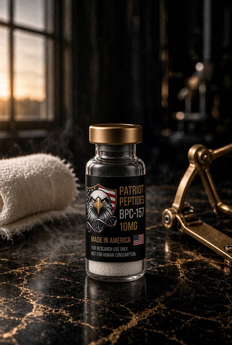
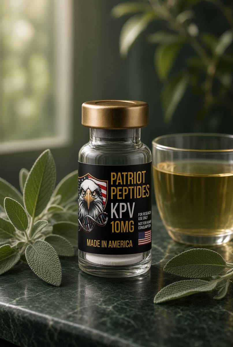
Research use only. Not a combined-use instruction.
Read next
45 min · long read · Peptide research
BPC-157: the pentadecapeptide that survived the stomach
A 15-residue fragment of a gastric protein, stable in acid, with a large preclinical literature on endothelium, tendon and nitric oxide. Here is the molecule, the papers, and what they actually mean.

48 min · long read · Peptide research
KPV: three residues, an NF-κB off-switch
The C-terminal tripeptide of α-MSH keeps much of the anti-inflammatory pharmacology and almost none of the tanning. Gut epithelium, PepT1, and a very small molecule.
More in this desk

53 min · long read · Peptide research
Recovery, GH pulses and the CJC / ipamorelin pair
Somatotrophs have two 'go' receptors. CJC without DAC is a slightly longer GHRH pulse. Ipamorelin is a selective ghrelin-receptor key that does not yank ACTH. Together they are the pair Bowers already showed is more than additive — not a gym protocol.
53 min · long read · Peptide research
How research peptides are made — and why HPLC actually matters
Solid-phase peptide synthesis builds a chain one residue at a time. HPLC then asks whether the main peak is what you think it is. ≥98% is not a slogan. It is a chromatogram.
52 min · long read · Peptide research
Fatigue, cellular energy and the NAD+ / MOTS-c neighbourhood
NAD+ is the rechargeable chip every cell spends on fuel and DNA repair — and the pool shrinks with age. MOTS-c is a mitochondrial 16-mer sent out under metabolic stress. Two answers to 'I have no energy' that are not coffee.
51 min · long read · Peptide research
Melanocortin circuits: pigment, appetite and PT-141
Melanotan II lights MC1, MC3, MC4 and MC5 — pigment plus the rest of the sheet. PT-141 is the free-acid cousin pointed at MC3 and MC4, the circuitry papers use for desire and energy, not skin colour. Same Arizona family. Different question.
Essays describe published research. They are not medical advice and they do not authorise human use of any catalogue item.