

Peptide research · 45 min · 9,965 words
BPC-157: the pentadecapeptide that survived the stomach
A 15-residue fragment of a gastric protein, stable in acid, with a large preclinical literature on endothelium, tendon and nitric oxide. Here is the molecule, the papers, and what they actually mean.
What this essay actually tells you
- BPC-157 is acid-stable because it is proline-rich. That's why oral gavage studies exist at all for a 15-mer. Most peptides would not survive the stomach long enough to be a story.
- Reported engagements: VEGFR2 phosphorylation, FAK–paxillin in endothelium and tenocytes, eNOS-dependent nitric-oxide tone. Name the assays. That's the file.
- Sikiric's Zagreb group built the largest corpus. Independent labs have reproduced endothelial migration more cleanly than claims that wander into the CNS. Read the independent ones first.
What this actually means
BPC-157 is a short piece of a stomach protein that acid and pepsin do not finish off, because the chain is full of proline. In rats it has been reported to shrink stomach lesions from anti-inflammatory drugs and alcohol. In dishes, blood-vessel cells migrate and tendon cells crawl, with a named growth-factor receptor and a grip kinase on the blots. A Zagreb laboratory spent thirty years arguing that the peptide resets nitric-oxide tone rather than simply raising it. That is a large, positive, published animal literature, and the interesting scientific sentence is smaller than the folklore: a 15-mer can be given by mouth in a rat and still be the 15-mer, and endothelium notices it. Independent labs have repeated the vessel and tendon-cell pieces more cleanly than the brain claims. We stock the named sequence because of that literature, not because of the healing story that grew up around it.
Most peptides you swallow never get past the stomach, and that's the first thing that makes this chain interesting. Pepsin — the acid protease that starts digesting protein at lunch — plus a pH that would unfold most globular proteins, plus a transit time measured in hours: that's the gauntlet for anything arriving as a chain of amino acids. Body protection compound-157 is a fifteen-residue fragment taken from a proteinaceous activity in human gastric juice, synthesised as Gly-Glu-Pro-Pro-Pro-Gly-Lys-Pro-Ala-Asp-Asp-Ala-Gly-Leu-Val, written GEPPPGKPADDAGLV, mass about 1419.5 daltons. Predrag Sikiric's group in Zagreb noticed that the fragment still cytoprotected mucosa after the juice had done its worst, and then spent three decades putting that 15-mer into injury models a gastroenterologist would recognise and into several a gastroenterologist would not. The origin is chemistry. Most fifteen-mers die in acid. This one is proline-rich, stiff, a poor pepsin substrate, and that is why oral gavage studies in rats exist at all. The rest of the literature — endothelium, tendon-to-bone, muscle crush, a nitric-oxide argument framed as a reset rather than a simple stimulation — follows from a stable chain that a vessel cell can notice.
In short. This peptide is a fifteen-amino-acid piece of a stomach protein that acid does not destroy. That chemistry is why rat studies could even give it by mouth.
We stock the named sequence because a literature exists: GEPPPGKPADDAGLV, HPLC-characterised, mass on the certificate, the pentadecapeptide the Zagreb papers and the independent endothelial papers actually used. That's a research object. It isn't a protocol, it isn't a licensed medicine, and it isn't a synonym for TB-500, which orbits an actin-binding motif and has its own page. Repair, when you sit down to measure it, is a named tissue, a named insult, a named readout, and a clock: gastric lesion area after indomethacin, load-to-failure of a tendon-to-bone junction at six weeks, tenocyte outgrowth from an explant, endothelial migration across a scratch in eight hours. Those are assays you can run. This piece names the molecule, the stomach that selected it, the single-laboratory programme that made it famous, the receptor tyrosine kinase independent labs can actually blot, and the central-nervous-system claims that have not earned the same outside check. No dosing here. The rodent models already ran; you'll find their clocks and endpoints in PubMed.
In short. We keep the named chain because published animal and cell work exists. That isn't a treatment plan, and it isn't the same molecule as the actin peptide people pair it with.
Sequence is identity, and identity is the first experiment. Fifteen residues is a short protein and a long peptide. Insulin is fifty-one. GLP-1 is thirty or thirty-one depending on the amide. A class-B GPCR ligand of this length usually has a defined fold and a receptor pocket it evolved to occupy. BPC-157 has no solved globular structure worth hanging a mechanism on. It's Pro-rich, acidic in the middle where two aspartates sit, and hydrophobic at the carboxyl end with leucine and valine. People have docked it, in silico, against VEGFR2 and against various extracellular-matrix proteins, and docking a floppy 15-mer is a way to generate a colourful figure, not a way to generate a Kd. The experimental engagements that have survived contact with other laboratories are cruder and better: endothelial cells migrate; VEGFR2 is internalised and phosphorylated; focal-adhesion kinase and paxillin light up; nitric-oxide tone in a vessel or a gut wall moves. Those are phosphorylation blots and organ-bath traces. They're not a crystal, and they're not a wellness caption.
In short. A peptide is its amino-acid sequence. This one is short, floppy, and not folded like insulin. The useful evidence is cell movement and phosphorylation, not a computer picture of it sitting on a receptor.
Cytoprotection is the ability of a mucosa to resist injury without a measurable change in acid secretion. The pentadecapeptide entered that conversation as a non-prostaglandin cytoprotectant. The vessel, not the parietal cell, is the room the cleaner papers actually occupy.— Reading Robert A, Szabo S, and the Sikiric gastric-lesion corpus against Hsieh et al. on VEGFR2 in endothelium.
The chain: GEPPPGKPADDAGLV
Write the residues in three-letter code and the chemistry is already visible. Glycine-glutamate, then three prolines in a row, then glycine-lysine-proline, then alanine-aspartate-aspartate-alanine, then glycine-leucine-valine. Four prolines in fifteen positions, clustered toward the amino end. Two consecutive aspartates in the middle. A hydrophobic carboxyl tail. Theoretical monoisotopic mass for the free peptide is 1419.5 daltons; the molecular formula is C62H98N16O22 if you write the uncharged chain. The lyophilised cake on a certificate will also report trifluoroacetate or acetate counter-ions depending on the cleavage and the salt exchange, and it's worth writing which. CAS 137525-51-0 is the registry number the catalogues use. Purity ≥98% HPLC is a peak, not a physiology. A deletion peptide missing one glycine can hide under a cheap ultraviolet trace and still wreck an assay, which is why the interesting chromatogram is the one with a mass on it, not the one with a pretty single peak at 214 nanometres.
In short. The chain is fifteen amino acids with four prolines, mass about 1,420 daltons. A lab certificate should show that mass and a clean peak. A pretty peak alone is not enough.
Body protection compound was the name Sikiric's group gave to a proteinaceous activity in human gastric juice that cytoprotected mucosa against a catalogue of insults. BPC-157 is the synthetic pentadecapeptide they took from that work and put into print, from the early 1990s, as a stable fragment that still did the job. The gastric juice protein from which the fragment was taken is not a hormone in the Bayliss and Starling sense. It is neither secretin nor gastrin. It doesn't occupy a class-B GPCR on a pancreatic cell and raise cyclic AMP. It's a cytoprotective peptide activity that a laboratory in Croatia chose to synthesise as a 15-mer and then spent a career dropping into every injury model the animal house would support. That career is the Sikiric corpus, and it's both the reason the molecule is famous and the reason we keep a pencil in hand. Hundreds of papers, one principal investigator as a constant, a recurring nitric-oxide theme, and a spread of endpoints that would make a multi-centre consortium blush.
In short. The original activity was noticed in stomach juice, not as a gut hormone that tells the pancreas to release insulin. A Croatian lab then made a fifteen-residue piece and tested it for decades.
A GPCR ligand of this length, when it is a real evolved ligand, usually arrives with a fold, a disulfide, or a lipid handle, and with a receptor it was selected to occupy. This 15-mer was selected by gastric juice, which is a different evolutionary pressure. Survival in acid is not occupancy at a seven-helix receptor. Survival in acid is a backbone that pepsin cannot comfortably cut and a chain that doesn't need a salt-bridge fold to remain a peptide. That distinction matters because the rest of this catalogue is full of true GPCR ligands — incretins, melanocortins, ghrelin mimetics — and it's tempting to file every lyophilised 15-mer as one more occupancy. VEGFR2, the receptor named in the cleaner BPC-157 papers, is a receptor tyrosine kinase — a single-pass transmembrane enzyme, not a seven-helix lock. The second-messenger diagram still earns a place later, because Akt, eNOS and nitric oxide are an amplification cascade. They're just not a Gs cascade. Name the lock before you name the downstream chemistry.
In short. Most short peptides here fit a seven-helix receptor. This one doesn't have to. Survival in stomach acid is a backbone fact, not a proof that it uses the same lock as a gut hormone.
Proline is the residue that does most of the telling. The side chain loops back onto the backbone nitrogen, so proline is both an amino acid and a fold-breaker. It starves several proteases of a comfortable cut site. It disfavours alpha helix. Three consecutive prolines in GEPPP are a local polyproline stretch; they don't present a classic scissile bond, and they make the amino-terminal half of the 15-mer look, to a protease, like a problem. The fourth proline, after the lysine, repeats the joke a little further along. Gastric lipase and the subsequent tryptic and chymotryptic gauntlet in the duodenum are the next tests, and they aren't nothing, but a chain this proline-rich is already a bad substrate before it leaves the stomach. That's why oral rodent studies exist at all. They aren't a marketing claim about bioavailability in a person. They're a consequence of a proline-rich 15-mer that gastric juice itself selected. Conflating gavage in a rat with a capsule in a person is how a stability fact becomes a wellness claim.
In short. Proline is a stiff amino acid that digestive enzymes cut poorly. Four of them in this short chain are why stomach juice doesn't chew it to pieces the way it chews most peptides.
Acid, pepsin, and why gavage exists
Pepsin is an aspartic protease with a preference for aromatic and hydrophobic residues, optimally active around pH 2. A peptide rich in proline and poor in phenylalanine and tyrosine is already a bad substrate. The stomach's pH 1.5 is not merely a number on a first-year slide. It protonates side chains, it unfolds anything that was relying on a salt bridge, and it hands the unfolded chain to pepsin. Most research peptides we lyophilise wouldn't survive that room as ligands. This one was taken from that room. The historical selection pressure is the entire reason the molecule escaped a gastroenterology footnote and walked into tendon papers: a chain that can be gavaged and still move a gastric-lesion endpoint has already told you it's still the chain when it meets the mucosa. Parenteral studies exist too, and they're a different pharmacokinetic object. Intraperitoneal, intramuscular, peri-tendinous — those routes skip the acid test that made the peptide interesting. They're lawful experiments. They're not the origin story, and we shouldn't write them as if they were.
In short. Stomach acid and pepsin destroy most short peptides. This one was taken from stomach juice and still works in mouth-dose rat studies. Injection studies skip that test and are a different experiment.
The gut lining is a sheet of polarised epithelial cells, a mucus layer, tight junctions, a basement membrane, and a lamina propria full of immune cells and a vascular bed that is never more than a few tens of micrometres away. Cytoprotection, in the language André Robert and Sandor Szabo taught the ulcer field, is the ability of a mucosa to resist injury without a measurable change in acid secretion. Prostaglandins were the first such story; misoprostol is the licensed descendant. BPC-157 entered that conversation as a non-prostaglandin cytoprotectant, and the Zagreb papers on NSAID gastropathy, alcohol lesions, and bile-reflux models are the original dataset. Independent groups have been thinner on the gastric endpoint than on the endothelial one, which is already a clue about how the literature aged. A peptide that protects a mucosa might do so by keeping epithelium stuck to basement membrane, by keeping blood flow, by damping an inflammatory burst, or by all three. Blood flow is the sentence that leads, almost immediately, to nitric oxide and to endothelium.
In short. The gut lining is a thin living wall with mucus, tight seals, and a rich blood supply. Early papers asked whether this peptide helped that wall resist injury without turning down acid.
Diagram
Mucus
MUC2 gel
The first argument a microbe has to win.
Epithelium
enterocyte · goblet · Paneth
One cell thick. The wall is the cell, not a fascia.
Tight junctions
claudin · occludin · ZO-1
The seal. ‘Leaky gut’ as a brand is not this protein list.
PepT1
SLC15A1
Oligopeptide transporter. Inflamed gut induces it. KPV can ride it.
Lamina propria
immune cells
Where NF-κB decisions become cytokines.
BPC-157 neighbourhood
NO · VEGFR2 · FAK
Cytoprotection, blood flow, how a damaged lining organises.
IBS is a Rome-criteria cluster. It does not name a receptor. The preclinical literature that named molecules for barrier tissue keeps coming back to BPC-157 and KPV — two ligands, one organ on a reading list, neither a gastroenterology appointment.
Once a stable peptide talks to endothelium, the rest of the injury catalogue is almost inevitable. A tendon-to-bone junction is a vascular problem and a collagen problem. A crushed muscle is a vascular problem, a myofibre problem, and a fibrosis problem if the clock runs wrong. A colon anastomosis is a vascular problem and a leak problem. A cornea isn't a gut, but it is an epithelium on a stroma with a migration clock. The Zagreb group put the 15-mer into all of those, and into a few places a sceptical reader wouldn't have started: nigrostriatal lesions, behavioural despair, inflammatory-bowel models that sit next to the original mucosa, periodontal defects. Breadth is a scientific virtue when each model is independently replicated. Breadth is a warning light when one laboratory is the constant and the endpoints multiply faster than the mechanistic papers. The gastric, vascular and tenocyte pieces are the ones a new bench should start with. They're closest to the chemistry that made the peptide interesting, and they're the pieces other people have actually run.
In short. Once a peptide talks to blood-vessel cells, labs try it in tendon, muscle and gut repairs. Some of those tests have been repeated outside the original group. Many have not.
Robert's 1979 demonstration that prostaglandins could protect gastric mucosa against necrotising agents, at doses that did not inhibit acid, is the conceptual ancestor of every cytoprotection paper that followed. The word is specific. It doesn't mean the peptide is an antacid. It doesn't mean the peptide is a proton-pump inhibitor. It means the lining holds against an insult that would otherwise strip it, while acid output stays on the graph. Szabo's work on sulfhydryls, vascular injury, and ethanol models added the blood-flow chapter that a purely epithelial story had been missing: early vascular stasis and endothelial damage precede the obvious mucosal necrosis in several of these models. That's why a gastric peptide that later shows up in an endothelial scratch assay isn't a category error. The stomach was always a vascular organ as well as a secretory one. The 15-mer's original endpoint was lesion area. The mechanistic neighbourhood that aged better is the capillary underneath the lesion.
In short. Cytoprotection means the lining resists damage without the drug having to shut off acid. Blood flow in that lining turned out to matter as much as the surface cells.
- Sequence
- GEPPPGKPADDAGLV
- Mass
- 1419.5 Da
- Prolines
- 4 of 15
- VEGFR2 (KDR)
- RTK, ~230 kDa mature
- FAK–paxillin
- adhesion kinase blot
- Independent replication
- angiogenesis, tenocytes
- Gavage in the rat
- µg–mg range in papers
- Certificate
- ≥98% HPLC plus mass
Fifteen residues. Four prolines. The chain gastric juice selected.
Free peptide. Certificate must also name the counter-ion.
The chemical reason pepsin and pH 1.5 do not finish the job.
Not a GPCR. Internalisation and phosphorylation are the endothelial node.
Tenocyte and endothelial migration. Inhibitors belong on the next well.
Cleaner than the CNS claims. Start here if you are a new bench.
Possible because the chain is still the chain. Not a human capsule claim.
A peak without a mass is a label. A label is not a ligand.
NSAID gastropathy and ethanol lesions
NSAID gastropathy is the original neighbourhood. Indomethacin, diclofenac, aspirin: cyclo-oxygenase inhibition, prostaglandin collapse, a mucosa that can no longer keep its mucus and bicarbonate and blood flow, lesions that a pathologist scores by area. Robert's cytoprotection was prostaglandin replacement. Sikiric's argument was that BPC-157 reduced lesion area in these models without being a prostaglandin, and that the protection reached intestinal and liver injuries that NSAIDs also cause. Those papers are numerous. They're also, as a body, light on randomisation detail and on the sort of blinding a modern ulcer paper would insist on. That isn't a reason to invent a conspiracy. It's a reason to treat effect sizes as provisional and to look for the groups that ran an NSAID lesion with a characterised 15-mer and a pre-registered scoring rule. The scientific value of the gastric work, for a peptide catalogue, isn't a human ulcer claim. It's the chemical fact that a 15-mer survived the organ that invented it, plus a pile of rodent lesion scores that a careful lab could re-run.
In short. The first papers were about stomach damage from anti-inflammatory drugs. The peptide seemed to shrink those lesions in rats. The studies are many, and the methods are not always as tight as a modern trial.
Ethanol is the other classic gastric insult, and it's dirtier than a cyclo-oxygenase inhibitor in a useful way. Absolute or high-percentage ethanol, instilled into a rat stomach, produces haemorrhagic lesions within minutes. The mechanism is a mix of direct epithelial necrosis, mucus disruption, and a vascular injury Szabo spent a career measuring: early stasis, endothelial damage, a mucosa that dies because blood stopped arriving. A peptide that shrinks those lesions could be an epithelial protectant, a vascular protectant, or both. The Zagreb ethanol papers report reduced lesion area and invoke the same nitric-oxide set-point they invoke everywhere else. Independent endothelial work, later and cleaner, gives you a reason to take the vascular half seriously without having to swallow the entire organoprotection philosophy. If you're going to re-run an ethanol model, score the lesions blind, state the ethanol concentration and the volume, and put an eNOS inhibitor arm next to the peptide arm. Otherwise you have a picture of a stomach and a caption.
In short. Alcohol poured into a rat stomach makes rapid bleeding lesions, partly because blood flow fails. Papers say this peptide shrinks those lesions. Score them blind and test the blood-flow idea directly.
Bile reflux, cysteamine duodenal ulcers, and the various pancreatitis and colitis models that sit next to the stomach in the Zagreb bibliography are the same sentence stretched along the gut tube. Each has a real clinical cousin. Each is also a model with its own confounders, its own scoring conventions, and its own history of peptides that looked miraculous in one laboratory and quiet in the next. I'm not going to narrate every insult. The pattern is the point. A proline-rich 15-mer that can be given by mouth, that reduces lesion area in NSAID and ethanol stomachs, and that later shows VEGFR2 and FAK activity in endothelium, is a coherent preclinical object. A 15-mer that also fixes pancreatitis, colitis, periodontal defects and a nigrostriatal lesion, in the same principal investigator's hands, is a programme that has outrun its mechanistic papers. Keep the coherent object. File the rest as hypotheses a different bench would have to own.
In short. The original lab tested the peptide in many gut injuries beyond simple stomach ulcers. The stomach, the vessel and the tendon cell are the parts other people have actually repeated.
The Sikiric corpus, and how a single-lab programme ages
Predrag Sikiric, University of Zagreb School of Medicine, and a rotating cast of colleagues — Seiwerth, Kokić, Drmić, Starešinić, the names recur — built, from about 1993, one of the largest preclinical peptide corpora in print. The early papers sit in Journal of Physiology and Pharmacology, Journal of Orthopaedic Research, Burns, Digestive Diseases and Sciences, and a long tail of specialty journals that will take an injury model. The through-line they themselves emphasise is organoprotection: a peptide that restores a physiological set-point after an insult, rather than a peptide that simply stimulates a receptor. The nitric-oxide system is the set-point they named most often. L-NAME, which inhibits nitric-oxide synthase, makes lesions worse; L-arginine, which feeds the enzyme, can make them better; BPC-157, in their hands, nudges the animal back toward the middle from either side. That's an unusual pharmacological claim. It's also a claim that demands, and has not always received, a dose–response, an eNOS phosphorylation blot, and an outside laboratory with no reason to protect the hypothesis.
In short. A Zagreb group spent thirty years testing this peptide in many injury models. They argue it resets nitric-oxide tone rather than simply raising it. That claim is interesting and still needs outside checks.
Organoprotection is a philosophy before it is a kinase. It says the peptide doesn't push a system in one direction; it restores a middle after an insult has shoved the animal off it. In practice that philosophy has to become an experiment: a two-sided perturbation, a middle you can measure, and a peptide arm that returns the measurement from both sides. Nitric oxide is almost the only named system in this corpus that could, in principle, support that design, because you can inhibit the synthase with L-NAME and you can feed it with L-arginine, and you can measure the gas, the cyclic GMP, the phospho-enzyme, or the lesion that sits downstream. Most of the Zagreb papers measure the lesion. Fewer measure the enzyme. That's how a philosophy outruns a blot. It's a lawful way to write a career. It's a frustrating way to hand a mechanism to the next laboratory. The next laboratory, if it is wise, will pick one side of the philosophy and blot it.
In short. The original group's big idea is that the peptide restores a balance after injury, rather than pushing one switch. In practice that idea still has to become a named measurement on a named enzyme.
The nitric oxide system is not simply stimulated or inhibited. In this literature the pentadecapeptide is asked to restore a set-point after the insult has shoved it — a claim that is either a deep pharmacology or a habit of language, and only a blot with L-NAME, L-arginine and an eNOS antibody can tell those apart.— Reading Sikiric P et al. on BPC-157 and the NO system, J Physiol Pharmacol and related Zagreb papers, 1990s–2020s.
How a single-laboratory programme ages is a sociology of science before it is a verdict on a peptide. Effect sizes stay large. Journals stay specialised. Reviewers are often colleagues in the same injury field. Negative results, if they exist, are quieter. None of that is fraud; all of it is how academic careers work when a compound is yours. The correction, which the field already knows how to apply, is independent replication of the pieces that would change a biochemist's week. Angiogenic and tenocyte-migration findings have had more of that correction than the more extravagant central-nervous-system claims — nigrostriatal protection, antidepressant-like behaviour, a wandering into models that a 15-mer from gastric juice had no particular chemical reason to enter. A peptide can have more than one job. It cannot have every job. When I sit with this literature I keep the stomach, the vessel, the tendon, and the nitric-oxide blot. I put the CNS papers in a pile labelled interesting, unreplicated, and not the reason the 15-mer is on a research shelf.
In short. One lab publishing for decades will look bigger than it is. Other labs have repeated the blood-vessel and tendon-cell pieces more cleanly than the brain claims.
Independent laboratories did not reproduce the entire Zagreb map, and nobody honest expected them to. What they did reproduce, in endothelium and in tendon fibroblasts, is enough to keep the 15-mer interesting as a probe. VEGFR2 phosphorylation and internalisation. Endothelial migration in a scratch or a Boyden chamber that a VEGFR2 inhibitor can blunt. Tenocyte outgrowth and a FAK–paxillin blot. Some anastomotic and fistula papers from groups adjacent to Zagreb. That's a real, if still preclinical, object. It's also a much smaller object than the healing-peptide story that grew up around it. The gap between those two objects is the gap this piece exists to keep open. The 15-mer on the shelf is GEPPPGKPADDAGLV, HPLC-characterised, the sequence those papers named. The papers are public. The blot is yours. Start with the node another laboratory has already occupied, not with the most exotic endpoint in the bibliography.
In short. Other groups have repeated the vessel-cell and tendon-cell findings. That is enough to keep the molecule interesting. It is not the same as the larger healing story around it.
Tendon-to-bone, muscle crush, and a vascular repair bed
Tendon-to-bone is the paper a sports-medicine thread actually wants. Chang, Tsai, Lin, Hsu and Pang, Journal of Applied Physiology 2011: pentadecapeptide BPC-157 promoted tendon outgrowth, cell survival and cell migration in a rat Achilles model, with FAK and paxillin implicated in the tenocyte. That paper is usable because it names a cell, a kinase, and a phenotype a dish can hold. Starešinić and the Zagreb group ran transected Achilles and quadriceps defects, load-to-failure, collagen organisation, and a vascular density in the repair bed that is the angiogenic half of the same story. A tendon heals slowly because it is poorly vascularised and because the collagen has to be laid in the line of force, not as a scar. Anything that persuades tenocytes to migrate, survive, and pull on a matrix, and that persuades a capillary to enter the defect, will move those endpoints in a rodent. Whether the 15-mer does that by VEGFR2, by FAK, by a nitric-oxide effect on the vessel, or by some combination, is the mechanistic question the better papers try to close with inhibitors.
In short. In rats, the peptide has been reported to help tendon cells crawl, survive and organise collagen, and to bring in small vessels. That is a rodent finding with named tests, not a sports protocol.
Load-to-failure at six weeks is a phenotype, and phenotypes are allowed, but they aren't a receptor. A repaired Achilles in a rat is a surgery, a housing condition, a loading history, a peptide exposure, and every systemic thing the animal did while the clock ran. The explant is closer to a mechanism: a piece of tendon on a dish, cells crawling off, a count, a kinase blot. Chang ran that neighbourhood. A lab that wants the 15-mer as a tenocyte probe should run the explant, the scratch, the phospho-FAK blot, and a collagen-gel contraction if they have the patience. A lab that wants a rodent biomechanics paper should budget for the six-week clock and for a surgeon who can make the defect the same way twice. Those are different grants. They're often written as one sentence, usually as healing. If you can't tell an explant from a load-to-failure curve, you're not yet ready to interpret either result, however pretty the trichrome.
In short. Watching tendon cells crawl off a scrap of tissue is closer to a mechanism than weighing a repaired tendon six weeks later. Both appear in the papers. They answer different questions.
Muscle crush is the third Zagreb staple. A mechanical insult, a haematoma, a myofibre necrosis, a satellite-cell response, a fibrosis if the clock runs wrong. Papers from that group report faster restoration of function, better fibre organisation, and again a vascular component. Crush is a dirty model, which is both its virtue and its vice: it looks like an injury a person might recognise, and it confounds everything at once. A clean myoblast migration assay in a dish, with the peptide characterised and a FAK inhibitor on the next well, is the experiment that would make a muscle claim mechanistic. Some of that work exists. Not enough of it exists outside the original laboratory. The pattern is now familiar, and it will keep being familiar: the in-vivo phenotype is large and repeated in-house; the molecular paper that would make a biochemist relax is thinner and later. That's how a single-lab programme ages, and it isn't unique to Zagreb. It is what happens when a compound is interesting, cheap to synthesise, and more fun to drop into a new injury than to sit with one blot until the kinase is named.
In short. Crushed muscle in a rat is a messy injury, and the original lab reported faster recovery. Messy injuries are hard to interpret. A clean cell-migration test would do more to name the job.
Vascular density in a repair bed is the sentence that ties tendon, muscle and gastric mucosa together without pretending they are one tissue. Granulation tissue that cannot recruit capillaries will not feed a fibroblast. A tendon graft that remains ischaemic will not organise collagen in the line of force. A mucosa whose capillary bed has stasised will necrose no matter how intact the surface mucus looked ten minutes earlier. VEGF biology already knew this. The BPC-157 literature's cleaner contribution is to put a proline-rich 15-mer onto that angiogenic programme via VEGFR2 internalisation and phosphorylation, with Akt and eNOS downstream, in endothelium a Western lab can buy. That's a smaller claim than organoprotection, and it's a more useful one, because a smaller claim can be falsified. SU5416, a VEGFR2 kinase inhibitor, or a blocking antibody, or an siRNA, should take the migration with it if the story is true. If it doesn't, the 15-mer is doing something else, and that something else would be the actual paper.
In short. New small vessels in an injured tissue are a shared need of stomach, tendon and muscle. The cleaner cell work puts this peptide on a named vessel-growth receptor you can block.
VEGFR2 is a receptor tyrosine kinase, not a GPCR
VEGFR2 is KDR, Flk-1, a receptor tyrosine kinase. It isn't a GPCR. Seven-helix receptors, G proteins, arrestin, cyclic AMP — that's a different superfamily, and this catalogue uses it for incretins and ghrelin mimetics and melanocortins. VEGFR2 is a single-pass transmembrane protein with an extracellular immunoglobulin-like ligand domain and an intracellular kinase. VEGF-A dimers it; the kinase domains autophosphorylate; phospholipase C-gamma, PI3K–Akt, Src, and a FAK neighbourhood are among the effectors; endothelial survival, migration and proliferation follow. Internalisation of VEGFR2 isn't merely turning the receptor off. Endocytic traffic of VEGFR2 is part of how the angiogenic signal is shaped, and Hsieh's BPC-157 paper sits on that traffic: the pentadecapeptide associated with VEGFR2 activation and internalisation, with Akt and eNOS downstream. You can disagree with a figure. You can't relabel an RTK as a GPCR because the rest of the catalogue is GPCRs. The second-messenger diagram earns a place here because Akt, eNOS and nitric oxide are an amplification cascade. They're just not a Gs cascade.
In short. The blood-vessel receptor named in the cleaner papers is an enzyme in the membrane, not the seven-helix locks most catalogue peptides use. Downstream, kinases and nitric oxide still amplify the signal.
Internalisation is a trafficking sentence. Ligand binds, the receptor clusters, clathrin or a non-clathrin path pulls a vesicle, endosomes sort the cargo for recycling or for lysosomal destruction, and some signalling continues from the endosome. For VEGFR2, the endosomal chapter isn't optional reading. Papers from the VEGF field — not the BPC field — spent years showing that where the receptor sits after the ligand arrives changes which effectors it sees. If a 15-mer promotes VEGFR2 internalisation and phosphorylation, the experiment that follows isn't a testimonial. It is: does a kinase-dead VEGFR2, or SU5416, or a VEGFR2 siRNA, take the endothelial migration with it? Hsieh's group used inhibitors. A new bench should use them too, and should put the characterised peptide on a mass spectrometer before the first well is seeded. VEGFR2 antibodies on a blot are cheap. Identity first. Then the kinase. Then the picture of cells closing a gap.
In short. When that receptor is pulled inside the cell, the growth signal can still run. The honest test is whether blocking the receptor also blocks the cell movement you think the peptide caused.
Hsieh, Liu, Wang and colleagues, Journal of Molecular Medicine 2017, tied BPC-157 to VEGFR2 activation and internalisation in endothelium, with downstream Akt and eNOS phosphorylation — a one-sided, angiogenic, RTK-shaped story that a Western lab can pick up without buying the entire organoprotection philosophy. That's how you want a large corpus to age: a few nodes other people can occupy, and a surrounding philosophy you are free to leave on the shelf. Occupancy here is a pharmacological word. It means the receptor is engaged, phosphorylated, trafficked. It doesn't mean the 15-mer has been shown, by a solved structure, to sit in the VEGF-A pocket. Direct binding of a floppy pentadecapeptide to VEGFR2 remains the weaker half of the Hsieh paper; the phosphorylation and the inhibitor-sensitive phenotype are the stronger half. A new experiment should pick which sentence it is testing. A pull-down or a surface-plasmon measurement would test binding. A phospho-blot plus SU5416 would test the pathway. Doing both and calling them a docking figure is how a neighbourhood gets louder than the data.
In short. A 2017 cell paper linked the peptide to a vessel-growth receptor being switched on and pulled inside, with named enzymes downstream. Binding is still the shakier half. The blot is the stronger half.
Diagram
× 1
Ligand
One peptide in one pocket. nM–µM. Shape, not a mood.
× 10–10²
G proteins
The occupied GPCR is a GEF. Each Gα is a catalyst.
× 10³–10⁴
cAMP / IP₃ / Ca²⁺
Adenylyl cyclase and PLC do not make one molecule. They make a cloud.
× 10⁴–10⁶
PKA / PKC / CaMK
Kinases phosphorylate many substrates per messenger.
× tissue
Secretion, transcription, motility
The organism-level readout. Still not a protocol.
This is the only magic, and it is not magic. A nanomolar ligand can move a micromolar messenger because enzymes sit between them. Desensitisation (GRK, β-arrestin, endocytosis) is how the cell refuses to let ‘more ligand’ mean ‘more signal’ forever.
Focal-adhesion kinase is the other named node, and it is how a tendon paper and an endothelium paper can share a kinase without sharing a tissue. FAK sits at integrin clusters where a cell grips extracellular matrix. Autophosphorylation at Tyr397 recruits Src; paxillin and p130Cas are phosphorylated; Rac and the actin machinery are told that the grip is real and that a protrusion may proceed. Chang's tenocyte work put FAK and paxillin on the BPC-157 page. Endothelial migration uses the same adhesion kinase because crawling is crawling. A peptide that lights FAK in two cell types is not therefore a universal repair juice. It's a peptide that may feed a kinase two cell types already use to move. The blot is phospho-FAK, phospho-paxillin, a FAK inhibitor — PF-573228 is the one people reach for — and a scratch that should stall when the kinase is blocked. If the scratch doesn't stall, FAK was a spectator, and you've learned something the paper you are citing did not. Spectator kinases are common. They're also publishable, if you write them down.
In short. A cell uses this kinase to know it is gripping the surface it wants to crawl on. Tendon cells and vessel cells both use it. Block it to see if the peptide needed it.
eNOS is endothelial nitric-oxide synthase, phosphorylated at Ser1177 by Akt among other kinases, and that phosphorylation is a standard Western readout of an angiogenic or shear-stress programme. Nitric oxide then is the gas: short-lived, locally acting, a relaxant of smooth muscle, a modulator of platelet and leukocyte stickiness, a player in VEGF's own vascular-permeability effects. Sikiric's reset language and Hsieh's eNOS phosphorylation can live in the same neighbourhood without being the same claim. One is a two-sided physiological set-point across organ systems. The other is a kinase on a blot in a human umbilical-vein endothelial cell. A new experiment should pick which sentence it is testing. Organ-bath aortic rings, L-NAME, haemoglobin as an NO scavenger, and an eNOS phospho-antibody will do for the vessel. A gastric-lesion score plus L-NAME will do for the mucosa. Doing both and calling them one mechanism is how two measurements get flattened into one caption. Keep them as two measurements. Write two figure legends.
In short. A blood-vessel enzyme makes nitric oxide, a short-lived gas that opens vessels. You can measure the enzyme on a blot or the gas by how a vessel relaxes. Pick one question per experiment.
Endothelial migration is the honest assay because it is cheap, visual, and inhibitor-friendly. Seed a monolayer. Wait until it is confluent. Scratch it with a pipette tip. Image at 0, 8, 12, 24 hours. The gap closes by cells crawling, and, if you let it go too long, by proliferation, which is why a mitomycin C arm exists in a careful protocol. Add the peptide at a stated concentration, from a stock whose identity you have confirmed by HPLC-MS, in a medium whose serum content you have written down because serum is a VEGF bath. Put SU5416 or a VEGFR2 blocking antibody in the next well. Put a FAK inhibitor in the one after that. If the 15-mer closes the gap and the inhibitors reopen it, you have a paper. If the 15-mer closes the gap and the inhibitors do nothing, you have a different paper, and you should write it rather than forcing VEGFR2 onto a result that did not ask for it. Either outcome is science. A closed gap and a caption that says healing isn't a paper. It's a photograph.
In short. The simple dish test is a scratch in a sheet of vessel cells. Time-lapse the gap. Add blockers of the named receptor or kinase. If the gap still closes, your story was wrong.
Tenocytes are not endothelial cells. They're the fibroblasts of tendon, a lineage that lives in a dense collagen hierarchy, sees load as its growth factor, and migrates poorly compared with a human umbilical-vein endothelial cell. Outgrowth from an explant, as Chang ran it, is a fair assay: a piece of tendon on a dish, cells crawling off, a count, a kinase blot. Serum, as ever, is a confounder, because serum is a growth-factor bath. So is the possibility that the 15-mer is doing something to the explant's residual vessels rather than to the tenocyte itself. A purified tenocyte monolayer, a defined medium, phospho-FAK, and a FAK inhibitor are how you close that gap. Until those wells exist, the honest sentence is that tendon cells in explant culture migrate more when the peptide is present, and that FAK and paxillin are implicated. Implicated is a real word. It's weaker than required, and it's stronger than a caption. Required is what the inhibitor arm is for.
In short. Tendon cells are not vessel cells. They crawl more slowly and live in dense collagen. Watching them leave a scrap of tendon is a fair test if you blot the grip kinase.
Nitric oxide as a set-point, not a donor
Nitric oxide is a real, named, assayable gas, and that's why the reset claim deserves more than a shrug. Endothelial nitric-oxide synthase makes NO from L-arginine; NO activates soluble guanylate cyclase; cyclic GMP relaxes smooth muscle; blood flow rises. Too little nitric oxide, and a mucosa or a tendon graft is ischaemic. Too much, and you have hypotension and a different pathology. L-NAME inhibits the synthase; L-arginine feeds it. Sikiric's group reported that BPC-157 counteracted both L-NAME and L-arginine derangements in lesion models, which is the reset. Independent endothelial papers have shown eNOS-dependent effects without always signing up to the two-sided reset. Those two sentences can both be true. A peptide could phosphorylate eNOS in a HUVEC and, in an animal, move lesion scores back toward the middle from both a synthase-blocked and an arginine-loaded state. It could also be that the two-sided animal result is a habit of how that laboratory writes injury papers. Only a blot with both perturbations, an eNOS antibody, and an outside group can tell those apart.
In short. The original group says the peptide restores nitric-oxide balance from both directions. Outside labs more often show a one-way vessel-growth path. Both can be measured. They are not automatically the same claim.
A nitric-oxide donor is a different object. Sodium nitroprusside, S-nitroso-N-acetylpenicillamine, a nitrate that a doctor actually prescribes — those dump the gas into the system and let the consequences follow. The Zagreb claim, read generously, is that the 15-mer is not that dump. It's a correction. Read less generously, correction is what you call a result when the error bars on both sides of a set-point are large and the scoring is unblinded. I don't know which reading is true. I know which experiment would tell me. Randomised, blinded gastric-lesion or aortic-ring work, L-NAME and L-arginine as two-sided perturbations, the characterised 15-mer in the middle, phospho-eNOS and a cyclic-GMP measurement as the mechanistic readouts, and a laboratory that did not spend thirty years on the hypothesis. Until that paper exists, the reset remains the most interesting sentence in the corpus and the least closed. Interesting and unclosed is a normal state in peptide physiology. It isn't a reason to write a protocol. It's a reason to blot.
In short. A nitric-oxide donor pours the gas in. The original papers claim something subtler: a return to the middle. That is still a hypothesis until someone outside the original lab measures the enzyme from both sides.
Cyclic GMP is the nucleotide that makes the gas a muscle-relaxant signal. Soluble guanylate cyclase binds NO at a haem; GTP becomes cyclic GMP; protein kinase G phosphorylates the targets that drop cytosolic calcium in smooth muscle. Sildenafil lives in this neighbourhood as a phosphodiesterase-5 inhibitor, which is how a bedroom drug and an ulcer peptide can share a second messenger without sharing a mechanism. If BPC-157's vascular effects run through eNOS, cyclic GMP should move, and a guanylate-cyclase inhibitor such as ODQ should blunt the organ-bath phenotype. That experiment is ordinary. It is also, relative to the size of the corpus, under-done in independent hands. A new bench that wants the nitric-oxide story should run the ring, the ODQ, the L-NAME, and the phospho-eNOS, and shouldn't also try to solve tendon outgrowth in the same week. One cascade per grant. The peptide will still be on the shelf when the rings are done.
In short. Nitric oxide relaxes muscle by raising a second messenger called cyclic GMP. If the peptide's vessel story is true, that messenger should move, and blocking it should blunt the effect.
What independent labs actually repeated
Replication is a sociological word and a methodological one, and the two shouldn't be confused. Methodologically, a replication is the same ligand, a stated concentration, a named cell or tissue, a named readout, and a result that a second kitchen can cook. Sociologically, a replication is a paper whose authors do not need the original hypothesis to be true. The BPC-157 literature is rich in the first sense inside Zagreb and thinner in the second sense outside it, except at two nodes. Endothelial migration and VEGFR2 traffic, Hsieh and neighbours. Tenocyte outgrowth and FAK–paxillin, Chang and neighbours. Those nodes are why a research catalogue keeps the 15-mer. They're also why a research catalogue does not keep a CNS legend. Nigrostriatal protection, behavioural despair, a wandering into seizure and multiple-sclerosis models — those papers exist, they're mostly in-house, and a 15-mer from gastric juice had no particular chemical reason to enter a synapse. A peptide can surprise you. Surprise isn't a substitute for an outside laboratory.
In short. Other labs have repeated the vessel-cell and tendon-cell work more cleanly than the brain-and-behaviour papers. That difference is how a careful reader ranks the claims.
The central-nervous-system claims are the ones a certain kind of reader wants, because a gut peptide that also fixes a nigrostriatal lesion would be a story, and stories travel. Dopamine neurons, behavioural despair assays, a handful of seizure models: the Zagreb group put the 15-mer into all of them and reported protection. Blood–brain barrier papers from the same programme argue that the peptide helps a damaged barrier rather than simply crossing a healthy one, which is at least a physiologically literate way to wander into the brain. Independent groups have not made this the node they chose to occupy. That may change. Until it does, the ranking I'd teach is: gastric chemistry first, endothelium and tenocyte next, nitric-oxide set-point as an open mechanistic claim, CNS as a pile labelled unreplicated. Ranking isn't a smear. Ranking is how you decide which blot to run on a Tuesday. The chemistry was always the stomach.
In short. Brain-and-mood papers from the original lab exist, and they travel well as stories. Other groups have not made those the findings they chose to repeat. Rank the stomach and the vessel first.
Angiogenesis, as a word, is doing too much work in captions and just enough work in a HUVEC scratch. New capillaries in a repair bed are a named, countable phenotype: CD31, von Willebrand factor, a vascular density in a section, a Matrigel plug if you still believe in Matrigel. Endothelial migration and proliferation are the cell-biological halves. Tube formation is a prettier half that is easier to over-interpret. The Hsieh paper's value is that it tied the prettier pictures to a receptor tyrosine kinase a biochemist already knew how to inhibit. That's the correction a large in-house corpus needs. It doesn't make the 15-mer VEGF. VEGF-A is a cystine-knot dimeric glycoprotein with a well-measured nanomolar Kd at VEGFR2. BPC-157 is a proline-rich 15-mer with a phosphorylation phenotype and an inhibitor-sensitive migration. Those can share a pathway without sharing a ligand identity. Writing them as synonyms is how two objects become one rumour. Write the pathway. Keep the ligands apart.
In short. New vessel growth is a real, countable event. This peptide is not the body's own vessel-growth factor. It may feed some of the same machinery.
Diagram
| Node | Catalogue | Conversation |
|---|---|---|
| GPCR | Ipamorelin, MT2, PT-141, retatrutide, CJC | Second messengers, secretion, appetite, pigment |
| RTK / IGF1R | IGF-1 LR3 | IRS–PI3K–Akt–mTOR and Shc–ERK |
| Cytokine receptor | Somatropin (HGH) | GHR–JAK2–STAT5b, hepatic IGF-1 |
| Cofactor | NAD+ | Sirtuins, PARPs, CD38, redox |
| Actin buffer | TB-500 / Tβ4 motif | G-actin sequestration, motility |
| Growth-factor-like | BPC-157 | VEGFR2 / FAK / eNOS neighbourhood |
| Copper ligand | GHK-Cu | Transcriptome shift in fibroblasts |
| MC fragment | KPV | NF-κB, PepT1, no pigment |
| Nuclear / pineal | Epithalon (AEDG) | TERT and melatonin literatures |
| mtORF peptide | MOTS-c | AMPK, folate–methionine cycle |
Each row is a different kind of molecular conversation. The catalogue peptides bind at these nodes; they are not interchangeable, and stacking them because a forum did mixes unrelated literatures.
TB-500 lives in a different building, and the map is how you keep from walking into it by accident. Full-length thymosin β4 is a forty-three-residue G-actin sequestering peptide; research TB-500 orbits the LKKTETQ actin-binding motif; Ac-SDKP is a third pharmacophore snipped from the N-terminus. The two showed up in injury models and both were short enough to lyophilise, so they got paired. Biochemistry didn't glue them. One talks, on the cleaner papers, to a receptor tyrosine kinase and to focal-adhesion kinase. The other parks actin monomers so a cell can push a lamellipodium. There's no heterodimer. There's no shared receptor. There's no paper in which a defined molar ratio of GEPPPGKPADDAGLV and LKKTETQ was the independent variable and a named kinase the dependent one. Phenotype rhyme — both look like repair in a rodent — is the weakest reason to co-administer two ligands, and the most common. The neighbouring essay on this desk takes the pair apart at catalogue length. This essay is the 15-mer alone.
In short. People pair this peptide with a different chain that helps cells crawl by holding spare actin. Shared injury stories are not a shared receptor. This piece stays with the stomach 15-mer.
What the molecule is not
It isn't a class-B GPCR ligand. Secretin, glucagon, GLP-1, GIP, and the parathyroid hormone family occupy seven-helix receptors that couple primarily to Gs and raise cyclic AMP. BPC-157 doesn't belong in that sentence, even though it is a gut-derived peptide of comparable length to some of those hormones. Origin in a gut juice is not occupancy at a gut-hormone receptor. The temptation to file it with the incretins is a filing error, and the second-messenger diagram in this essay is there to show amplification after an RTK, not to smuggle the 15-mer into a GPCR superfamily. If someone publishes a convincing Gs-coupled receptor for GEPPPGKPADDAGLV, the map will move. Until then, the lock that independent labs can name is VEGFR2, and VEGFR2 is a kinase. A GPCR diagram would be dishonest here. Honesty, in a catalogue that is full of true GPCRs, is knowing when not to reach for the seven-helix cartoon.
In short. This is not one of the gut hormones that raise cyclic AMP through a seven-helix receptor. The named lock in the better papers is a different kind of membrane enzyme.
It isn't a solved ligand at VEGFR2 either, and that sentence has to sit next to the previous one without cancelling it. Phosphorylation of VEGFR2, internalisation of VEGFR2, and an inhibitor-sensitive endothelial phenotype are pathway evidence. They aren't a crystal, a measured Kd, or a competition with VEGF-A that would make a pharmacologist relax. Docking a floppy 15-mer into the VEGF-binding immunoglobulin domains is a figure-making exercise. Floppy peptides dock everywhere if you let them. The honest experimental ladder is identity of the chain, a phospho-VEGFR2 blot, a kinase-dead or inhibited receptor, and only then a binding measurement if you have the biophysics. Skipping to the docking figure is how a paper looks mechanistic on a slide and remains unmechanistic in a lab meeting. Hsieh's phosphorylation data are the reason the RTK sentence is in this essay. Hsieh's docking, if any, is not. Keep the blot. Leave the cartoon on the server.
In short. Even the vessel-receptor story is a pathway story, not a proof that the peptide sits in the same pocket as the body's own vessel-growth factor. Phosphorylation plus a blocker is the evidence.
It isn't, except as folklore, a human medicine. The published literature is a large preclinical corpus: gastric lesions, tendon, endothelium, a long tail of in-house injury models. Preclinical is a class of evidence, not a missing Phase 3 that someone forgot to run. People have taken unlicensed 15-mers. People take all sorts of unlicensed peptides. That fact doesn't convert a rodent lesion score into a clinical endpoint, and it doesn't convert a HUVEC scratch into a dosing schedule. I'm not going to tell you how many milligrams, how many weeks, or what a person should expect from a rat Achilles. Those sentences are protocols, and protocols for unlicensed peptides belong in a different kind of document. The papers that already ran the models wrote their methods: gavage or injection, species, clock, endpoint. Go there if you're running an experiment. Stay here if you wanted the molecule distinguished from the rumour.
In short. The published work is animal and cell work, which is a real class of evidence, not a failed human trial. This piece will not turn those papers into a dosing schedule.
Identity first, then the blot
HPLC-MS is how you know which chain you reconstituted. A nickname on a cap is how you pretend you already know. Mass 1419.5 for the free 15-mer; a trifluoroacetate adduct if the cleavage left TFA in the cake; a sodium or potassium adduct if the buffer was careless. Purity ≥98% at 214 nanometres is necessary and not sufficient. Co-eluting deletion peptides, truncated sequences, and the occasional wrong chain from a sloppy synthesis will sit under a cheap UV peak and still give you a phenotype, which is then a phenotype of the wrong molecule. The certificate should carry the sequence, the mass, the HPLC trace, and the counter-ion. A conscientious bench repeats the mass on its own instrument before the first well is seeded. That isn't fussiness. That's the difference between a ligand and a rumour in a vial. We synthesise the named 15-mer. We put the sequence and the mass on the certificate. We don't put a healing percentage on it, because a healing percentage isn't a property of a pentadecapeptide. It's a property of a model, a dose, a clock, and a control group.
In short. The lab certificate should show the sequence, the mass and a clean peak. Repeat the mass yourself before the first dish. A healing percentage is not a property of a chain of amino acids.
Dose in a dish is a concentration, not a milligram rumour. Endothelial and tenocyte papers that are worth citing state nanomolar-to-micromolar ranges and, when they are careful, a dose–response. Serum concentration in the medium has to be written down, because serum is VEGF, FGF, and a hundred other reasons a scratch will close. A zero-peptide well in the same serum is the control; a boiled-peptide well is a better one; a scrambled 15-mer is the one a reviewer should demand and often does not get. In the animal, gavage and parenteral routes are different pharmacokinetic objects, and the Zagreb papers use both, sometimes in the same bibliography without always making the distinction the centre of the figure. A new experiment picks one route, states it, and does not analogise from rat gavage to a human capsule on the strength of proline. Proline is why gavage was possible. Possibility isn't a bioavailability table. Possibility is a reason the model exists.
In short. In a dish, write the concentration and what else is in the medium. In an animal, mouth-dose and injection are different experiments. Surviving acid is not the same as a measured uptake table.
Inhibitors are how a pathway becomes a pathway. SU5416 or a more modern VEGFR2 kinase inhibitor for the endothelial claim. PF-573228 for FAK. L-NAME and L-arginine for the nitric-oxide claim, plus ODQ if you want the cyclic-GMP chapter. A VEGFR2 blocking antibody or an siRNA if you want to be unkind to your own hypothesis, which is the correct attitude. Run them on the next well, not in a different paper two years later. If the phenotype dies with the inhibitor, the named node was causal in that assay. If the phenotype lives, the named node was a spectator, and you should say so rather than reaching for a more exotic receptor. Spectator results are how literatures get honest. They're also how a 15-mer that really does something via a different kinase gets the chance to show it. Forcing every closed scratch through VEGFR2 is how an RTK story becomes a reflex. Reflexes are for spines. Papers are for disconfirmable sentences.
In short. Put blockers of the named receptor or kinase in the next well. If the effect survives the blocker, the story was wrong, which is still a useful result.
Collagen organisation is the tendon endpoint a polarised-light microscope can actually see. Birefringence under picrosirius red, fibre alignment relative to the line of force, a scar that is a scar rather than a tendon: those are scores, and they want blinding as badly as a gastric-lesion area wants it. Vascular density in the same section, CD31 or a lectin, is the angiogenic half. Load-to-failure is the biomechanical half, weeks later, confounded by everything. A paper that reports all three without an inhibitor is a phenotype paper. A paper that reports the explant, phospho-FAK, and a FAK inhibitor is a mechanism paper that happens to be about tendon. This literature has more of the first than of the second, which is normal for an injury peptide and still a reason to be picky about which figure you photocopy onto a grant. Photocopy Chang. Photocopy Hsieh. Read Sikiric as the surrounding library, not as a substitute for those nodes. Surrounding libraries are allowed to be large. They aren't allowed to be the only citation.
In short. In tendon, you can score how neatly collagen lines up, how many small vessels arrived, and how much load the repair will take. The cell-and-kinase papers are the ones to copy first.
How to read the papers, and what this desk will not do
Keep the assays you can name. VEGFR2 phosphorylation and internalisation. Endothelial migration that a VEGFR2 inhibitor can blunt. FAK and paxillin in a tenocyte. Organised collagen and vascular density in a tendon-to-bone defect, read as phenotype. Gastric lesion area after NSAID or ethanol insult, read as the origin story and as a pile of rodent scores a careful lab could re-run. eNOS phosphorylation as the one-sided angiogenic readout; the two-sided nitric-oxide reset as an open claim. Those findings are why the 15-mer is on the shelf. File as unreplicated the CNS tour, the most extravagant organoprotection captions, and any sentence that converts a HUVEC scratch into a human protocol. Filing isn't throwing away. Papers can move from the unreplicated pile to the occupied node when someone else runs them. Until then, ranking is the job, and ranking is how you decide which blot to run on a Tuesday.
In short. Keep the tests you can name: vessel-cell movement, a named receptor, tendon-cell crawl, stomach-lesion area in rats. File the brain claims as unrepeated until another lab owns them.
The neighbouring essays on this desk take the molecule's cousins and its physics. BPC-157 and TB-500: two different ideas of repair — the joint piece, written so the pair cannot collapse into a stack. TB-500 and thymosin β4: repair is a cytoskeletal programme — Goldstein, Safer, Huff, the actin buffer, Ac-SDKP as a third pharmacophore. GHK-Cu sits in an adjacent room with copper and a fibroblast array. The living-cell essays — occupancy and amplification, actin as a city-scale object, the gut as a wall — are the physics underneath the vial. A research peptide is a few nanometres of chain. The cell it meets is a packed interior with ten billion proteins. Scale was always the point. Folklore is a way of not looking at scale. Read the joint essay if you arrived from a thread that sold two lyophilised cakes as one job. Read this one if you wanted the stomach, the 15-mer, and the receptor tyrosine kinase without the actin analogue in the next sentence.
In short. Separate essays cover the actin peptide and the pair that folklore glued together. This one is the stomach 15-mer on its own, and the cell-scale physics sits underneath both.
I'm not going to dose. I won't tell you how to combine vials, how many milligrams, how many weeks, or what a person should expect from a rodent tendon or a rodent stomach. I won't launder a HUVEC scratch into a clinic. I won't pretend that a single-laboratory programme is a multi-centre trial, or that an unreplicated nigrostriatal paper is a reason to put a gastric 15-mer into a synapse. The papers that already ran the models wrote their methods. Go there if you're running an experiment. Stay here if you wanted the chemistry, the corpus, and the ranking. The listing is a lyophilised research peptide, HPLC-characterised, labelled for laboratory use only. It isn't a licensed medicine. It's a probe: GEPPPGKPADDAGLV, 1419.5 daltons, proline-rich, acid-stable, a gastric juice fragment whose cleaner outside papers live at VEGFR2, FAK–paxillin and eNOS. Research use only, as the legal class of the reagent — not as a refrain in the physiology, which was always the interesting part.
In short. This is not a dosing guide and not a medicine. The vial is a lab reagent with a certificate. The interesting part was always which job the chain can actually do in a named test.
Questions the essay actually answers
- What is BPC-157’s sequence, and why does that matter?
- GEPPPGKPADDAGLV, fifteen residues, mass 1419.5 Da, CAS 137525-51-0. Four prolines clustered toward the amino end, two aspartates in the middle, a hydrophobic carboxyl tail. Sequence is identity: the papers named this chain, and the vial should be this chain, with a mass on the certificate so you can check.
- Why does stomach acid not destroy it?
- Pepsin prefers aromatic and hydrophobic residues and works around pH 2. A proline-rich 15-mer is a poor substrate and a stiff backbone that does not need a salt-bridge fold to remain a peptide. That is why oral gavage studies in rats exist at all. It is chemistry, not a human capsule claim, and not a bioavailability table.
- Is BPC-157 the same as TB-500?
- No. BPC-157 is a gastric pentadecapeptide with a VEGFR2 and nitric-oxide story. TB-500 orbits the actin-binding motif of thymosin β4, a G-actin sequestering peptide. They get bundled as a pair because both showed up in injury models. Biochemistry does not bundle them. Two certificates, two masses, two jobs. The neighbouring essay takes the pair apart.
- Is VEGFR2 a GPCR? How does BPC-157 talk to cells?
- VEGFR2 (KDR/Flk-1) is a receptor tyrosine kinase, not a seven-helix GPCR. Hsieh et al., Journal of Molecular Medicine 2017, associated BPC-157 with VEGFR2 activation and internalisation, with Akt and eNOS downstream. Chang et al., Journal of Applied Physiology 2011, put FAK and paxillin on the tenocyte side. The lock is an RTK and an adhesion kinase, not Gs.
- What does the Sikiric group actually claim?
- Organoprotection from a gastric 15-mer, with a recurring argument that the nitric-oxide system is reset rather than simply stimulated: L-NAME and L-arginine derangements both nudged back toward a set-point. Hundreds of papers, one laboratory as a constant, NSAID and ethanol gastric lesions as the original neighbourhood, then tendon-to-bone and muscle crush. Independent groups have reproduced angiogenic and tenocyte-migration pieces more cleanly than the CNS claims.
- Has BPC-157 been tested in people?
- The published literature is a large preclinical corpus — gastric lesions, tendon, endothelium — which is exactly the work this vial is for. Preclinical is a class of evidence, not a missing Phase 3. Unlicensed human use is not a trial. This listing is the named 15-mer from that literature, a research reagent, not a medicine.
- What have independent laboratories actually repeated?
- Endothelial migration and VEGFR2 traffic; tenocyte outgrowth and FAK–paxillin. Some anastomotic work from groups adjacent to Zagreb. That is a real, still-preclinical object, and a much smaller one than the healing-peptide story around it. The CNS papers remain largely in-house. Ranking those nodes is how you decide which blot to run.
- What is the nitric-oxide ‘reset’ in one sentence?
- Sikiric’s group argues the pentadecapeptide restores a physiological NO set-point after an insult, counteracting both L-NAME (synthase block) and L-arginine (substrate load), rather than simply raising or lowering the gas. Hsieh’s eNOS phosphorylation is a one-sided, angiogenic neighbour of that claim, not a proof of the two-sided philosophy. A blot with both perturbations would tell them apart.
- How would a lab actually use this vial?
- Confirm mass and HPLC first. Then a named cell, a named insult, a named readout, a clock, and an inhibitor arm: HUVEC scratch plus SU5416; tenocyte explant plus a FAK inhibitor; gastric-lesion area plus L-NAME if you are re-running the origin story. Identity, then the blot, then the prettier picture. The rodent models already ran; their clocks and endpoints are in PubMed.
- Is this a medicine or a protocol?
- Neither. The listing is a lyophilised, HPLC-characterised research peptide for laboratory assays. It is not a licensed medicine, not a dosing schedule, and not a pair with TB-500. The physiology was the interesting part; the close is the legal class of the reagent.
Hypothetical research reconstitution
How this vial is typically mixed
Hypothetical research reconstitution for the named catalogue vial. Not a protocol, not medical advice, not a use instruction. These amounts sit in published and commonly cited laboratory ranges. The vial is labelled for research use only — not for human or veterinary administration.
BPC-157
10mg
Mix with 2 ml bacteriostatic water → 5 mg/ml · 5,000 mcg/ml
- Hypothetical aliquot
- 250 mcg
- 0.05 ml · 5 units on a U-100 syringe
- How often
- Once or twice daily
- 2–4 weeks in the papers that actually run a course
Bench steps
- Let the vial sit until it is no longer cold to the touch.
- Wipe the stopper with 70% isopropyl alcohol. Let it dry.
- Draw 2 ml bacteriostatic water (0.9% benzyl alcohol).
- Run the water slowly down the inside glass — do not blast the cake.
- Roll between finger and thumb until the cake is gone. Do not shake.
- Label the date. Store the solution at 2–8 °C. Do not freeze. Use within 30 days unless the note below says otherwise.
Stable in bacteriostatic water in the fridge. 500 mcg is the upper end of what most bench notes call a daily aliquot; 250 mcg is the usual starting mark.
Bacteriostatic water and sterile syringes ship with peptide orders over £75. Kit details · 10 ml bacteriostatic water
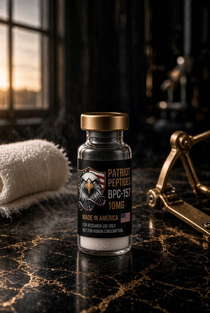
The molecule in the essay
The same published structure the essay describes — HPLC-characterised.
Read next

49 min · long read · Peptide research
TB-500 and thymosin β4: repair is a cytoskeletal programme
Cells crawl by parking and releasing actin monomers. Thymosin β4 is the buffer. TB-500 is the research analogue built around that idea — characterised, named, on the bench.
48 min · long read · Peptide research
BPC-157 and TB-500: two different ideas of repair
A gastric pentadecapeptide and an actin-binding motif. Related in folklore, unrelated in mechanism — two of the most interesting repair probes in the catalogue.
More in this desk

53 min · long read · Peptide research
Recovery, GH pulses and the CJC / ipamorelin pair
Somatotrophs have two 'go' receptors. CJC without DAC is a slightly longer GHRH pulse. Ipamorelin is a selective ghrelin-receptor key that does not yank ACTH. Together they are the pair Bowers already showed is more than additive — not a gym protocol.
53 min · long read · Peptide research
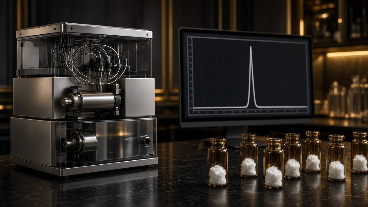
How research peptides are made — and why HPLC actually matters
Solid-phase peptide synthesis builds a chain one residue at a time. HPLC then asks whether the main peak is what you think it is. ≥98% is not a slogan. It is a chromatogram.
52 min · long read · Peptide research
Fatigue, cellular energy and the NAD+ / MOTS-c neighbourhood
NAD+ is the rechargeable chip every cell spends on fuel and DNA repair — and the pool shrinks with age. MOTS-c is a mitochondrial 16-mer sent out under metabolic stress. Two answers to 'I have no energy' that are not coffee.
51 min · long read · Peptide research
Melanocortin circuits: pigment, appetite and PT-141
Melanotan II lights MC1, MC3, MC4 and MC5 — pigment plus the rest of the sheet. PT-141 is the free-acid cousin pointed at MC3 and MC4, the circuitry papers use for desire and energy, not skin colour. Same Arizona family. Different question.
Essays describe published research. They are not medical advice and they do not authorise human use of any catalogue item.