

Peptide research · 46 min · 10,206 words
Melanocortins: from tanning receptors to inflammatory off-switches
MC1R makes pigment. MC4R runs appetite — and its loss is a monogenic obesity syndrome. KPV keeps the anti-inflammatory tail of α-MSH. MT2 is the pan-agonist that does not know subtle.
· updated
What this essay actually tells you
- α-MSH is cleaved from POMC. Five melanocortin GPCRs (MC1R–MC5R) run pigment, appetite, inflammation and sebum via Gs–cAMP. One precursor. Five jobs.
- MC4R loss-of-function is a monogenic obesity syndrome. That's how seriously the hypothalamus takes this family, and why a pan-agonist is never 'just a tan'.
- Melanotan II is a cyclic pan-agonist from Hadley's Arizona programme. KPV is the C-terminal tripeptide that calms NF-κB without most of the tan. Same family. Different fragment. Different question.
What this actually means
One precursor protein, POMC, is chopped into ACTH and α-MSH, a thirteen-residue tanning hormone that turned out to run much more than tan. Five G-protein-coupled receptors, MC1R through MC5R, read that family. MC1R on melanocytes decides eumelanin versus pheomelanin; the red-hair variants (R151C, R160W, D294H) are why some people burn and why melanoma risk travels with that colouring. MC4R in the hypothalamus is an appetite brake: loss-of-function mutations are the most common monogenic obesity, and setmelanotide (Imcivree, FDA 2020) is an MC4R agonist licensed for POMC and leptin-receptor deficiency. Melanotan II, built in Hadley's Arizona programme as a sunless-tanning analogue, occupies almost every receptor in the family (pigment, appetite, flushing, arousal) because that's what a pan-agonist does. KPV is the opposite design: the last three amino acids of α-MSH, a fragment that calms NF-κB in gut and skin models without most of the tan.
A tanning hormone that also runs appetite, sebum and an inflammatory off-switch sounds like three stories. It's one precursor. Pro-opiomelanocortin, POMC, is a polyprotein — a long chain that's cut into several hormones — written in corticotrophs of the anterior pituitary, in melanotrophs of the intermediate lobe, and in a thin population of hypothalamic neurons, then cut by prohormone convertases into the peptides a textbook actually names. In a corticotroph the principal product is ACTH, thirty-nine residues, the ligand that tells the adrenal cortex to make cortisol. In a melanotroph, and in those hypothalamic cells, the same precursor is cut further to α-melanocyte-stimulating hormone: thirteen residues, acetylated at the amino terminus, amidated at the carboxyl terminus, sequence SYSMEHFRWGKPV. The last three letters of that sequence are lysine-proline-valine. That fragment will return as KPV, and it isn't a cosmetic afterthought. Five G-protein-coupled receptors, MC1R through MC5R, read this family. They're primarily Gs-coupled, so occupancy raises cyclic AMP. What the cell does with that rise depends on which receptor you occupied and where it lives, which is the whole charm of a shared lock class.
In short. POMC is cut into ACTH and α-MSH. Five receptors read those pieces. Pigment is only one of the jobs.
Melanocytes make eumelanin when MC1R is occupied and working. Paraventricular hypothalamic neurons brake appetite when MC4R is occupied and working. Sebaceous glands write sebum when MC5R is occupied. Macrophages and keratinocytes can quiet NF-κB when the C-terminal fragment of α-MSH is present, and that quieting doesn't require the tanning receptor. Treating the family as a sunbed story is how you miss four of those five sentences. The Arizona programme that built Melanotan I and Melanotan II in the 1980s and 1990s was, in its own words, a photoprotection project. The clinic then noticed appetite falling, faces flushing, and arousal rising, because a cyclic analogue that occupies MC1, MC3, MC4 and MC5 at once will do those things. Palatin later took a close cousin, bremelanotide, to an FDA label for hypoactive sexual desire. Rhythm took an MC4R agonist, setmelanotide, to an FDA label for rare genetic obesity. The family left the tanning booth years ago. What follows is the precursor, the five receptors, the licensed cousins, the pan-agonist, and the tripeptide that was designed by subtraction. It's physiology, at the length the family actually needs.
In short. The same hormone family runs pigment, appetite, sebum and some inflammatory switches. A tanning analogue was how the clinic first noticed the rest.
Farooqi and O'Rahilly put MC4R in the obesity clinic. Setmelanotide put an agonist on an FDA label. Anyone still treating this family as a tanning story has not been reading.— Farooqi IS, Keogh JM, Yeo GS, Lank EJ, Cheetham T, O'Rahilly S. Clinical spectrum of obesity and mutations in the melanocortin 4 receptor gene. N Engl J Med. 2003; 348: 1085–1095.
POMC is a polyprotein. Convertases decide which hormone you get.
The POMC gene sits on the short arm of chromosome 2, at 2p23.3. The primary translation product is a few hundred amino acids with paired basic residues marking the cut sites. Prohormone convertase 1/3, the enzyme PC1, is the first pair of scissors in a corticotroph: it releases ACTH and β-lipotropin. Prohormone convertase 2, PC2, is the second pair, abundant in melanotrophs and in hypothalamic POMC neurons. PC2 takes ACTH and cuts it to a shorter N-terminal piece that peptidylglycine α-amidating monooxygenase, PAM, amidates, and that an N-acetyltransferase acetylates. The product of those two modifications is α-MSH. γ-MSH comes from the N-terminal fragment. β-endorphin comes from β-lipotropin. One gene, a handful of convertases, a tissue-specific peptide cocktail. A corticotroph isn't a melanotroph wearing a different hat. The enzymes are different, the products are different, and the receptors those products occupy are different. If a protocol writes 'POMC peptides' as if they were interchangeable, it hasn't yet chosen an experiment, because the scissors decide which hormone you actually made.
In short. One gene is cut differently in different cells. Pituitary corticotrophs make ACTH. Other POMC cells make α-MSH, the thirteen-residue tanning hormone.
α-MSH is Ac-SYSMEHFRWGKPV-NH2. Thirteen residues is a small hormone, and almost every position has been mutated by someone with a reason. The N-terminal acetyl and the C-terminal amide aren't decorations. They slow exopeptidases and they change potency at the receptors. The core pharmacophore is His-Phe-Arg-Trp, residues six to nine, the HFRW tetrapeptide that melanocortin receptors actually want in the pocket. That's the tanning pharmacophore, the appetite pharmacophore, the sebum pharmacophore: one motif, five receptors, different tissues. The C-terminal tripeptide, Lys-Pro-Val, is what remains when you throw the core away. Medicinal chemistry is often addition. KPV is subtraction. Keep the tail. Lose most of the pigment, most of the MC4R appetite agonism, most of the autonomic circus a pan-agonist brings to a first-in-human room. What remains is small enough to be a PepT1 substrate and stubborn enough, in the papers that bother to measure it, to suppress NF-κB in epithelium and in macrophages. The fragment isn't a weaker α-MSH. It's a different question, which is why it gets its own vial.
In short. α-MSH is thirteen amino acids. The middle four turn pigment and appetite receptors on. The last three, KPV, are the anti-inflammatory tail.
Hypothalamic POMC neurons sit in the arcuate nucleus, next door to AgRP/NPY neurons that do the opposite job. Leptin, from adipose tissue, excites the POMC cells and silences the AgRP cells. Insulin does some of the same. The POMC cells release α-MSH onto MC4R in the paraventricular nucleus and in the brainstem; the AgRP cells release agouti-related peptide, an inverse agonist at MC3R and MC4R, plus GABA. Appetite, in this neighbourhood, is a melanocortin tone, not a mood. Break POMC, or break the convertase PCSK1, or break the leptin receptor upstream, or break MC4R downstream, and the brake fails. That's why a pigment-hormone family became an obesity-genetics family, and why a cyclic peptide that occupies MC4R could become a licensed medicine for people whose genes already failed on that path. The arcuate anatomy is the reason a tanning analogue made volunteers less hungry. It was never a side-effect in the sense of an accident. It was a receptor the ligand could not help occupying, sitting next door to the one Arizona had come for.
In short. Brain cells that make α-MSH sit next to cells that block the same receptors. Leptin leans on that seesaw. That is how this family became an appetite story.
- POMC precursor
- ~241 aa
- α-MSH
- 13 residues, Ac-SYSMEHFRWGKPV-NH2
- KPV
- 3 residues, ~342 Da
- Melanotan II
- ~1024 Da, cyclic heptapeptide
- MC1R–MC5R
- five class-A GPCRs
- RHC alleles
- R151C, R160W, D294H
- Setmelanotide
- Imcivree, FDA 2020
- Bremelanotide
- Vyleesi, FDA 2019
One gene, tissue-specific convertases, several hormones.
Acetylated, amidated. HFRW is the core. KPV is the tail.
Lys-Pro-Val. NF-κB fragment. PepT1 substrate.
Ac-Nle-cyclo[Asp-His-D-Phe-Arg-Trp-Lys]-NH2. Pan-agonist.
Primarily Gs–cAMP. Tissue writes the phenotype.
Hypomorphic MC1R. Pheomelanin, burns, melanoma risk.
MC4R agonist for POMC / PCSK1 / LEPR deficiency.
PT-141. Licensed for HSDD. Close cousin of MT2.
Diagram
MC1R
melanocyte
Eumelanin vs pheomelanin. Red-hair alleles. Afamelanotide’s receptor.
MC3R
hypothalamus
Energy balance. Occupied by MT2 because MT2 occupies almost everything.
MC4R
hypothalamus
Appetite brake. Loss-of-function obesity. Setmelanotide (FDA 2020).
MC5R
sebaceous
Sebum. A first-class output people still treat as a footnote.
KPV
tail of α-MSH
NF-κB off-switch. PepT1 uptake. Designed not to tan.
MT2
pan-agonist
Ac-Nle-cyclo[Asp-His-D-Phe-Arg-Trp-Lys]-NH2. Pigment, appetite, flushing, arousal.
α-MSH is Ac-SYSMEHFRWGKPV-NH2. The last three letters are KPV. Melanotan II is what a pan-agonist looks like. KPV is the design move in the other direction. Match the ligand to the question.
Five GPCRs, primarily Gs, and the tissue writes the rest
The melanocortin receptors are class-A, rhodopsin-like GPCRs: seven transmembrane helices, an extracellular face that binds peptide, an intracellular face that acts as a guanine-nucleotide exchange factor for a heterotrimeric G protein. There are five of them in mammals, numbered MC1R to MC5R in the order they were cloned, which is not the order of their physiological importance and is a standing irritation if you teach the family. They share the HFRW pocket. They don't share tissue. MC1R lives on melanocytes, and also on macrophages and keratinocytes, which is the sentence the inflammation literature needs. MC2R is the ACTH receptor on the adrenal cortex; it wants ACTH, it needs the accessory protein MRAP, and α-MSH and Melanotan II are poor ligands for it. MC3R and MC4R live in hypothalamus and elsewhere in the brain, with some peripheral expression that a review will argue about. MC5R lives on exocrine glands, sebaceous epithelium in particular. Five locks. One ancestral key. The phenotype is the lock's address, not a mystic property of the peptide. That's a teaching sentence I keep coming back to, because it stops pigment stealing the whole family.
In short. Five related receptors. Same style of lock, different rooms. Melanocytes, adrenal, brain appetite circuits, and oil glands are the main rooms.
Roger Cone, Kathleen Mountjoy and colleagues cloned MC1R from melanoma RNA in 1992 and then the rest of the family in short order, which is why the numbering is cloning order rather than physiology order. MC1R was the MSH receptor everyone already believed in. MC2R was the ACTH receptor the adrenal had been using since the 1950s. MC3R, MC4R and MC5R were the surprises: three more GPCRs with the same HFRW pocket, sitting in brain and in exocrine glands, waiting for someone to knock them out. The knockout mice did the teaching. Mc4r-null mice eat, grow and become obese in a way that looks like the later human heterozygous loss. Mc1r-null mice (extension, the old coat-colour locus) are yellow because pheomelanin wins. Mc5r-null mice have a sebum defect. Mc3r-null mice have a subtler energy-balance phenotype. That's how a pigment-receptor cloning paper became an obesity-genetics paper, and how a Tucson tanning analogue found extra receptors to occupy. A family you can clone, knock out and occupy is a family a catalogue can name without inventing a job, which is a quieter achievement than it sounds.
In short. The five receptors were cloned in the 1990s. Knockout mice then showed pigment, appetite and sebum as separate jobs of the same family.
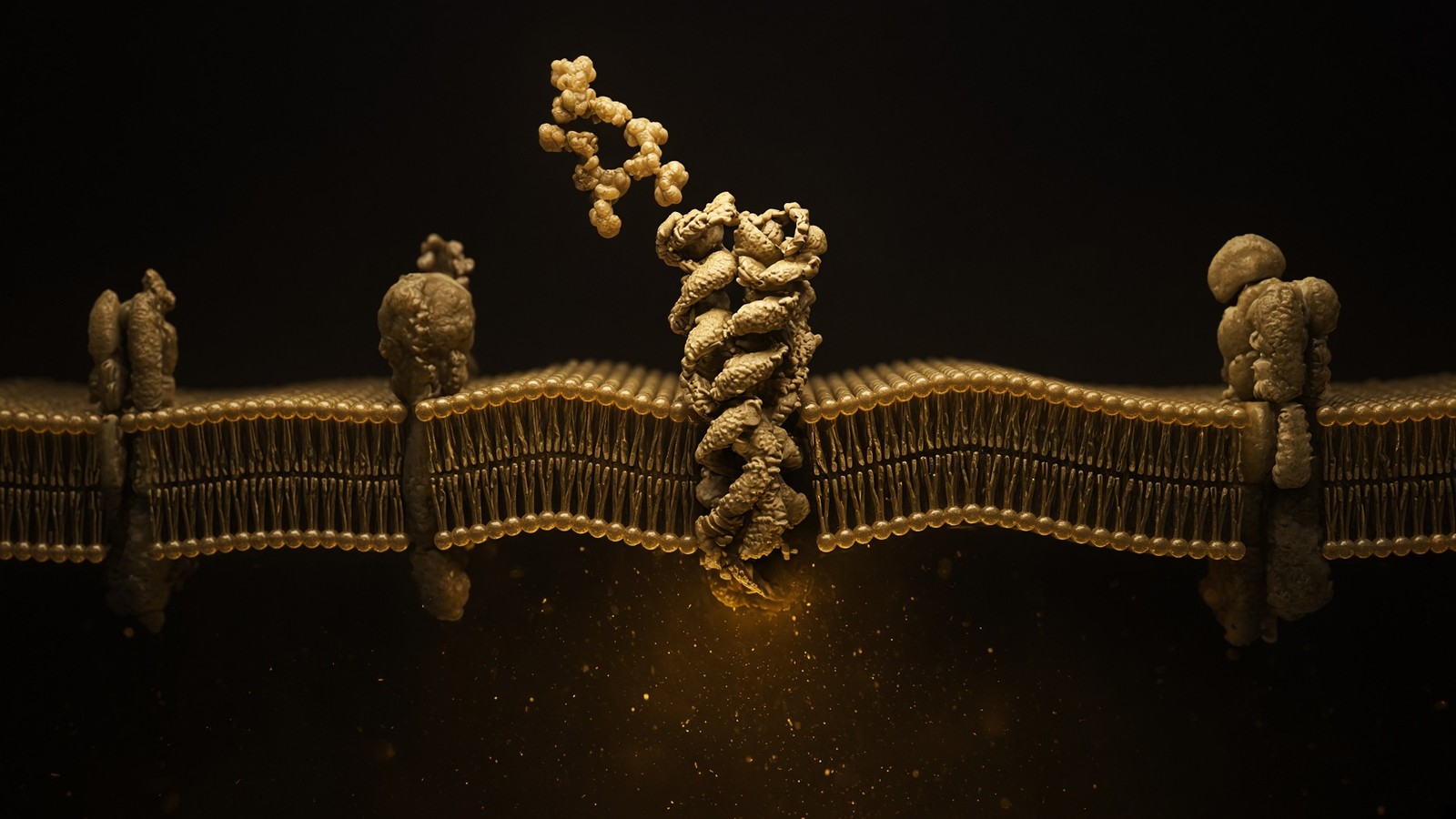
Gs is the default G protein. Occupancy rearranges the helices. The cytoplasmic face catalyses GDP-to-GTP exchange on Gαs. Gαs-GTP finds adenylyl cyclase. Cyclase makes cyclic AMP from ATP. Protein kinase A reads the cAMP, phosphorylates CREB and a list of other substrates, and the cell writes whatever programme that receptor, in that tissue, is for. In a melanocyte the programme is MITF, then tyrosinase, then eumelanin. In a paraventricular neuron the programme is a change in excitability and in peptide release that the organism experiences as satiety. In a sebocyte the programme is lipid synthesis for sebum. The second messenger is the same nucleotide. The invoice is not. Some of these receptors also recruit β-arrestin, and some of them couple in part to other G proteins in particular assays, and the biased-agonism literature exists. For a working map, Gs–cAMP is the sentence you write first. Lefkowitz and Kobilka's chemistry Nobel in 2012 was for this architecture in general, not for melanocortins in particular. The architecture is why a nanomolar peptide can move a micromolar messenger, which still feels slightly like cheating until you remember the enzymes in between.
In short. These receptors mostly raise cAMP inside the cell. The same messenger means pigment in skin, fullness in brain, oil in a gland — depending on the receptor.
Diagram
Outside
Peptide ligand
Named sequence in the nM–µM pocket. Shape complementarity, not vibes. A 15-mer and a 4-mer do not fit the same hole.
Membrane
7-TM receptor
Helices rearrange. The cytoplasmic face becomes a GEF for a heterotrimeric G protein (Gs, Gi, Gq, G12/13).
Inside
Second messengers
cAMP, IP₃, Ca²⁺, β-arrestin. One occupied receptor can spawn thousands of messenger molecules. That is amplification.
~800 GPCRs in the human genome. Seven transmembrane helices, an extracellular ligand pocket, an intracellular G-protein handshake. Catalogue neighbours: ipamorelin at GHSR, PT-141/MT2 at melanocortin receptors, retatrutide at GLP-1R/GIPR/GCGR.
Amplification is arithmetic, not a metaphor. One occupied receptor can load tens to hundreds of G proteins because the receptor is a catalyst, not a stoichiometric sponge. Each Gαs-GTP can occupy adenylyl cyclase long enough to make many cAMP molecules. Each cAMP-activated PKA can phosphorylate many substrates. A nanomolar occupancy in the pocket can therefore raise a micromolar messenger in the cytosol, which is the only magic in this family and is not magic. Local nanodomains — AKAPs, phosphodiesterases sitting next to the cyclase — mean a cAMP rise next to a channel is not a cAMP rise next to the nucleus. That's why two ligands at the same receptor can write different intracellular sentences if they prefer different nanodomains or different arrestin versus G-protein balances. Melanotan II and bremelanotide share a ring and don't write identical sentences. Desensitisation is the other half: GRKs phosphorylate the occupied receptor, β-arrestin binds, the receptor is pulled inward. More ligand is not more signal forever. A first-hour flush in a melanocortin study is often that machinery, not a reason to double the concentration.
In short. One receptor can make a cloud of cAMP because enzymes sit in between. The cell then turns the receptor off, so more ligand is not more signal forever.
Diagram
× 1
Ligand
One peptide in one pocket. nM–µM. Shape, not a mood.
× 10–10²
G proteins
The occupied GPCR is a GEF. Each Gα is a catalyst.
× 10³–10⁴
cAMP / IP₃ / Ca²⁺
Adenylyl cyclase and PLC do not make one molecule. They make a cloud.
× 10⁴–10⁶
PKA / PKC / CaMK
Kinases phosphorylate many substrates per messenger.
× tissue
Secretion, transcription, motility
The organism-level readout. Still not a protocol.
This is the only magic, and it is not magic. A nanomolar ligand can move a micromolar messenger because enzymes sit between them. Desensitisation (GRK, β-arrestin, endocytosis) is how the cell refuses to let ‘more ligand’ mean ‘more signal’ forever.
- Ligand in the extracellular pocket. Shape complementarity. HFRW for the full agonists; KPV is a different, smaller question.
- Seven helices rearrange. The cytoplasmic face becomes a GEF for Gs.
- Gαs-GTP activates adenylyl cyclase. cAMP rises. PKA and EPAC read it.
- The tissue writes the phenotype: MITF and tyrosinase, satiety, sebum, or, for the fragment, IκB preservation.
- GRK and β-arrestin desensitise. Endocytosis. The signal is a pulse unless the ligand is built not to leave.
MC1R: eumelanin, pheomelanin, and the alleles that burn
MC1R on a melanocyte is the pigment receptor the public already thinks it knows. Occupancy, α-MSH or a synthetic agonist, raises cAMP. PKA phosphorylates CREB. CREB, with other inputs, writes MITF, microphthalmia-associated transcription factor, the master transcription factor of the melanocyte. MITF writes TYR, tyrosinase, and TYRP1 and DCT (TYRP2), the enzymes that take tyrosine through DOPA and dopaquinone into the melanin polymers. When the cAMP signal is strong, the polymer is eumelanin: brown-black, a broad UV absorber, photoprotective in the ordinary sense that a physical chemist would accept. When the cAMP signal is weak, dopaquinone is diverted toward cysteine conjugates and the polymer is pheomelanin: red-yellow, a poorer absorber, and in some assays a source of reactive oxygen rather than a shield. The switch isn't a lifestyle. It is a receptor, a cyclic nucleotide, a transcription factor and a pair of polymer chemistries. A brochure that says 'melanin' as if there were one pigment hasn't looked at a melanosome. Two polymers, two colours, two kinds of protection, one receptor deciding which way the chemistry runs.
In short. MC1R tells pigment cells to make dark eumelanin. If the receptor is weak, they make red-yellow pheomelanin instead, which protects less well against ultraviolet light.
The red-hair-colour alleles are the cleanest genotype-to-phenotype story in human pigmentation, and people are still surprised by it. R151C, R160W and D294H are the common European hypomorphs: missense changes that leave a receptor on the surface but blunt the cAMP response to α-MSH. Heterozygotes tan poorly. Compound heterozygotes and homozygotes are the classic red-hair, fair-skin, freckled, burns-rather-than-tans phenotype. Valverde, Rees, Sturm, Duffy, Palmer — the association papers of the 1990s and 2000s — mapped that colouring onto those alleles with a clarity most complex traits never get. The receptor is still a receptor. A hypomorph isn't a deletion. Residual signalling, ligand-independent constitutive activity (MC1R has some), and the rest of the pigment genome (OCA2, SLC45A2, TYR itself) still move the phenotype. But if you want a single-gene sentence about why some skin never browns, MC1R is that sentence. Agouti signalling protein, ASIP, is the endogenous inverse agonist at MC1R; the yellow mouse is ASIP overexpressed, and the yellow is pheomelanin winning because the receptor is blocked.
In short. Common red-hair versions of MC1R answer poorly to the hormone. Fair skin, freckles and burning follow. That is one of the cleanest gene-to-colour stories we have.
Melanoma statistics travel with that colouring, and the travel isn't only 'these people burn, therefore they get more ultraviolet, therefore they get more melanoma'. They do burn. Cumulative UV is a first-class cause. There is also a pheomelanin-intrinsic story: Mitra, Fisher and colleagues, in a mouse with a red-hair Mc1r allele, showed oxidative DNA damage and melanoma risk that did not require ultraviolet, which is a disturbing paper if you had hoped pigment chemistry was only a filter. Human epidemiology is messier than a genetically red mouse. MC1R variants raise melanoma risk in fair-skinned cohorts after you adjust, imperfectly, for sun exposure and for naevus count. The practical sentence for a melanocortin page is simpler than the oncology: a hypomorphic MC1R tilts the polymer toward pheomelanin, tilts the skin toward burning, and travels with melanoma risk. Afamelanotide was built, in part, as a photoprotective agonist for skin that tans poorly. Erythropoietic protoporphyria, the licensed indication, is a different phototoxicity — ferrochelatase deficiency, protoporphyrin in the skin, pain in visible light — but the receptor is the same MC1R, and eumelanin is still the pigment you are trying to write.
In short. Red-hair MC1R variants travel with higher melanoma risk. Some of that is burning. Some of it may be the red pigment itself making oxidative damage.
MC1R isn't only a melanocyte protein. Macrophages express it. Keratinocytes express it. Some endothelial and immune cells express it. α-MSH has an anti-inflammatory literature on those cells that predates KPV as an isolated reagent: reduced NF-κB activity, reduced adhesion-molecule transcription, reduced cytokine output. Luger's dermatology group in Münster spent years on that, with Brzoska and Böhm, in keratinocyte and melanocyte systems. Getting's group, at the William Harvey, spent years on melanocortin anti-inflammatory tone in macrophages and in rodent inflammation models, with a particular interest in MC3R as well as MC1R. The full hormone occupies the tanning receptor and the inflammatory receptors because they are the same ancestral GPCR family. That's a feature if you want both. It's a confound if you wanted IκB without MITF. The reason KPV exists as a research object is that the inflammatory papers survived subtraction of the HFRW core, including in systems where MC1R was absent or blocked. Pigment wants a different ligand. Inflammatory tone in a barrier tissue wants the fragment, rather than a pan-agonist that will also tan, suppress appetite and surprise the autonomic nervous system.
In short. MC1R also sits on immune cells, so the full hormone can calm inflammation and tan at once. The tripeptide was kept for the calming, without most of the tan.
MC2R is the adrenal, and the pan-agonist mostly leaves it alone
MC2R is the ACTH receptor. It lives on zona fasciculata cells of the adrenal cortex. Occupancy raises cAMP, PKA phosphorylates the machinery that moves cholesterol to CYP11A1, and cortisol is written. The receptor is fussy. It wants ACTH, not α-MSH, and it needs melanocortin-2 receptor accessory protein, MRAP, to traffic and to signal. Familial glucocorticoid deficiency type 1 is MC2R loss-of-function; type 2 is MRAP. Melanotan II, for all its promiscuity at MC1, MC3, MC4 and MC5, is a poor MC2R agonist, which is why a pan-melanocortin probe doesn't routinely present as an ACTH analogue. That selectivity is luck as much as design, and it's worth keeping on the page so nobody files 'melanocortin agonist' under 'steroidogenesis'. ACTH is a melanocortin. It isn't the melanocortin this catalogue is holding. The corticotroph, the adrenal, the circadian cortisol curve — those are a different page, a different axis, a different vial if a vial were even the point. Name MC2R so you can leave it, then return to the four receptors the lactam actually occupies, which are already enough to fill a first-in-human room.
In short. MC2R is the adrenal ACTH receptor and needs a helper protein. Melanotan II barely touches it, which is why a tanning probe is not a cortisol drug.
MC3R and MC4R: energy balance, and the obesity gene that is not rare enough
MC3R is the quieter of the two central receptors and isn't a footnote. It sits on POMC neurons themselves, among other sites, and functions in part as an inhibitory autocrine brake: α-MSH released from the neuron can occupy MC3R on the same population and temper the output. Knockout mice have a metabolic phenotype that is not identical to MC4R knockout — more of a shift in body composition and feed efficiency than the frank hyperphagia of MC4R loss. Human MC3R variants exist and are argued over; they haven't given us a licensed agonist of their own. Melanotan II occupies MC3R because Melanotan II occupies almost everything in the family. That's a feature of a pan-agonist and a nuisance if you wanted to know which receptor moved feeding. Bremelanotide leans toward MC3 and MC4, which is why the sexual-function literature sits on those two and not on the melanocyte. Energy balance is a circuit. MC3R is one node. MC4R is the node the clinic actually met, and once you've met it you understand why a tanning analogue was never going to be only a tanning analogue.
In short. MC3R helps the brain's energy circuit and partly brakes the cells that make α-MSH. It is real. MC4R is the one obesity genetics made famous.
MC4R is the hypothalamic receptor that made this family a metabolic subject. It is expressed on paraventricular neurons, on brainstem neurons that run autonomic outflow, and in a list of other nuclei a neuroanatomist can recite. Occupancy by α-MSH is an appetite brake and a push on energy expenditure. Occupancy by AgRP is the opposite: AgRP is an inverse agonist, not merely a blocker, because MC4R has constitutive activity and AgRP turns that down. Yeo, Farooqi, O'Rahilly and colleagues at Cambridge showed, in the late 1990s and then in a New England Journal paper in 2003, that heterozygous loss-of-function mutations in MC4R are the most common single-gene cause of severe obesity in the cohorts that bother to sequence it. A few percent of severe early-onset obesity. Dominant with incomplete penetrance. Hyperphagia from childhood, increased lean mass, accelerated linear growth, high bone mineral density — a phenotype that isn't 'they eat too much' as a moral sentence. The neuron is an appetite brake. Break the receptor and the brake fails. That's a first-class melanocortin output. A tanning page that leaves it out hasn't described the family.
In short. MC4R in the hypothalamus is an appetite brake. Inherited damage to one copy is the most common single-gene cause of severe obesity.
The 2003 paper is worth naming because it is the document, not a tweet. Farooqi, Keogh, Yeo, Lank, Cheetham, O'Rahilly: Clinical spectrum of obesity and mutations in the melanocortin 4 receptor gene, New England Journal of Medicine, 348: 1085–1095. They sequenced MC4R in people with severe obesity, found a spectrum of heterozygous mutations, showed that the mutations that wrecked receptor function in a dish tracked with the clinical severity, and put a tanning-hormone receptor in the endocrine clinic. Subsequent work filled in the signalling: Gs, cAMP, but also a β-arrestin and MAPK conversation that some obesity mutations hit more than others, which is why not every missense is the same disease. Rare homozygous or compound-heterozygous complete loss is more severe. The common heterozygous LoF is already enough to be the leading monogenic obesity. Polygenic obesity is still most of obesity. MC4R isn't 'the' obesity gene. It is the most common of the rare, severe, early ones, and it is a melanocortin receptor, which is the only reason it belongs here rather than in a general genetics lecture. That's a distinction worth keeping kind.
In short. Farooqi and O'Rahilly sequenced MC4R in severe obesity and showed the broken receptors matched the broken appetite. That paper moved this family into the clinic.
POMC deficiency is the rarer, cleaner experiment of nature. If you cannot make the ligand, MC4R never sees α-MSH, MC1R never sees it either, and ACTH is missing so the adrenal never sees that. The child is ACTH-deficient, obese, and often red-haired, because the pigment receptor and the appetite receptor lost the same peptide. PCSK1 deficiency wrecks the convertase that cuts POMC and proinsulin and a list of other prohormones; obesity is part of a broader endocrine mess. Leptin-receptor deficiency wrecks the upstream signal that tells POMC neurons to fire. Three genetic lesions, one pathway, one licensed peptide that was built to occupy MC4R anyway. Setmelanotide, RM-493, Imcivree, Rhythm Pharmaceuticals: a cyclic octapeptide MC4R agonist. Kühnen, Krude and colleagues treated POMC-deficient patients and published the weight loss in the New England Journal in 2016. The FDA licensed it in 2020 for chronic weight management in people six and older with POMC, PCSK1 or LEPR deficiency, later adding Bardet-Biedl syndrome. When a tanning-hormone family has an FDA-approved obesity drug, the family left the booth. Setmelanotide is that drug. Melanotan II is not. Same ancestral family, different selectivity, different legal class.
In short. If the body cannot make α-MSH, you get obesity, cortisol failure and often red hair together. Setmelanotide is the licensed MC4R agonist for those rare pathway diseases.
Leptin is the upstream signal that made the arcuate melanocortin circuit a textbook. Friedman cloned leptin from the ob mouse; the receptor lives on POMC neurons and on AgRP neurons. Leptin rising, as the warehouse fills, excites POMC cells so they release α-MSH onto MC4R, and silences AgRP cells so the inverse agonist withdraws. Starvation is the opposite: POMC quiet, AgRP loud, appetite up. That's why leptin-receptor deficiency looks like POMC deficiency looks like MC4R deficiency, one floor down each time, and why setmelanotide can bypass a broken leptin receptor or a missing ligand and still occupy the last receptor. MRAP2, a relative of the adrenal accessory protein, helps MC4R traffic and signal; some rare obesity mutations hit MRAP2 instead of the receptor itself. The circuit is allowed to have more than one named protein. A pan-agonist given to a volunteer still occupies MC4R on this circuit, which is why appetite moved in Arizona. It also occupies MC1R, MC3R and MC5R, which is why appetite was never the only thing that moved. Selectivity is which protein you occupy. A pan-agonist refuses that choice on purpose.
In short. Leptin talks to brain cells that release α-MSH onto the appetite receptor. Break leptin, the hormone, or MC4R, and the same brake fails. Setmelanotide occupies the last step.
MC5R writes sebum, and inverse agonists prove the receptors were on
MC5R is the sebum receptor. It is expressed on sebaceous glands and on other exocrine epithelia. Knockout mice have a defect in water repulsion and thermoregulation that traces to altered sebum, which is a sentence that makes sense once you remember that fur is a hydrophobic coat a gland has to oil. Human sebocytes express MC5R; α-MSH can change their lipid programme in culture. This is a first-class melanocortin output that people still treat as a footnote because it doesn't photograph as a tan and doesn't hospitalise as obesity. A pan-agonist will occupy MC5R because that's what pan means. Whether a given flushing or a given change in skin oil in an old Arizona volunteer study was MC5R, MC1R, autonomic, or all three isn't a question a comment thread gets to answer. Name the receptor. Put it on the map. Then stop treating glandular epithelium as an embarrassing extra. Five receptors were cloned because five receptors exist, and sebum is one of the jobs the family actually does, even if it never made a magazine cover.
In short. MC5R sits on oil glands. The family does sebum as well as pigment and appetite. A pan-agonist will touch that receptor too.
Constitutive activity is the other fact this family teaches well. MC4R signals somewhat without ligand; AgRP is an inverse agonist that turns that basal signal down, not a competitive antagonist that merely keeps α-MSH off. ASIP does the equivalent at MC1R, which is why the yellow mouse is yellow and fat: the same inverse agonist, overexpressed, blocks pigment at MC1R and blocks satiety at MC4R. A ligand that reduces constitutive activity is doing work even when the agonist is absent. That's why 'blocker' is a sloppy word here, and why you have to say agonist, antagonist or inverse agonist on purpose. The endogenous pair is α-MSH on and AgRP/ASIP off, a push-pull the arcuate already runs as anatomy. Synthetic peptides sit on that seesaw. Melanotan II is a heavy on. Setmelanotide is an on aimed at MC4R. KPV is barely on the seesaw at all, which is the design. Inverse agonism is also why some MC4R obesity mutations that wreck constitutive activity can look different from mutations that wreck α-MSH binding. The receptor was never a simple off/on light switch. It was a dimmer with a basal setting.
In short. These receptors are partly on even without hormone. AgRP and agouti protein turn that basal signal down. That is inverse agonism, not a simple blockade.
Arizona built a photoprotection peptide and the clinic read the rest of the sheet
Mac Hadley and Victor Hruby, University of Arizona, spent the 1980s and 1990s making melanocortin analogues that would raise eumelanin without ultraviolet. Fair MC1R-variant skin tans poorly and burns well; a peptide that occupied MC1R and wrote eumelanin was, on paper, photoprotection you could dose. The linear analogue [Nle4, D-Phe7]α-MSH replaced methionine with norleucine so oxidation would not wreck the chain, and replaced L-phenylalanine at position 7 with D-phenylalanine so proteases would hesitate and potency would rise. That molecule is Melanotan I, later afamelanotide, later Scenesse. Clinuvel took it to an implant. The EMA licensed it for erythropoietic protoporphyria in 2014; the FDA followed in 2019. EPP is ferrochelatase deficiency, protoporphyrin IX in erythrocytes and skin, burning pain in visible light. Eumelanin in the epidermis is a filter for that light. The implant is a licensed photoprotective melanocortin agonist in a defined disease. It is Melanotan I, not Melanotan II. Different molecule, different label, different legal class. The research pan-agonist on a catalogue is the cyclic cousin, and the cyclic cousin is a different experiment, which is a kindness to both molecules if you let it be.
In short. Arizona made a linear tanning analogue to raise dark pigment without sun. That analogue, afamelanotide, became a licensed implant for a rare light-pain disease.
Melanotan II is Ac-Nle-cyclo[Asp-His-D-Phe-Arg-Trp-Lys]-NH2. Norleucine again, D-phenylalanine again, and a lactam bridge between aspartic acid and lysine that pins the HFRW core in a turn the receptor likes. Super-agonist is the word the Arizona papers used, and they meant it: potency at MC1R, MC3R, MC4R and MC5R that made the linear parent look polite. MC2R, as above, is mostly spared. Enzymatic resistance is the other point of the D-amino acid and the cycle. A linear α-MSH in plasma is a short conversation; the cyclic lactam is a longer one. Hruby's chemistry and Hadley's physiology were a collaboration of the sort that still looks good on a paper: a named sequence, a named cyclisation, a named receptor sheet, a named set of rodent pigment and behaviour assays. Dorr, Lines, Levine and colleagues then put the peptide into a small human study, Life Sciences 1996, and wrote down what happened. Pigment rose. Nausea happened. Stretching and yawning happened. Spontaneous erection happened. Appetite moved. A pan-agonist in a volunteer is a pan-agonist. The group did not hide the extras. That honesty is why the later sexual-function programme had a paper trail instead of a rumour.
In short. Melanotan II is a small cyclic peptide that turns on four of the five receptors at once. Early volunteers tanned, felt sick, flushed, and noticed arousal and a quieter appetite.
A superpotent cyclic melanotropic peptide in a pilot phase-I study did what a pan-agonist does: pigment, nausea, stretching, yawning, erection. The extras were data, not a scandal to be edited out.— Dorr RT, Lines R, Levine N, et al. Evaluation of melanotan-II, a superpotent cyclic melanotropic peptide in a pilot phase-I clinical study. Life Sci. 1996; 58: 1777–1784.
Those extras map onto receptors if you let them. Pigment is MC1R on melanocytes. Appetite is MC3R and MC4R in hypothalamus. Flushing is vascular and autonomic, a mixture a first-hour study isn't designed to dissect. Arousal — stretching, yawning, spontaneous erection in men — is central melanocortin tone, MC4R heavily implicated, with a spinal and autonomic half that the sexual-function papers spent the next two decades on. None of that was 'off-target' in the medicinal-chemistry sense of hitting a different protein family. It was on-family, on-purpose promiscuity. A pan-agonist is a tool for asking whether a phenotype is melanocortin-gated at all. It is a poor tool for asking which subtype did the work. If you want subtype, you change the molecule, or you use a knockout, or you use an antagonist, or you pick the licensed cousin that was taken through to a label on one job. Melanotan II remains the research pan-agonist because that's the question it answers. Subtle is a different vial, and KPV is the design move that went looking for subtle on purpose.
In short. The extra effects were the other melanocortin receptors doing their jobs. A pan-agonist is how you ask whether the family is involved, not which member did it.
A bench that claims a melanocortin result still has to pick an assay. cAMP, in a cell line that actually expresses the subtype you named, is occupancy as a nucleotide: ELISA, HTRF, a GloSensor, the old radioimmunoassay. Pigment, in melanocytes or in a melanoma line, is MITF and tyrosinase and a melanin pellet you can see. Feeding, in a rodent, is grams over hours after a central or peripheral dose, with the usual caveat that nausea looks like satiety if you aren't watching. NF-κB, for the fragment, is a reporter, a p65 immunofluorescence, an IκB blot, a cytokine ELISA. Those four readouts aren't interchangeable. A cAMP rise in an MC1R line isn't a quieter DSS colon. A feeding suppression isn't a tan. Writing 'melanocortin activity' without naming the assay hasn't yet done the experiment. Concentration matters: nanomolar at a GPCR, micromolar if you're asking a transporter to carry a tripeptide, and a rumour dose isn't a Kd. Spare receptors mean EC50 can sit below Kd. Species matters: rodent MC3R and human MC3R aren't a copy-paste. Write the assay. Then write the ligand.
In short. cAMP, pigment, feeding and NF-κB are four different measurements. A melanocortin paper has to name which one it ran, and at which receptor.
PT-141 is the close cousin that got a sexual-desire label
Take the Melanotan II ring and leave the C-terminus as a carboxylic acid instead of an amide, and the literature shifts. Less MC1, more MC3 and MC4. Pigment recedes as the question; central melanocortin circuitry becomes the question. Palatin named the structure bremelanotide. The research code that stuck is PT-141. The chemistry is still a cyclic heptapeptide with norleucine, D-phenylalanine and the Asp-Lys lactam. The C-terminal amide-to-acid change is a small edit with a large effect on which receptor the ring prefers, which is a useful lesson in how little sequence it takes to change a family portrait. Early development chased male erectile function and then moved, for regulatory and clinical reasons, to acquired, generalised hypoactive sexual desire disorder in premenopausal women. The FDA licensed Vyleesi in 2019 as an on-demand subcutaneous injection. Nausea is common. That's the medicine, in its indication, with its pharmacovigilance and its device. A research vial of the named heptapeptide is a laboratory ligand. We are not Palatin. The two objects share a backbone idea. They don't share a legal class, and that's a distinction a chemist and a regulator can both live with.
In short. PT-141 is Melanotan II's ring with the end left as an acid. It prefers the brain receptors over pigment, and bremelanotide became a licensed desire medicine.
The licensed cousins, taken together, are the proof of the family. Afamelanotide occupies MC1R for photoprotection in EPP. Bremelanotide occupies MC3R/MC4R for a defined desire indication. Setmelanotide occupies MC4R for rare genetic obesity on the POMC pathway. Three peptides, three jobs, three labels, three companies, three decades of chemistry that started as a tanning analogue on a Tucson bench. That's success as a pharmacologist means it: the ancestral ligand was real, the receptors were clonable, the analogues were makeable, and at least three of the phenotypes were large enough and clean enough to survive a regulator. Melanotan II is none of those three medicines. It is the unselective parent those programmes learned from, still the right probe when a bench wants to know whether a melanocortin phenotype is ligand-gated at all, before anyone spends a year on a subtype-selective analogue. Pigment in melanocytes, cAMP in MC4R-expressing neurons, feeding suppression in rodents: those are fair assays. They are why the pan-agonist stays in a research catalogue. They aren't a sunbed, and they aren't Vyleesi in a different bottle.
In short. Three licensed drugs came out of this family: a light-pain implant, a desire injection, a rare-obesity agonist. Melanotan II is the unselective research parent, not those medicines.
Nausea is the melanocortin extra a first-hour study should expect and a paper should name. MC4R sits in the brainstem as well as in the paraventricular hypothalamus, including neighbourhoods that run aversive and emetic tone. Area postrema and related nuclei are how a peptide that never needed to be a gut hormone can still make a volunteer reach for a basin. Afamelanotide, bremelanotide and setmelanotide all carry gastrointestinal events on their labels; Melanotan II did in Arizona. That's on-family pharmacology, not a contaminant. Stretching and yawning are the other brainstem signature, old in the α-MSH literature, easy to joke about, still a real central readout. A feeding study that doesn't watch for malaise will count nausea as satiety and publish a cleaner curve than the animal lived. A human study that doesn't list nausea has probably not given a melanocortin agonist, or has not asked. The extras are data. Write them down, as Dorr did, and then decide whether your question can survive them. A pigment assay in a dish doesn't have a brainstem. A rodent feeding assay does.
In short. Melanocortin agonists often cause nausea because appetite receptors also sit in brainstem areas that trigger queasiness. That is the family, not a dirty sample.
KPV is the design move in the other direction
Subtract α-MSH until three residues remain: lysine-proline-valine, the C-terminal tail, often itself amidated in the reagents people actually weigh. The HFRW core is gone. Most of the MC1R tanning potency is gone. Most of the MC4R appetite agonism is gone. What remains, in the papers that measured it, is an anti-inflammatory fragment. Luger's group, in keratinocytes and in skin inflammation models, showed that α-MSH and C-terminal fragments still calm inflammatory transcription. Getting's group, in macrophages and in rodent inflammation, showed melanocortin peptides reducing leukocyte recruitment and cytokine output, and spent time on which receptor, if any, the smaller fragments still needed. The master switch those assays keep reading is NF-κB: the transcription factor that puts TNF, IL-6, IL-1β and a list of adhesion molecules on the page. KPV reduces NF-κB reporter activity, preserves IκB, and reduces p65 nuclear translocation in keratinocytes and in macrophages. That's a small, precise, interesting reagent. It is a weak pigment signal, which is the point of the subtraction, not a failure of it.
In short. KPV is the last three amino acids of α-MSH, kept because they still calm inflammatory genes after the tanning piece is removed.
NF-κB, in working English, is a dimer — classically p65/p50 — held in the cytoplasm by IκB. Inflammatory signals, TLRs, TNF receptor, IL-1 receptor, converge on IκB kinase. IKK phosphorylates IκB. Phosphorylated IκB is ubiquitinated and eaten by the proteasome. Freed p65 goes to the nucleus and writes the inflammatory cassette. An agent that preserves IκB, or that otherwise keeps p65 out of the nucleus, turns that cassette down. Glucocorticoids do a version of this, among other things, and they are hormones with a very large invoice. KPV is a tripeptide with a much smaller invoice and a much smaller literature. The reported mechanism in epithelium and myeloid cells is IκB preservation and reduced p65 translocation, with downstream falls in TNF, IL-6 and IL-1β transcripts. Replication quality varies by lab, as it does in all peptide inflammation work. The mechanistic core — NF-κB down, in more than one cell type — has been seen more than once, which is the most we ask of a three-residue literature before we're willing to keep the vial on a map. Three residues, a switch, a quieter cassette. Enough to be interesting.
In short. NF-κB is the switch that turns inflammatory genes on. KPV papers say the fragment helps keep that switch off by protecting the protein that holds it in the cytoplasm.
Getting's inflammation papers are the macrophage half. Melanocortin peptides, including smaller fragments, reduce neutrophil recruitment in rodent models, reduce cytokine output from macrophages, and in some assays still work when MC1R is absent or blocked, which is the sentence that makes KPV more than a weak α-MSH. MC3R on macrophages is one proposed contributor in the full-peptide experiments. The fragment's residual activity in MC1R-null or antagonist-blocked systems is the intellectual reason to use KPV instead of α-MSH or Melanotan II when pigment and MC4R appetite circuitry would confound the assay. A pan-agonist in a colitis model will occupy MC1R, MC3R, MC4R and MC5R, move feeding, move pigment in any melanocytes that happen to be in the dish, and give you a phenotype you can no longer assign. The tripeptide was built, by deletion, to ask a narrower question: can this tail calm NF-κB in a barrier tissue without lighting the rest of the sheet? The answer in the better papers is yes, with the usual caveats of concentration, model and readout. That's enough to keep the fragment as its own object.
In short. Some of the calming still happens when the tanning receptor is missing. That is why the fragment is a different tool from the full hormone or from Melanotan II.
PepT1 is why a tripeptide can be an intestinal story
Dalmasso, Charrier-Hisamuddin, Nguyen, Yan, Sitaraman and Merlin, Gastroenterology 2008: PepT1-mediated tripeptide KPV uptake reduces intestinal inflammation. PepT1 is SLC15A1, a proton-coupled oligopeptide transporter on the apical membrane of small-intestinal enterocytes, built to absorb dipeptides and tripeptides from digested protein. Inflammation induces it, including in colon, which isn't its quiet-state address. A tripeptide that is a PepT1 substrate can therefore enter epithelium by a nutrient transporter, not only by a melanocortin receptor. That's a pharmacokinetic gift for a colitis model, and it is why DSS-colitis papers keep appearing in this literature. Dextran sodium sulphate wrecks the colonic epithelium in a mouse; disease activity, myeloperoxidase, inflammatory transcripts are the usual readouts. KPV has been reported to reduce those readouts, including after oral delivery, consistent with PepT1 uptake in inflamed gut. Oral, for a tripeptide, is a coherent design in a tissue that already expresses the transporter. It isn't a reason to call the fragment a gastroenterology appointment, and we won't. It's a reason the fragment has a door the longer analogues don't.
In short. Gut cells have a transporter, PepT1, that swallows small peptides from food. Inflamed gut makes more of it. KPV can ride that transporter into the lining.
SLC15A1 is a twelve-transmembrane proton symporter. The proton-motive force at the brush border, acid outside relative to cytosol, drives di- and tripeptides in. Substrate size is the point: this isn't a GPCR pocket, it is a nutrient hole that wants short peptides. KPV fits. α-MSH doesn't, not as a thirteen-mer. Melanotan II doesn't. The transporter is why the fragment and the pan-agonist aren't interchangeable even before you look at receptor pharmacology. A colitis paper that can't name PepT1 hasn't yet explained how an oral tripeptide would get in. A skin paper doesn't need PepT1; keratinocytes and the inflammatory literature there are a different door, closer to Luger than to Merlin. Two tissues, one fragment, two entry stories. The shared intracellular sentence is NF-κB. The shared design sentence is subtraction of the tanning core. Everything else — transporter versus receptor, colon versus epidermis, DSS versus a cytokine-stimulated keratinocyte — is the experiment you actually ran. Name the door. Then the quieter cassette makes sense as cargo or as a ligand, depending on the tissue.
In short. PepT1 only carries very short peptides, so KPV can use it and the longer tanning analogues cannot. That is one reason the fragment is its own experiment.
Diagram
Mucus
MUC2 gel
The first argument a microbe has to win.
Epithelium
enterocyte · goblet · Paneth
One cell thick. The wall is the cell, not a fascia.
Tight junctions
claudin · occludin · ZO-1
The seal. ‘Leaky gut’ as a brand is not this protein list.
PepT1
SLC15A1
Oligopeptide transporter. Inflamed gut induces it. KPV can ride it.
Lamina propria
immune cells
Where NF-κB decisions become cytokines.
BPC-157 neighbourhood
NO · VEGFR2 · FAK
Cytoprotection, blood flow, how a damaged lining organises.
IBS is a Rome-criteria cluster. It does not name a receptor. The preclinical literature that named molecules for barrier tissue keeps coming back to BPC-157 and KPV — two ligands, one organ on a reading list, neither a gastroenterology appointment.
DSS-colitis is a model, and models lie in structured ways. Dextran sodium sulphate is a chemical injury, not Crohn's disease and not ulcerative colitis as a person has them. Barrier disruption, innate inflammation, a histology score, a colon length, a myeloperoxidase number: those are the endpoints. Translation from that mouse to a human inflammatory bowel disease is a long walk that this fragment hasn't finished, and we won't pretend otherwise. What the model is good for is asking whether a named tripeptide, given into a wrecked epithelium that has induced PepT1, changes inflammatory transcription and tissue injury in a direction the NF-κB story predicted. Several groups have reported that it does. Replication is uneven, as peptide inflammation work is. The mechanistic core has been seen more than once. That's the claim. IBS, as a Rome-criteria cluster, doesn't name a receptor and doesn't name PepT1, which is why a page that cares about occupancy will not file KPV under irritable bowel as a product. Adjacent biology is allowed. Indication is earned in a different building, with a different kind of evidence.
In short. Mouse colitis models are chemical injury, not human IBD. They are fair for asking whether KPV calms a wrecked lining. They are not a treatment claim.
The gut barrier, drawn properly, is mucus (MUC2), a one-cell-thick epithelium, tight junctions (claudin, occludin, ZO-1), and an immune lamina propria underneath. 'Leaky gut' as a brand isn't that protein list. PepT1 sits on the apical membrane and, in inflammation, where it isn't always invited. NF-κB decisions in the epithelium and in the macrophages of the lamina propria become cytokines, and cytokines become the histology. KPV enters that stack as a tripeptide cargo and as an NF-κB quieting. BPC-157, a gastric pentadecapeptide with a cytoprotection, nitric-oxide and VEGFR2 literature, sits on the same organ as a different ligand. The two are neighbours on a reading list. They aren't a stack, not a protocol, and not interchangeable because both have been typed next to the word gut. Name the transporter for KPV. Name VEGFR2 and eNOS for BPC-157. Then you have two mechanisms. Until then you have two interesting fragments sharing an organ, which is already more useful than a blended caption.
In short. The lining is mucus, a single cell layer, tight seals, and immune cells underneath. KPV and BPC-157 both appear in gut papers. They bind different things.
Three benches keep showing up for the fragment, and they aren't the same room. A keratinocyte, cytokine-stimulated, is Luger's neighbourhood: NF-κB reporter, IκB blot, IL-6 and TNF transcripts, sometimes a scratch assay someone will over-read as repair. A macrophage, LPS-stimulated, is Getting's neighbourhood: cytokine ELISA, leukocyte recruitment in a rodent if the paper leaves the dish. An enterocyte, or a DSS colon, is Merlin's neighbourhood: PepT1 at the apical membrane, myeloperoxidase, a histology score. Shared intracellular sentence: p65 stays out of the nucleus more than it did. Different entry, different confounders, different concentrations. A keratinocyte doesn't need PepT1 to see a tripeptide you put in the medium; an oral colon study does. A macrophage study that can't say whether MC1R or MC3R or neither carried the effect hasn't finished the receptor work. Three benches, one fragment. Write which bench. Then write whether the fragment got in as cargo or sat on a GPCR, or whether you don't yet know. Not knowing is allowed. Blending the three rooms is how the fragment gets oversold.
In short. KPV papers use skin cells, immune cells, or gut lining. The shared finding is quieter inflammatory genes. How the tripeptide got in differs by tissue.
MC1R-independent activity is the point of the fragment
If KPV still worked only through MC1R, there would be little reason to isolate it. You'd use α-MSH, or afamelanotide, or Melanotan II, and you'd accept the pigment. Several groups have shown residual anti-inflammatory activity in MC1R-null cells, in antagonist-blocked systems, or at concentrations and in tissues where a tanning assay would have been silent. The molecular identity of every remaining target isn't a solved crystal structure. Intracellular uptake via PepT1, subsequent IκB effects that don't require a GPCR at the plasma membrane, and residual activity at other melanocortin receptors have all been proposed. Dependence on MC1R is incomplete. That incompleteness is the design. It's why a lab that wanted NF-κB in epithelium without MITF in melanocytes reaches for the tripeptide. It's why a lab that wanted eumelanin reaches for a full agonist and leaves KPV in the freezer. Subtype-selective tools and receptor-null systems are how you find out which remaining target did the work in your assay. The catalogue can't do that experiment for you. It can put both sequences on the shelf with masses attached, which is the useful half of the job.
In short. KPV still calms inflammation in some systems that lack the tanning receptor. That leftover activity is why the fragment exists as its own reagent.
Antimicrobial-adjacent effects have been described for α-MSH fragments, as they have for other cationic short peptides. Lysine at the N-terminus of KPV is a positive charge; membranes of some microbes notice positive charges. That literature is real and smaller than the NF-κB literature, and it is easy to over-read into a caption. A tripeptide isn't a hospital antibiotic. It isn't a reason to file KPV under infection. Mention it so a paper that saw a microbial readout isn't surprised, then return to the inflammatory transcription that is the fragment's actual job here. Keratinocyte work, macrophage work, PepT1 work, DSS work: those are the four pillars. Pickart-style transcriptomes they are not. Broad microarray claims belong to GHK-Cu, next door, with their own replication argument. KPV is smaller than that claim and should stay smaller. Three residues, a transporter, an NF-κB cassette. Enough. The temptation to make a three-residue story larger than the papers is real, and the papers are already interesting without the extra width.
In short. Some short α-MSH pieces also bother microbes, as many small positive peptides do. That is a side note. The main KPV papers are about inflammatory gene-switching.
Two named sequences, two questions, one ancestral hormone
The catalogue lists KPV and Melanotan II as named sequences because they are the two design directions this family actually taught. Melanotan II is the cyclic pan-agonist: Ac-Nle-cyclo[Asp-His-D-Phe-Arg-Trp-Lys]-NH2, ten milligrams, HPLC-characterised, the Arizona molecule that occupies MC1, MC3, MC4 and MC5. You use it when the question is whether a phenotype is melanocortin-gated at all — pigment in a melanocyte, cAMP in an MC4R line, feeding in a rodent — before anyone claims a subtype. KPV is Lys-Pro-Val, ten milligrams, the C-terminal tripeptide, the NF-κB fragment, the PepT1 substrate. You use it when pigment would confound the assay and when the tissue of interest is epithelium or macrophage. PT-141, bremelanotide as a named heptapeptide, sits between them as the MC3/MC4-preferring ring. Three ligands, one family, three questions. A bench that opens the pan-agonist and hopes for a clean IκB readout hasn't matched the ligand to the question. A bench that opens the tripeptide and hopes for a tan hasn't read the subtraction. Match the ligand. Then the family starts to look like a toolkit rather than a single trick.
In short. Melanotan II asks what the whole receptor family does at once. KPV asks what the anti-inflammatory tail does without the tan. They are different tools.
Identity is a chromatogram and a mass, not a nickname. KPV is three residues, 342 daltons if you weigh the free tripeptide, a short retention on reverse-phase C18, a main peak that should dominate if the cake is what the label says. Melanotan II is a cyclic heptapeptide, about 1024 daltons, a lactam that a linear deletion peptide will not copy, a different peak. HPLC area-percent without mass spectrometry can still be a clean peak of the wrong chain. A missed coupling in solid-phase synthesis makes an N-minus-one contaminant that looks almost right on a cheap UV scan. That's why the interesting number isn't 'did we make a peptide' but 'what else is in the peak', and why a certificate that names sequence, mass and purity is the invoice a research ligand owes you. Nicknames rotate every season. The sequence does not care. Ac-Nle-cyclo[Asp-His-D-Phe-Arg-Trp-Lys]-NH2 is Melanotan II. Lys-Pro-Val is KPV. If the peak isn't that mass, you're holding a different experiment, and that's a useful thing to find out before the first well is seeded.
In short. KPV and Melanotan II are named chains with named masses. A clean HPLC peak still needs a mass to prove it is the right peptide.
Licensed neighbours are how you keep the legal classes honest. Afamelanotide is Scenesse, an implant, EPP, Clinuvel, MC1R-forward photoprotection. Bremelanotide is Vyleesi, an on-demand injection, HSDD, Palatin, MC3/MC4-forward. Setmelanotide is Imcivree, a daily injection, POMC/PCSK1/LEPR deficiency and later Bardet-Biedl, Rhythm, MC4R-forward. Those are medicines, in indications, with regulators attached. The research sequences are laboratory ligands with masses and chromatograms. Patriot Peptides synthesises named structures and puts the papers next to the vials. We are not Clinuvel, not Palatin, not Rhythm, not Eli Lilly, and not a tanning shop. A Phase 1 Arizona paper from 1996 is a paper. A 2019 or 2020 FDA label is a label. A lyophilised cake with a sequence on the certificate is a cake. Reading them on the same afternoon is encouraged. Stapling them into a protocol isn't. The physiology doesn't change because someone wanted a shorter path between a receptor and a person. The receptors will still be Gs-coupled in the morning, whether or not anyone opened a vial.
In short. Three medicines already came from this receptor family, each for one job. The research peptides are the named laboratory sequences, not those products.
What a melanocyte, a paraventricular neuron, a sebocyte and a macrophage share is a GPCR architecture and an ancestral ligand. What they don't share is a phenotype. cAMP is the common coin. MITF, satiety, sebum and IκB are the jobs. The HFRW core spends that coin at four receptors with enthusiasm. The KPV tail spends almost none of it on pigment and still shows up in the inflammatory transcription assays. Farooqi and O'Rahilly spent the coin at MC4R and found childhood obesity. Hadley and Hruby spent it across the sheet and found pigment plus the extras a pan-agonist owes you. Dalmasso and Merlin spent the tail through PepT1 and found a quieter DSS colon. Luger spent it on keratinocytes and found NF-κB down. The family is large enough for all of those papers. It isn't large enough to excuse using the wrong ligand. Match the receptor to the tissue. Match the ligand to the receptor. Then run the assay you actually designed. That's the whole teaching point, and it's kinder than it sounds, because the family will do the rest if you let it.
In short. Same messenger, different jobs in different cells. Use the pan-agonist for the whole family, the tail for inflammation, and the licensed drugs only as the papers and labels describe them.
A family, not a tanning recipe
We're not here to tell you how to tan, how to quiet a gut, how to move desire, or how to occupy MC4R in a person whose appetite has outrun a diet. Those are protocols, indications, or wishes, and they aren't the job. The papers that already ran melanocyte cAMP assays, DSS colons, knockout feeding studies and first-in-human Arizona rooms are in PubMed. Go there if you're running an experiment. Stay here if you wanted the family explained: one precursor, five receptors, a pan-agonist, a tripeptide, three licensed cousins, and a pair of research sequences with masses. Dosing, stacking the tail with the pan-agonist, and filing a lyophilised cake under photoprotection or gastroenterology are category errors we won't make. Research-use-only, as the legal class of the reagent, not as a refrain in the physiology. The receptors will still be Gs-coupled in the morning, whether or not anyone opened a vial. That's a better close than a shopping list, and it leaves the family looking the size it actually is.
In short. This is the biology of the family, not instructions for using it. The papers already contain the methods. The vials are laboratory sequences with names and masses.
The neighbouring pages are the rest of the map. How peptides talk to cells: occupancy, amplification, arrestin — the GPCR physics this family sits on, with KPV's PepT1 exception drawn in. KPV, the anti-inflammatory tripeptide: the fragment in more detail, the DSS papers, the IκB sentence. Melanotan II, the pan-agonist: Arizona, the extras, the licensed cousins as proof. Innate immunity, when that Cell-desk piece lands: NF-κB and inflammasomes as the floor KPV actually occupies. Pathophysiology as a stack: receptor, cell, tissue, organism, so a tanning analogue that also cuts appetite is no longer a surprise. This piece is the family portrait. POMC is chopped. Five receptors read the pieces. MC1R writes eumelanin or, if the allele is RHC, fails to. MC4R is an appetite brake whose loss is the commonest monogenic severe obesity. Melanotan II occupies almost the whole sheet. KPV is the tail that was kept for NF-κB. The papers are public. The sequences are named. Match the ligand to the question, and the family starts to look like science rather than a booth.
In short. Read the KPV essay, the Melanotan II essay, and the receptor essay next. This one is the family: precursor, five receptors, pan-agonist, tail.
α-MSH is Ac-SYSMEHFRWGKPV-NH2. The last three letters are KPV. Melanotan II is what a pan-agonist looks like. The fragment is the design move in the other direction. Pigment and NF-κB are different experiments.
- POMC is cut to ACTH in corticotrophs and to α-MSH in melanotrophs and hypothalamic neurons. α-MSH is Ac-SYSMEHFRWGKPV-NH2. The tail is KPV.
- Five class-A GPCRs, MC1R–MC5R, primarily Gs–cAMP. Tissue writes the phenotype: pigment, adrenal (MC2R/ACTH), energy, sebum.
- MC1R: MITF, tyrosinase, eumelanin versus pheomelanin. RHC alleles R151C, R160W, D294H. Melanoma risk travels with that colouring.
- MC4R: hypothalamic appetite brake. Farooqi and O'Rahilly: heterozygous LoF is the commonest monogenic severe obesity. Setmelanotide (Imcivree, FDA 2020) for POMC / PCSK1 / LEPR deficiency.
- Melanotan I is afamelanotide (Scenesse) for EPP. Melanotan II is the cyclic pan-agonist at MC1/3/4/5. PT-141 / bremelanotide (Vyleesi, FDA 2019) is the MC3/MC4-preferring cousin for HSDD.
- KPV: Luger, Getting, NF-κB / IκB in keratinocytes and macrophages. Dalmasso 2008: PepT1 uptake, DSS-colitis. MC1R-independent activity is the point of the fragment.
- Two research sequences, two questions. The pan-agonist asks whether the family is involved. The tail asks about inflammatory tone without the tan.
Questions the essay actually answers
- Is Melanotan II a licensed tanning medicine?
- No. Afamelanotide (Melanotan I, Scenesse) is licensed for erythropoietic protoporphyria as a photoprotective implant. Bremelanotide (PT-141, Vyleesi) is licensed for hypoactive sexual desire. Setmelanotide (Imcivree) is licensed for rare MC4R-pathway obesity. Melanotan II is the research pan-agonist from the same Arizona programme: Ac-Nle-cyclo[Asp-His-D-Phe-Arg-Trp-Lys]-NH2, the unselective parent those medicines learned from.
- Why does MC4R belong in a tanning-hormone essay?
- Because α-MSH occupies it. It is the hypothalamic appetite brake. Heterozygous loss-of-function is the most common monogenic cause of severe obesity (Farooqi and O'Rahilly, NEJM 2003). Setmelanotide is the licensed agonist for people whose POMC, PCSK1 or leptin-receptor genes already failed upstream. Appetite is a first-class melanocortin output.
- Does KPV tan?
- Not in any way that would make it an MC1R tanning probe. The tanning pharmacophore of α-MSH is the His-Phe-Arg-Trp core, upstream of KPV. The tripeptide's published job is NF-κB suppression, including via PepT1-mediated uptake in gut models. That is why a lab reaches for it when pigment would confound the assay.
- What is POMC, and how does it become α-MSH?
- Pro-opiomelanocortin is a polyprotein from a gene on 2p23.3. Prohormone convertase 1/3 in corticotrophs yields ACTH. Convertase 2 in melanotrophs and hypothalamic POMC neurons, plus acetylation and amidation, yields α-MSH: Ac-SYSMEHFRWGKPV-NH2. The last three residues are KPV. Tissue-specific cutting is the whole point of a polyprotein.
- What are the red-hair-colour alleles?
- The common European hypomorphic MC1R variants: R151C, R160W and D294H. They blunt cAMP signalling, so pheomelanin wins over eumelanin. Fair skin, freckles, poor tanning, burning. Melanoma risk travels with that colouring, from ultraviolet and, in at least some models, from pheomelanin chemistry itself.
- How is setmelanotide different from Melanotan II?
- Setmelanotide (Imcivree) is an MC4R-forward cyclic peptide licensed in 2020 for obesity caused by POMC, PCSK1 or LEPR deficiency, later Bardet-Biedl. Melanotan II is a pan-agonist at MC1, MC3, MC4 and MC5, a research sequence, not that medicine. Same ancestral family. Different selectivity, different legal class, different job.
- What is PepT1, and why does KPV care?
- PepT1 is SLC15A1, a proton-coupled di- and tripeptide transporter on enterocytes, induced in inflammation. Dalmasso et al., Gastroenterology 2008, showed KPV can ride it into intestinal epithelium and reduce DSS-colitis injury. Thirteen-residue α-MSH and cyclic Melanotan II do not fit that hole. The transporter is a reason the fragment is its own experiment.
- What is the relationship between Melanotan II and Vyleesi?
- Bremelanotide (Vyleesi, FDA 2019) is PT-141, the C-terminal free-acid analogue of the Melanotan II ring, preferring MC3 and MC4 over pigment. Palatin took it to a hypoactive-sexual-desire label. Catalogue Melanotan II is the unselective amidated parent. Catalogue PT-141 is the named heptapeptide as a laboratory ligand, not Palatin's medicine.
- Why would a lab pick KPV instead of Melanotan II?
- When the question is inflammatory tone in epithelium or macrophages and pigment, appetite or autonomic extras would confound the readout. KPV keeps NF-κB suppression, including MC1R-independent activity, and can enter gut lining on PepT1. Melanotan II occupies the whole sheet on purpose. Match the ligand to the question.
- Does occupying a melanocortin receptor require the peptide to enter the cell?
- For MC1R through MC5R, no. They are GPCRs: occupancy outside rearranges helices, Gs runs, cAMP rises inside. KPV is the exception that proves the rule — a tripeptide that can also be cargo on PepT1, with intracellular NF-κB consequences that do not all require the tanning receptor. Name the lock. Then ask whether cargo is even the point.
Hypothetical research reconstitution
How these vials are typically mixed
Hypothetical research reconstitution for the named catalogue vial. Not a protocol, not medical advice, not a use instruction. These amounts sit in published and commonly cited laboratory ranges. The vial is labelled for research use only — not for human or veterinary administration.
KPV
10mg
Mix with 2 ml bacteriostatic water → 5 mg/ml · 5,000 mcg/ml
- Hypothetical aliquot
- 250–500 mcg
- 0.05–0.10 ml · 5–10 units on a U-100 syringe
- How often
- Once or twice daily
- 2–4 weeks
Bench steps
- Let the vial sit until it is no longer cold to the touch.
- Wipe the stopper with 70% isopropyl alcohol. Let it dry.
- Draw 2 ml bacteriostatic water (0.9% benzyl alcohol).
- Run the water slowly down the inside glass — do not blast the cake.
- Roll between finger and thumb until the cake is gone. Do not shake.
- Label the date. Store the solution at 2–8 °C. Do not freeze. Use within 30 days unless the note below says otherwise.
α-MSH tripeptide. Same reconstitution arithmetic as BPC-157. Some barrier-tissue papers also dissolve it for well work rather than a drawn aliquot.
MT-2
10mg
Mix with 2 ml bacteriostatic water → 5 mg/ml · 5,000 mcg/ml
- Hypothetical aliquot
- 100–250 mcg
- 0.02–0.05 ml · 2–5 units on a U-100 syringe
- How often
- Every other day, or 3× weekly
- Until the pigment question in the assay is answered; then a hold
Bench steps
- Let the vial sit until it is no longer cold to the touch.
- Wipe the stopper with 70% isopropyl alcohol. Let it dry.
- Draw 2 ml bacteriostatic water (0.9% benzyl alcohol).
- Run the water slowly down the inside glass — do not blast the cake.
- Roll between finger and thumb until the cake is gone. Do not shake.
- Label the date. Store the solution at 2–8 °C. Do not freeze. Use within 30 days unless the note below says otherwise.
Start at 100 mcg in the notes that bother to titrate. Nausea is the classic first-hour finding in the melanocortin literature. Protect from light.
PT-141
10mg
Mix with 2 ml bacteriostatic water → 5 mg/ml
- Hypothetical aliquot
- 0.5–1.0 mg
- 0.10–0.20 ml · 10–20 units on a U-100 syringe
- How often
- As required in the bremelanotide literature; not a daily molecule
- Per experiment, not a calendar cycle
Bench steps
- Let the vial sit until it is no longer cold to the touch.
- Wipe the stopper with 70% isopropyl alcohol. Let it dry.
- Draw 2 ml bacteriostatic water (0.9% benzyl alcohol).
- Run the water slowly down the inside glass — do not blast the cake.
- Roll between finger and thumb until the cake is gone. Do not shake.
- Label the date. Store the solution at 2–8 °C. Do not freeze. Use within 30 days unless the note below says otherwise.
Free-acid analogue of MT-II. Palatin’s research dose around 1.75 mg is a licensed-medicine figure, not a shop instruction. 0.5 mg is where most bench notes start.
Bacteriostatic water and sterile syringes ship with peptide orders over £75. Kit details · 10 ml bacteriostatic water
The vials this essay sits on
Named sequences the essay maps — KPV, MT2, PT-141. Hypothetical research neighbourhood, not a protocol, not a medicine. One press puts every in-stock vial in the bag.
 Research only
 Research onlyOut of stock
Melanocortin
MT2
10 mg Melanotan II. Cyclic pan-melanocortin super-agonist.
4.6(720)
39 browsing this now · 3 purchased in the last 24 hours
10mg
£20.00
 Made in USA
Research use only. Not a combined-use instruction.
Read next

48 min · long read · Peptide research
KPV: three residues, an NF-κB off-switch
The C-terminal tripeptide of α-MSH keeps much of the anti-inflammatory pharmacology and almost none of the tanning. Gut epithelium, PepT1, and a very small molecule.
48 min · long read · Peptide research
Melanotan II: a pan-melanocortin agonist with a clinical cousin
Built in Arizona as a sunless-tanning analogue, MT2 lights MC1, MC3, MC4 and MC5. The FDA-approved relative is bremelanotide. The research reagent is the unselective parent.
More in this desk

53 min · long read · Peptide research
Recovery, GH pulses and the CJC / ipamorelin pair
Somatotrophs have two 'go' receptors. CJC without DAC is a slightly longer GHRH pulse. Ipamorelin is a selective ghrelin-receptor key that does not yank ACTH. Together they are the pair Bowers already showed is more than additive — not a gym protocol.
53 min · long read · Peptide research
How research peptides are made — and why HPLC actually matters
Solid-phase peptide synthesis builds a chain one residue at a time. HPLC then asks whether the main peak is what you think it is. ≥98% is not a slogan. It is a chromatogram.
52 min · long read · Peptide research
Fatigue, cellular energy and the NAD+ / MOTS-c neighbourhood
NAD+ is the rechargeable chip every cell spends on fuel and DNA repair — and the pool shrinks with age. MOTS-c is a mitochondrial 16-mer sent out under metabolic stress. Two answers to 'I have no energy' that are not coffee.
51 min · long read · Peptide research
Melanocortin circuits: pigment, appetite and PT-141
Melanotan II lights MC1, MC3, MC4 and MC5 — pigment plus the rest of the sheet. PT-141 is the free-acid cousin pointed at MC3 and MC4, the circuitry papers use for desire and energy, not skin colour. Same Arizona family. Different question.
Essays describe published research. They are not medical advice and they do not authorise human use of any catalogue item.