

Peptide research · 49 min · 10,680 words
Skin, collagen, hair — copper peptide, GLOW and the inflammation tail
GHK-Cu escorts copper into a fibroblast so collagen can be finished. GLOW puts that tripeptide in a cake with BPC-157, TB-500 and KPV. KPV on its own is the anti-inflammatory tail without the tan. Matrix literature, not a moisturiser.
What this essay actually tells you
- GHK-Cu is three letters and a metal: copper into the fibroblast without a Fenton spill. Pickart, then the arrays. Not hydrolysed collagen in coffee.
- GLOW 80mg is 50mg GHK-Cu plus 10mg each of BPC-157, TB-500 and KPV. A cap-count decision. Not a cosmetic brand.
- KPV on skin is keratinocyte NF-κB without pigment. Adjacent barrier literature, different ligand from the copper tripeptide.
What this actually means
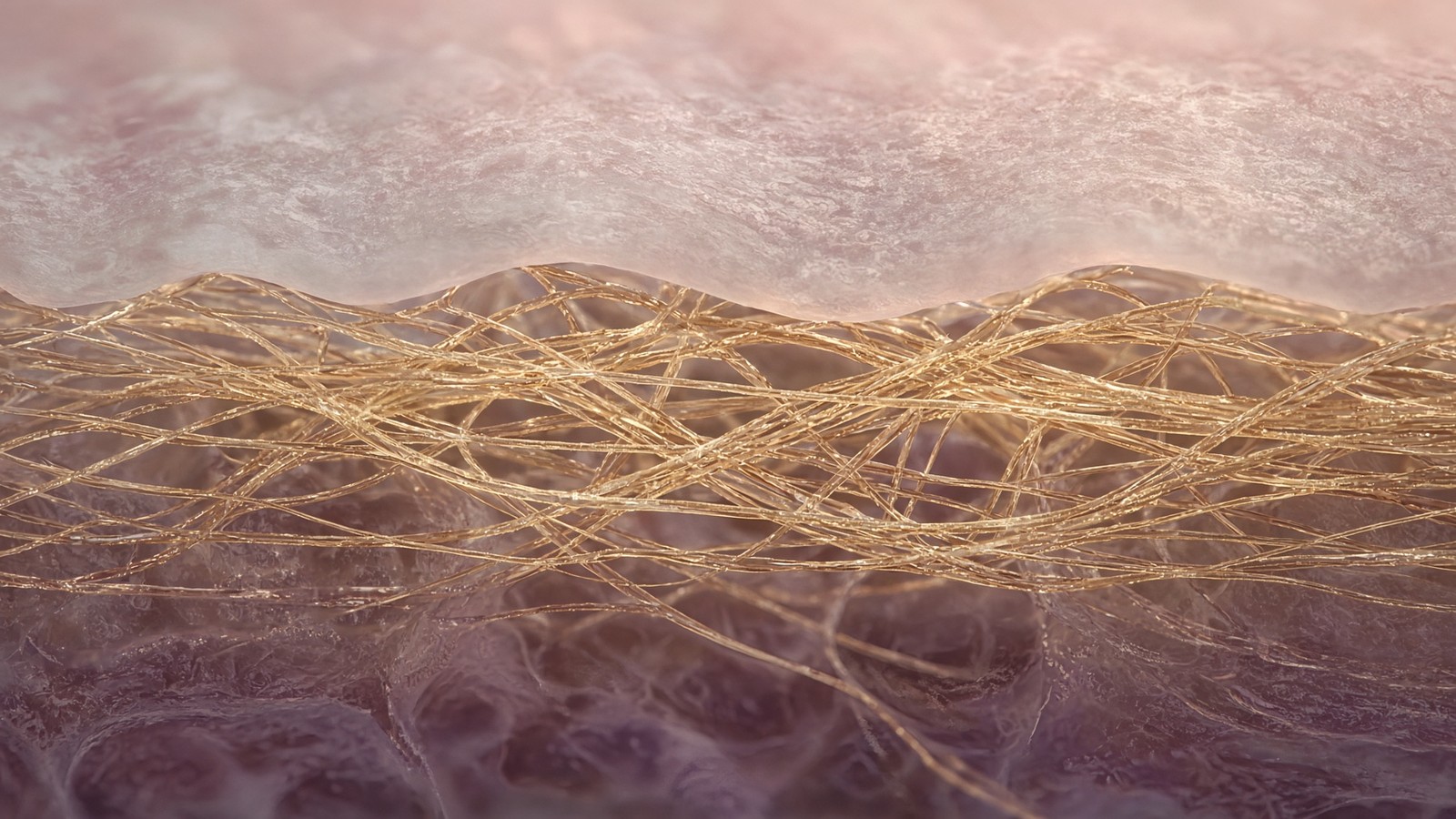
Skin is a matrix problem before it's a mirror problem. Collagen has to be cross-linked; that reaction wants copper; free copper also burns tissue. GHK is the tripeptide that grabs Cu²⁺ and walks it in. Pickart watched old culture start to write protein like young culture. Later arrays shifted toward repair and away from fibrosis. Hair-follicle papers sit on the same copper-and-matrix logic. GLOW is that tripeptide plus the three fragments we already sell as singles — BPC, TB-500, KPV — in one cake, because a bench that wants the neighbourhood shouldn't have to open four caps. KPV on its own is the NF-κB quieting without pigment. None of this is a cream.
Skin is a matrix problem before it is a mirror problem. Type I collagen has to be written, hydroxylated, secreted, packed into a 67-nanometre D-period, and then covalently cross-linked; that last reaction wants copper; free copper also burns tissue through Fenton chemistry. Three characterised objects sit on that tissue in the papers, and they don't do the same job. GHK-Cu is glycyl-L-histidyl-L-lysine holding Cu²⁺ in a square-planar grip so a fibroblast can receive the metal without a hydroxyl-radical mill in the medium. GLOW, as listed, is an 80 milligram cake: 50 milligrams of that copper tripeptide plus 10 milligrams each of BPC-157, TB-500 and KPV. KPV on its own is lysine-proline-valine, the C-terminal tripeptide of α-melanocyte-stimulating hormone, studied in keratinocytes as an NF-κB quieting without a tan. A cream and a scoop of hide digest are different objects again. We'll unbundle the three named sequences as chemistry, not as a glow.
In short. Skin searches land on a copper tripeptide, a four-chain blend, and an anti-inflammatory tail. They share a tissue in the library, not a mechanism.
Grocery-aisle English ruined the word peptide, and hydrolysed collagen did most of the public damage. A tub of hide or fish-skin digest lists grams, a flavour, and an average fragment size of two to five kilodaltons, and it calls the contents peptides because the pieces sit in the same mass band as some hormones. A research vial lists a one-letter code, a calculated mass, and a chromatogram on which a main peak can be pointed at. Type I collagen is a heterotrimeric triple helix, two α1(I) chains and one α2(I) chain, each about a thousand residues, glycine every third position, hydroxyproline holding the three strands. Industrial collagen peptides are an enzymatic hydrolysate of that helix: thousands of overlapping fragments, batch-variable, no single HPLC peak that means one molecule. GHK-Cu is three letters and a metal, CAS 89030-95-5, a complex mass of about 340.7 grams per mole. Both objects are amino-acid chains joined by amides. That's where the kinship ends. The neighbouring collagen-powder essay is the long form of the split. This page is the tissue page: the cable, the copper enzyme that finishes it, the plasma tripeptide, the inflammation tail, and the four-sequence cake.
In short. Collagen powder is chopped-up helix, a mixture, food. GHK-Cu is one named chain plus copper, with a mass and a chromatogram. The shared word is an accident.
GLOW exists in the catalogue because a bench that reads repair-neighbourhood papers will meet four named sequences and shouldn't have to open four caps. Fifty milligrams of GHK-Cu, ten milligrams of the gastric 15-mer BPC-157, ten milligrams of the actin-adjacent thymosin analogue TB-500, ten milligrams of KPV: an 80 milligram lyophilised cake. Those four chains are already sold as singles. The blend doesn't merge their mechanisms. Copper delivery doesn't become actin sequestration by sharing a stopper, and an NF-κB fragment doesn't become a VEGFR2 ligand by sitting in the same vial. It isn't a cosmetic brand. It is a cap-count decision. If a paper only wanted the copper, the GHK-Cu vial is still there. If it only wanted the quieting, KPV is still there. Forum language will call the cake a stack and a glow-up. Biochemistry will call it four ligands, four certificates, four readouts. We'll keep those jobs apart, because a blend that can't name its four chains is just a white powder with a hopeful caption.
In short. Four named sequences packed together are still four jobs. Packing them does not make them one pathway.
KPV on skin is a different sentence from GHK-Cu on a fibroblast, and the difference is the whole reason to keep the tail on this page rather than bury it in a blend caption. α-MSH is thirteen residues of pigment, appetite and inflammatory quieting. The tanning pharmacophore sits in the middle, His-Phe-Arg-Trp, the tetrapeptide melanocortin receptors actually want in the pocket. Keep the last three letters. Lose most of the tan. Luger, Brzoska, Haycock and Moustafa put that tripeptide into keratinocytes and showed NF-κB p65 staying out of the nucleus after TNF, with IκB preserved, and with residual activity when MC1R was blocked. That's barrier-epithelium inflammation without pigment. Adjacent literature to the copper-matrix story, because both papers mention skin. Different ligand. Different assay. A keratinocyte isn't a dermal fibroblast. NF-κB isn't lysyl oxidase. Three residues on each chain is an accident of length, not a kinship. Mapping the neighbourhood isn't treating a person, and a characterised cake is a reagent. Licensed aesthetic medicine, retinoids, and a clinic plasma device still live where they lived when this page is closed.
In short. KPV is three letters from a tanning hormone, used because it quiets inflammatory genes in skin lining without much pigment. That is not the copper peptide.
Collagen has to be cross-linked. That reaction wants copper. Free copper also burns tissue. GHK is how plasma already solved that logistics problem in three residues. A hydrolysate in coffee is a different object wearing the same English syllable.— Reading of Pickart’s isolation and Maquart’s fibroblast papers against the industrial collagen-peptide specification.
Type I collagen is a helix. A hydrolysate is a population.
Type I collagen is a heterotrimer: two α1(I) chains, gene COL1A1, and one α2(I) chain, gene COL1A2. Each chain’s helical domain is about a thousand residues. The exact census is a little longer once you count the N- and C-telopeptides that sit outside the helix and do the cross-link chemistry, and longer still in the procollagen that left the endoplasmic reticulum with registration peptides still attached. The two α1 chains and the one α2 chain wind around each other, a right-handed triple helix built from three left-handed polyproline-II-like strands. Ramachandran’s group in Madras, and Rich and Crick in Cambridge, argued the geometry in the 1950s; we've been measuring it ever since. The repeating triplet is Gly-X-Y. Glycine is the only side chain small enough — a hydrogen — to sit at the sterically crowded interior. X is often proline. Y is often hydroxyproline, written after translation by prolyl 4-hydroxylase, a 2-oxoglutarate-dependent dioxygenase that needs iron, ascorbate and molecular oxygen. Scurvy is the existence proof: without vitamin C the hydroxylase stalls, under-hydroxylated chains fail to form a stable helix, and connective tissue falls apart. A food hydrolysate of the same protein has thrown that specification away.
In short. Type I collagen is two matching chains and one slightly different chain, each about a thousand amino acids, packed with glycine every third residue.
Biosynthesis is a factory with named rooms, and it's worth walking once so the coffee digest can be seen as the opposite process. COL1A1 and COL1A2 are transcribed, spliced, translated into the rough endoplasmic reticulum. Signal peptides come off. Prolyl and lysyl hydroxylases modify the chains. HSP47, the collagen-specific chaperone, walks the folding. The C-propeptides register the two α1 chains and the one α2 chain so the triple helix zips from the C-terminus toward the N-terminus. The molecule moves through Golgi, is secreted as procollagen, and extracellular proteinases — ADAMTS-2, BMP-1/tolloid — cut the N- and C-propeptides. Only then is the helix a collagen molecule that can pack into a fibril. Lysyl oxidase, later, writes the covalent cross-links. A food factory starts at the other end: a tissue that already did all of that, hide or bone or fish skin, is denatured and then cut. Gelatin is the denatured helix. Collagen peptides, in the industrial sense, are gelatin cut smaller so the average piece dissolves in cold liquid and doesn't gel. Nobody ran COL1A1. Nobody formed a helix. Nobody asked lysyl oxidase to do anything. The letters are recycled. The object is new, and it's a mixture.
In short. Cells build collagen through a long, named pipeline of folding, cutting and cross-linking. A food factory starts from finished tissue and digests it.
Once the processed helices pack side by side, they don't pack in register. Each molecule is staggered by 67 nanometres relative to its neighbours, the D-period Hodge and Petruska described, a number electron microscopy and low-angle X-ray scattering have been returning for decades. The stagger produces a gap region and an overlap region, the banding you can see on a well-prepared fibril, and it's also where the cross-links sit. Molecules pack into fibrils, tens to hundreds of nanometres across. Fibrils pack into fibres. Dermis writes a weave more isotropic than tendon, type I and type III together, a mechanical environment fibroblasts feel through integrins. Type III is the wound and reticular partner; type IV is the basement-membrane network at the dermoepidermal junction, not a fibril at all. A peptide that increases collagen, as a hydroxyproline number in a dish, hasn't yet told you which architecture it's serving. A powder that supplies glycine, proline and hydroxyproline hasn't told you anything about architecture. The D-period, the fibre, the dermis: those are cell and enzyme and time, not a scoop.
In short. Collagen molecules pack with a 67-nanometre stagger to make a fibril. Drinking small fragments doesn't rebuild that stagger in skin.
The industrial product is an enzymatic hydrolysate, and the honest name is the one a paper would use. Endopeptidases — often bacterial proteases chosen for price and for the fragment-size they leave — cut the denatured helix until the average piece is two to five kilodaltons, twenty to fifty residues if you pretend the pieces are uniform, which they aren't. The distribution has a tail of di- and tripeptides and a tail of larger leftovers. Batch to batch, the distribution moves. Source to source — bovine versus marine — the residue census moves, because fish type I isn't cow type I in every triplet, and because skin isn't bone. There's no single sequence. There's no one fragment that is the product. A certificate that listed every species in a hydrolysate would be a proteomics paper, and it would be out of date when the next lot ran. What the tub lists instead is grams, a flavour, sometimes a source, sometimes an average molecular weight. Those are food-label facts. They aren't a one-letter code or an HPLC main peak.
In short. The digest is a mixture whose average size is two to five kilodaltons. The pieces change with the batch and the animal. There is no one chain to name.
Nutrition papers on collagen hydrolysates are allowed to be interesting, and they aren't this ligand. Iwai, Hata and Sato showed that Pro-Hyp and other di- and tripeptides appear in human plasma after oral collagen peptides, at micromolar concentrations, with a time course of hours. Some fibroblast and chondrocyte papers then add those dipeptides to a dish and report a modest change in hyaluronan or in a collagen transcript. That's a food-derived peptide story, and it has a literature. It's still a population of fragments, still not Gly-His-Lys·Cu²⁺, and still not a copper stoichiometry. Confusing a plasma Pro-Hyp peak after a scoop with Pickart’s plasma GHK number is how a grocery aisle stole a laboratory isolation. One is a digestion product of a thousand-residue helix. The other is a circulating tripeptide isolated because old liver tissue in culture synthesised protein like young tissue, usually carrying copper. Amino acids are shared. Identity is not. If a tub lists grams and a flavour, you're holding food. If a vial lists a sequence, a metal, a mass and a purity, you're holding a reagent. Mixing the words is how a laboratory term walked into a supermarket.
In short. Some collagen-powder dipeptides do appear in blood after a drink. That nutrition story is real, and it's still not the named copper tripeptide.
Diagram
- Amino acid~110 DaTwenty side chains. The alphabet.
- Peptide bondamide, planarCarboxyl carbon to the next nitrogen. Resonance holds it flat.
- Oligopeptide< ~20 residuesMost hormones and fragments. GHK is three. KPV is three.
- Polypeptide20–50+Insulin 51. GLP-1 31. Retatrutide is a designed chain in this band.
- Proteinfolded machineHaemoglobin, a GPCR, lysyl oxidase. Tertiary structure worth drawing.
Insulin (Banting and Best, 1921) was the first peptide anyone bothered calling a medicine. A collagen hydrolysate is food. A named sequence with a mass and a chromatogram is a research peptide. The shared word is the accident.
Lysyl oxidase finishes the gel. The metal is not optional.
Collagen that has just been secreted isn't yet a cable. The triple helix of type I is a hydrogen-bonded rope, glycine every third residue, hydroxyproline holding the three chains, a beautiful structure that will still slide if you pull on it. Load-bearing connective tissue requires covalent cross-links between chains and between fibrils. Lysyl oxidase (LOX) and the LOX-like enzymes LOXL1 through LOXL4 do that job. They oxidatively deaminate the ε-amino group of specific lysine and hydroxylysine residues in collagen and elastin, leaving an aldehyde: allysine or hydroxyallysine. Those aldehydes then condense — aldol products, Schiff bases, and, with time and further chemistry, the pyridinolines of mature collagen and the desmosines of elastin. The enzyme carries copper and a lysine tyrosylquinone cofactor, LTQ, which is built in place from a tyrosine and a lysine once copper is bound. No copper, no LTQ, no aldehyde, no cross-link. A culture deprived of copper doesn't make a dermis. It makes a weak gel. That sentence is older than GHK and doesn't depend on any microarray. It's why a copper tripeptide and a tub of hydrolysate get mentioned at the same dinner, and why that mention is a muddle.
In short. Lysyl oxidase uses copper to turn lysine in collagen and elastin into aldehydes that cross-link. Without the metal you get a weak gel, not a load-bearing matrix.
The LOX family isn't one enzyme with one tissue. LOX itself is the canonical collagen and elastin cross-linker, secreted as a proenzyme, activated by BMP-1/tolloid proteases that cut the propeptide. LOXL1 is particularly tied to elastin and to the elastic-fibre failures of pseudoexfoliation glaucoma. LOXL2 has a different reputation: fibrosis, tumour stroma, a target people have tried to antibody. The family sits on the same metal and the same chemistry, then writes different pathologies depending on which isoform is loud and where. Cross-links that fail give you fragile tissue. Cross-links that run too hard give you a stiff, fibrotic one. TGF-β induces LOX. Hypoxia induces LOX. β-Aminopropionitrile, the lathyrism toxin from sweet pea, inhibits the enzyme; animals and people exposed to it get fragile collagen, aneurysms, a skeleton that won't hold. Menkes disease, a failure of the copper-transporting ATPase ATP7A, starves secreted copper enzymes and produces a connective-tissue phenotype that looks, in places, like a cross-link failure. Three existence proofs, three different inputs, one tissue that falls apart when the cable isn't finished. A copper-delivery reagent sits on that node, not above it.
In short. Five related enzymes cross-link matrix. Too little copper-dependent activity leaves tissue fragile; too much helps fibrosis. GHK-Cu sits on that node, not above it.
A human adult carries something on the order of 100 milligrams of copper, most of it bound. Ceruloplasmin is the major plasma carrier. Albumin binds copper at an N-terminal motif. CTR1 imports it; Atox1, ATP7A/B, CCS and the SCO proteins hand it on. There is, by design, almost no free copper in a healthy cytosol. Rae, O’Halloran and colleagues measured the free Cu⁺ concentration as vanishingly low, buffered by a surplus of ligands. That's the point of a chaperone network. The metal is too useful and too dangerous to be allowed to wander. The hazard is Fenton chemistry, named for the iron reaction and equally available to copper: Cu⁺ plus hydrogen peroxide yields Cu²⁺, a hydroxide, and a hydroxyl radical. The hydroxyl radical is indiscriminate. It attacks bases, lipids, protein side chains. Superoxide can reduce Cu²⁺ back to Cu⁺, so a small pool of free copper plus a little peroxide becomes a catalytic radical factory. GHK is a small circulating complex that sits on the same chemical problem those proteins were built to solve: move copper, don't spill it.
In short. Cells keep almost no free copper, because loose copper plus peroxide makes hydroxyl radicals. Dedicated proteins, and GHK, exist to move the metal without that spill.
Superoxide dismutase is the other copper job, and it belongs on a skin-matrix page because a fibroblast that can't mop the first radical won't keep the matrix it just wrote. Superoxide is oxygen plus an extra electron, O₂•⁻, the first leak of the respiratory chain. SOD catalyses the disproportionation, two superoxide and two protons to oxygen and hydrogen peroxide. SOD1 is the cytosolic Cu/Zn enzyme; the copper is the redox-active metal at the active site, the zinc is structural, and CCS loads the copper. A cell that can't place copper on SOD1 is a cell whose first antioxidant enzyme is disarmed. Feed copper carelessly and you may raise LOX and SOD while also raising hydroxyl radical from the unbound fraction. Feed it as GHK-Cu and the hypothesis is that the bound fraction dominates. Testing that hypothesis is a ROS readout plus an activity readout, in the same dish, with a copper-salt arm that will almost certainly look dirtier on the ROS side. Older papers reported antioxidant effects in peroxide, consistent with shielding, with SOD-related activity, or with a sacrificial ligand. Those are different mechanisms.
In short. SOD1 is the cytosolic enzyme that turns superoxide into peroxide and oxygen. It needs copper at the active site. Delivery to that enzyme is a second job for the same metal.
Diagram
- GHKGly-His-LysPickart, 1970s plasma fraction. ~200 ng/ml at 20; ~80 ng/ml at 60.
- Cu²⁺ complexsquare-planarHistidine imidazole + backbone nitrogens. The product is the complex.
- Lysyl oxidaseLOX / LOXLCopper enzyme. No metal, no collagen cross-links, a weak gel.
- SOD1Cu/ZnFirst mop for superoxide. Same metal, different job.
- Free Cu²⁺FentonCu⁺ + H₂O₂ → HO·. Delivery without a redox-active pool is the point.
Pickart and Margolina’s microarrays claim thousands of transcripts. Modern RNA-seq with multiple-testing correction is the replication the claim still owes. Copper delivery to LOX and SOD would remain interesting if the spreadsheet shrinks.
Pickart’s tripeptide, and why the metal is the product
In the early 1970s Loren Pickart was fractionating human plasma for a reason that still sounds almost rustic. Cultured liver tissue from older donors synthesised protein in a pattern that looked tired. Albumin-rich fractions from young plasma, added to those cultures, shifted the pattern toward the youthful one. The active piece, once the fractionations had been honest enough, was three amino acids: glycine, histidine, lysine. GHK. It usually arrived with a copper ion attached, which turned out not to be a contaminant. Pickart and Thaler published the growth-modulating plasma tripeptide in Nature New Biology in 1973. The observation refused to go away, which is more than most 1970s peptide lore managed. Three residues is small enough to be underestimated and chemically tidy enough to survive scepticism. A defined ligand, a metal, a dish, a protein-synthesis readout. That's an origin story a paper can still stand next to, fifty years on, without needing a cream or a testimonial to hold it up. Isolating it from albumin-rich plasma, before cheap mass spectrometry, meant chromatography and bioassay. The field kept it because the same tripeptide sat next to copper enzymes that finish matrix.
In short. In the 1970s a chemist found a three-amino-acid piece of blood that made old liver cells make protein like young ones. It usually carried copper.
Pickart reported plasma GHK at roughly 200 nanograms per millilitre at age twenty and about 80 nanograms per millilitre at age sixty. Those are his numbers, and they should be treated as such: a named measurement from the laboratory that isolated the peptide, not a multi-centre reference interval. Even on that caveat they're worth holding, because they put a clock on a circulating ligand rather than a rumour. Two hundred nanograms per millilitre of a 340-dalton complex is a nanomolar neighbourhood. Sixty-year-old plasma, on the same method, is holding less than half. Restoration stories write themselves from that arithmetic, and most of them should be sent back. Restoring a circulating tripeptide isn't the same work as restoring a dermis, a follicle, or a liver. It's a reason to look. The look has to be an assay with copper stoichiometry declared, a cell type named, and a readout that isn't a photograph of someone’s cheek. Ageing changes a thousand other plasma peptides. GHK is the one this page can name, weigh, and complex with a metal. Why it falls is a separate question, and none of the usual accounts has been closed.
In short. Pickart measured about 200 nanograms of GHK per millilitre of plasma at twenty, and about 80 at sixty. A falling number is a reason to look, not a restoration plan.
Cu²⁺ is a d⁹ ion with a strong preference for square-planar or distorted octahedral geometry. GHK offers it a tridentate grip: the N-terminal nitrogen of glycine, a deprotonated peptide-bond nitrogen, and the imidazole nitrogen of histidine. Crystal structures and solution studies put the copper in a square-planar complex, with water or another oxygen often completing the plane or sitting at a longer axial distance. The lysine side chain is usually not the fourth ligand in the simple 1:1 complex; it's a charge and a handle. That geometry is why the complex is stable enough to keep free Cu²⁺ from roaming and labile enough, in the right neighbourhood, to hand the metal to an enzyme that wants it. Stability and lability aren't opposites here. They are the two requirements of a chaperone. Too tight and the enzyme never sees the ion. Too loose and the ion is a Fenton reagent in the buffer. Call it an ionophore and you have said something true and incomplete. Call it a ligand in its own right and you have said the other half. Distinguishing those needs a copper-matched control and a GHK-without-copper arm. Most older papers didn't run both.
In short. GHK holds Cu²⁺ in a square-planar grip through histidine and backbone nitrogens. Tight enough to stop free copper roaming, loose enough to hand the metal on.
Maquart, Pickart, Borel and colleagues showed, in 1988, that the GHK-Cu complex stimulated collagen synthesis in fibroblast cultures. That paper is still the one to hand a sceptic. The readout was collagen, the cells were fibroblasts, the ligand was the copper complex. Wegrowski, Maquart and Borel followed with sulphated glycosaminoglycan synthesis, the proteoglycan half of the same neighbourhood. Those are protein and carbohydrate outputs, not a 4,000-gene spreadsheet. They are also the findings that don't require you to believe anything extraordinary about transcription. A fibroblast given copper in a form it can use, in a medium that may have been marginally copper-limited, will finish more matrix. Decorin, the small leucine-rich proteoglycan that binds collagen fibrils and sequesters TGF-β, sits on the same street. SPARC, secreted protein acidic and rich in cysteine, also called osteonectin, is a matricellular protein that sits at the interface of cells and matrix and is one of the transcripts later arrays put on the up list. The 1980s biochemistry didn't need those names to be true. Desmosine, pyridinoline, or a LOX activity assay would finish the sentence that paper started. Until they do, the honest caption is more collagen made.
In short. In 1988 the copper complex made cultured fibroblasts produce more collagen. That finding doesn't require a giant gene story. It requires copper getting to the matrix enzymes.
Most ligands occupy a receptor and move a pathway. GHK-Cu’s published microarrays claim something ruder: on the order of thousands of human transcripts, a shift toward a younger, less fibrotic, more DNA-repair-competent pattern. Collagen genes up. Decorin up. TIMPs up. DNA-repair transcripts up. SOD up. MMP-1 and MMP-3 down. A fibrinogen and metastatic cassette down. TGF-β and integrin signalling in fibroblasts moved in parallel. Pickart, Vasquez-Soltero and Margolina put those lists into a series of reviews and papers in the 2000s and 2010s, drawing on Affymetrix-era profiles of cultured human cells treated with the complex. Breadth is an extraordinary claim. A 2000s microarray was a miracle and a mess: compressed dynamic range, cross-hybridisation, probe-set annotation lagging the genome, batch effects, fold-change cut-offs without an adjusted p-value. Some of those tables contained real biology. Collagen, TIMP, MMP, SOD, decorin aren't random. They are the neighbourhood the 1988 biochemistry already pointed at. RNA-seq with copper stoichiometry declared, GHK-without-copper and copper-without-GHK arms, and a false-discovery-rate threshold is the replication the claim still owes. Until it exists, copper delivery to LOX and SOD is enough, and the transcriptome-reset remains a hypothesis.
In short. The unusual claim is breadth: thousands of genes, repair up, some destruction down. That's much larger than copper reaching lysyl oxidase, and it still needs a modern repeat.
A hair follicle is a matrix organ with a clock. Anagen is the growth phase, catagen the regression, telogen the rest. The dermal papilla is a cluster of specialised fibroblasts at the base of the follicle; it inducts the epithelium, sets follicle size, and is the mesenchymal half of the conversation that decides whether a follicle stays in anagen. The extracellular neighbourhood of the papilla is collagen, proteoglycans, laminin, a basement membrane, and a copper-dependent cross-link census like any other connective-tissue niche. Copper peptides entered the hair literature because papilla cells in culture respond to them on growth and on the size of the follicle they will support, in models that people who run hair assays already know how to over-interpret. Follicle organ-culture, whisker-pad models, and dermal-papilla proliferation assays are real tools. Anagen programmes include VEGF, IGF-1, several Wnts, and a suppression of TGF-β that would otherwise push the follicle toward catagen. That TGF-β sentence should sound familiar; it's the same factor the dermal-fibroblast arrays were arguing about. AHK-Cu is the cousin some hair papers name instead; it isn't GHK-Cu. Name the three letters, the metal, and anagen length.
In short. Hair follicles grow, rest and shrink on a clock. The dermal papilla is a cluster of fibroblasts whose matrix GHK-Cu has been studied in. That's a culture literature, not a density claim.
Elastin is the other LOX substrate, and it's the one ageing skin actually misses. Tropoelastin is secreted, aligned on a fibrillin microfibril scaffold, and then cross-linked by LOX family members into desmosine and isodesmosine, the tetrafunctional residues that make elastin a rubber. Adult dermis makes almost no new elastin under ordinary conditions. Solar elastosis is a mess of damaged elastic material, not a fresh network. A type I hydrolysate doesn't contain tropoelastin sequence in any useful sense; hide is collagen, not elastic-fibre feedstock, and the digest is still a mixture. Cosmetic copy that slides from collagen peptides to elasticity is sliding from a type I hydrolysate to a different protein with a different gene, a different cross-link, and a different developmental window. GHK-Cu, as a copper donor, can in principle sit on elastin cross-linking as well as collagen cross-linking, because LOX does both. That's still a copper-enzyme sentence, not a powder sentence, and not a photograph of a face. Answering the elastin question with desmosine, a LOX activity assay, and a defined cell type is a paper. Keep the proteins named: collagen I, elastin, LOX, GHK-Cu.
In short. Elastin is a different rubbery protein that also needs copper cross-links. Adult skin barely makes new elastin. A collagen powder isn't that protein.
- GHK-Cu
- 3 residues, ~340.7 Da
- Plasma GHK at 20 years
- ~200 ng/ml
- Plasma GHK at 60 years
- ~80 ng/ml
- Type I α chain
- ~1000 residues
- Hydrolysate piece
- 2–5 kDa average
- D-period
- 67 nm
- GLOW cake
- 80 mg
- KPV
- 3 residues, ~342 Da
Gly-His-Lys·Cu²⁺. CAS 89030-95-5. Square-planar. One HPLC peak.
Pickart’s number. A nanomolar neighbourhood.
Same method, less than half. A clock, not a protocol.
COL1A1 / COL1A2. Gly-X-Y. Two α1(I), one α2(I) in the helix.
Thousands of overlapping fragments. No single sequence. Food.
Hodge–Petruska stagger. A fibril fact, not a powder fact.
50 mg GHK-Cu, 10 mg BPC-157, 10 mg TB-500, 10 mg KPV. Cap-count.
Lys-Pro-Val. α-MSH C-terminus. NF-κB down. Different ligand.
KPV on keratinocytes is NF-κB, not a tan
KPV is lysine-proline-valine. Three residues, a mass of 342.4 grams per mole, CAS 67724-34-9, the C-terminal tripeptide of α-melanocyte-stimulating hormone. α-MSH itself is thirteen residues, acetylated at the amino terminus, amidated at the carboxyl terminus, sequence Ac-SYSMEHFRWGKPV-NH2, a product of pro-opiomelanocortin once the convertases have finished with ACTH. The last three letters of that sequence are the molecule this heading is about. Medicinal chemistry is often addition: a D-amino acid here, a lactam bridge there, a pan-agonist that occupies four receptors because the chemist refused to pick one. KPV is subtraction. Keep the tail. Lose most of the pigment, most of the MC4R appetite agonism, most of the autonomic extras a cyclic analogue brings into a first-in-human room. The core pharmacophore of α-MSH is His-Phe-Arg-Trp, residues six to nine, the HFRW tetrapeptide that melanocortin receptors actually want in the pocket. Melanotan II was built around a cyclic version of that core. KPV is what remains when you throw the core away. That's a precise, small, interesting reagent. It isn't a weaker α-MSH. It's a different question, and the question on this page is keratinocyte inflammatory transcription, not a sunbed and not a copper enzyme.
In short. KPV is the last three amino acids of the tanning hormone α-MSH. Labs keep the anti-inflammatory tail and drop most of the pigment.
Nuclear factor kappa-light-chain-enhancer of activated B cells is a transcription-factor family, not a mood. In the canonical pathway the heterodimer that matters is p65/RelA with p50. In a resting cell that dimer sits in the cytosol, bound to IκBα, the inhibitor whose name is the whole mechanism: inhibitor of κB. A receptor signal — TNF receptor 1, IL-1 receptor, a Toll-like receptor — converges on the IκB kinase complex, IKKα/IKKβ/NEMO. IKK phosphorylates IκBα on serines 32 and 36. The phosphodegron is recognised by a β-TrCP ubiquitin ligase, IκBα is proteasomally destroyed, and p65 goes to the nucleus. On the DNA it writes TNF, IL-6, IL-1β, IL-8, COX-2, a cassette of adhesion molecules and chemokines, the inflammatory page a barrier tissue turns to when the lining is under attack. Sen and Baltimore named the factor in B cells in 1986. If a peptide paper says anti-inflammatory and can't name IκB, it has described a feeling. Preservation of IκB is the mechanistic sentence Luger’s dermatology group and Getting’s inflammation group kept returning to. α-MSH, and then the C-terminal tripeptide, reduced IκB degradation after TNF, IL-1β or lipopolysaccharide, so p65 nuclear translocation fell and the cytokine transcripts fell. That's the off-switch.
In short. NF-κB is a gene switch for inflammatory proteins. A partner called IκB holds it in the cytosol. Destroy IκB and the switch enters the nucleus and writes cytokines.
Keratinocytes are the skin half of the KPV sandbox, and they aren't melanocytes and not dermal fibroblasts. A keratinocyte is the barrier cell of epidermis; it expresses MC1R at low level in several reports, it runs a great deal of NF-κB when TNF or IL-1 hits it, and it is the cell Haycock and Moustafa used when they asked whether α-MSH and KPV could stop p65 after TNF. HaCaT, the spontaneously immortalised human keratinocyte line, is the usual workhorse, with the usual caveats of a line that isn't primary and isn't a reconstructed epidermis. Contact-hypersensitivity models in mice, from Luger's group, put the tripeptide into a living ear or a living back and asked about sensitisation and elicitation, which is a different experiment from a luciferase well. Brzoska, Luger, Böhm, Haycock, Moustafa: those are the papers to start with. Several groups have shown residual activity when MC1R is missing or blocked. That incomplete dependence is the intellectual reason to reach for the fragment instead of α-MSH or Melanotan II when a tan, an appetite crash or a melanocortin circus would confound the assay. A living ear is a barrier assay with a named hapten.
In short. Skin lining cells are one sandbox: after an inflammatory push, the tripeptide can keep NF-κB down. That is a dish or a mouse-ear assay, not a cream.
Adjacent barrier literature is why KPV keeps turning up on a skin page and on a gut page, and adjacency isn't identity. Dalmasso, Merlin and colleagues showed that the tripeptide can ride PepT1 (SLC15A1), the H⁺-coupled oligopeptide transporter intestine already uses for dietary di- and tripeptides and induces in inflammation. That's why DSS-colitis models keep appearing: the ligand can get into epithelium through a nutrient door. Keratinocytes aren't enterocytes. PepT1 isn't the main door of epidermis. The shared biochemistry is NF-κB in a barrier sheet, not a shared transporter, and not a shared copper enzyme. The irritable-bowel essay on this desk is the organ portrait for gut: a Rome-criteria cluster, a one-cell lining, BPC-157 on vessels and nitric oxide, KPV on IκB and PepT1. This page is the dermis-and-epidermis portrait: GHK-Cu on fibroblasts and lysyl oxidase, KPV on keratinocyte NF-κB, GLOW as the cake that happens to hold both. File pigment under a different ligand. File copper delivery under GHK-Cu. File IκB preservation under KPV. Three drawers. A search bar that puts them on the same afternoon hasn't merged the drawers, and a paper that can't say which drawer it opened hasn't chosen.
In short. KPV also enters inflamed gut lining on a food-peptide transporter. Skin lining is a different door. The shared fact is a quieter inflammatory switch, not a shared lock.
Three residues on GHK and three residues on KPV is an accident of length, and treating them as a class of skin tripeptides is how a grocery word eats a ligand. One holds a metal in a square-planar grip and talks to lysyl oxidase and SOD1 in a fibroblast. One is a melanocortin tail that leaves IκB standing in a keratinocyte. One has a histidine imidazole as the chemistry. One has a proline kink and a lysine charge as the chemistry. They don't share a receptor. They don't share a metal. They don't share a chromatogram except in the trivial sense that both absorb at 214 nanometres because both have peptide bonds. Calling both copper-free tripeptides a class is a category error even before you notice that GHK-Cu isn't copper-free. GLOW lyophilises them together with two longer chains, which is a logistics decision a bench can want and a mechanism a blot can't merge. If a paper needs the copper complex, it needs stoichiometry, without a melanocortin fragment as a confound. Blend papers have to name four ligands and four readouts. That request answers itself.
In short. Two different three-letter peptides sit on skin papers. One carries copper to matrix enzymes. One quiets an inflammatory gene switch. Length is not kinship.
Diagram
MC1R
melanocyte
Eumelanin vs pheomelanin. Red-hair alleles. Afamelanotide’s receptor.
MC3R
hypothalamus
Energy balance. Occupied by MT2 because MT2 occupies almost everything.
MC4R
hypothalamus
Appetite brake. Loss-of-function obesity. Setmelanotide (FDA 2020).
MC5R
sebaceous
Sebum. A first-class output people still treat as a footnote.
KPV
tail of α-MSH
NF-κB off-switch. PepT1 uptake. Designed not to tan.
MT2
pan-agonist
Ac-Nle-cyclo[Asp-His-D-Phe-Arg-Trp-Lys]-NH2. Pigment, appetite, flushing, arousal.
α-MSH is Ac-SYSMEHFRWGKPV-NH2. The last three letters are KPV. Melanotan II is what a pan-agonist looks like. KPV is the design move in the other direction. Match the ligand to the question.
Most of the anti-inflammatory activities of α-MSH can be attributed to its C-terminal tripeptide KPV. Pigment is the obstacle that made the fragment interesting. IκB is the protein that made it mechanistic. Copper is a different invoice.— Brzoska T, Luger TA, Maaser C, Abels C, Böhm M. α-Melanocyte-stimulating hormone and related tripeptides. Endocr Rev. 2008; 29: 581–602. Reading against Haycock and Moustafa on p65 in keratinocytes, and against Pickart on GHK-Cu.
GLOW is a cake of four named sequences
Write the masses out before anyone is allowed a glow-up caption. GHK-Cu is Gly-His-Lys·Cu²⁺, about 340.7 daltons, 50 milligrams in the cake. BPC-157 is GEPPPGKPADDAGLV, fifteen residues, 1419.5 daltons, CAS 137525-51-0, 10 milligrams. TB-500 is a research analogue around thymosin β4’s LKKTETQ actin-binding motif; native Tβ4 is forty-three residues, about 4.96 kilodaltons, and the analogue’s certificate has to say which chain you actually reconstituted; 10 milligrams. KPV is Lys-Pro-Val, about 342 daltons, 10 milligrams. Four sequences. Four masses. One lyophilised 80 milligram cake. HPLC will tell you the chains are in the aliquot if the method is built to see them; a forum caption will tell you they're one skin protocol. The chromatogram is the better witness. The point of the blend, as a catalogue object, is that a bench reading the repair neighbourhood — matrix copper, gastric cytoprotection and vessels, actin-buffered crawl, keratinocyte NF-κB — shouldn't have to guess which white cake is which, and shouldn't have to open four stoppers to put four named ligands on the same afternoon’s ice. Convenience is a real virtue in a cold room. Convenience isn't a heterodimer, not a shared receptor, and not a published factorial.
In short. GLOW is fifty milligrams of the copper tripeptide plus ten milligrams each of three other named chains. Four masses, not one mechanism.
BPC-157 in that cake is a gastric 15-mer, proline-rich, stubborn in pepsin and pH 1.5, isolated as a fragment of a gastric-juice protein that survived acid. The Zagreb corpus under Sikiric put it into NSAID and alcohol gastric lesions, then into endothelium, tendon, a nitric-oxide argument. The cleaner independent papers live at VEGFR2, FAK–paxillin and eNOS. Chang, Tsai and colleagues, Journal of Applied Physiology 2011, is the tenocyte-outgrowth paper a tendon page has to cite. That's a vessel-and-cytoprotection neighbourhood. The two sequences keep turning up next to skin searches because internet culture bundled repair with whatever short chain had an injury paper. Unbundling is the job. A denser bed of small vessels in a defect can be repair biology. A denser bed of small vessels in photoaged dermis can be part of the problem. Phenotype rhyme is the weakest reason to co-administer two ligands. There's no factorial of the copper complex and the 15-mer on a named kinase. Adjacent tissue is lawful. A shared lock is a claim that hasn't been made.
In short. BPC-157 is a fifteen-amino-acid stomach fragment with vessel and nitric-oxide papers. Same repair shelf as GHK-Cu. Different lock. Different blot.
TB-500 in that cake is a laboratory analogue built around thymosin β4, the principal G-actin sequestering peptide of animal cells. Native Tβ4 is forty-three residues, intrinsically disordered, acetylated at the amino-terminal serine, mass about 4.96 kilodaltons. Daniel Safer showed in 1991 that the abundant actin-sequestering peptide Fx and thymosin β4 were the same molecule. Cells crawl by parking and releasing actin monomers. Left unbuffered, a cytoplasm full of G-actin would polymerise wherever a barbed end presented itself. Thymosin β4 binds the monomer one-to-one and holds it off the filament until profilin, a local drop, or a nucleating machine asks for it. LKKTETQ, residues 17–23, is the principal actin-binding motif; Ac-SDKP, the N-terminal tetrapeptide, is a third pharmacophore, anti-fibrotic, not an actin buffer. Research TB-500, depending on the certificate, orbits the motif or is the full analogue. Scratch assays, corneal alkali injury, dermal re-epithelialisation: those are crawl-and-close experiments. Full-length Tβ4 went into human ophthalmic and dermal studies under the RegeneRx RGN programmes; mixed, not empty. Actin sequestration isn't copper delivery. A lamellipodium isn't a lysyl-oxidase aldehyde. Putting the analogue in the same cake as GHK-Cu doesn't teach a fibroblast to confuse those jobs. This paragraph is the unmix.
In short. TB-500 is built around a protein that holds spare actin so a cell can crawl. That holding job isn't copper delivery and not an inflammatory-gene switch.
The internet bundled the four because all of them had preclinical injury or matrix literatures, all of them were short enough to synthesise on a solid-phase rig, all of them were unlicensed, and all of them photographed well as a stack. Skin threads wanted a copper peptide, a vessel peptide, a crawl peptide and a quieting peptide, and didn't want to learn four mechanisms. A single lyophilised cake with one reconstitution instruction is a commercial object. It isn't a biochemical object. There's no heterodimer. There's no shared receptor across copper chaperone, VEGFR2, G-actin and IκB. There's a shared customer: a laboratory that reads barrier and repair papers and would rather open one cap. Customer isn't mechanism. A wound bed does need matrix, a vessel, a crawling cell and a quieter inflammatory transcription, so pairing the ideas is a fair hypothesis. The hypothesis is lawful. The leap from hypothesis to a fixed quartet of research peptides, at folkloric ratios, in the absence of a factorial experiment, is not. The factorial is each ligand alone, the quartet, and neither, on a named readout. Until that figure exists, the pairing is a rhyme.
In short. People bundled them because all four showed up in injury or matrix stories and all four were easy to make. Shared rumour isn't a shared receptor.
A blend is a convenience. A stack, in forum language, is a protocol. This catalogue will sell the convenience to a laboratory that can name the four chains. It won't launder it into a protocol. Blend papers, if anyone writes them, have to name four ligands and four readouts: LOX or collagen for the copper complex, phospho-VEGFR2 or an endothelial scratch for the 15-mer, a G/F-actin ratio or a pyrene-actin curve for the analogue, IκB and nuclear p65 for the tail. A hydroxyproline number on a mixed dish is a soup. A photograph of a face is a different legal object. If a paper needs only the copper, it should weigh GHK-Cu, declare moles of peptide and moles of Cu²⁺, and leave the gastric fragment in the freezer. If it needs the neighbourhood as four named sequences, the 80 milligram cake saves four caps and still requires four assays. Related in folklore. Unrelated in mechanism. Four certificates, or one certificate that lists four masses. Either way the names have to stay on the page. GLOW is 50 milligrams of Pickart's complex plus 10 milligrams each of three other sequences, freeze-dried together. That sentence is the blend.
In short. Packing four named ligands together is a convenience for a bench. It is not a recipe, and it does not merge four blots into one juice.
Diagram
BPC-157: Pro-rich, acid-stable, Sikiric corpus. VEGFR2 internalisation, FAK–paxillin, eNOS-dependent NO tone. A cytoprotection story that escaped the stomach.
TB-500: cytoskeletal buffer. Injury releases Tβ4 extracellularly; VEGF, MMPs and keratinocyte migration follow. SDKP is a separate N-terminal anti-fibrotic pharmacophore. Two literatures, two jobs.
Thymosin β4 is the principal G-actin sequestering peptide. TB-500 is built around the LKKTETQ motif. BPC-157 is a gastric 15-mer (GEPPPGKPADDAGLV) that talks to VEGFR2 and focal adhesions. Related in folklore. Unrelated in mechanism.
Diagram
| Node | Catalogue | Conversation |
|---|---|---|
| GPCR | Ipamorelin, MT2, PT-141, retatrutide, CJC | Second messengers, secretion, appetite, pigment |
| RTK / IGF1R | IGF-1 LR3 | IRS–PI3K–Akt–mTOR and Shc–ERK |
| Cytokine receptor | Somatropin (HGH) | GHR–JAK2–STAT5b, hepatic IGF-1 |
| Cofactor | NAD+ | Sirtuins, PARPs, CD38, redox |
| Actin buffer | TB-500 / Tβ4 motif | G-actin sequestration, motility |
| Growth-factor-like | BPC-157 | VEGFR2 / FAK / eNOS neighbourhood |
| Copper ligand | GHK-Cu | Transcriptome shift in fibroblasts |
| MC fragment | KPV | NF-κB, PepT1, no pigment |
| Nuclear / pineal | Epithalon (AEDG) | TERT and melatonin literatures |
| mtORF peptide | MOTS-c | AMPK, folate–methionine cycle |
Each row is a different kind of molecular conversation. The catalogue peptides bind at these nodes; they are not interchangeable, and stacking them because a forum did mixes unrelated literatures.
Clinic is next door, not in the vial
NEOGEN nitrogen plasma, in the clinic essay next door, denatures collagen with a pulse of ionised N₂ and then waits ninety days for fibroblasts to write a new band. Heat-shock protein 47, TGF-β1, MMP clearance of the denatured gel, TIMP trying to keep that clearance from becoming a hole: the histology is textbook wound healing with a thermal start. GHK-Cu’s fibroblast arrays move collagen, TIMPs and MMPs. That's why the two essays are filed as neighbours. Neighbouring is a reading list. It isn't a bowl. Nobody is pouring a research complex into a CE-marked handpiece, and a thermal denaturation isn't a copper-delivery assay. CELL STORY and Dermalux at eLIVEate sit on adjacent biology — micro-injury, mitochondrial light. Wunsch and Matuschka, 2014, reported that red and near-infrared LED phototherapy increased collagen and elastin and reduced MMP-1 in human skin after a course, which is a dermal readout of a cytochrome c oxidase instruction, not a ligand. The clinic owns the energy settings and the consent. The catalogue owns characterised sequences. There's no commission on those bookings and no facial mixed into a cake. The shared cell is a fibroblast.
In short. A clinic plasma device and a light bed both sit on fibroblast collagen biology. That's a reason to read those essays, not a reason to mix them with these ligands.
Retinol, tretinoin, licensed botulinum toxin, licensed hyaluronic-acid fillers, and the medicines a dermatologist writes for acne or for inflammatory dermatoses are a different legal class from a lyophilised research peptide. They have indications, risk registers, and, in the retinoid case, decades of AP-1 and MMP-1 biochemistry that Fisher, Voorhees and colleagues put on photoaged dermis in the 1990s. Ultraviolet B induces interstitial collagenase in fibroblasts through AP-1, and the enzyme then nicks type I fibrils at a single site, after which gelatinases finish the job. Fragmented collagen, a wrecked mechanical environment, a fibroblast that then writes less new collagen because the wrecked matrix tells it, via integrins, to stop. GHK-Cu’s arrays putting MMP-1 down and collagen up are sitting on photoageing biochemistry whether the reviews said so or not. Sitting on isn't reversing. An AP-1 blot, an MMP-1 ELISA on conditioned medium from UV-treated fibroblasts, and a copper-matched control would be the paper. A cream with a sun-damage claim is not. Licensed aesthetic medicine lives in a clinic and a formulary. These vials are the named sequences those preclinical papers used. Keep the jobs. Bundling a paper and an appointment is how a reagent becomes a medical claim.
In short. Retinol, fillers and clinic devices are licensed or CE-marked objects with their own evidence. These sequences are research ligands. Sitting next to photoageing biochemistry isn't a sunscreen.
TGF-β is the double-edged growth factor of this tissue, and a scar page that skips it hasn't started. In a clean wound it recruits fibroblasts, induces collagen and LOX, and helps close the defect. In a chronic wound or a fibrotic organ the same factor keeps writing matrix until the organ is a scar. Dose, duration, context, and which TGF-β isoform, which receptor, which Smad versus non-Smad arm: the difference between healing and fibrosis isn't a different molecule. It's the same molecule left on too long, or heard in a cell that has already senesced, or heard without the decorin that would have buffered it. Integrin signalling is the mechanical half of the same conversation. Fibroblasts feel the stiffness of the matrix they sit on through integrins, focal-adhesion kinase, and a cytoskeletal argument TB-500’s essay treats from the actin side. A stiffer matrix makes more TGF-β available from the latent complex, a feed-forward that fibrosis researchers know by heart. Pickart’s arrays reported TGF-β and integrin signalling in fibroblasts shifting rather than simply amplifying. Reset is a strong verb. Shifted in a wound-resolution direction in a 2000s microarray is the verb the data will carry.
In short. TGF-β heals a wound and, left on, scars an organ. The copper-peptide arrays claim those signals were shifted. That's a strong word, and a scar isn't a cream.
Assays that name the ligand
GHK acetate without copper is a tripeptide. Copper sulphate without GHK is a metal salt. GHK-Cu is the complex. Those three reagents won't give the same result in a fibroblast, a papilla cell, or a LOX activity assay, and running only one of them is how a literature becomes uninterpretable. Serum-containing medium already carries copper on albumin and ceruloplasmin; the amount varies by batch. A GHK experiment in 10 percent foetal bovine serum is, among other things, a copper-reallocation experiment whose baseline metal the authors may not have measured. Chelex-treated medium, defined copper, preformed complex, and a stated molar ratio are the minimum adult methods. Identity tests belong on the reagent before they belong on the lysate. Reverse-phase HPLC at 214 or 220 nanometres sees peptide bonds. Mass spectrometry asks whether the mass is the mass of GHK, of GHK-Cu, of a dimer, of a deletion peptide from a missed coupling. Copper content can be measured by ICP or by a careful ultraviolet-visible spectrum of the d–d band; the square-planar complex isn't colourless in concentrated solution. Certificates that list a sequence and a purity without a metal number have characterised a peptide, not the complex.
In short. GHK alone, copper alone, and GHK-Cu are three different experiments. Check the chromatogram, the mass, and the copper content before the cell assay.
Doses in the culture literature wander, and molarity is the difference between a restoration and a spill. Nanomolar, if you're trying to look like Pickart’s plasma. Micromolar, if you're trying to look like a 1988 collagen-synthesis paper that needed a signal a hydroxyproline assay could see. High micromolar, if someone has imported a cosmetic-serum percentage into a dish without converting it. Those aren't the same experiment, and a transcriptome at 10 micromolar isn't a restoration of a 200 nanogram-per-millilitre plasma tone. Copper toxicity also has a dose. SOD1 and LOX want metal; mitochondria and DNA don't want a Fenton mill. A concentration series isn't optional if the claim is a reset rather than a poison. Report the molarity of the complex, not a percentage, not a milligram per millilitre without a molecular weight, and not a drop of stock. Read out enzyme activity — LOX, SOD — as well as transcripts. A collagen I ELISA or a hydroxyproline measurement so the protein exists, not only the message. MMP-1 in the conditioned medium. BAPN as the LOX-inhibitor control finishes a cross-link story; more collagen protein with a weak gel does not.
In short. Culture doses range from plasma-like nanomolar to high micromolar. Report molarity of the complex, and measure enzyme activity as well as gene messages.
KPV assays are a different panel, and borrowing GHK’s panel is how neighbourhoods collapse. IκBα protein by Western, with a phospho-IκBα blot if you want the degron. p65 nuclear translocation by fractionation or by microscopy. An NF-κB luciferase if you have the line. TNF, IL-6, IL-1β, IL-8 at transcript and at protein. MC1R protein, an antagonist, a null cell, a cyclic-AMP assay: those are the receptor set, and the reason to pick the fragment is that a useful fraction of the phenotype survives when that set is closed. PepT1 is the gut-door set; keratinocytes aren't obliged to use it, and a skin paper that claims PepT1 without showing the transporter has borrowed a colitis methods line. Gly-Sar competition and SLC15A1 knockdown belong in the gut essay. On epidermis the honest controls are the receptor block, the IκB blot, and a keratinocyte rather than a fibroblast, because a fibroblast NF-κB story is a different cell’s inflammatory cassette. HaCaT is allowed as a scout. Primary keratinocytes or a reconstructed epidermis are how you stop a line’s idiosyncrasy becoming a mechanism. A cytokine ELISA without IκB is a phenotype. Phenotypes are scouts, not a mechanism title.
In short. For KPV, measure the inhibitor protein, the transcription factor in the nucleus, and a cytokine, in skin lining cells. Block the pigment receptor so you know which door you used.
GLOW as a single juice is the experiment this page exists to stop. Four ligands in one well, one hydroxyproline number, a closed scratch, a quieter luciferase, a hopeful caption: that's a soup, and soups don't replicate. If the blend is the object, the design is a factorial, or at least a dissection. Each named sequence at the mass ratio the cake actually contains, plus the cake, plus vehicle, on four readouts that can tell the four jobs apart. Phospho-VEGFR2 and SU5416 for the 15-mer. Pyrene-actin or a G/F ratio, and a scrambled motif, for the analogue. LOX activity and a copper-salt arm for the complex. IκB and an MC1R block for the tail. If only the soup moves and the singles do not, you have a contamination, a metal, or a result that won't travel. If one single moves the readout you cared about, you have learned which chain you actually wanted, and the other three were folklore in the well. Identity first, then the blot. Mass the reconstituted stock: lyophilised peptides are hygroscopic, and labels smudge. A certificate without four masses is a label.
In short. Do not add the blend to a dish and take one number. Test each named chain, and the mix, on assays that can tell copper, vessels, actin and NF-κB apart.
Hydrolysed collagen is the negative control the word peptide still needs, and a skin-matrix essay that skips it'll lose the argument to a tub. Put a 2–5 kilodalton hydrolysate, at a gram-per-litre nutrition concentration and at a micromolar-fragment concentration, next to nanomolar GHK-Cu, in the same fibroblast, with copper declared. The hydrolysate may feed amino acids. It may even move a transcript if Pro-Hyp is in the dish, as some nutrition papers report. It won't hold Cu²⁺ in a square-planar grip, won't show a single HPLC peak of 340.7 daltons, and won't be KPV. That figure, run once, would teach more than another review that uses peptide as a synonym for both objects. Iwai and Sato can sit on the same bench as Pickart and Maquart. They can't sit on the same chromatogram. Count the sequences. If you can't count them, you aren't holding GHK-Cu, and you aren't holding KPV, and you aren't holding GLOW. You are holding food. Food is allowed to be interesting. It isn't allowed to borrow a CAS number. The neighbouring collagen-powder essay is the long form of that control.
In short. Put collagen powder next to the copper tripeptide in the same dish if you need to see the difference. One is a mixture. The other is one chain and a metal.
Photoaged dermis and the senescent secretory phenotype are the two ageing programmes that keep chewing the same matrix, and both belong on the assay list if the claim is a counter-secretome. Ultraviolet B, AP-1, MMP-1, nicked type I, gelatinases, a wrecked integrin conversation: Fisher and Voorhees. Campisi’s SASP includes those MMPs, inflammatory cytokines, and TGF-β family members, from cells that refuse to die. A ligand claimed to quiet MMP-1 and raise TIMP is sitting next to both literatures whether anyone files it there or not. Sitting next to isn't a senolytic, and it isn't a sunscreen. The experiment is an MMP-1 ELISA on conditioned medium from UV-treated fibroblasts, and a separate plate of senescent fibroblasts scored for SASP cytokines, each with copper-matched controls, each with a GHK-without-copper arm. p16 is a different invoice. Dasatinib plus quercetin is a different invoice. GHK-Cu isn't either of those. If the transcriptional claim survives RNA-seq, it would sit next to SASP as a counter-secretome, not as a way of killing the cell. Senolytics kill the cell. A matrix-repair transcriptome would ask the cell to shut up and rebuild. Those are different pharmacological ideas.
In short. Sun-damaged skin and senescent cells both turn on enzymes that cut collagen. GHK-Cu arrays sit next to those pathways. Sitting next to a pathway isn't a proof and not a senolytic.
- Name the reagent: GHK-Cu, KPV, BPC-157, TB-500, or the 80 mg cake with four masses. Not copper peptide and not collagen powder.
- For GHK-Cu, declare moles of peptide and moles of Cu²⁺. Preformed complex, or mixed in the medium. Measure baseline copper if serum is present.
- Run GHK-without-copper and copper-without-GHK, or say why you did not. Activity readouts for LOX or SOD beat a pathway score.
- For KPV, show IκB, nuclear p65, and an MC1R control, in keratinocytes. PepT1 is a gut door unless you have shown it on the sheet you used.
- For GLOW, do not take a soup number. Factorial or dissection: four ligands, four readouts that can tell the jobs apart.
- If you claim a transcriptome, use RNA-seq and a false-discovery-rate threshold, not a 2004 fold-change cut-off. Hydrolysate is a control, not a synonym.
What the reading list is, once the cream is off
Strip the moisturiser, the hair photograph and the 4,000-gene adjective, and the tissue is still a matrix problem. Dermis is type I and type III collagen, a 67-nanometre D-period, proteoglycans, a copper-dependent cross-link census, and a fibroblast that has opinions about TGF-β and about how stiff the neighbourhood is. Epidermis is a keratinocyte sheet that runs NF-κB when TNF or a hapten hits it, and that isn't a melanocyte. Hair is a follicle with a dermal papilla, which is to say another fibroblast cluster on a clock. Hydrolysed collagen in coffee is a 2–5 kilodalton population of type I fragments with no single HPLC peak; it is food, it may even be interesting food, and it isn't a ligand. Lysyl oxidase finishes the gel and needs copper to build its lysine tyrosylquinone cofactor. Free copper Fenton-chemistries the neighbourhood. Those sentences don't require a peptide catalogue. They are why a copper-binding plasma tripeptide, an anti-inflammatory melanocortin tail, and a four-sequence cake keep landing on the same search. The tissue is real. The search is allowed to be generous. The page isn't allowed to pretend the ligands are one object.
In short. Skin is a built matrix, a lining that can inflame, and a hair follicle on a clock. Collagen powder is food. The enzymes that finish collagen need copper and do not need a scoop.
Three jobs, once you've taken the brochure off. GHK-Cu is Gly-His-Lys·Cu²⁺, Pickart 1973, Maquart 1988, a square-planar delivery of copper into a fibroblast without a Fenton spill, a later microarray census that may or may not survive RNA-seq, hair-papilla models as a second tissue with the same matrix logic. KPV is Lys-Pro-Val, the C-terminus of α-MSH, Luger and Haycock on keratinocyte NF-κB, IκB preserved, MC1R not required for all of it, adjacent barrier literature, a different ligand from the copper tripeptide. GLOW is 50 milligrams of the first plus 10 milligrams each of BPC-157, TB-500 and KPV, an 80 milligram cake, a cap-count decision, not a cosmetic brand, not a heterodimer, not a published factorial. BPC-157 is GEPPPGKPADDAGLV, vessels and nitric oxide, a gastric 15-mer. TB-500 orbits LKKTETQ, a G-actin buffer, a crawl. Four named sequences in the cake, two of them also sold as the singles this page is really about, all of them characterised lyophilised solids. A moisturiser, a hydrolysate, a senolytic, a hair-density claim and a licensed retinoid are other objects. The live question is the map. The live chemistry is the complex, the tail, and the honesty to keep them apart when they share a stopper.
In short. Under the marketing: a copper-carrying plasma peptide, an anti-inflammatory tail without a tan, and four named sequences packed together. Keep those three objects.
What a modern replication would look like, in working English, isn't mysterious, and it is still the experiment the field owes. A single authenticated human dermal-fibroblast line, a defined passage window because senescent fibroblasts write a different matrix. Medium with measured copper. GHK-Cu at two concentrations that bracket Pickart's plasma nanomolar and the higher doses culture papers used. Arms: complex, GHK, Cu²⁺, vehicle. Twenty-four and forty-eight hour RNA-seq, plus a LOX activity assay and a SOD activity assay on parallel plates. A collagen I ELISA or hydroxyproline so the protein exists. MMP-1 in the conditioned medium. A second donor line. Pre-registered primary endpoints: COL1A1, COL3A1, DCN, MMP1, TIMP1, SOD1, and a global false-discovery rate on the rest. For the tail, a keratinocyte plate, not the same fibroblast: IκB, nuclear p65, an MC1R block, IL-8 in the medium. For the cake, a factorial or a dissection, not a soup. That programme is expensive and boring and would settle more than another review. Until someone runs it, the live literature is Maquart 1988, the LOX and SOD enzymology, Pickart's plasma numbers, a microarray census, and keratinocyte IκB. Hypothesis-generating is a respectable status, not a finished map.
In short. A fair modern test would measure copper, compare three reagents, and read both gene messages and enzyme activity in defined fibroblasts, with a separate keratinocyte plate for KPV.
The public papers are a fortnight of evenings, not a guru. Pickart and Thaler, 1973, the isolation. Maquart, Pickart, Borel, FEBS Letters 1988, collagen synthesis in fibroblasts. Wegrowski on glycosaminoglycans. Pickart, Vasquez-Soltero and Margolina, 2015 and 2018, the array reviews, to be read as a census. Rae and O'Halloran on free copper being vanishingly low. Fisher and Voorhees on UV, AP-1 and MMP-1 in photoaged dermis. Campisi on SASP, so the scissors stay named. Brzoska, Luger, Endocrine Reviews 2008, and Haycock on p65 in keratinocytes, so the tail isn't a copper caption. Dalmasso, Gastroenterology 2008, PepT1, so the gut door stays on the gut page. Safer 1991, thymosin β4 as the G-actin sequesterer. Chang 2011, BPC-157 and tenocyte outgrowth, so the 15-mer has a tissue paper that isn't a face. Iwai and Sato, so the hydrolysate has its own plasma dipeptides and can't steal Pickart's. Hodge and Petruska, so the D-period stays a fibril fact. That's a reading list. The restoration headlines and the glow-up captions will still be there when you come back, and they'll look smaller. Read the figures, the doses, and whether the control was the metal or a tub of powder.
In short. A short stack of named papers covers the isolation, the collagen dish, the copper hazard, the keratinocyte switch, the actin buffer and the powder. Read those before any headline.
What a sceptical colleague should leave with is a topology, not a shopping list. Type I collagen is a Gly-X-Y triple helix that becomes a cable only when lysyl oxidase, a copper enzyme, writes the cross-links. A hydrolysate of that helix is food, 2–5 kilodaltons, no single peak. GHK-Cu is the plasma tripeptide that holds Cu²⁺ square-planar and walks it into a fibroblast without a Fenton spill; Pickart isolated it, Maquart showed collagen synthesis, later arrays claimed a spreadsheet, RNA-seq is still owed. KPV is the anti-inflammatory tail of α-MSH, IκB, keratinocyte p65, a different ligand, adjacent barrier literature, no tan required. GLOW is 50 milligrams of the copper complex plus 10 milligrams each of BPC-157, TB-500 and KPV, a cake of four named sequences, a cap-count, not a brand. Hair-follicle papers sit on the same copper-and-matrix logic as the fibroblast work. Clinic plasma and light sit on adjacent fibroblast biology and are different legal objects. If your experiment needs the copper, weigh the complex and declare the metal. If it needs the tail, blot IκB in a keratinocyte. If it needs four named sequences on one afternoon's ice, the cake exists.
In short. Leave with the map: a copper-finished collagen cable, a named copper tripeptide, an inflammation tail without pigment, and four sequences packed together rather than merged.
Research-use-only. Not for human consumption, not a medicine, not a moisturiser. The lyophilised GHK-Cu, the 80 milligram GLOW cake, and the KPV listing are laboratory reagents, HPLC-characterised, labelled for in-vitro work: a fibroblast, a keratinocyte, a LOX tube, an IκB blot, a papilla assay whose ligand you can name. The physiology in the paragraphs above is public, cited, and older than the vials. Use it to design the experiment you have the controls for, with the metal declared, the cell type named, and the four chains kept apart even when they share a stopper. Read Pickart, read Maquart, read Luger, then weigh the cake. The currency of a dermis is a cross-linked helix. The ligand that escorts the metal, the tail that quiets a keratinocyte, and the blend that packs four sequences are three objects you can actually hold. Time, in skin, is also photoageing and SASP and a hundred other clocks. These three you can weigh.
In short. These are research chemicals for experiments, not medicines and not creams. The biology is public. Weigh the named chain, declare the metal, and keep the claim the size of the chromatogram.
Questions the essay actually answers
- Is GHK-Cu a collagen supplement?
- No. It's a copper-binding tripeptide, Gly-His-Lys · Cu²⁺, studied in fibroblast and hair-follicle models. Collagen cross-linking is one copper-dependent process it touches. Hydrolysed collagen powder is a food mixture with no single sequence.
- Why does the copper matter?
- Lysyl oxidase needs copper to finish collagen and elastin. SOD1 needs copper to mop superoxide. Free Cu²⁺ catalyses Fenton chemistry. GHK holds the ion in a square-planar complex so those enzymes can receive metal without a redox-active pool. Leave the copper out and you're studying GHK, not GHK-Cu.
- Is GLOW a skincare product?
- No. It's a lyophilised blend of four named research peptides: 50 mg GHK-Cu, 10 mg BPC-157, 10 mg TB-500 and 10 mg KPV. An 80 mg cake so a bench that wants all four doesn't open four caps. Labelled for laboratory use. Not a cosmetic brand.
- Why include KPV on a skin essay?
- Keratinocyte NF-κB work is skin-lining inflammation without pigment. Luger, Brzoska, Haycock and Moustafa put the α-MSH tail on IκB and p65. Adjacent to the copper-matrix story. Different ligand. Different assay.
- Will KPV tan keratinocytes?
- That isn't its job. The melanocortin-receptor pharmacophore of α-MSH sits in the His-Phe-Arg-Trp core, upstream of the tail. Residual anti-inflammatory activity when MC1R is blocked is the intellectual reason to pick KPV instead of Melanotan II for an NF-κB assay.
- Did the microarray reset really move thousands of genes?
- That's Pickart and Margolina’s published claim, from Affymetrix-era profiles. Modern RNA-seq with proper multiple-testing correction is the replication the claim still owes. Copper delivery plus TGF-β modulation would remain interesting if the spreadsheet shrinks.
- Does GHK-Cu grow hair?
- Dermal-papilla and follicle organ-culture models exist, sitting on the same copper-and-matrix logic as the fibroblast work. That's a literature. It isn't a density claim and not a shampoo. Anagen assays name the ligand; photographs of scalps do not. AHK-Cu is a related tripeptide some hair papers use instead — name the three letters.
- How is GLOW different from the singles?
- The singles are one sequence each. GLOW is 50 mg of the copper complex plus 10 mg each of BPC-157, TB-500 and KPV, freeze-dried together. Four mechanisms. Cap-count, not a merged pathway. If you only wanted copper, the GHK-Cu vial is still there. If you only wanted the quieting, KPV is still there.
- What should a paper declare?
- For GHK-Cu: moles of GHK and moles of Cu²⁺, whether the complex was preformed, baseline copper in the medium, and a copper-salt arm plus a GHK-without-copper arm. Activity readouts for LOX or SOD beat a pathway score. For KPV: IκB, nuclear p65, an MC1R control, a keratinocyte. For GLOW: four masses, four readouts, not a soup number.
- Is this a medicine or a moisturiser?
- Neither. The listings are lyophilised research solids, HPLC-characterised, for laboratory assays. Cosmetic copper-peptide products, collagen powder, clinic plasma devices and any licensed aesthetic medicine are different objects, different labels, different evidence.
Hypothetical research reconstitution
How these vials are typically mixed
Hypothetical research reconstitution for the named catalogue vial. Not a protocol, not medical advice, not a use instruction. These amounts sit in published and commonly cited laboratory ranges. The vial is labelled for research use only — not for human or veterinary administration.
GHK-Cu
100mg
Mix with 5 ml bacteriostatic water → 20 mg/ml
- Hypothetical aliquot
- 1–2 mg
- 0.05–0.10 ml · 5–10 units on a U-100 syringe
- How often
- Once daily
- 4–8 weeks
Bench steps
- Let the vial sit until it is no longer cold to the touch.
- Wipe the stopper with 70% isopropyl alcohol. Let it dry.
- Draw 5 ml bacteriostatic water (0.9% benzyl alcohol).
- Run the water slowly down the inside glass — do not blast the cake.
- Roll between finger and thumb until the cake is gone. Do not shake.
- Label the date. Store the solution at 2–8 °C. Do not freeze. Use within 30 days unless the note below says otherwise.
The solution is blue if the copper is on the peptide. Grey or colourless is the wrong cake. 100mg wants 5 ml or the syringe marks get silly.
GLOW
80mg (50 GHK-Cu / 10 BPC-157 / 10 TB-500 / 10 KPV)
Mix with 4 ml bacteriostatic water → 20 mg/ml of the blend
- Hypothetical aliquot
- 5 mg of blend (≈ 0.25 ml)
- 0.25 ml · 25 units on a U-100 syringe
- How often
- Once daily, or five days on / two off
- 4–6 weeks
Bench steps
- Let the vial sit until it is no longer cold to the touch.
- Wipe the stopper with 70% isopropyl alcohol. Let it dry.
- Draw 4 ml bacteriostatic water (0.9% benzyl alcohol).
- Run the water slowly down the inside glass — do not blast the cake.
- Roll between finger and thumb until the cake is gone. Do not shake.
- Label the date. Store the solution at 2–8 °C. Do not freeze. Use within 30 days unless the note below says otherwise.
Four sequences, one cake. You cannot dose the four independently from this vial. Blue from the copper. Fridge.
KPV
10mg
Mix with 2 ml bacteriostatic water → 5 mg/ml · 5,000 mcg/ml
- Hypothetical aliquot
- 250–500 mcg
- 0.05–0.10 ml · 5–10 units on a U-100 syringe
- How often
- Once or twice daily
- 2–4 weeks
Bench steps
- Let the vial sit until it is no longer cold to the touch.
- Wipe the stopper with 70% isopropyl alcohol. Let it dry.
- Draw 2 ml bacteriostatic water (0.9% benzyl alcohol).
- Run the water slowly down the inside glass — do not blast the cake.
- Roll between finger and thumb until the cake is gone. Do not shake.
- Label the date. Store the solution at 2–8 °C. Do not freeze. Use within 30 days unless the note below says otherwise.
α-MSH tripeptide. Same reconstitution arithmetic as BPC-157. Some barrier-tissue papers also dissolve it for well work rather than a drawn aliquot.
Bacteriostatic water and sterile syringes ship with peptide orders over £75. Kit details · 10 ml bacteriostatic water
The vials this essay sits on
Named sequences the essay maps — GHK-Cu, GLOW, KPV. Hypothetical research neighbourhood, not a protocol, not a medicine. One press puts every in-stock vial in the bag.
 Research onlyOut of stock
Copper complex
GHK-Cu
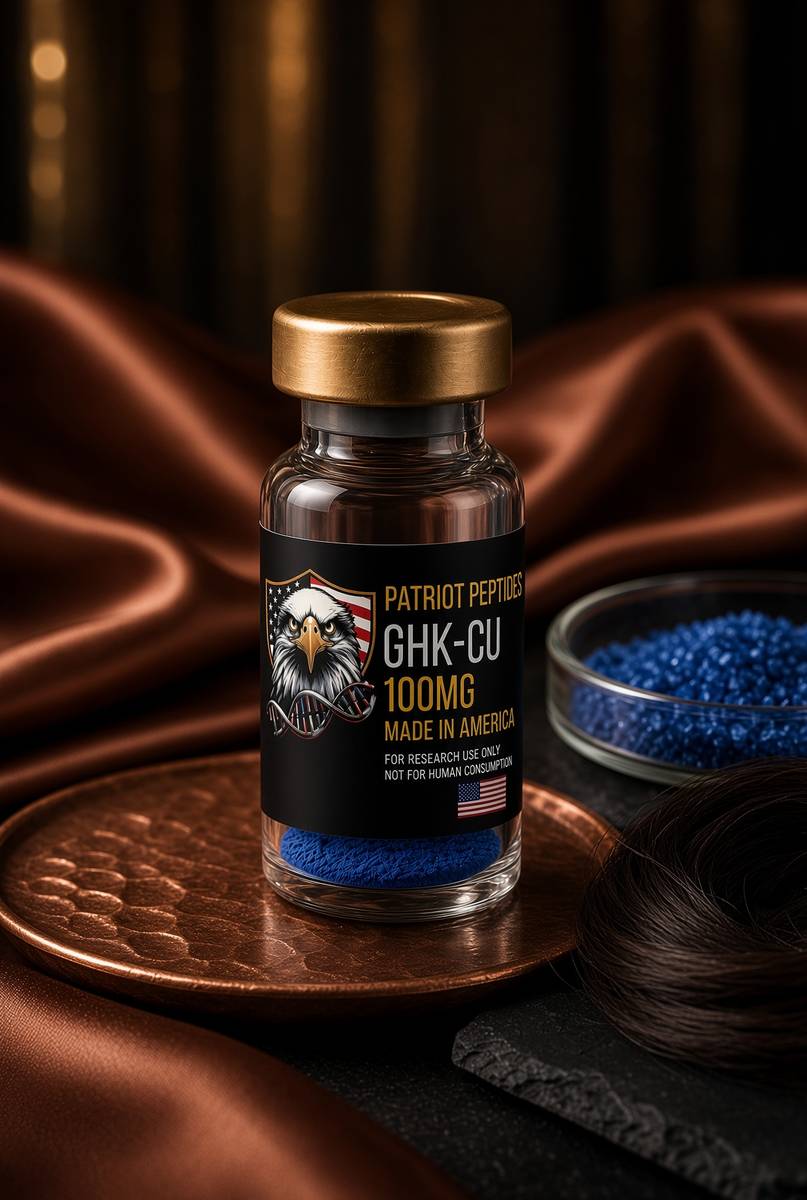
100 mg GHK-Cu. Pickart’s copper tripeptide, lyophilised.
4.9(590)
83 browsing this now · 5 purchased in the last 24 hours
100mg
£35.00
 Made in USAOut of stock
Copper complex
GLOW
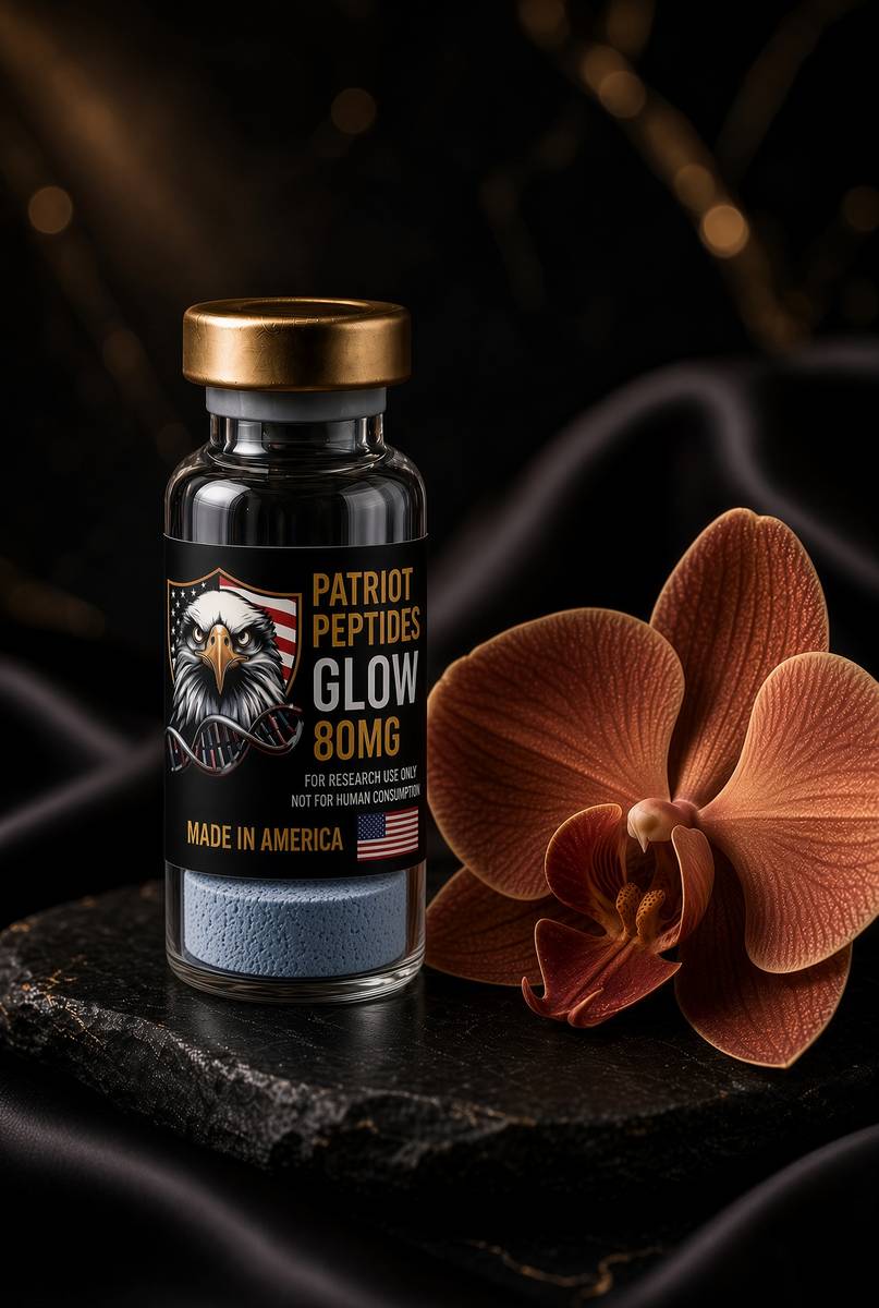
80 mg blend — 50 mg GHK-Cu, 10 mg BPC-157, 10 mg TB-500, 10 mg KPV.
4.8(548)
99 browsing this now · 5 purchased in the last 24 hours
80mg
£65.00
 Research only
Research use only. Not a combined-use instruction.
Read next
45 min · long read · Peptide research
GHK-Cu: a copper tripeptide that rewrites a spreadsheet of genes
Pickart isolated GHK from plasma fractions that made old liver tissue synthesise proteins like young tissue. The tripeptide holds copper. The live claim is a transcriptome, not a moisturiser.
48 min · long read · Peptide research
KPV: three residues, an NF-κB off-switch
The C-terminal tripeptide of α-MSH keeps much of the anti-inflammatory pharmacology and almost none of the tanning. Gut epithelium, PepT1, and a very small molecule.
49 min · long read · Peptide research
Collagen powder is not a research peptide
Type I collagen is a thousand-residue triple helix. Industrial ‘collagen peptides’ are a 2–5 kDa hydrolysate of that helix — thousands of fragments, no single HPLC peak. GHK-Cu is three letters and a metal.
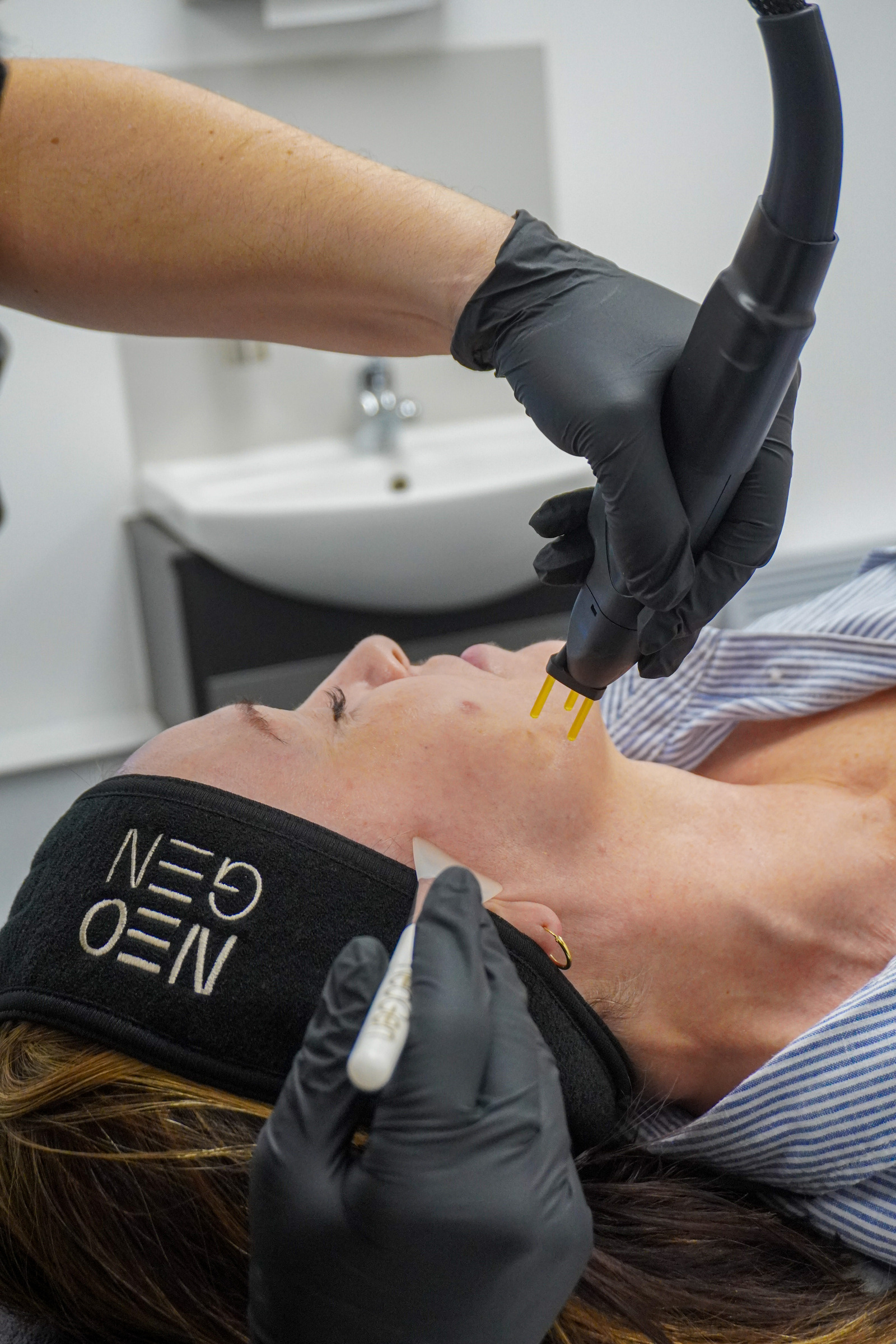
45 min · long read · Clinic
NEOGEN nitrogen plasma: what the energy actually does to skin
NEOGEN plasma at eLIVEate Me in Buckinghamshire. Nitrogen plasma denatures collagen without a laser chromophore. A course of three, then 90 days of rebuild.
More in this desk

53 min · long read · Peptide research
Recovery, GH pulses and the CJC / ipamorelin pair
Somatotrophs have two 'go' receptors. CJC without DAC is a slightly longer GHRH pulse. Ipamorelin is a selective ghrelin-receptor key that does not yank ACTH. Together they are the pair Bowers already showed is more than additive — not a gym protocol.
53 min · long read · Peptide research
How research peptides are made — and why HPLC actually matters
Solid-phase peptide synthesis builds a chain one residue at a time. HPLC then asks whether the main peak is what you think it is. ≥98% is not a slogan. It is a chromatogram.
52 min · long read · Peptide research
Fatigue, cellular energy and the NAD+ / MOTS-c neighbourhood
NAD+ is the rechargeable chip every cell spends on fuel and DNA repair — and the pool shrinks with age. MOTS-c is a mitochondrial 16-mer sent out under metabolic stress. Two answers to 'I have no energy' that are not coffee.
51 min · long read · Peptide research
Melanocortin circuits: pigment, appetite and PT-141
Melanotan II lights MC1, MC3, MC4 and MC5 — pigment plus the rest of the sheet. PT-141 is the free-acid cousin pointed at MC3 and MC4, the circuitry papers use for desire and energy, not skin colour. Same Arizona family. Different question.
Essays describe published research. They are not medical advice and they do not authorise human use of any catalogue item.