

Peptide research · 34 min · 7,389 words
The blood-brain barrier: why a dish is not a brain
Brain capillaries are sealed by claudin-5, wrapped by astrocyte endfeet, and pumped by P-glycoprotein. A plastic dish is not that wall. Semax and Selank have papers; DSIP has a finding story. None of that is a measured ticket from blood into brain interstitial fluid.
What this essay actually tells you
- The wall is specialised endothelium: claudin-5 tight junctions, astrocyte endfeet, pericytes, luminal P-gp. A Transwell is not that unit.
- Most research peptides are polar, charged and protease food. Semax and Selank have papers — neighbourhoods, not a Kp,uu stamp through claudin-5.
- DSIP is WAGGDASGE, isolated from blood leaving a sleeping brain. A finding story is not an influx assay and not a hypnotic.
What this actually means
The blood–brain barrier is specialised capillary endothelium: tight junctions (claudin-5), astrocyte endfeet, pericytes, and efflux pumps such as P-glycoprotein. It is not a rumour and not a customs officer. Cerebrospinal fluid is not plasma and not brain interstitial fluid. Most research peptides are too large, too polar and too short-lived to cross; a dish of endothelial cells is a model, often a leaky one. Semax (MEHFPGP) and Selank (TKPRPGP) have BDNF and GABA-tone literatures. Those literatures are not a measured ticket through claudin-5, and they are not a nootropic protocol. DSIP (WAGGDASGE) was isolated from blood leaving a sleeping brain — a finding story, not an influx assay. We stock the named sequences as research reagents. Occupancy of a receptor does not require tourism. Research use only.
The blood–brain barrier is specialised capillary endothelium, and once you have that picture the rest of the page gets easier. Tight junctions seal the cells together. Astrocyte endfeet wrap the vessel. ATP-binding cassette efflux pumps sit on the blood face and throw many compounds back out. The whole arrangement lives inside a neurovascular unit that a dish of immortalised cells has never quite reproduced. A Transwell insert at ten nanomolar is an assay. A brain is about eighty-six billion neurons, a matching census of glia, roughly a litre of cerebral blood a minute, and a barrier that evolved to keep the weather of plasma out of the neuropil. Most peptides in a research catalogue do not have a published, quantitative ticket across that barrier. Semax and Selank have a literature. DSIP has a finding story. Neither is a passport stamp. We stock characterised sequences for a bench. We do not stock a way into your head. Anatomy first. Then the papers. Then the legal class of the vial.
In short. The barrier is anatomy, not a mood. A dish is an assay. Most peptides do not hold a ticket across. Research sequences are not a passport.
A dish is not a brain
The folk model is a tiny chain that slips through a membrane, finds the interesting bit of a neuron, and changes a mood. It is a children's book, and we can put it down. Capillary endothelial cells in cortex are continuous, not fenestrated. The paracellular cleft is closed by tight junctions so thoroughly that horseradish peroxidase, which leaks freely from muscle capillaries, stops at the brain endothelial cell as if it had hit glass. Reese and Karnovsky showed that in 1967 with electron microscopy and a peroxidase that anybody could buy. Brightman and Reese confirmed the junctions the same decade. Everything written since about peptides for the brain that does not start from those micrographs is writing fiction on top of a photograph. A monolayer on plastic can look a bit like an endothelium. It does not have the basement membrane, the pericyte coverage, the polarised astrocyte endfeet, the shear of blood, or the efflux repertoire of a working neurovascular unit. Treating the two as interchangeable is how an assay becomes a shop page.
In short. Brain capillaries are sealed continuous endothelium. A plastic monolayer is not that wall. Reese and Karnovsky already took the photograph.
Occupancy of a receptor is a different page, already written on this desk: how peptides talk to cells. A ligand can occupy a GPCR on a peripheral cell, on a circumventricular organ that lacks a full barrier, or — in the rare, measured, paper-backed case — on a cell inside the parenchyma after a real crossing. Those three sentences are not synonyms. Forums flatten them into it works on the brain. We will not. Semax is an ACTH(4–10) analogue with a Pro-Gly-Pro tail and a BDNF neighbourhood in nerve-cell papers. Selank is a tuftsin analogue with the same tail and a GABA and enkephalin-tone neighbourhood. DSIP is Trp-Ala-Gly-Gly-Asp-Ala-Ser-Gly-Glu, isolated from blood leaving a sleeping rabbit brain. Isolation from venous outflow is a finding. It is not an influx assay, not a Kp,uu, and not a reason to call a nonapeptide a hypnotic. Research use only, throughout. The kit reconstitutes a cake. It does not authorise a mammal, and it does not punch a hole in claudin-5.
In short. Occupancy, circumvention and crossing are three different sentences. Semax, Selank and DSIP have papers. Papers are not a stamp in a passport.
A blood–brain barrier is not a single structure. It is an endothelial phenotype, a set of junctions, a set of transporters, and a conversation with the cells that wrap the vessel.— After Abbott, Zlokovic, Daneman — the reviews, not a forum.
What Ehrlich actually saw
Paul Ehrlich, late nineteenth century, injected aniline dyes into animals and noticed that most organs stained and the brain did not. That observation is still mis-sold as he discovered the blood–brain barrier. He discovered a staining pattern. Max Lewandowsky used the phrase Bluthirnschranke. Edwin Goldmann, one of Ehrlich's students, did the complementary experiment: dye into cerebrospinal fluid stained brain and spared the rest of the body. Together those two directions — blood to brain refused, CSF to brain permitted — are the first clean map. The barrier is directional, tissue-specific, and not a mystic force-field. It is also not absolute. Circumventricular organs (area postrema, median eminence, organum vasculosum of the lamina terminalis, subfornical organ, pineal, and the messy neighbour that is choroid plexus) have fenestrated capillaries on purpose, because the brain needs to taste plasma somewhere. A peptide that occupies a receptor in area postrema has not crossed the BBB. It has visited a shop with the shutters open. The distinction is the whole subject.
In short. Ehrlich saw a staining pattern, not a force-field. Some brain windows are open on purpose. Visiting those is not the same as crossing the wall.
The modern object is the neurovascular unit, a phrase that has earned its keep. Endothelium is the wall you can photograph. Pericytes sit in the basement membrane at a coverage the rest of the body does not match. Astrocyte endfeet plaster the abluminal surface and polarise aquaporin-4 toward it. Microglia and neurons talk to the vessel on a slower clock. Daneman, Prat, Armulik and colleagues spent the 2000s making pericytes non-optional: coverage correlates with barrier tightness; loss correlates with leak. Zlokovic's reviews put the unit into neurodegeneration without flattening it into a caption. Abbott's 2010 Neurobiology of Disease paper remains the cleanest single map of structure and function we will point a reader at. If a catalogue page says crosses the BBB and does not mention endothelium, tight junctions, efflux, or a number, it has not described a barrier. It has described a hope. Named cells, named pumps, a number. That is a barrier sentence. Hope is not.
In short. The wall is a unit: endothelium, pericytes, endfeet. Named reviews already exist. A shop page that skips them is selling hope.
Endothelium, not a fence cartoon
A peripheral continuous capillary already has an endothelium. What the brain does is a change of job description. Brain microvascular endothelial cells are thin, tightly joined, rich in mitochondria, poor in caveolae relative to muscle, and polarised: luminal membrane facing blood, abluminal facing the basement membrane and the endfeet. The polarisation is not decoration. GLUT1, also called SLC2A1, is how glucose enters; it sits both sides, and GLUT1 deficiency is a real paediatric epilepsy syndrome, which is how you know the transporter is not optional. LAT1, SLC7A5, heterodimerised with 4F2hc, is the large-neutral-amino-acid carrier that L-DOPA uses, which is why a small, LAT1-substrate drug can be a Parkinson's medicine and a random heptapeptide cannot borrow the same sentence. Transferrin receptor 1 mediates iron. Insulin receptor, leptin receptor, and a short list of others have a receptor-mediated transcytosis literature that William Pardridge spent a career trying to turn into Trojan-horse biologics. That literature is a delivery-engineering argument. It is not a general amnesty for research peptides.
In short. Brain endothelium is polarised and transporter-rich. Glucose and L-DOPA have named carriers. A random heptapeptide does not inherit those tickets.
Thickness of the endothelial cell at the capillary is on the order of a few hundred nanometres. The lumen is just wide enough for erythrocytes to deform through. Transit time for a red cell is a second or so. In that second, a solute in plasma either has a transporter, a lipophilic transcellular path, or it does not cross. Paracellular flux, the default in fenestrated gut or kidney capillaries, is the path the tight junction exists to close. Molecular weight, hydrogen-bond count, polar surface area, ionisation at pH 7.4, plasma protein binding, and metabolic half-life are the boring variables. Peptides fail most of them at once: they are large by small-molecule standards, polar, full of hydrogen-bond donors, charged at physiological pH, and lunch for peptidases in plasma and at the endothelial surface. A fifteen-residue gastric fragment and a seven-residue ACTH analogue do not become honorary diazepam because someone wrote nootropic on a forum. Size is not a mood. It is daltons. A second is all most solutes get. That is plenty of time to be refused.
In short. A second of capillary transit is all most solutes get. Peptides are large, polar, charged and protease food. Those four facts close the door.
- Neurons in a human brain
- ~86 billion
- Cerebral blood flow
- ~750 ml/min
- Capillary endothelial thickness
- ~200–300 nm
- Tight-junction cleft
- effectively closed
- P-gp (ABCB1)
- luminal efflux
- CSF volume
- ~150 ml
- Typical research peptide
- 0.8–2 kDa
- In vivo BBB TEER
- ~1,500–2,000 Ω·cm²
Azevedo, Herculano-Houzel. Not a hundred billion. Not a Transwell.
About a seventh of cardiac output at rest. The wall sees the plasma constantly.
A few hundred nanometres of cell, then the junction, then the basement membrane.
Horseradish peroxidase stops here. Reese and Karnovsky, 1967.
ATP-driven. Many xenobiotics go in and come straight back out.
Turned over three or four times a day. Not plasma. Not brain interstitial fluid.
Semax and Selank are heptapeptides. DSIP is a nonapeptide. Polar. Charged. Not diazepam.
Many cell-culture models sit far below that. Leak is not a phenotype. It is a failure of the model.
Tight junctions, named
A tight junction is not glue. It is a belt of transmembrane proteins that stitch adjacent endothelial membranes into a seal, backed by a cytoplasmic plaque that ties the belt to the actin cytoskeleton. Claudins are the seal. Occludin is the regulator that everyone measured first because the antibody existed. Junctional adhesion molecules, JAMs, sit in the neighbourhood. Zonula occludens proteins — ZO-1, ZO-2, ZO-3 — are the plaque. In brain endothelium the star claudin is claudin-5. Nitta, Furuse, Tsukita and colleagues deleted it in the mouse in 2003 and watched a size-selective leak: small tracers got through; larger ones still did not. That is the experiment that turned the barrier into a protein. If your in-vitro model does not express claudin-5 in a continuous junctional pattern, you do not have a barrier, you have a sieve with a press release. VE-cadherin at the adherens junction is necessary but not sufficient. People who write tight junctions and cannot name claudin-5 have named a textbook chapter, not a molecule. Name the protein. Then talk about the wall.
In short. Claudin-5 is the seal protein of brain endothelium. Delete it and small tracers leak. A model without it is a sieve, not a barrier.
The seal is regulated, which is not the same as optional. Inflammatory cytokines, VEGF, some pathogens, and ischaemia can loosen junctions; that is pathology, and it is measured as a drop in transendothelial electrical resistance and a rise in permeability to labelled tracers. Pharmacologists have spent decades trying to open the barrier on purpose, briefly, for drug delivery — hyperosmotic mannitol in a catheterised carotid is the old neurosurgical trick; focused ultrasound with microbubbles is the newer one. Both are procedures with a literature and a risk register. Neither is a property of a lyophilised cake. A research peptide does not modulate the BBB because a forum needs it to. If a paper shows a change in occludin phosphorylation in a particular cell line, that is a cell-line result. It is not a visa, and it is not a protocol for a person. We will not launder a phosphorylation blot into a delivery claim. Pathology and a neurosurgical trick are both real. A freeze-dried sequence is neither of them.
In short. Junctions can loosen in disease or under a neurosurgical trick. That is pathology or a procedure. It is not a property of a research cake.
Astrocyte endfeet and the rest of the unit
Astrocytes do not make the barrier. Endothelium makes the barrier. What astrocytes do is induce and maintain the endothelial phenotype, and what their endfeet do is cover the abluminal surface so thoroughly that older textbooks drew the barrier as a glial wall and were wrong in a useful direction. Polarised aquaporin-4 on the endfoot is how water moves at the gliovascular interface; the same polarisation is disturbed in some epilepsies and after injury. Kir4.1, the inward-rectifier potassium channel, sits there too, buffering K⁺ so that a firing neuropil does not pickle the cleft. The endfoot is a homeostatic machine, not a second endothelium. When people say the astrocytes are the BBB, they are repeating a mid-century error that electron microscopy retired. The correct sentence is longer: endothelium is the seal and the transporter sheet; astrocytes tell it to stay specialised and handle the water and potassium; pericytes, as Armulik showed, are required for tightness at a level the field had under-sold. Three cell types. One unit. A dish that plates only endothelium is already missing two of the speakers.
In short. Endothelium seals. Astrocyte endfeet induce, cover, and handle water and potassium. Pericytes keep it tight. A dish of one cell type is already missing the conversation.
Pericytes in brain capillaries cover a larger fraction of the abluminal surface than pericytes in muscle or skin. They sit inside the basement membrane. They are not smooth muscle; they are a related mural cell with a contractile literature that is still being argued about at the capillary level. What is not argued about is the barrier correlation. Armulik, Genové, Betsholtz: pericyte deficiency increases permeability. Daneman's developmental work put pericytes on the timeline of barrier genesis. A model that never met a pericyte can still be a useful screen. It cannot be cited as the BBB without a wince. The basement membrane itself — collagen IV, laminin, nidogen, heparan sulphate proteoglycans — is a third physical filter and a signalling board. Matrix metalloproteinases that chew it in stroke and in some tumour microenvironments are why a leaky barrier in disease is not a delivery strategy you would copy on purpose. Copying pathology is how you write a cartoon about opening the barrier with peptides. We will not write that cartoon. Tightness is a unit property. Disease leak is not a method.
In short. Brain pericytes are part of tightness, not decoration. The basement membrane is a filter. Disease leak is not a delivery method anyone should copy.
P-glycoprotein and the other pumps
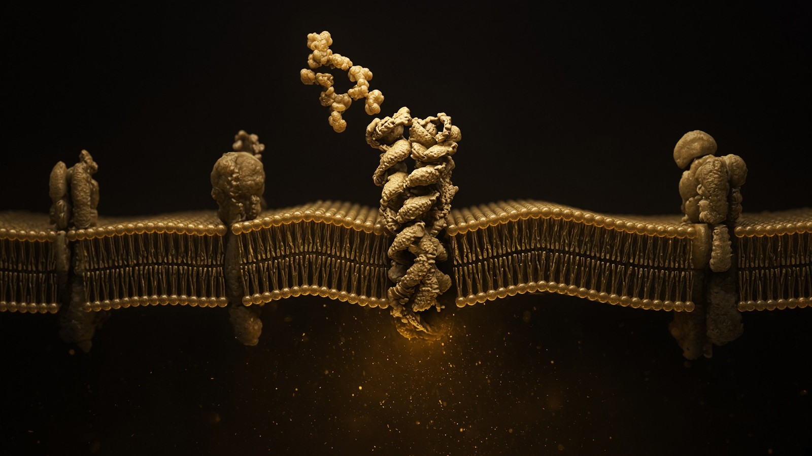
Suppose, generously, that a peptide or a small analogue gets into the endothelial cell. The cell is not a waiting room. P-glycoprotein — ABCB1, MDR1, the same pump oncology spent thirty years hating — sits on the luminal membrane of brain endothelium and throws a startling fraction of xenobiotics back into blood. It spends ATP. It is not a metaphor. Schinkel's mdr1a knockout mice, in the 1990s, showed brains that accumulated ivermectin, loperamide, digoxin and a list of other substrates to levels that would have been a trivia question in the wild-type. Cordon-Cardo and Gottesman had already put the protein at the barrier by immunohistochemistry. If your compound is a P-gp substrate, the endothelial cytoplasm is a revolving door. BCRP, ABCG2, does overlapping work. MRP family members, ABCC1, ABCC4 and others, sit with different substrate preferences and, in some cases, on the abluminal face. Together they are why lipid solubility, the old medicinal-chemist's passport, is not a passport. Lipophilic and a P-gp substrate is how you get a tour of an endothelial cytoplasm and nothing else.
In short. P-gp sits on the blood face of brain endothelium and pumps substrates back out. Getting into the endothelial cell is not the same as reaching a neuron.
Peptides are not classic P-gp substrates in the way cyclosporine and some tyrosine-kinase inhibitors are. That is not a compliment. It usually means they never got far enough into the bilayer or the cell for the pump to become the rate-limiting step. The rate-limiting step was the membrane, the junction, or the peptidase. Cyclosporine is a cyclic peptide and a famous P-gp substrate; that is a special case with a special structure, and it is a warning, not a template. People who say peptides cannot be pumped so they must get through have inverted the logic. The pump is for the things that can enter. The things that cannot enter do not need a pump. They need a transporter, a Trojan-horse receptor, or an honest admission. The catalogue's heptapeptides and nonapeptide have not been handed a Schinkel-style knockout paper that puts them in parenchyma at a measured unbound concentration. Until someone publishes that experiment, the adult sentence is: not shown. Not shown is not shown not to. It is also not a caption.
In short. Most research peptides never get far enough in for P-gp to be the problem. Absence of a pump story is not evidence of crossing. Not shown is the adult caption.
CSF is not plasma, and CSF is not brain
Cerebrospinal fluid is made at the choroid plexus, a folded epithelium sitting on a fenestrated capillary bed. That is the blood–CSF barrier, and it is a different object from the blood–brain barrier. The endothelium at choroid plexus is leaky on purpose; the tight junctions sit in the choroid epithelium, which secretes CSF. Composition is not plasma. Protein is much lower. Potassium is held in a narrower band. Some transmitters and peptides are measurable here because the plexus put them there, or because they arrived from brain interstitial fluid along a long, slow drainage path, or because a lumbar puncture sampled a compartment that is not the synapse you cared about. A CSF concentration is a number. It is not a brain interstitial concentration, and it is not a receptor occupancy at a hypothalamic nucleus. Pharmacologists who still treat lumbar CSF as a surrogate for striatal unbound drug are making a 1970s approximation that microdialysis and Kp,uu,brain were invented to retire. We will not un-retire it for a research peptide.
In short. CSF is made at choroid plexus, a different barrier. A CSF number is not a brain-interstitial number and not occupancy at the receptor you meant.
Plasma is the other confusion, and it is an easy one to make if you only have a blood tube. A peptide concentration in plasma after an administration you are not going to perform, because this is a research vial, would still not be a brain concentration. Plasma protein binding, peptidases — DPP-4 for incretins; a crowd of other proteases for everything else — first-pass if anyone was foolish enough to imagine oral heptapeptides, and the barrier itself sit between the two numbers. Unbound brain concentration over unbound plasma concentration, Kp,uu,brain, is the ratio that means anything for occupancy of a central receptor. Total brain homogenate is a trap: you have mashed endothelium, parenchyma, blood left in capillaries, and bound drug, and then you have called the mash brain. Autoradiography of a labelled peptide that lights up the circumventricular organs and the choroid has described the open windows, not a tour of cortex. Read the figure before you read the abstract. Abstracts are where CNS penetration goes to become a personality.
In short. Plasma is not brain. Homogenate is not interstitial fluid. A labelled peptide lighting up open windows has not toured cortex. Read the figure.
- Blood–brain barrier: continuous capillary endothelium, claudin-5, P-gp, GLUT1, LAT1. The wall of the neuropil.
- Blood–CSF barrier: fenestrated choroid capillaries plus tight choroid epithelium. Makes CSF. Different junctions, different job.
- Circumventricular organs: fenestrated on purpose so the brain can taste plasma. Area postrema, median eminence, OVLT, SFO, pineal.
- Brain interstitial fluid: the actual extracellular fluid of parenchyma. Not CSF. Not plasma. Microdialysis if you are serious.
- Lumbar CSF: a convenient sample. A long way from a hypothalamic synapse, in space and in time.
Why most peptides never get a ticket
Medicinal chemistry learned, slowly and at great expense, which small molecules get into brain: modest size, few hydrogen-bond donors, a logP in a boring middle band, not a strong P-gp substrate, not too ionised, not glued to albumin so tightly that the unbound fraction is a rounding error. Lipinski's rule of five was never a brain rule, but the brain is stricter than the gut. Peptides violate the spirit of those rules as a class. A typical 7–15 residue chain is 800–1,800 daltons, with a backbone that is a hydrogen-bond factory, side chains that are charged at pH 7.4 — histidine, glutamate, lysine, arginine; look at Semax, look at Selank, look at DSIP — and a half-life in plasma measured in minutes unless someone has already done the analogue chemistry that incretin medicines did with fatty-acid acylation and DPP-4 resistance. That analogue chemistry is a half-life trick for a peripheral hormone receptor. It is not a BBB ticket. Semaglutide's central appetite effects are still argued as vagal, area-postrema, and some receptor-mediated stories, not as a general proof that peptides wander into cortex.
In short. Brain-penetrant small molecules are a narrow club. Peptides fail size, polarity, charge and half-life at once. An incretin headline is not a ticket for a heptapeptide.
There are exceptions, and they are named, which is how you know they are exceptions. Receptor-mediated transcytosis of transferrin-receptor antibodies is a delivery field with companies attached. Insulin has a receptor at the barrier and a transport literature; that does not make every peptide insulin. Leptin has a transport literature that saturates and breaks in obesity, which is a plot, not a product. Cell-penetrating peptides — TAT, penetratin, polyarginine — are a laboratory trick for cargo in a dish and a toxicology problem in a mammal; they are not in the catalogue. Cyclic peptides with the right lipophilicity and intramolecular hydrogen bonds, cyclosporine the eternal example, can behave more like small molecules and then often meet P-gp. Intranasal delivery is a separate argument: olfactory and trigeminal routes, a small surface, a large claim-to-data ratio, and a literature that is honest when it reports both the labelled peptide in olfactory bulb and the absence of the same signal in deep parenchyma. Intranasal is not a synonym for in the hippocampus. Selling that synonym is selling a route as a result.
In short. Named exceptions exist: transferrin Trojan horses, insulin, some cyclics, a cautious nasal literature. Exceptions are named because they are not the class.
- Paracellular: closed by claudin-5 and company. Default peptide path is this path, and this path is shut.
- Passive transcellular: needs lipophilicity the average peptide does not have. Backbone hydrogen bonds like water more than they like bilayer.
- Carrier-mediated: GLUT1, LAT1 and friends. Your sequence has to look like their substrate. ACTH fragments and tuftsin analogues do not.
- Receptor-mediated transcytosis: transferrin receptor, insulin receptor, a short list. Engineering problem, not a default property of a research cake.
- Adsorptive transcytosis: cationic proteins sticking to the luminal surface. A messy, saturable, often toxic neighbourhood. Not a catalogue strategy.
- Efflux: P-gp, BCRP, MRPs. For the few that get in, a revolving door back to blood.
- Metabolism: peptidases in plasma, at the endothelium, in the parenchyma. A sequence that never arrives has still been eaten.
Models, TEER, and the honesty of a leak
In-vitro blood–brain barrier models are useful the way all models are useful: they answer the question they were built to answer, and they lie when you ask a different one. A Transwell with Caco-2 is a gut screen someone forgot to relabel. A Transwell with immortalised brain endothelial cells — hCMEC/D3 is the one everybody has used and then complained about — can give you a number for apparent permeability. Primary bovine or porcine brain endothelial cells, often co-cultured with astrocytes, get closer. iPSC-derived brain microvascular endothelial-like cells are the current hope, with TEER values that sometimes approach in-vivo ranges and a differentiation protocol that is a paper in itself. TEER, transendothelial electrical resistance, is the quick electrical proxy for tightness. In vivo, the barrier sits somewhere around 1,500–2,000 Ω·cm² or higher depending on who measured and how. Many published BBB models sit at a few hundred, which is a leaky epithelium doing its best. Apparent permeability, Papp, of a paracellular marker such as Lucifer yellow or sucrose tells you whether the junctions are actually closed. If sucrose crosses, your peptide crossing is a hole, not a mechanism.
In short. A Transwell is a model. TEER and sucrose permeability tell you if it is tight. A leaky monolayer that a peptide crosses has not demonstrated a barrier ticket.
Even a good model is still a dish. It does not have the shear of blood unless you added a pump. It does not have the pericyte coverage of cortex unless you plated them and proved it. It does not have the unbound-fraction politics of plasma. It does not have a neuron on the other side that you can honestly call a circuit. Co-culture with astrocytes improves junctions in many protocols; it still is not a brain. Organ-chips with microfluidic channels are better showmanship and sometimes better biology. They remain chips. The translational gap from a Papp in a well to a Kp,uu,brain in a rodent is the gap this page exists to keep visible. A research catalogue that sells peptides is allowed to read those papers. It is not allowed to caption a vial with a Transwell result as if the well were a mammal. HPLC tells you which chain you have. TEER tells you whether a monolayer is tight. Neither tells you that a heptapeptide sat at a cortical synapse. Collapsing those three numbers into one caption leaves the anatomy behind.
In short. Even a tight well is still a well. Shear, pericytes, plasma binding and a circuit are missing. A vial caption is not allowed to forget that.
Semax and Selank: literature is not a stamp
Semax is Met-Glu-His-Phe-Pro-Gly-Pro. That is ACTH(4–10) rewritten with a Pro-Gly-Pro tail so that peptidases do not eat it quite as fast. The sequence is MEHFPGP. The Russian literature — Ashmarin, Myasoedov, and a school that treated ACTH fragments as more than pigment and cortisol — sits on nerve-cell transcription: BDNF, TrkB, copper-zinc superoxide dismutase, a set of antioxidant enzymes. There are rodent papers. There are cell papers. There is a registered medicine in the Russian Federation that we do not sell, does not import, and will not hide behind. What there is not, on a till in the United Kingdom labelled research use only, is a passport stamp. A BDNF blot in a cultured neuron treated with Semax is occupancy or a transcriptional neighbourhood in a dish. It is not evidence that a lyophilised cake, reconstituted in bacteriostatic water, crossed claudin-5 in a human and sat down at TrkB in prefrontal cortex. Those are different experiments. Only one of them has been done in the papers we can point at without wincing. The other is a forum.
In short. Semax is MEHFPGP, an ACTH fragment analogue with a BDNF neighbourhood in papers. A blot in a dish is not a human crossing of claudin-5.
Selank is Thr-Lys-Pro-Arg-Pro-Gly-Pro. Tuftsin, TKPR, plus the same Pro-Gly-Pro tail. Tuftsin was an immunomodulatory tetrapeptide from the IgG heavy chain before it was a nootropic rumour. The Selank papers sit on GABAergic tone and on how long enkephalins last — peptidase politics, not a benzodiazepine binding site. Seredenin's school is the name to look up if you actually want the primary literature rather than a Reddit summary. Again: cell papers, rodent papers, a Russian pharmacy fact that is not a UK medicine. Again: not a licensed anxiolytic, not a protocol, not a demonstration that the heptapeptide is sitting in amygdala after a peripheral administration you are not licensed to perform. Sharing a tail with Semax is chemistry. Sharing a vial in the catalogue is a cap-count decision for a bench that wants both named sequences on one certificate. Sharing a receptor is false. They do not. ACTH-fragment neighbourhood is not tuftsin neighbourhood. Collapsing them into the nootropic peptide is how a search bar writes pharmacology.
In short. Selank is TKPRPGP, a tuftsin analogue with GABA and enkephalin-tone papers. Same tail as Semax, different lock. Not an anxiolytic and not a shared receptor.
Nootropic is a marketing word. Aniracetam was a marketing word with a slightly older passport. BDNF is a neurotrophin with a receptor, TrkB, and a physiology. GABA is a transmitter with ionotropic and metabotropic receptors and a well-described inhibitory job. We will use the second two and leave the first on the forum where it belongs. A research vial of the pair is two characterised heptapeptides, HPLC on the certificate, made as a blend because the paper trails are adjacent in search queries and a bench should not have to guess which white cake is which. Adjacent in search is not adjacent in a synapse. The occupancy page on this desk already told you that a peptide is a ligand, not a mood that enters a cell and does repair. This barrier page is the sequel for anyone who then says ah, but the brain. Most ligands never see the brain. Some see a circumventricular organ. A few have a measured Kp,uu. Semax and Selank, as stocked here, are research sequences with named literatures. The literature is not a stamp. We will sell you the sequences. We will not design your maze, your blot, or your story about focus.
In short. Nootropic is marketing. BDNF and GABA are biology. This vial is two named heptapeptides for a bench. The papers are not a stamp and not a protocol.
Diagram
Outside
Peptide ligand
Named sequence in the nM–µM pocket. Shape complementarity, not vibes. A 15-mer and a 4-mer do not fit the same hole.
Membrane
7-TM receptor
Helices rearrange. The cytoplasmic face becomes a GEF for a heterotrimeric G protein (Gs, Gi, Gq, G12/13).
Inside
Second messengers
cAMP, IP₃, Ca²⁺, β-arrestin. One occupied receptor can spawn thousands of messenger molecules. That is amplification.
~800 GPCRs in the human genome. Seven transmembrane helices, an extracellular ligand pocket, an intracellular G-protein handshake. Catalogue neighbours: ipamorelin at GHSR, PT-141/MT2 at melanocortin receptors, retatrutide at GLP-1R/GIPR/GCGR.
DSIP: a finding story, not an influx assay
Delta sleep-inducing peptide is Trp-Ala-Gly-Gly-Asp-Ala-Ser-Gly-Glu. WAGGDASGE. Nine residues, 849 daltons, a lot of glycine, an aspartate and a glutamate, a tryptophan at the N-terminus. Schoenenberger and Monnier isolated it from the cerebral venous blood of rabbits during thalamic stimulation that produced delta-wave sleep, and they published in the 1970s. The name describes the finding. That is the whole original claim, and it is a better claim than most peptides get: they found a sequence in a specific biofluid in a specific state. What the name does not describe is a mechanism, a receptor that has been cloned and agreed, a Kp,uu,brain, or a licensed hypnotic. Later animal work sits on sleep architecture and on HPA-axis markers, mixed in the way that field is mixed. Van Cauter's sleep-restriction work still explains more next-day glucose noise than any nonapeptide caption. We said so on the sleep page. We will say so here. Isolation from blood leaving a brain is not proof that the peptide had a job inside that brain, and it is not proof that a synthetic nonapeptide injected somewhere else will take the same path in reverse.
In short. DSIP was found in blood leaving a sleeping brain. A finding story is not an influx assay, not a cloned receptor, and not a hypnotic.
Venous effluent is downstream. Things found there may have been made in brain and dumped, made in the periphery and never entered, made in the periphery and entered and left, or ridden along as a fragment of something larger. Peptide isolation in the 1970s was heroic chemistry with the resolution of its decade. Sequences get revised. Contaminants happen. The DSIP literature has had arguments about whether the nonapeptide is a physiological sleep factor or a named curiosity that assays sometimes move. That argument is allowed. What is not allowed is to reverse the arrow: found in brain venous blood during sleep, therefore this vial crosses the barrier and induces sleep. Found-in is not went-into. Effluent is not influent. A sleep-architecture paper in a rodent, if you have one with proper scoring and a vehicle control, is a sleep-architecture paper in a rodent. It is not a human hypnotic, not a replacement for CBT-I, and not a reason to skip the apnoea question. This listing is WAGGDASGE, freeze-dried, HPLC-characterised, research use only. The dark bedroom remains the more powerful reagent, and it is not for sale.
In short. Venous effluent is downstream. Found-in is not went-into. A named nonapeptide is a research sequence. A dark bedroom is still the stronger reagent.
Occupancy does not require tourism
The occupancy page said this once, and the barrier page has to say it again because the brain is where people forget. Most peptide ligands occupy a protein on the cell surface. The information that crosses the plasma membrane is conformation: a GPCR's helices rearrange, a G protein spends GTP, a second messenger rises. The ligand, in the boring and correct case, is still outside, or is on its way to a lysosome after the receptor was internalised. A neuron is a cell. It has a surface. If a peptide could reach that surface, occupancy would be occupancy. The blood–brain barrier is the reason most of them cannot reach that surface from plasma. That is a pharmacokinetic wall, not a philosophical one. Peripheral occupancy remains available: pituitary, enteric nervous system, nodose ganglion, immune cells, endothelium itself, circumventricular organs. A GH secretagogue literature is a pituitary literature. An incretin literature is a gut–pancreas–brainstem literature, and the brainstem bit is often area postrema and nucleus tractus solitarius, which is not the brain in the way a forum means the brain. Naming the cell is the whole game.
In short. A peptide usually occupies a surface lock. The barrier stops most of them reaching neuronal surfaces from plasma. Peripheral and circumventricular occupancy still count as occupancy.
Diagram
| Node | Catalogue | Conversation |
|---|---|---|
| GPCR | Ipamorelin, MT2, PT-141, retatrutide, CJC | Second messengers, secretion, appetite, pigment |
| RTK / IGF1R | IGF-1 LR3 | IRS–PI3K–Akt–mTOR and Shc–ERK |
| Cytokine receptor | Somatropin (HGH) | GHR–JAK2–STAT5b, hepatic IGF-1 |
| Cofactor | NAD+ | Sirtuins, PARPs, CD38, redox |
| Actin buffer | TB-500 / Tβ4 motif | G-actin sequestration, motility |
| Growth-factor-like | BPC-157 | VEGFR2 / FAK / eNOS neighbourhood |
| Copper ligand | GHK-Cu | Transcriptome shift in fibroblasts |
| MC fragment | KPV | NF-κB, PepT1, no pigment |
| Nuclear / pineal | Epithalon (AEDG) | TERT and melatonin literatures |
| mtORF peptide | MOTS-c | AMPK, folate–methionine cycle |
Each row is a different kind of molecular conversation. The catalogue peptides bind at these nodes; they are not interchangeable, and stacking them because a forum did mixes unrelated literatures.
Area postrema is the example that earns its own paragraph because it is where weight-loss medicines and nausea live, and where people then say see, peptides act on the brain. Area postrema is a circumventricular organ. Its capillaries are fenestrated. It tastes plasma so that the animal can vomit in time. GLP-1 receptors there are a real pharmacology. They are not a proof that the same peptide sat in insular cortex. Median eminence is where hypothalamic releasing hormones enter pituitary portal blood; fenestrated on purpose. Pineal is outside the barrier in the way that matters for melatonin dumping into CSF and blood. If your assay is a circumventricular receptor, you may not need a barrier ticket. If your caption then says crosses the BBB, you have upgraded a fenestrated capillary into a continuous one to make the sentence prettier. We will not pretty it. Semax's nerve-cell papers are mostly dishes and tissue, not a mapped circumventricular story. Selank's GABA-tone papers are not an area-postrema story. DSIP's isolation from cerebral venous blood is not a circumventricular story either. Three different objects. Three different missing tickets.
In short. Circumventricular organs taste plasma on purpose. That is not the BBB. Upgrading a fenestrated window into a crossing claim is how a caption cheats.
What the catalogue is actually for
Patriot Peptides is a research-reagent catalogue. The Semax and Selank listing is two named heptapeptides, twenty milligrams, freeze-dried together, HPLC-characterised, made in the United States, labelled for laboratory research only. The DSIP listing is ten milligrams of WAGGDASGE, the same legal class, the same honesty about what a sequence is. Neither vial is a nootropic protocol. Neither is an anxiolytic. Neither is a sleeping tablet. Neither is a claim that claudin-5 sat down and opened because a search query contained the word focus. A bench that wants to treat a neuronal culture, a receptor assay, a peptidase assay, or a carefully designed animal study under the licence that animal study requires, can buy a characterised chain instead of a mystery powder. That is the job. The journal exists so that the same person can read what the barrier actually is before they write a sentence they will have to retract. We would rather lose the caption than lose the anatomy. Sequences for a bench. A wall that stays a wall.
In short. These vials are characterised research sequences. Not a nootropic, not an anxiolytic, not a hypnotic. The journal is the anatomy, not a use instruction.
Reconstitution is mass divided by volume, bacteriostatic water in the kit, a laboratory solvent, a label that is not being coy. HPLC-MS is how we know which chain we shipped. Occupancy, if you have a receptor and an assay, is a concentration, a Kd, a cell type. Crossing, if you have a barrier and an assay, is a Papp against a paracellular marker, or a Kp,uu in an animal, or a microdialysis number, or an imaging study that survived its controls. Transcriptional neighbourhoods — BDNF, GABA-tone, sleep architecture — are papers. Papers are not stamps. The occupancy page, the sleep page, and the Semax/Selank page on this desk are the adjacent reading. This one is the wall. A dish of endothelial cells can be a good wall or a bad wall. A brain is not a dish. A person is not a brain in the way a caption wants. Thirty-six trillion cells, a barrier that keeps plasma out of neuropil, and a research cake that occupies one lock in one assay if you set the assay up. That is the whole offering. It is enough. It is not a passport.
In short. Measure crossing with crossing assays, occupancy with occupancy assays, sequences with HPLC. A person is not a dish. A cake is not a passport.
We characterise ligands. We do not punch holes in claudin-5, and we do not caption a finding story as an influx.
If you came here from a search for nootropic, brain fog, anxiety, or sleep, the map is this: two Russian heptapeptides with BDNF and GABA-tone literatures, and a nonapeptide isolated during delta sleep, sitting on a till that will not pretend the blood–brain barrier is a rumour. Licensed medicines for anxiety, attention and insomnia live in a clinic and a formulary. Dark rooms and a stable clock still do more work than a nonapeptide. A named receptor and a named cell type still do more work than a forum. Read Abbott. Read Reese and Karnovsky. Read Schoenenberger and Monnier as a finding, not as a visa. Read Ashmarin and Seredenin as neighbourhoods, not as stamps. Then, if the experiment you actually have is a dish, a blot, a binding curve, design that experiment. We will sell you the sequence. We will not sell you a brain. Research use only. The barrier was not consulted, and it would have said no. That last sentence is anatomy, not a joke.
In short. Searches for focus, anxiety or sleep land on named sequences and named papers. They do not land on a barrier ticket, a medicine, or a protocol.
The numbers, once more, because captions forget them
A human brain is about 1.4 kilograms, 86 billion neurons, as many non-neuronal cells, a cerebral blood flow of roughly 750 millilitres a minute, a CSF volume of 150 millilitres turned over three or four times a day, and an endothelial surface area that Pardridge used to quote around 12–20 square metres depending on the textbook. Capillary transit is about a second. Tight junctions close the paracellular path. P-gp and BCRP sit on the luminal membrane. GLUT1 and LAT1 are the honest tickets for glucose and large neutral amino acids. Claudin-5 is the named seal. Astrocyte endfeet cover the abluminal face with AQP4 polarised toward it. Pericytes keep the unit tight. CSF is not plasma. CSF is not interstitial fluid. A Transwell at a few hundred ohm-square-centimetres is not 2,000. A heptapeptide is about 800 daltons of polar backbone. A nonapeptide is similar. Plasma peptidases do not care about your search query. Occupancy of a GPCR is a surface story. Crossing is a barrier story. Isolation from venous blood is a finding story. Three stories. One till.
In short. Brain, flow, CSF, junctions, pumps, named transporters, polar peptides. Three stories — occupancy, crossing, finding — and none of them is a protocol.
We will end where the title started. A dish is a controlled lie that sometimes tells the truth about a molecule. A brain is an organ with a barrier that evolved to keep the dish's simplifications out. Mixing the two is the original sin of translational captions. Semax does not become a nootropic because a neuron in a well transcribed BDNF. Selank does not become an anxiolytic because a GABA-tone paper exists in a journal a forum did not read. DSIP does not become a night's sleep because a Swiss physiologist found a nonapeptide in rabbit venous blood in 1977. The sequences are real. The papers are real. The barrier is real. The legal class of the vial is research use only. Hold those four facts at once and you have this page. Drop one of them and you have a shop we would not buy from. Patriot Peptides would rather be dry, precise, and slightly rude about anatomy than fluent in a language that pretends claudin-5 is a rumour. The blood–brain barrier is why a dish is not a brain. Reese and Karnovsky already took the photograph. We are only reading it out loud.
In short. Sequences, papers, barrier and legal class: hold all four. A dish that transcribed BDNF has not opened claudin-5. We will not pretend otherwise.
Questions the essay actually answers
- Do research peptides cross the blood–brain barrier?
- As a class, no, not in any way you could caption a vial with. Brain capillary endothelium is sealed by claudin-5, polarised, and pumped by P-gp and BCRP. Typical 7–15 residue peptides are polar, charged and protease food. Named exceptions (LAT1 substrates, some Trojan-horse constructs, a few cyclics) are named because they are not the class. Semax, Selank and DSIP do not arrive with a published Kp,uu,brain that we will print as a stamp.
- What is P-glycoprotein doing at the barrier?
- P-gp (ABCB1/MDR1) sits on the luminal membrane of brain endothelium and spends ATP to throw many xenobiotics back into blood. Schinkel's mdr1a knockout mice showed the brain accumulating ivermectin, loperamide and others. Getting into the endothelial cell is not reaching a neuron. Many peptides never get far enough in for P-gp to be the rate-limiting step; the membrane and the peptidase already said no.
- Is Semax a nootropic that enters the brain?
- No. Semax is MEHFPGP, an ACTH(4–10) analogue with a Pro-Gly-Pro tail. The papers sit on BDNF, TrkB and antioxidant-enzyme transcription in nerve cells. That is a literature. It is not a nootropic product, not a UK medicine, and not a measured passport through claudin-5. The catalogue stocks it with Selank as a characterised research pair. Research use only.
- Does DSIP's name mean it crossed into a sleeping brain?
- No. Schoenenberger and Monnier isolated WAGGDASGE from cerebral venous blood during delta-wave sleep. Venous effluent is downstream. Found-in is not went-into. The name describes a finding, not a permeability coefficient, not a cloned receptor, and not a hypnotic. This listing is that nonapeptide, freeze-dried, for a bench.
- Why isn't a CSF concentration a brain concentration?
- CSF is made at choroid plexus — the blood–CSF barrier, a different wall. Protein is lower than plasma; the composition is not interstitial fluid of parenchyma. Lumbar CSF is a convenient sample a long way from a hypothalamic synapse. Unbound brain concentration over unbound plasma (Kp,uu,brain), or microdialysis, is what occupancy of a central receptor actually wants. Homogenate is a mash. Abstracts blur this. Figures should not.
- Are tight junctions ever open?
- They can loosen in inflammation, ischaemia and some tumours; that is pathology. Hyperosmotic mannitol and focused ultrasound are procedures with risk registers. Claudin-5 knockout made a size-selective leak in a mouse (Nitta 2003). None of that is a property of a lyophilised research peptide. A forum that needs the barrier open has not described a mechanism. It has described a wish.
- Is a Transwell insert a blood–brain barrier?
- No. It is a model. TEER and sucrose (or Lucifer yellow) permeability tell you whether the monolayer is tight. Many immortalised lines sit far below in-vivo resistance. If a paracellular marker crosses, your peptide 'crossing' is a hole. Even a good co-culture is still a dish: incomplete shear, incomplete pericytes, no circuit. Useful. Not a brain.
- Why stock Semax, Selank and DSIP at all, then?
- Because they are named sequences with named literatures a bench actually reads — BDNF/TrkB and GABA-tone for the heptapeptide pair, a 1977 isolation and later architecture papers for the nonapeptide. A characterised cake beats a mystery powder. Stocking a ligand is not captioning a visa, not writing a nootropic protocol, and not selling a hypnotic. HPLC on the certificate. Claudin-5 still closed. Research use only.
Hypothetical research reconstitution
How these vials are typically mixed
Hypothetical research reconstitution for the named catalogue vial. Not a protocol, not medical advice, not a use instruction. These amounts sit in published and commonly cited laboratory ranges. The vial is labelled for research use only — not for human or veterinary administration.
Semax / Selank
20mg
Mix with 2 ml bacteriostatic water → 10 mg/ml of the pair
- Hypothetical aliquot
- 200–600 mcg
- 0.02–0.06 ml · 2–6 units on a U-100 syringe
- How often
- Once or twice daily
- 10–14 days on, then a pause, in the original Russian notes
Bench steps
- Let the vial sit until it is no longer cold to the touch.
- Wipe the stopper with 70% isopropyl alcohol. Let it dry.
- Draw 2 ml bacteriostatic water (0.9% benzyl alcohol).
- Run the water slowly down the inside glass — do not blast the cake.
- Roll between finger and thumb until the cake is gone. Do not shake.
- Label the date. Store the solution at 2–8 °C. Do not freeze. Use within 30 days unless the note below says otherwise.
The literature on these two is often intranasal, not a drawn subcutaneous mark. Same water, same fridge. Short courses, not a forever molecule.
DSIP
10mg
Mix with 2 ml bacteriostatic water → 5 mg/ml · 5,000 mcg/ml
- Hypothetical aliquot
- 100–300 mcg
- 0.02–0.06 ml · 2–6 units on a U-100 syringe
- How often
- Once daily, evening
- 7–14 nights, then a pause
Bench steps
- Let the vial sit until it is no longer cold to the touch.
- Wipe the stopper with 70% isopropyl alcohol. Let it dry.
- Draw 2 ml bacteriostatic water (0.9% benzyl alcohol).
- Run the water slowly down the inside glass — do not blast the cake.
- Roll between finger and thumb until the cake is gone. Do not shake.
- Label the date. Store the solution at 2–8 °C. Do not freeze. Use within 30 days unless the note below says otherwise.
Delta-sleep peptide. Night-time aliquot in the papers that bother with a clock. Fridge. Short runs.
Bacteriostatic water and sterile syringes ship with peptide orders over £75. Kit details · 10 ml bacteriostatic water
The vials this essay sits on
Named sequences the essay maps — Semax + Selank, DSIP. Hypothetical research neighbourhood, not a protocol, not a medicine. One press puts every in-stock vial in the bag.
 Made in USAOut of stock
Neuropeptide
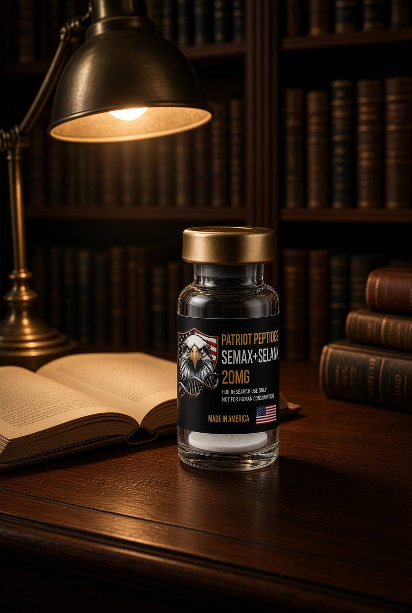
Semax + Selank
20 mg Semax + Selank — two Russian heptapeptides, one lyophilised vial.
4.7(548)
21 browsing this now · 2 purchased in the last 24 hours
20mg
£40.00
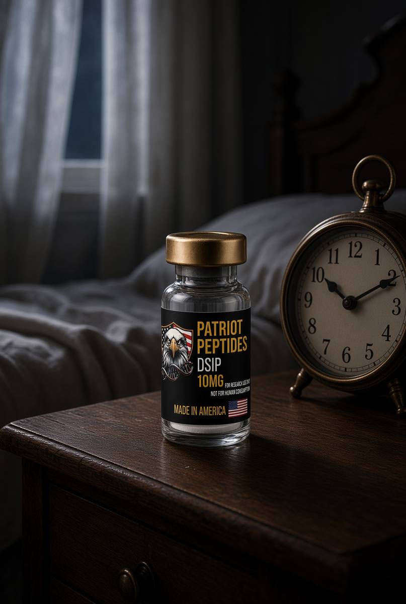 Made in USAOut of stock
Neuropeptide
DSIP
10 mg DSIP — the nonapeptide isolated during slow-wave sleep.
4.7(521)
42 browsing this now · 1 purchased in the last 24 hours
10mg
£25.00
Research use only. Not a combined-use instruction.
Read next
70 min · long read · The living cell
How peptides talk to cells: occupancy, amplification, arrestin
A peptide is a ligand. Most of the catalogue binds a GPCR on the cell surface: one occupancy, then enzymes make thousands of second messengers. That amplification is real, and it is not magic. Desensitisation is why more ligand is not more signal forever.
43 min · long read · Peptide research
Focus, stress tone and the Semax / Selank pair
Semax is an ACTH fragment analogue studied for BDNF. Selank is a tuftsin analogue studied for GABA and enkephalin tone. They share a tail and a country, not a receptor. One vial, two paper trails — not a nootropic, not an anxiolytic.
More in this desk

53 min · long read · Peptide research
Recovery, GH pulses and the CJC / ipamorelin pair
Somatotrophs have two 'go' receptors. CJC without DAC is a slightly longer GHRH pulse. Ipamorelin is a selective ghrelin-receptor key that does not yank ACTH. Together they are the pair Bowers already showed is more than additive — not a gym protocol.
53 min · long read · Peptide research
How research peptides are made — and why HPLC actually matters
Solid-phase peptide synthesis builds a chain one residue at a time. HPLC then asks whether the main peak is what you think it is. ≥98% is not a slogan. It is a chromatogram.
52 min · long read · Peptide research
Fatigue, cellular energy and the NAD+ / MOTS-c neighbourhood
NAD+ is the rechargeable chip every cell spends on fuel and DNA repair — and the pool shrinks with age. MOTS-c is a mitochondrial 16-mer sent out under metabolic stress. Two answers to 'I have no energy' that are not coffee.
51 min · long read · Peptide research
Melanocortin circuits: pigment, appetite and PT-141
Melanotan II lights MC1, MC3, MC4 and MC5 — pigment plus the rest of the sheet. PT-141 is the free-acid cousin pointed at MC3 and MC4, the circuitry papers use for desire and energy, not skin colour. Same Arizona family. Different question.
Essays describe published research. They are not medical advice and they do not authorise human use of any catalogue item.