

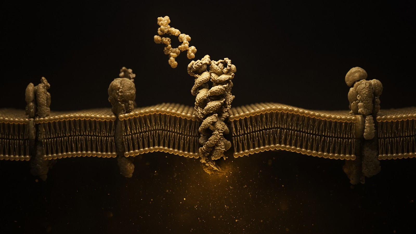
The living cell · 70 min · 15,291 words
How peptides talk to cells: occupancy, amplification, arrestin
A peptide is a ligand. Most of the catalogue binds a GPCR on the cell surface: one occupancy, then enzymes make thousands of second messengers. That amplification is real, and it is not magic. Desensitisation is why more ligand is not more signal forever.
What this essay actually tells you
- A peptide is a ligand. ~800 human GPCRs; one occupancy can spawn thousands of cAMP or IP₃ molecules. That amplification is enzymatic, not mysterious.
- Catalogue map: ipamorelin at GHSR (Raun 1998), CJC at GHRHR, MT2/PT-141 at melanocortins, retatrutide at GLP-1R/GIPR/GCGR, IGF-1 LR3 at IGF1R, somatropin at GHR–JAK2–STAT5b, KPV via PepT1.
- Desensitisation (GRK, β-arrestin, endocytosis) is why more ligand is not more signal forever. A 10 nM dish is not a person. The vial is labelled for laboratory use.
What this actually means
Most of the catalogue is a key for a lock on the cell surface. The lock is usually a GPCR: seven helices in the membrane, a pocket outside, a G protein inside. Occupancy rearranges the helices. The G protein spends GTP. Adenylyl cyclase or phospholipase C runs. cAMP or Ca²⁺ rises by orders of magnitude because one receptor can load many G proteins and each enzyme can make many messengers. Then kinases. Then a secretory granule, a transcriptional programme, a change in excitability. IGF-1 LR3 is a different lock (a receptor tyrosine kinase). Somatropin is a cytokine-receptor lock (JAK–STAT). KPV can cheat via PepT1 as well as melanocortin neighbourhoods. NAD+ is not a receptor ligand; it is a cofactor. GHK-Cu is a copper ligand with a fibroblast transcriptome, not a seven-helix story. TB-500’s LKKTETQ binds actin, which is a cytoskeleton, not a receptor. If you cannot name the lock, you do not yet have a mechanism. A dish at ten nanomolar is not a person: diffusion, proteases, DPP-4, plasma binding and first-pass sit between the two. The fatty-acid handle on an incretin analogue is a half-life trick for a medicine; this research vial is the sequence, not the pen. HPLC is how we know which chain we are talking about. Research use only.
Diagram
Outside
Peptide ligand
Named sequence in the nM–µM pocket. Shape complementarity, not vibes. A 15-mer and a 4-mer do not fit the same hole.
Membrane
7-TM receptor
Helices rearrange. The cytoplasmic face becomes a GEF for a heterotrimeric G protein (Gs, Gi, Gq, G12/13).
Inside
Second messengers
cAMP, IP₃, Ca²⁺, β-arrestin. One occupied receptor can spawn thousands of messenger molecules. That is amplification.
~800 GPCRs in the human genome. Seven transmembrane helices, an extracellular ligand pocket, an intracellular G-protein handshake. Catalogue neighbours: ipamorelin at GHSR, PT-141/MT2 at melanocortin receptors, retatrutide at GLP-1R/GIPR/GCGR.
Lefkowitz and Kobilka got the 2012 chemistry Nobel for this family, and they deserved it. GPCRs — G protein-coupled receptors — are how photons, odours, neurotransmitters, chemokines, glycoprotein hormones and a large fraction of peptides tell a cell that something happened outside. About a third of approved small-molecule drugs hit them. Peptide ligands hit them too, which is why incretin biology remade endocrinology and why a lyophilised analogue can be a serious research object rather than a spa treatment. About 800 human genes. Seven helices in a five-nanometre wall. The ligand, in the ordinary case, stays outside. The information is a shape change. I find that quietly thrilling, and then we can name the locks this page actually uses. About 800 human genes encode them. Class A is crowded; class B is the peptide-hormone neighbourhood this till actually uses. We'll walk occupancy, amplification, arrestin, then the exceptions. Research use only sits at the close, as the legal class of the reagent.
In short. GPCRs are how a cell learns that something happened outside, and peptides occupy them. That's why a lyophilised analogue is a research object, not a spa treatment.
We're looking at the occupancy essay. A peptide, in the sense we use the word, is a ligand. It occupies a protein. The protein changes shape, or it recruits a partner, or it ferries a cargo, or it hands over a metal, or it buffers a filament. Downstream of that handshake there may be a thousand second messengers, a kinase cascade, a granule that fuses, a gene that is read. None of that requires the peptide to ‘enter the cell and do repair’. Most of the catalogue never needs to cross the bilayer as the message. The message is conformation. If that sentence sounds too small for the marketing, it is because the marketing was always too large for the chemistry. Most of the catalogue never needs to cross the bilayer as the message. The message is conformation. If that sentence sounds too small for the marketing, it's because the marketing was always too large for the chemistry. Lefkowitz spent a career on that handshake. We'll spend this page naming the locks.
In short. A peptide is a ligand: it occupies a protein, and the protein changes shape. The message is conformation, not a tourist crossing the membrane to do repair.
A ligand is not a visitor
The folk model is a tiny chain that slips through the membrane, finds the broken bit, and mends it. It's a children’s book. Membranes are a five-nanometre hydrophobic wall. A typical 10–40 residue peptide is charged, hydrogen-bonded to water, and has no business in the hydrocarbon core unless it is a designed cell-penetrating sequence, a transporter substrate, or a very small, unusually behaved fragment. GPCRs exist precisely so the cell doesn't have to let the ligand in. The extracellular face binds. The helices rearrange. The intracellular face becomes a guanine-nucleotide exchange factor. Information crossed. The peptide, in the boring and correct case, is still outside, or is on its way to a lysosome after the receptor was internalised as a complex. That's occupancy. It isn't tourism. That's occupancy. It isn't tourism. GPCRs exist precisely so the cell doesn't have to let the ligand in. The extracellular face binds. The helices rearrange. Information crossed. The peptide, in the boring and correct case, is still outside, or on its way to a lysosome after the receptor was internalised as a complex.
In short. Most peptides never need to enter: they bind outside, the helices rearrange, and information crosses as shape. Occupancy isn't tourism through a five-nanometre wall.
Walk the catalogue with that sentence in hand and most of it sits down. Ipamorelin occupies GHSR, extracellularly. Modified GRF(1–29) occupies GHRHR, extracellularly. PT-141 and melanotan II occupy melanocortin receptors, extracellularly. Retatrutide occupies GLP-1R, GIPR and GCGR, extracellularly. Somatropin occupies GHR, extracellularly. IGF-1 LR3 occupies IGF1R, extracellularly. The information is the shape change. The flood is inside. If you need the peptide itself to be in the cytosol to ‘work’ hasn't yet named the lock, or has named one of the exceptions, which we'll name honestly rather than use as a smear across the whole till. The information is the shape change. The flood is inside. If you need the peptide itself to be in the cytosol to work, you haven't yet named the lock, or you've named one of the exceptions, which we'll name honestly rather than smear across the whole till. Ipamorelin, GRF, PT-141, retatrutide, somatropin, IGF-1 LR3: occupancy, not tourism.
In short. Ipamorelin, GRF, PT-141, retatrutide, somatropin and IGF-1 LR3 occupy an extracellular face; the flood is inside. If you need the peptide in the cytosol, you haven't named the lock.
The exceptions aren't a mood. They're different proteins. KPV, Lys-Pro-Val, the C-terminal tripeptide of α-MSH, has a literature as cargo on PepT1 (SLC15A1), a proton-coupled di- and tripeptide transporter. That's uptake. The peptide can cross. Downstream there is an NF-κB literature, which is transcription-factor politics, not a tan. GHK-Cu is a copper ligand: glycine-histidine-lysine holding Cu²⁺ in a square-planar complex. The tripeptide is a chaperone for a metal that enzymes actually want; the metal has an intracellular life; the receptor story, such as it is, is messier than a GPCR and lives in fibroblast arrays. NAD+ is 663 daltons of cofactor, not a GPCR ligand. How a dinucleotide’s salvage intermediates get into a cell is a transport literature (CNTs, ENTs, NMN handling that is still being argued) and isn't this essay’s handshake. Epithalon’s nuclear and pineal claims — TERT, melatonin amplitude — are the other exception, and they belong in the nucleus essay, where a promoter is the thing being accused. We won't launder a tetrapeptide’s gene-level literature into a membrane-occupancy story just to keep the catalogue tidy.
In short. The exceptions are different proteins, not a mood. KPV is cargo on PepT1; GHK-Cu is a copper ligand; NAD+ is a cofactor; Epithalon belongs in the nucleus essay.
If a supplier can't tell you which of those sentences applies, they're selling a sequence and a mood. A sequence is a chemical fact. A mood isn't a blot. The rest of this page is the locks this catalogue actually uses, with the arithmetic of amplification, the politics of arrestin, and the reasons a well at 10 nM isn't a mammal. Research use only, throughout. The kit reconstitutes a cake. It doesn't authorise a body. Kd, EC50, a named G protein, a named cell type: those are the grown-up nouns. We'll walk them in that order, and we'll keep the exceptions named as exceptions rather than smeared across the till. Occupancy θ equals ligand over ligand plus Kd, if the isotherm is simple. At the Kd you have half the receptors bound. Spare receptors and desensitisation then decide what that fraction actually does. Name those nouns and a well at 10 nM stops pretending to be a person.
In short. A sequence is a chemical fact; a mood isn't a blot. This page names the locks, the amplification, and why a well at 10 nM isn't a mammal.
Occupancy is a number, not a mood
Kd, Ki, EC50, IC50. If a paper doesn't have one of those, it doesn't yet have a lock-and-key story. Occupancy θ = [L] / ([L] + Kd) for a simple binding isotherm. At [L] = Kd you have half the receptors bound. Spare receptors mean you can get a full downstream response with a fraction occupied — amplification again. Desensitisation means the same occupancy tomorrow isn't the same response: GRKs phosphorylate the C-terminus, β-arrestin binds, the receptor is pulled into clathrin-coated pits, dephosphorylated in endosomes, recycled or sent to lysosomes. A ‘more is better’ story that ignores desensitisation is how you write a cartoon. Spare receptors mean you can get a full downstream response with a fraction occupied — amplification again. Desensitisation means the same occupancy tomorrow isn't the same response: GRKs phosphorylate the C-terminus, β-arrestin binds, the receptor is pulled into clathrin-coated pits. A more-is-better story that ignores desensitisation is how you write a cartoon.
In short. Occupancy is a fraction bound, not a feeling: at the Kd, half the receptors are occupied. Spare receptors and desensitisation then decide what that fraction actually does.
Affinity isn't efficacy. A ligand can bind tightly and do nothing useful (antagonist), bind tightly and do a bit (partial agonist), bind modestly and do a lot (high-efficacy agonist with spare receptors), or bind and prefer one intracellular partner over another (biased agonist). The internet collapses those four into ‘potent’, which is a word that has a definition and is almost never being used that way. Potency, properly, is a position on a concentration axis — EC50, IC50 — and it is downstream of occupancy plus efficacy plus the assay’s particular effector. A cAMP assay and an arrestin recruitment assay of the same ligand at the same receptor can disagree by orders of magnitude. That disagreement is data. It's also why we won't print a single ‘strength’ for a catalogue peptide and call it science. A cAMP assay and an arrestin recruitment assay of the same ligand at the same receptor can disagree by orders of magnitude. That disagreement is data. It's also why we won't print a single strength for a catalogue peptide and call it science. Affinity is how tightly it binds. Efficacy is what the occupied receptor then does.
In short. Affinity is how tightly a ligand binds; efficacy is what the occupied receptor then does. Collapsing both into ‘potent’ is how the internet writes pharmacology.
Receptor number is the forgotten integer. A typical mammalian cell might display 10³ to 10⁵ copies of a given GPCR, depending on type, state and antibody. An olfactory neuron is a specialist and can be denser; a cell that barely transcribes the gene is a rounding error. Spare receptors — the old Stephenson/Nickerson observation, cleaned up by Furchgott — mean that a full tissue response can be reached when only a slice of the receptor pool is occupied, because the cascade saturates before the binding curve does. Knock down the receptor number (irreversible antagonist, knockdown, disease) and the EC50 drifts right; the tissue has lost its spare capacity. Two laboratories reporting different potencies for the same ligand may be reporting different receptor densities, different G-protein stoichiometries, different PDE activity, different assay times. Occupancy is a number. The number lives in a cell.
In short. A typical cell displays thousands to a hundred thousand copies of one GPCR. Spare receptors mean a full response can come from a slice occupied.
- Human GPCR genes
- ~800
- Helices in the membrane
- 7
- Receptors on one cell
- 10³–10⁵
- Occupancy at Kd
- 50%
- G proteins per occupied receptor
- tens–hundreds
- Messenger rise
- nM → µM
About half are olfactory. The peptide catalogue lives in the other half, and only in a handful of those.
One bundle. The information that crosses is the arrangement of those helices.
A typical census for one GPCR type. Not a million. Not ten.
θ = [L]/([L]+Kd). Half bound is a definition, not a feeling.
The receptor is a catalyst, not a stoichiometric pairing. Rhodopsin is the extreme.
A nanomolar occupancy can move a micromolar second messenger. That is the only magic.
Tissue is the other forgotten word. GHSR on a somatotroph is a GH-release story. GHSR in the hypothalamus and on vagal afferents is an appetite story. GLP-1R on a β-cell is insulin. GLP-1R in the brainstem is aversive and satiety politics. MC1R on a melanocyte is pigment. MC4R in the paraventricular hypothalamus is energy and, in the licensed bremelanotide literature, sexual function. The ligand doesn't know which tissue it landed in. The cell does. A catalogue that lists a peptide as ‘for’ a body part has already left pharmacology. The ligand doesn't know which tissue it landed in. The cell does. A catalogue that lists a peptide as for a body part has already left pharmacology. GHSR on a somatotroph is GH; the same receptor on a vagal afferent is appetite. GLP-1R on a β-cell is insulin; in the brainstem it's satiety and aversion. Name the cell.
In short. The ligand doesn't know which tissue it landed in; the cell does. GHSR on a somatotroph is GH; the same receptor on a vagal afferent is appetite.
The GPCR superfamily, because most of the till lives here
The human genome encodes on the order of 800 GPCRs. Fredriksson’s GRAFS classification (glutamate, rhodopsin, adhesion, frizzled/taste2, secretin) and the older A/B/C/F scheme are two maps of the same continent. Class A, rhodopsin-like, is the crowded city: monoamine receptors, opioid receptors, chemokine receptors, the ghrelin receptor, the melanocortin receptors, hundreds of olfactory GPCRs that we'll never stock. Class B, secretin-like, is a smaller, structurally distinct neighbourhood with a large N-terminal extracellular domain that catches peptide hormones: secretin itself, GHRH, GLP-1, GIP, glucagon, PTH, calcitonin, CRF. Class C is glutamate-like, a Venus-flytrap extracellular domain, dimers by default: mGluRs, GABA-B, calcium-sensing receptor. Class F is frizzled and smoothened, Wnt and Hedgehog politics, not a peptide-hormone till. Adhesion GPCRs (sometimes class B2) are a third of the non-olfactory census and almost none of the catalogue. If a forum says ‘peptides bind GPCRs’ it has named a superfamily the way a tourist names Europe.
In short. The human genome encodes about 800 GPCRs, class A crowded, class B the peptide-hormone neighbourhood this catalogue actually uses. Naming the superfamily isn't naming a lock.
Historically, something like 35% of approved small-molecule drugs have targeted GPCRs. That number moves as oncology antibodies and kinase inhibitors take share, but the family is still the densest pharmacological neighbourhood in the pharmacopoeia. The reason is structural and historical at once: the ligand-binding face is outside, so you can reach it from the blood; the signalling is catalytic, so a small occupancy moves a large messenger; the family is large, so there are many locks; and twentieth-century pharmacology found the monoamine receptors first and never really left. Peptide GPCRs were harder. Peptides are bigger, more polar, more protease-food, worse oral drugs. Insulin snuck through as a hormone replacement. The incretin decade is what happens when chemistry finally solves half-life (fatty-acid acylation, albumin as a slow-release partner, DPP-4 resistance) and a class-B receptor turns out to move body weight into the range of surgery. The research vial in the catalogue isn't that medicine. It's the sequence the papers drew.
In short. GPCRs remain the densest pharmacological neighbourhood because the lock is outside and the signalling is catalytic. Peptide ligands were harder: polar, protease-food, poor oral drugs.
The peptide GPCRs this catalogue actually uses are a short list, and we'll walk it. GHSR (ghrelin receptor, GHS-R1a): class A, ipamorelin. GHRHR: class B, modified GRF(1–29), the pulse without DAC. MC1R, MC3R, MC4R, MC5R: class A, melanotan II as a pan-agonist, PT-141 as the MC3/MC4 neighbour of licensed bremelanotide. GLP-1R, GIPR, GCGR: class B, retatrutide as a unimolecular triple agonist. That's the GPCR till. Everything else is a different lock, and pretending otherwise is how you get NAD+ described as a ‘receptor ligand’ by someone who hasn't opened a textbook since A-level. That's the GPCR till. Everything else is a different lock, and pretending otherwise is how you get NAD+ described as a receptor ligand by someone who hasn't opened a textbook since A-level. We'll walk GHSR, GHRHR, the melanocortins, and the incretin–glucagon trio. Then the exceptions, named as exceptions.
In short. The catalogue’s GPCRs are a short list: GHSR, GHRHR, the melanocortins, and the incretin–glucagon trio. Everything else is a different lock, and NAD+ isn't on the list.
- Class A, rhodopsin-like: GHSR (ipamorelin); MC1/3/4/5 (melanotan II, PT-141). Ligand in a pocket among the helices and extracellular loops.
- Class B, secretin-like: GHRHR (CJC without DAC); GLP-1R, GIPR, GCGR (retatrutide). Large N-terminal ECD catches the peptide; the N-terminus of the ligand then dips into the transmembrane bundle.
- Class C, glutamate-like: not a catalogue neighbourhood. Venus-flytrap domain, constitutive dimers.
- Class F, frizzled: Wnt and smoothened. Not a peptide-hormone lock in the catalogue.
- Not GPCRs: IGF1R (RTK), GHR (cytokine receptor), PepT1 (transporter), NAD+ (cofactor), GHK-Cu (copper ligand), TB-500 (actin motif).
The handshake: pocket, helices, GEF
A GPCR in the inactive state has a ligand-facing pocket that isn't quite right, a bundle of seven helices packed so that the intracellular face won't do catalysis, and a heterotrimeric G protein nearby that is holding GDP in Gα like a clenched fist. Ligand occupancy rearranges the pocket. The rearrangement is transmitted through the helices. The famous move, crystallised by Kobilka’s group on β2AR and then seen across the family, is an outward swing of transmembrane helix 6 on the intracellular side, opening a cavity that Gα’s C-terminus can occupy. The receptor, now in the active-ish ensemble, acts as a guanine-nucleotide exchange factor: GDP leaves Gα, GTP enters because GTP outnumbers GDP in cytosol, and the heterotrimer comes apart into Gα-GTP and Gβγ. Both pieces can talk to effectors. That's the handshake. The ligand didn't enter. GTP did the work.
In short. Ligand occupancy rearranges seven helices, and the receptor then loads a G protein with GTP. The ligand didn't enter; GTP did the work.
Class A peptide receptors (GHSR, melanocortins) bind ligand in a pocket formed by the transmembrane bundle and the extracellular loops, a somewhat amine-receptor-like geometry with extra room for a peptide. Class B receptors do a two-step that the structures have made almost boring: the C-terminal half of the peptide hormone binds the large N-terminal extracellular domain, raising the local concentration of the peptide’s N-terminus, which then inserts into the transmembrane bundle and does the activating work. That's why class-B ligands are longer (GLP-1, GIP, glucagon, GHRH) and why a 39-residue triple agonist can be a single chain that still occupies three related pockets. It's also why truncating a class-B peptide from the N-terminus is how you make an antagonist, a fact the PTH and GLP fields learned the hard way. That's why class-B ligands are longer — GLP-1, GIP, glucagon, GHRH — and why a 39-residue triple agonist can be a single chain that still occupies three related pockets. Truncating a class-B peptide from the N-terminus is how you make an antagonist, a fact the PTH and GLP fields learned the hard way.
In short. Class A peptide receptors bind in a pocket among the helices. Class B receptors catch the peptide on a large extracellular domain, then dip its N-terminus into the bundle.
Gα comes in families, and the families write different intracellular sentences. Gs stimulates adenylyl cyclase: ATP to cAMP, then PKA and EPAC. Gi inhibits adenylyl cyclase, lowering cAMP, and Gβγ from Gi-coupled receptors can open GIRK potassium channels, which is how many inhibitory synapses actually quiet a neuron. Gq activates phospholipase C-β: PIP2 is cleaved to IP3 and DAG; IP3 opens IP3 receptors on the ER and Ca²⁺ floods the cytosol; DAG and Ca²⁺ recruit PKC. G12/13 talk to Rho GEFs and the actin cytoskeleton, a slower, shape-changing sentence. One receptor can couple to more than one Gα. GHSR is Gq-leaning with Gi in the mix and a constitutive murmur even without ligand. GHRHR is a Gs specialist. Melanocortin receptors are Gs. GLP-1R is Gs, with a β-arrestin literature that incretin chemists argue about for a living. The G protein isn't flavour text. It's which second messenger you get.
In short. Gs raises cAMP, Gi lowers it, and Gq raises calcium via IP₃. The G protein isn't flavour text; it is which second messenger you get.
- Ligand occupies the extracellular pocket (class A: bundle and loops; class B: ECD then TM insertion).
- Helices rearrange. TM6 swings out on the inside. The intracellular cavity opens.
- The active receptor is a GEF for a heterotrimeric G protein waiting with GDP in Gα.
- GDP out, GTP in. Gα-GTP and Gβγ dissociate and find effectors.
- Gs: adenylyl cyclase, cAMP, PKA. Gi: cyclase down. Gq: PLC, IP3, DAG, Ca²⁺, PKC. G12/13: Rho.
- GTP is hydrolysed. RGS proteins hurry it. The heterotrimer reassembles. The receptor, if still occupied, can do it again — until GRK and arrestin say otherwise.
Constitutive activity is the awkward cousin. Some GPCRs, GHSR among them, leak a little GEF activity with no ligand in the pocket. Inverse agonists quiet that leak; neutral antagonists occupy the pocket without changing the leak much. The ghrelin receptor’s constitutive noise is a published fact, not a personality. It's one reason GHSR pharmacology isn't a simple on/off switch, and one reason a ‘ghrelin mimetic’ is a phrase that needs a functional assay attached. Occupancy of a noisy receptor is a shift in an already-running conversation. It is one reason GHSR pharmacology isn't a simple on/off switch, and one reason a ghrelin mimetic is a phrase that needs a functional assay attached. Occupancy of a noisy receptor is a shift in an already-running conversation. Inverse agonists quiet the leak; neutral antagonists occupy the pocket without changing it much. The leak is published, not a personality.
In short. Some GPCRs signal a little with no ligand in the pocket. Occupancy of a noisy receptor is a shift in an already-running conversation, not an on/off switch.
Name the receptor, the G protein, the messenger, and the cell type. Four words. Without them you are holding a sequence, not a mechanism.
Amplification arithmetic: the only magic, and it is not magic
One occupied receptor is a catalyst. It isn't consumed by the handshake. While ligand is bound and arrestin hasn't yet arrived, the receptor can load G protein after G protein with GTP. Tens to hundreds is the ordinary range people quote; rhodopsin, which is a GPCR that caught a photon rather than a peptide, can activate on the order of hundreds of transducins per second in a rod outer segment, and the phosphodiesterase cascade below that hydrolyses 10⁵ cGMP molecules per photon. That's the textbook extreme, and it is why you can see in the dark. Peptide GPCRs are less theatrical and the same shape of story. Each Gαs-GTP can bind an adenylyl cyclase. Each cyclase can convert hundreds of ATP molecules to cAMP per second while the Gα is GTP-bound. Each cAMP can, in the PKA tetramer, contribute to releasing catalytic subunits that phosphorylate many substrates. Three catalytic steps. Occupancy is approximately binary for a given receptor molecule. Output is a flood.
In short. One occupied receptor is a catalyst: it can load many G proteins, and each enzyme then makes many messengers. Occupancy is roughly binary; output is a flood.
Do the volume arithmetic once, because it is the sentence that justifies a nanomolar ligand. A typical mammalian cell is on the order of a picolitre. A micromolar solution in a picolitre is on the order of 10⁶ molecules (Avogadro does this in his sleep: 10⁻⁶ mol·L⁻¹ × 10⁻¹² L × 6 × 10²³ ≈ 6 × 10⁵). Basal cAMP is often tens of nanomolar. A Gs-coupled stimulus can take it into the micromolar. You don't need a mole of ligand to make a mole of messenger. You need a few thousand occupied receptors, a few thousand cyclases, and a few seconds. The ligand can sit at nanomolar — at the Kd of a decent peptide GPCR — and the messenger can sit at micromolar. That's amplification. That's why occupancy of 10⁴ receptors can run a cell. That's the only magic, and it is stoichiometry plus catalysis.
In short. A nanomolar occupancy can raise a micromolar second messenger because the receptor and the cyclase are both catalytic. That's amplification: stoichiometry, not magic.
The same arithmetic is why ‘more ligand’ saturates. Once the receptors are occupied, extra ligand is a spectator. Once the cyclases are busy, extra Gα is a queue. Phosphodiesterases are chewing cAMP while cyclases are making it; the steady state is a fight, not a bucket. AKAPs nail PKA next to particular substrates, so a cAMP nanodomain next to a calcium channel isn't a cAMP nanodomain next to a transcription factor. Global cAMP, the number a crude kit reports, is a city-wide average of neighbourhood arguments. If you treat a fold-change in whole-cell cAMP as ‘the signal’ hasn't met an AKAP. The flood is real. It's also local, opposed, and saturable. Amplification isn't infinity. AKAPs nail PKA next to particular substrates, so a cAMP nanodomain next to a calcium channel isn't a cAMP nanodomain next to a transcription factor. Global cAMP, the number a crude kit reports, is a city-wide average of neighbourhood arguments. The flood is real. It's also local, opposed, and saturable. Amplification isn't infinity.
In short. Once receptors are occupied, extra ligand is a spectator. The cAMP flood is real, but local, opposed by phosphodiesterases, and saturable.
Diagram
× 1
Ligand
One peptide in one pocket. nM–µM. Shape, not a mood.
× 10–10²
G proteins
The occupied GPCR is a GEF. Each Gα is a catalyst.
× 10³–10⁴
cAMP / IP₃ / Ca²⁺
Adenylyl cyclase and PLC do not make one molecule. They make a cloud.
× 10⁴–10⁶
PKA / PKC / CaMK
Kinases phosphorylate many substrates per messenger.
× tissue
Secretion, transcription, motility
The organism-level readout. Still not a protocol.
This is the only magic, and it is not magic. A nanomolar ligand can move a micromolar messenger because enzymes sit between them. Desensitisation (GRK, β-arrestin, endocytosis) is how the cell refuses to let ‘more ligand’ mean ‘more signal’ forever.
Second messengers are the volume knob
cAMP: adenylyl cyclase makes it, phosphodiesterases destroy it, PKA and EPAC read it. Local nanodomains (AKAP scaffolds) mean a cAMP rise next to the channel isn't a cAMP rise next to the nucleus. Calcium: IP₃ receptors, ryanodine receptors, plasma-membrane channels, pumps that spend ATP to put it back, calmodulin, calcineurin, synaptotagmin. A neuron’s millisecond calcium isn't a hepatocyte’s. DAG: PKC. cGMP: nitric oxide, natriuretic peptides, PKG. One ligand, a flood, then a cell-type-specific dictionary. That dictionary is why the same GPCR in two tissues is two physiologies. A neuron's millisecond calcium isn't a hepatocyte's. DAG: PKC. cGMP: nitric oxide, natriuretic peptides, PKG. One ligand, a flood, then a cell-type-specific dictionary. That dictionary is why the same GPCR in two tissues is two physiologies. Flatten them into cAMP is good and you've joined a brochure. Local nanodomains do the actual reading.
In short. cAMP, calcium, DAG and cGMP are the volume knobs after occupancy. The same nucleotide in two tissues is two physiologies, because each cell keeps a different dictionary.
On a somatotroph, Gs-coupled GHRHR raising cAMP is a permission slip for L-type calcium channels, a phosphorylation of proteins at the granule, and a transcriptional nudge to GH via Pit-1 on a longer clock. Gq-coupled GHSR raising IP₃ and Ca²⁺ from stores is a different permission slip onto the same granules. The two messengers converge, which is why Bowers’ synergy is a second-messenger fact and not a smoothie. On a pancreatic β-cell, Gs-coupled GLP-1R raising cAMP is an amplification of glucose-stimulated insulin secretion — PKA and EPAC both have jobs at the granule and at the channel — which is why incretins are glucose-dependent and why a low glucose is supposed to keep the system from dumping insulin into a vacuum. On a melanocyte, Gs-coupled MC1R raising cAMP is a switch toward eumelanin via MITF. Same nucleotide. Three dictionaries. If you flatten them into ‘cAMP is good’, you have joined a wellness brochure and left the building.
In short. cAMP on a somatotroph is a GH-release permission slip; on a β-cell it amplifies insulin; on a melanocyte it switches pigment. Flatten that into ‘cAMP is good’ and you have left the building.
Calcium deserves its own paragraph because people treat it as a synonym for ‘the cell got excited’. Resting cytosolic Ca²⁺ is ~100 nM. The ER and the outside world are ~1 mM. That 10,000-fold gradient is a cocked gun. IP₃ opens a store. Voltage opens a plasma-membrane channel. The gun fires, local Ca²⁺ near the mouth of a channel can be tens of micromolar for milliseconds, and then SERCA and PMCA spend ATP putting it back, and mitochondria buffer the excess because they can. Calmodulin reads the Ca²⁺ and talks to kinases and phosphatases. Synaptotagmin reads it at a synapse and fuses a vesicle. Calcineurin reads it and dephosphorylates NFAT, which is a transcriptional sentence. The same ion, at least four clocks. Gq-coupled peptide receptors (GHSR, and others not in the catalogue) are an IP₃ tap on that gun. They aren't the gun.
In short. Resting cytosolic calcium is a hundred nanomolar; the stores sit at millimolar. Gq-coupled receptors are an IP₃ tap on that cocked gun, not the gun itself.
Desensitisation: why more ligand is not more signal forever
A GPCR that stayed GEF-active for as long as ligand was present would be a badly designed radio, stuck on. The off-switch is phosphorylation and arrestin. GRKs (GPCR kinases; GRK2 and GRK3 are the widely expressed pair, GRK4–6 more specialised) phosphorylate serine and threonine residues on the receptor’s C-terminus and intracellular loops, preferentially when the receptor is in the active conformation — that is, they punish occupancy. β-arrestin-1 and β-arrestin-2 (arrestin-2 and arrestin-3 in the older numbering; visual arrestin is a retinal specialist) bind the phosphorylated tail and, sterically, the G-protein site. GEF activity drops. That's homologous desensitisation, and it can happen in seconds to minutes. Heterologous desensitisation is PKA or PKC phosphorylating a receptor it doesn't belong to, a cross-talk mute. Both exist. Neither is optional flavour. Heterologous desensitisation is PKA or PKC phosphorylating a receptor it doesn't belong to, a cross-talk mute. Both exist. Neither is optional flavour. GRK2 and GRK3 are the widely expressed pair; they punish occupancy, preferentially when the receptor is in the active conformation. The radio that stayed on would be a badly designed radio.
In short. A GPCR that stayed on for as long as ligand was present would be a radio stuck on. Phosphorylation and arrestin mute the occupied receptor within minutes.
Arrestin is also an adaptor for endocytosis. It recruits clathrin and AP2. The receptor, often still ligand-bound, is pulled into a clathrin-coated pit, pinched off by dynamin, and delivered to an early endosome. From there the story splits. Some receptors are dephosphorylated, stripped of ligand by the endosomal pH, and recycled to the surface, resensitised, ready for another occupancy. Some are sorted to lysosomes and destroyed, which is down-regulation on a hours-to-days clock, a loss of receptor number rather than a temporary mute. Some, infamously the β2-adrenergic receptor and several peptide receptors including a GLP-1R literature, keep signalling from the endosome — a second wave of G protein or of arrestin-scaffolded MAPK that isn't the plasma-membrane sentence. Internalisation is therefore not simply ‘off’. It's a trafficking choice with signalling consequences. Papers that treat internalisation as a synonym for desensitisation are one step too crude, and we have all written that sentence at least once.
In short. Arrestin also pulls the receptor into a coated pit. Internalisation isn't simply ‘off’: some receptors recycle, some are destroyed, some keep signalling from the endosome.
This is why a continuous occupancy isn't a continuous output. A somatotroph exposed to a GHRH analogue in a pulse writes GH pulses; the same analogue glued to albumin (the DAC trick) flattens the occupancy into a drip, and the desensitisation and transcriptional machinery write a different programme. Veldhuis spent a career on that distinction. An incretin receptor occupied all day by a fatty-acylated agonist is, by design, living in the desensitisation-and-recycling regime of that receptor; the medicine literature argues about how much arrestin recruitment you want. A research vial of the sequence isn't an invitation to find out in a mammal. It's a defined ligand for a defined assay, and the assay has a time axis. Ignore the time axis and you will discover desensitisation by accident, which is the most expensive way to learn Lefkowitz. A research vial of the sequence isn't an invitation to find out in a mammal. It's a defined ligand for a defined assay, and the assay has a time axis. Ignore the time axis and you'll discover desensitisation by accident, which is the most expensive way to learn Lefkowitz. Veldhuis spent a career on pulse versus drip.
In short. A pulse of occupancy writes a pulse of output; a drip writes a different programme. Ignore the time axis and you will discover desensitisation by accident.
Biased agonism: G versus arrestin, and the honesty not to fake a profile
Two ligands can occupy the same GPCR and prefer different intracellular partners. That's biased agonism, also called functional selectivity, and it is no longer a boutique idea: Kenakin spent years making the quantitative language; Lefkowitz’s group showed that arrestin isn't merely a desensitiser but a scaffold for MAPK and other pathways; the incretin field now argues about G-biased versus arrestin-biased GLP-1 analogues as if they were different drugs, because they might be. The physical picture is ugly and probably true: the receptor is an ensemble of conformations, not a two-state switch; ligands stabilise different slices of the ensemble; those slices dock G protein and arrestin with different efficiencies. A ‘full agonist’ in a cAMP assay can be a wallflower in an arrestin-recruitment assay. The bias factor is a ratio of those efficacies, and it is assay-dependent, cell-dependent, and easy to overclaim.
In short. Two ligands can occupy the same GPCR and prefer different intracellular partners. A full agonist in a cAMP assay can be a wallflower for arrestin.
We won't invent bias profiles for catalogue peptides that don't have them. Retatrutide and the broader incretin set have a published literature on relative Gs versus arrestin engagement at GLP-1R, GIPR and GCGR; that literature is a medicine-chemistry argument, with numbers that belong to those papers and not to a research-vial page. Ipamorelin was chosen, in Raun 1998, for a physiological bias — GH versus ACTH and prolactin — which is a tissue-level selectivity, not a purified G-versus-arrestin factor. PT-141 and melanotan II share a cyclic hexapeptide core and differ at the C-terminus (free acid versus amide); they don't write identical melanocortin sentences, and the licensed neighbour (bremelanotide) was developed toward MC3/MC4 rather than pigment. That's ligand-dependent receptor choice and some bias, not a Kenakin table we're going to fabricate. If a page here ever prints a bias factor, it will have a citation. Until then, the honest sentence is: bias is real, it matters, and most of the till hasn't been profiled in the way a modern GPCR chemist would profile a clinical candidate.
In short. Most catalogue peptides have no published G-versus-arrestin profile, and we won't invent one. Bias is real; a factor printed here will have a citation.
There's a cheaper cousin of bias that still matters: receptor selectivity. Ipamorelin occupies GHSR and doesn't drag the HPA axis the way GHRP-6 did. PT-141 prefers the MC3/MC4 neighbourhood over MC1 pigment relative to melanotan II. Retatrutide occupies three class-B receptors with one chain, which is engineered polypharmacology, the opposite of selectivity and still a design. KPV isn't a pan-melanocortin agonist in the MT2 sense; it is a tripeptide with an anti-inflammatory literature and a transporter. Selectivity is which protein you occupy. Bias is which partner that protein then prefers. Both are how two sequences that look related on a poster write different intracellular sentences. Treating them as interchangeable is how you fail the oral exam this page is quietly setting. Selectivity is which protein you occupy. Bias is which partner that protein then prefers. Both are how two sequences that look related on a poster write different intracellular sentences. Treating them as interchangeable is how you fail the oral exam this page is quietly setting. Name the lock first. Then we can argue about partners.
In short. Selectivity is which protein you occupy; bias is which partner that protein then prefers. Treating related sequences as interchangeable is how you fail the oral exam.
The catalogue, lock by lock
Enough family portrait. The till is finite. Each row is a different conversation, and the only honest catalogue is the one that names the lock, the G protein where there is one, the messenger, and the cell type the literature actually used. What follows is that list. It isn't a protocol. It isn't a ranking. It is occupancy, itemised. GHSR, GHRHR, the melanocortins, the incretin–glucagon trio, then IGF1R, GHR, PepT1, NAD+, actin, copper. Different proteins. Different sentences. Once you can name the lock, a lot of captions get easier to put down. That's the whole point of walking the family first. We'll take them in that order, without ranking them, because ranking is how a till becomes a forum. A named G protein is a second messenger. A named cell type is a physiology. Skip either and you have a binding story, not a signalling story. That's the oral exam.
In short. Each row on the till is a different conversation. The honest catalogue names the lock, the G protein, the messenger, and the cell type.
Diagram
| Node | Catalogue | Conversation |
|---|---|---|
| GPCR | Ipamorelin, MT2, PT-141, retatrutide, CJC | Second messengers, secretion, appetite, pigment |
| RTK / IGF1R | IGF-1 LR3 | IRS–PI3K–Akt–mTOR and Shc–ERK |
| Cytokine receptor | Somatropin (HGH) | GHR–JAK2–STAT5b, hepatic IGF-1 |
| Cofactor | NAD+ | Sirtuins, PARPs, CD38, redox |
| Actin buffer | TB-500 / Tβ4 motif | G-actin sequestration, motility |
| Growth-factor-like | BPC-157 | VEGFR2 / FAK / eNOS neighbourhood |
| Copper ligand | GHK-Cu | Transcriptome shift in fibroblasts |
| MC fragment | KPV | NF-κB, PepT1, no pigment |
| Nuclear / pineal | Epithalon (AEDG) | TERT and melatonin literatures |
| mtORF peptide | MOTS-c | AMPK, folate–methionine cycle |
Each row is a different kind of molecular conversation. The catalogue peptides bind at these nodes; they are not interchangeable, and stacking them because a forum did mixes unrelated literatures.
Ghrelin receptor — ipamorelin
GHSR is a class-A GPCR, Gq and Gi, constitutively a bit noisy, endogenous ligand ghrelin — 28 residues, octanoylated on serine 3 by GOAT, ghrelin O-acyltransferase. Ipamorelin is a pentapeptide, Aib-His-D-2-Nal-D-Phe-Lys-NH2, chosen by Raun and colleagues in 1998 for GH release with little ACTH and prolactin. The older GHRPs dragged the HPA axis. The lock is the same. The selectivity is the point. Howard, Smith and colleagues cloned the receptor in 1996; Kojima and Kangawa found the endogenous ligand in 1999. Chemists had occupied the pocket for a decade before the body introduced itself. Read Raun once if you read one GHS-R paper. A later ileus programme didn't meet its primary endpoint, which is a useful reminder that a clean GH secretagogue isn't a universal gut drug and isn't a medicine in the catalogue. The sequence is Aib-His-D-2-Nal-D-Phe-Lys-NH2. The paper is European Journal of Endocrinology, 1998. Selectivity as data.
In short. GHSR is a class-A GPCR whose endogenous ligand is ghrelin. Ipamorelin occupies the same lock, chosen in 1998 for GH release with little ACTH or prolactin.
A little history, because the pentapeptide didn't fall out of a gym. Bowers’ GHRP-6 (His-D-Trp-Ala-Trp-D-Phe-Lys-NH2) was the synthetic surprise of the 1980s: a hexapeptide that released GH by a route that wasn't GHRH. Hexarelin followed, still dirty on ACTH and prolactin. Howard, Smith and colleagues cloned GHS-R in 1996; Kojima, Kangawa and colleagues found ghrelin, the endogenous ligand, in 1999, a stomach peptide that needed an octanoyl on serine 3 to work, installed by GOAT (ghrelin O-acyltransferase). The receptor had been occupied by chemists for a decade before the body introduced itself. Raun, Hansen, Johansen and the Novo Nordisk group published ipamorelin in the European Journal of Endocrinology in 1998: GH release in vitro and in vivo, dose–response tables that showed a flat ACTH and prolactin line at GH-effective concentrations, unlike GHRP-6. Selectivity as data. If you only read one GHS-R paper, read this one. A later ileus programme (postoperative gastrointestinal recovery) didn't meet its primary endpoint, which is a useful reminder that a clean GH secretagogue isn't a universal gut drug and not a medicine in the catalogue.
In short. Chemists occupied GHSR for a decade before the body introduced ghrelin. Raun’s 1998 tables showed GH without the ACTH drag, selectivity as data.
On the somatotroph, GHSR is a Gq-leaning receptor: PLC, IP₃, Ca²⁺ from intracellular stores, PKC in the mix, a Gi component, arrestin as for any self-respecting class-A GPCR. The calcium rise amplifies GH-granule exocytosis, especially when GHRHR has already raised cAMP. The receptor is also in hypothalamus and on vagal afferents, which is why ghrelin is an orexigenic signal and why a ‘GH secretagogue’ is never only a GH secretagogue. Ipamorelin occupies that lock. It doesn't occupy the motilin receptor in any way that made Raun’s tables interesting. Oral bioavailability is poor, as for most pentapeptides; that is a pharmacokinetic fact, not a suggestion. Research use is the label. The sequence is Aib-His-D-2-Nal-D-Phe-Lys-NH2. The paper is 1998. The receptor is also in hypothalamus and on vagal afferents, which is why ghrelin is an orexigenic signal and why a GH secretagogue is never only a GH secretagogue. Ipamorelin occupies that lock. Oral bioavailability is poor, as for most pentapeptides; that's a pharmacokinetic fact, not a suggestion. Research use is the label. The paper is 1998.
In short. On a somatotroph, GHSR raises calcium from stores. The same receptor sits in hypothalamus, so a GH secretagogue is never only a GH secretagogue.
GHRH receptor — CJC without DAC
GHRHR is class B, Gs, cAMP, PKA, L-type calcium, GH granule exocytosis. Native GHRH is 44 residues and DPP-IV clips it in minutes. Modified GRF(1-29) — CJC without DAC — keeps the pulse. DAC, Drug Affinity Complex, maleimidopropionyl, would conjugate to albumin and flatten the pulse into a drip. We stock the pulse because Veldhuis spent a career showing GH is a digital signal. Flatten it and the liver writes a different IGF-1 programme. Guillemin and Schally's 44-mer, the first 29 residues retaining most of the activity, D-Ala2 against the clip: that's the analogue as chemistry, not as a protocol. Research use only. The receptor is a Gs specialist on a somatotroph. A somatotroph is a three-input device: GHRH on, ghrelin on, somatostatin off. Occupying one input is an experiment. Occupying two as a treatment plan is how the axis got noisy. Somatostatin from hypothalamic periventricular neurons occupies SSTR2 and SSTR5 and is the physiological off-switch. Hold the three.
In short. GHRHR is class B and Gs: occupancy raises cAMP, then GH granules fuse. We stock the analogue without the albumin handle because GH is a pulse, not a drip.
Guillemin and Schally’s GHRH was a 44-residue hypothalamic peptide; the first 29 residues (GRF 1–29, sermorelin in another life) retain most of the activity. DPP-IV (DPP-4) clips at Ala2, which is why native GHRH is a terrible reagent and why the modified analogue carries substitutions — D-Ala2 among them — that make it a poorer substrate. Gln8, Ala15, Leu27 complete the usual tetra-substituted Mod GRF 1–29 story. The receptor, on the somatotroph, is a class-B Gs specialist: occupancy, cAMP, PKA, phosphorylation of L-type Ca²⁺ channels, a Ca²⁺ influx that isn't the Gq store-release of GHSR, fusion of GH granules, and on a slower clock a Pit-1-dependent transcriptional nudge to the GH gene. Somatostatin, from hypothalamic periventricular neurons, occupies SSTR2/5 and is the physiological off-switch. A somatotroph is a three-input device: GHRH on, ghrelin on, somatostatin off. Experiments that occupy one input are valid. Experiments that occupy two and pretend it is a treatment plan are how the axis got a bad name.
In short. A somatotroph is a three-input device: GHRH on, ghrelin on, somatostatin off. Occupying one input is an experiment; occupying two as a treatment plan is how the axis got noisy.
DAC is a different molecule. Maleimidopropionyl attached to the analogue conjugates to albumin Cys34 and stretches occupancy into a multi-day flat line. That's a half-life trick, chemically honest, pharmacologically a different question: pulse versus drip. We stock the analogue without that handle because the literature that made GHRH interesting is a pulsatility literature. The liver’s IGF-1, ALS and IGFBP3 programme notices the pattern of GH occupancy at GHR, not merely the area under a curve. A research vial of modified GRF(1–29) is a Gs-coupled class-B ligand with a short clock. It isn't CJC-1295 with DAC. It isn't sermorelin as a licensed diagnostic. It isn't a protocol for a mammal. It's the pulse, characterised, for a bench that wanted the pulse. The liver's IGF-1, ALS and IGFBP3 programme notices the pattern of GH occupancy at GHR, not merely the area under a curve. A research vial of modified GRF(1–29) is a Gs-coupled class-B ligand with a short clock. It isn't CJC-1295 with DAC. It isn't sermorelin as a licensed diagnostic. It isn't a protocol for a mammal. It's the pulse, characterised.
In short. DAC glues the analogue to albumin and flattens occupancy into a multi-day line. That's a different molecule; we stock the pulse.
Two ons on one somatotroph — Bowers’ synergy
Cyril Bowers showed, in the years when GHRP-6 was still a curiosity, that a GHRH analogue plus a ghrelin-mimetic GHRP releases more GH than the arithmetic sum of either ligand. J Clin Endocrinol Metab, 1990, with Thorner: GHRP acts synergistically with GHRH in normal men. The receptor logic is the second-messenger logic already named. GHRHR raises cAMP. GHSR raises Ca²⁺ from stores. The granules listen to both. Give GHRH alone, you get a pulse that still lives under somatostatin’s thumb and under whatever ghrelin tone the stomach is offering. Give a GHRP alone, you get a pulse that still wants GHRH in the background — which is why GHRP effects shrink if you strip GHRH. Give both, and the output is super-additive, not because the peptides are magic together, but because cAMP and calcium are. That's synergy as a receptor fact, not a smoothie.
In short. GHRHR raises cAMP; GHSR raises calcium; the granules listen to both. Bowers’ synergy is a second-messenger fact, not a smoothie and not a protocol.
Ipamorelin plus modified GRF(1–29) is the catalogue’s version of that two-input experiment: a selective GHSR agonist (Raun) plus a DPP-IV-resistant GHRH analogue without albumin glue (the pulse). The receptor logic is solid. The second messengers converge. What we don't have, and won't pretend to have, is a tidy human trial that isolates Bowers’ synergy with exactly these two analogues, at research-grade purity, with somatostatin tone measured. Occupying two inputs is a design choice in an assay. Translating that into a protocol-shaped object is how this literature got noisy. Research use only. The pair is two locks, two G proteins, one secretory cell. Name them that way and the stack is a mechanism. Name them as a ‘GH stack’ and you're in a forum. Occupying two inputs is a design choice in an assay. Translating that into a protocol-shaped object is how this literature got noisy. Research use only. The pair is two locks, two G proteins, one secretory cell. Name them that way and the stack is a mechanism. Name them as a GH stack and you're in a forum.
In short. Ipamorelin plus modified GRF is two locks, two G proteins, one secretory cell. The receptor logic is solid; a protocol-shaped object is how this literature got noisy.
Melanocortin receptors — PT-141, the licensed neighbour
MC1R pigment, MC3R and MC4R energy and sexual function, MC5R exocrine. Endogenous ligands are POMC fragments: α-MSH, ACTH. The family is class A, Gs, cAMP. Agouti and AgRP are the endogenous antagonists (inverse agonists, more carefully) at MC1 and MC4, which is why pigment and appetite are both melanocortin politics and why a yellow mouse is a genetics lesson. PT-141 is bremelanotide’s research-sequence neighbour: a cyclic heptapeptide, the free acid, biased in the physiological sense toward MC3/MC4 rather than the pigment receptor. Bremelanotide itself is a licensed medicine in some jurisdictions (Vyleesi, hypoactive sexual desire disorder, Palatin). We don't sell that product. We name the neighbour so the receptor story isn't a secret, and so nobody confuses a characterised research cyclic peptide with a prescription. We don't sell that product. We name the neighbour so the receptor story isn't a secret, and so nobody confuses a characterised research cyclic peptide with a prescription. Agouti and AgRP are the endogenous inverse agonists at MC1 and MC4, which is why pigment and appetite are both melanocortin politics. A yellow mouse is a genetics lesson.
In short. The melanocortin family is class A and Gs. PT-141 occupies the MC3/MC4 neighbourhood as a free-acid neighbour of licensed bremelanotide, which we don't sell.
The cyclic lactam bridge is the Hadley/Molinoff trick that made melanotan II a better tool than linear α-MSH, which DPP-IV and NEP treat as a snack. PT-141 keeps the ring and presents a free acid at the C-terminus rather than an amide. Same ring, different C-terminus, different sentence: less pigment relative to the pan-agonist, more of the MC3/MC4 conversation. That's ligand-dependent receptor occupancy, which is the adult version of ‘it doesn’t tan as much’. The intracellular sentence at MC4R is still Gs–cAMP in the neurons that have it; the tissue is hypothalamus and spinal sites the sexual-function literature actually named, not a magazine. Occupancy of MC4R is also an energy-balance conversation (the MC4R-null mouse is obese; setmelanotide is a licensed MC4R agonist for rare genetic obesity). PT-141’s development chose a different endpoint. Two tissues, one family, different medicines, and a research vial that is none of those medicines.
In short. Same ring as melanotan II, different C-terminus, different sentence: less pigment, more of the MC4 conversation. Two tissues, one family, and a research vial that is none of those medicines.
Melanotan II — pan MC1/3/4/5
Melanotan II is a cyclic heptapeptide pan-agonist (amide) at MC1, MC3, MC4 and MC5. That's the point and the problem. MC1R on melanocytes: cAMP, MITF, eumelanin, the tanning literature that made the analogue famous and then infamous. MC4R: energy, sexual function, the overlap with PT-141. MC3R: a quieter energy and inflammatory neighbourhood. MC5R: exocrine glands, a sentence almost nobody who buys a ‘tan peptide’ has ever read. A pan-agonist is a tool for a pharmacologist who wants the whole family on at once, or a historical object from the University of Arizona group that was trying to make a pigment ligand and found the other receptors coming along for the ride. It isn't a precision instrument. Treating MT2 and PT-141 as interchangeable because they share a ring is how you fail the exam. Treating either as a treatment plan is how you leave pharmacology.
In short. Melanotan II is a pan-agonist at MC1, MC3, MC4 and MC5: that is the point and the problem. Treating it as interchangeable with PT-141 is how you fail the exam.
KPV isn't the third musketeer. Lys-Pro-Val is the C-terminal tripeptide of α-MSH. It has an anti-inflammatory literature — NF-κB — and a PepT1 uptake literature, and it doesn't run the pigment programme. We'll give it its own heading under transporters, because that's the honest lock. Parking it here is the only warning the melanocortin section needs: three sequences, three conversations, one POMC ancestor. Melanocortin juice isn't a mechanism. MC1R is pigment. MC4R is energy and, in the licensed neighbour, sexual function. KPV is cargo on a proton-coupled transporter. Hold the three apart and the family stays a family. We'll park the uptake story under PepT1, where Dalmasso and Merlin actually put it, and we'll keep the NF-κB cassette as a transcriptional readout rather than a tan. Three residues. A proton-coupled pore. A transcription-factor literature. That's already more mechanism than a musketeer caption ever earned.
In short. KPV is the C-terminal tripeptide of α-MSH, with a transporter literature and an NF-κB literature. It isn't a third musketeer, and melanocortin juice isn't a mechanism.
Incretin and glucagon receptors — retatrutide
GLP-1R, GIPR, GCGR: class B GPCRs on β-cells, brain, adipose, liver. Retatrutide, LY3437943, is a fatty-acylated 39-residue triple agonist. Albumin binding via the fatty acid is the half-life trick. The published research structure is what we sell as a US-made, HPLC-MS characterised vial. It isn't Mounjaro, not Zepbound, not a Lilly pen, not a licensed medicine. Occupancy of three receptors is the mechanism. Weight is the floor-8 readout in the NEJM paper. Those are different sentences. Coskun and the Lilly group published the molecule; Jastreboff, Kaplan, Frias and colleagues published the Phase 2 weight-loss paper. That paper is a medicine paper. It isn't a use instruction for a research vial. Walk the three locks, because a triple agonist is three occupancy stories wearing one chain. GLP-1R on the β-cell is Gs and cAMP and glucose-dependent insulin. GIPR is an incretin in its own right. GCGR on the hepatocyte is glycogenolysis in the textbook and energy expenditure in the triple-agonist argument. One chain. Three pockets.
In short. Retatrutide occupies three class-B GPCRs with one fatty-acylated chain. Occupancy is the mechanism; weight is a later readout; the vial isn't a pen.
Walk the three locks, because a triple agonist is three occupancy stories wearing one chain. GLP-1R on the β-cell: Gs, cAMP, amplification of glucose-stimulated insulin secretion, a gastric-emptying brake, brainstem satiety and aversion. Native GLP-1 is a DPP-4 snack (half-life minutes); the analogue literature from exenatide through liraglutide and semaglutide is a half-life literature, fatty acids and albumin, not a new receptor. GIPR on the β-cell: also Gs, an incretin in its own right, adipose and bone conversations the field spent a decade arguing about (GIP antagonism versus agonism: tirzepatide settled that argument in one direction, for now). GCGR on the hepatocyte: Gs again, glycogenolysis and gluconeogenesis in the textbook, energy expenditure and lipid oxidation in the triple-agonist argument. One 39-residue chain, a fatty-acyl handle, three class-B pockets. Coskun and the Lilly group published the molecule; Jastreboff, Kaplan, Frias and colleagues published the Phase 2 weight-loss paper in the New England Journal of Medicine in 2023: 24.2% mean weight loss at 12 mg, 48 weeks. That paper is a medicine paper on an investigational product. It isn't a use instruction for a research vial. The vial is the published backbone, US-synthesised, characterised, labelled for the bench.
In short. GLP-1R, GIPR and GCGR are three occupancy stories wearing one chain. The NEJM paper is a medicine paper, not a use instruction for a research vial.
Fatty-acid acylation is the half-life trick worth naming once, because it is chemistry, not branding. A C16–C20 fatty acid, sometimes via a spacer, lets the peptide bind albumin. Albumin is abundant, long-lived, and too big to be filtered quickly. The unbound fraction is the fraction that occupies receptors and that DPP-4 can still see; the bound fraction is a circulating depot. Semaglutide, tirzepatide and retatrutide all use a version of this trick. The research vial is the sequence, including the handle if the published structure has one. It isn't the pen. It isn't the autoinjector. It isn't the licensed product’s excipients, device, or regulatory dossier. If you order a lyophilised triple agonist and think you've bought SURMOUNT, you haven't read the label, the paper, or this paragraph. The research vial is the sequence, including the handle if the published structure has one. It isn't the pen. It isn't the autoinjector. It isn't the licensed product's excipients, device, or regulatory dossier. If you order a lyophilised triple agonist and think you've bought SURMOUNT, you haven't read the label, the paper, or this paragraph.
In short. A fatty-acid handle lets the peptide bind albumin, turning a circulating protein into a slow-release depot. The research vial is the sequence, not the pen.
Receptor tyrosine kinases — IGF1R and the analogue that asks the kinase
A receptor tyrosine kinase, not a GPCR. Ligand-induced dimerisation, autophosphorylation, IRS proteins, PI3K–Akt–mTOR and Shc–ERK. Native IGF-1 is buffered by IGFBP1 through IGFBP6. Long R3 IGF-1 is an 83-residue analogue with collapsed binding-protein affinity, so more ligand reaches the kinase. That's the entire design. It isn't more GH. It is a different lock on a different floor. GH occupies GHR and asks the liver to make IGF-1; the analogue occupies IGF1R and skips the liver's opinion. Different assay, different risks in any literature that takes mitogenic signalling seriously. The research vial is the analogue. It isn't somatropin. It isn't a secretagogue. IGF1R is a heterotetramer, a close cousin of the insulin receptor. Occupancy brings two kinases together; they phosphorylate each other and recruit IRS and Shc. The insulin receptor prefers the metabolic side; IGF1R leans mitogenic, which is why the oncology literature watches this receptor with a long face. Hybrid receptors exist and confuse assays.
In short. IGF1R is a receptor tyrosine kinase, not a GPCR. Long R3 IGF-1 was designed so more ligand reaches that kinase, which isn't more GH.
IGF1R is a heterotetramer, α2β2, a close cousin of the insulin receptor, with a cysteine-rich extracellular α chain that binds ligand and a β chain that carries the kinase. Occupancy of two α chains brings the β kinases together; they phosphorylate each other; phosphotyrosines become docking sites for SH2-domain proteins. IRS-1 and IRS-2 are the metabolic adaptors: PI3K, PIP3, PDK1, Akt, then mTORC1 (growth, translation, autophagy mute) and the GLUT4 / glycogen story in the tissues that have it. Shc is the mitogenic adaptor: Grb2, Sos, Ras, Raf, MEK, ERK, a transcriptional and proliferative sentence. The insulin receptor prefers the IRS side; IGF1R talks to both and leans mitogenic, which is why IGF-1 is a growth factor and not a second insulin, and why the oncology literature watches this receptor with a long face. Hybrid insulin/IGF-1 receptors exist and confuse assays. Nothing in this family is monogamous.
In short. Occupancy of IGF1R brings two kinases together; they phosphorylate each other and recruit adaptors. Nothing in this family is monogamous.
Native IGF-1, 70 residues, doesn't wander free. IGFBP-1 through IGFBP-6 bind it with nanomolar-to-picomolar affinities and hold most of the circulating pool in a ternary complex with ALS (acid-labile subunit) that is too big to leave the vessel quickly. The buffer is the point: a growth factor that was fully free at endocrine concentrations would be a mitogenic accident. Proteases and binding-protein affinities then release a local free fraction. Long R3 IGF-1 (an N-terminal 13-residue extension plus Glu3Arg) was designed so that buffer collapses. More free analogue reaches IGF1R. That's the entire design, and it is a kinase-occupancy design, not a ‘more GH’ design. GH occupies GHR and asks the liver to make IGF-1; the analogue occupies IGF1R and skips the liver’s opinion. Different lock, different floor, different assay, different risks in any literature that takes mitogenic signalling seriously. The research vial is the analogue. It isn't somatropin. It isn't a secretagogue. It's an RTK ligand with the binding proteins shown the door.
In short. Native IGF-1 is buffered by binding proteins; Long R3 was designed so that buffer collapses. That's a different lock from GH, on a different floor.
Amplification still happens, just with a different arithmetic. One occupied IGF1R dimer is a kinase that can phosphorylate many IRS molecules while it stays active; each PIP3 is a docking site; Akt is a node with many substrates (TSC2, FOXO, GSK3, AS160). The cascade saturates. Spare receptors exist here too. Desensitisation is internalisation, phosphatase attack (PTP1B and friends), serine phosphorylation of IRS that uncouples it, and the slower transcriptional down-regulation of the receptor. The folk model of ‘IGF-1 LR3 enters the cell and builds muscle’ has skipped the receptor, the cascade, the buffer, and the cell type. Occupancy of a tyrosine kinase is the mechanism. The rest is a forum. Desensitisation is internalisation, phosphatase attack, serine phosphorylation of IRS that uncouples it, and the slower transcriptional down-regulation of the receptor. The folk model of IGF-1 LR3 entering the cell and building muscle has skipped the receptor, the cascade, the buffer, and the cell type. Occupancy of a tyrosine kinase is the mechanism.
In short. One occupied IGF1R can phosphorylate many downstream proteins; occupancy of a tyrosine kinase is the mechanism. ‘Enters the cell and builds muscle’ has skipped the receptor.
Cytokine receptors — GHR, JAK2, STAT5b, the 191
GHR is a class-I cytokine receptor: one transmembrane helix, no kinase of its own, JAK2 rented as the enzyme, STAT5b as the transcription factor that writes hepatic IGF-1, ALS and IGFBP3. MAPK and PI3K get a look in because nothing in signalling is monogamous. The ligand is 191 residues, the hormone the axis is named for, recombinant, a research aliquot, not a prescription. Occupancy rearranges two receptor chains; the JAK2 molecules trans-phosphorylate; STAT5b goes to the nucleus. That's a cytokine handshake, not a seven-helix GEF. Secretagogues occupy pituitary GPCRs and ask for release. Somatropin occupies GHR and skips the pituitary's opinion. Two locks. Two cells. One axis. Pulsatility again: GH in a healthy adult is a series of pulses, larger in deep sleep. Pulses write one hepatic programme; continuous occupancy writes another, SOCS2 leaning on the brake. A research aliquot is 191 residues of that ligand. It isn't a secretagogue and it isn't a protocol. Research use only on the solid.
In short. GHR is a cytokine receptor, not a GPCR: one ligand, a dimer, JAK2, STAT5b, then hepatic IGF-1. The 191-residue chain is a research aliquot, not a prescription.
GHR doesn't have a kinase domain. It rents one. Two receptor chains, each already associated with a JAK2 molecule, are rearranged by one somatropin occupying a site on the first chain and then a different site on the second — one ligand, a dimer, an asymmetric handshake that Brooks and Waters spent years pinning down against an older ‘dimerisation from monomers’ cartoon. JAK2 molecules trans-phosphorylate. STAT5b is recruited to phosphotyrosines, phosphorylated, dimerises, goes to the nucleus, and transcribes IGF-1, ALS, IGFBP3, and a longer list in hepatocytes. That's the endocrine GH axis as a cytokine-receptor story: occupancy in liver, a transcription factor, a second hormone (IGF-1) into the blood. Muscle and bone have GHR too; the relative contribution of endocrine IGF-1 versus autocrine/paracrine IGF-1 versus GHR's own non-STAT pathways is a review article, not a one-liner. Brooks and Waters pinned the handshake down.
In short. GHR has no kinase of its own; it rents JAK2. Occupancy rearranges two chains, STAT5b goes to the nucleus, and the liver transcribes IGF-1.
Pulsatility again. GH in a healthy adult is a series of pulses, larger in deep sleep, sexually dimorphic in rodents in a way that taught the field that STAT5b occupancy can be a digital signal: pulses write one hepatic programme, continuous occupancy writes another (SOCS2 feedback, receptor down-regulation, a different transcript). A research aliquot of somatropin is 191 residues of that ligand, recombinant, the sequence the receptor actually wants. It isn't a secretagogue. Secretagogues occupy GHRHR and GHSR on the somatotroph and ask the pituitary to release what it has. Somatropin occupies GHR and skips the pituitary’s opinion. Two different locks, two different cells, one axis. Mixing them in a sentence is how GH got sloppy as a word. A research aliquot of somatropin is 191 residues of that ligand, recombinant, the sequence the receptor actually wants. It isn't a secretagogue. Secretagogues occupy GHRHR and GHSR on the somatotroph and ask the pituitary to release what it has. Somatropin occupies GHR and skips the pituitary's opinion. Two different locks, two different cells, one axis.
In short. GH is a series of pulses. Secretagogues occupy pituitary GPCRs and ask for release; somatropin occupies GHR and skips the pituitary’s opinion.
SOCS2 is the off-switch worth naming. STAT5b induces it; SOCS2 then feeds back on GHR/JAK2. That's desensitisation in cytokine-receptor clothing: not GRK and arrestin, a transcriptional brake with a ubiquitin-ligase edge. The axis expects to be pulsed so that the brake has time to ease. Continuous occupancy leans on the brake. Anyone designing an assay with somatropin is designing a time course, whether they admit it or not. The 191-residue chain is a cytokine-receptor ligand. The kit doesn't make it a medicine. Research use only. The axis expects to be pulsed so that the brake has time to ease. Continuous occupancy leans on the brake. If you're designing an assay with somatropin, you're designing a time course, whether you admit it or not. The 191-residue chain is a cytokine-receptor ligand. The kit doesn't make it a medicine. Research use only.
In short. SOCS2 is the off-switch: occupancy induces it, then it feeds back on the receptor. Continuous occupancy leans on that brake.
Transporters — PepT1, and KPV as cargo
PepT1, SLC15A1, in enterocytes and some immune cells is a proton-coupled di- and tripeptide transporter. KPV's uptake literature uses it. That's a transporter, not a receptor: the peptide can cross. Treating KPV as a melanocortin tan is how you fail an oral exam. Treating a transporter substrate as the peptide that enters and repairs is the folk model putting on a lab coat. SLC15A1's day job is dietary di- and tripeptides across the apical membrane of small-intestinal enterocytes, driven by the proton gradient the sodium–proton exchanger maintains. It is promiscuous on purpose. KPV is Lys-Pro-Val, three residues, a legal cargo. Name the pore. Then name the transcriptional readout. Valacyclovir and some β-lactams smuggled through the same pore, which is why medicinal chemists love it. KPV is three residues and a legal cargo, not a melanocortin pan-agonist in the MT2 sense. Name PepT1, name NF-κB, and the tripeptide sits down. Call it a tan and you've left the exam.
In short. PepT1 is a transporter, not a receptor: KPV can cross. Treating it as a melanocortin tan, or as the peptide that enters and repairs, is how you fail the exam.
SLC15A1 is a twelve-transmembrane proton-peptide symporter of the POT family. Its day job is dietary di- and tripeptides across the apical membrane of small-intestinal enterocytes, driven by the proton electrochemical gradient the apical sodium–proton exchanger maintains. It's promiscuous on purpose: hundreds of di/tripeptides, plus peptide-like drugs (valacyclovir, some β-lactams) that medicinal chemists smuggled through the same pore. KPV is Lys-Pro-Val, three residues, a legal cargo. Dalmasso, Charrier-Hisamuddin, Merlin and colleagues showed PepT1-mediated KPV uptake and a reduction in intestinal inflammatory markers in models, sitting next to an NF-κB literature for the tripeptide. That's a transporter-plus-transcription-factor story. It isn't MC1R. It isn't pigment. It isn't ‘melanocortin juice, smaller’. Dalmasso, Charrier-Hisamuddin, Merlin and colleagues showed PepT1-mediated KPV uptake and a reduction in intestinal inflammatory markers in models, sitting next to an NF-κB literature for the tripeptide. That's a transporter-plus-transcription-factor story. It isn't MC1R. It isn't pigment. It isn't melanocortin juice, smaller.
In short. PepT1’s day job is dietary di- and tripeptides across the gut; KPV is legal cargo. That's a transporter-plus-transcription story, not MC1R and not pigment.
NF-κB is a transcriptional programme, floor 2 of the pathophysiology stack. In the resting cell, IκB holds p65/p50 in the cytosol. Inflammatory stimuli (TLRs, TNF, IL-1) activate IKK, IκB is phosphorylated and degraded, NF-κB enters the nucleus, and a cytokine cassette is transcribed. KPV’s literature claims a quieting of that cassette. The lock that got the peptide in, in the gut papers, is PepT1. There may also be melanocortin-neighbourhood effects at the cell surface; the point is that the dominant published uptake path is a transporter, and the dominant published readout is transcriptional. Two different proteins, one tripeptide. Name both. Don't launder them into a tan, and don't launder them into a protocol. Research use only. The sequence is Lys-Pro-Val. There may also be melanocortin-neighbourhood effects at the cell surface; the point is that the dominant published uptake path is a transporter, and the dominant published readout is transcriptional. Two different proteins, one tripeptide. Name both. Don't launder them into a tan, and don't launder them into a protocol. Research use only. The sequence is Lys-Pro-Val.
In short. KPV’s literature claims a quieting of NF-κB; the lock that got it in, in the gut papers, is PepT1. Two proteins, one tripeptide: name both.
Cofactor pockets — NAD+, which is not a GPCR
NAD+ is nicotinamide adenine dinucleotide, 663 daltons, a hydride-carrying cofactor that got promoted to a signalling budget. It isn't a receptor ligand. Dehydrogenases use it as a redox coin (NAD+ ⇌ NADH) to feed Complex I. Sirtuins (SIRT1–7) consume it as a co-substrate to deacylate lysines, releasing nicotinamide and O-acyl-ADP-ribose. PARP1 polymerises ADP-ribose onto DNA-damage foci and can drain millimolar NAD+ in minutes after a genotoxic hit. CD38 is a NADase whose expression climbs with age and inflammation. Salvage through NAMPT is the kinetic bottleneck in most mammalian cells. The pool is a budget being raided by two kinds of enzyme and refilled by one. Occupancy, if you insist on the word, is occupancy of those enzyme active sites by a cofactor, not occupancy of a seven-helix bundle by a peptide. The pool is a budget being raided by two kinds of enzyme and refilled by one. Occupancy, if you insist on the word, is occupancy of those enzyme active sites by a cofactor, not occupancy of a seven-helix bundle by a peptide. Salvage through NAMPT is the kinetic bottleneck. The neighbouring NAD essay is the map. This paragraph is why it isn't a GPCR.
In short. NAD+ is a cofactor, not a GPCR ligand. Occupancy, if you insist, is occupancy of sirtuin, PARP and CD38 active sites by a dinucleotide, not a seven-helix pocket.
Transport is a different literature, and we won't flatten it. Cells do not, as a rule, sip millimolar extracellular NAD+ and call it a day. Nicotinamide, nicotinamide riboside, NMN, and the transporters and ectoenzymes that handle them (CNTs, ENTs, CD73, CD38, the SLC12A8 argument) are the actual uptake conversation, and it is still being fought in public. The 1000 mg vial in the catalogue is lyophilised β-NAD+ for the bench, the same cofactor the sirtuin, PARP and CD38 papers assay. eLIVEate’s intramuscular NAD+ is a different product, a different till, a different company. Same carbon skeleton. Different law. Neither is a GPCR ligand, and this occupancy essay mentions the cofactor so that nobody files it under ‘peptides bind receptors’ and walks away feeling informed. Same carbon skeleton. Different law. Neither is a GPCR ligand, and this occupancy essay mentions the cofactor so that nobody files it under peptides bind receptors and walks away feeling informed. Nicotinamide, nicotinamide riboside, NMN, and the transporters that handle them are the actual uptake conversation, and it's still being fought in public.
In short. Cells don't sip millimolar extracellular NAD+ and call it a day. The 1000 mg vial is a bench cofactor, not a GPCR ligand and not a clinic appointment.
Cytoskeleton — TB-500, which is not a receptor either
Thymosin β4 is a 43-residue G-actin-sequestering peptide. LKKTETQ, residues 17–23 in the usual numbering, is the actin-binding motif. TB-500, in the research-chemical sense, is that motif’s neighbourhood: a fragment people use as a cytoskeletal tool. Actin isn't a receptor. It's the filament system that makes a cell hold shape, crawl, pinch, and present a leading edge. A protein that buffers G-actin (the monomer) changes the polymerisation equilibrium, and a cell that was trying to move or to remodel a junction notices. Goldstein’s thymosin literature, the wound-repair models, the endothelial migration assays — those are cytoskeletal and repair-biology arguments. They aren't GEF arguments. If you list TB-500 next to ipamorelin as ‘another peptide receptor ligand’ hasn't looked at the motif. Goldstein's thymosin literature, the wound-repair models, the endothelial migration assays — those are cytoskeletal and repair-biology arguments. They aren't GEF arguments. If you list TB-500 next to ipamorelin as another peptide receptor ligand, you haven't looked at the motif. Actin is a filament. LKKTETQ is how a fragment holds the monomer.
In short. TB-500's motif binds actin, which is a filament, not a receptor. Listing it next to ipamorelin as another peptide receptor ligand skips the sequence.
BPC-157, while we're in the neighbourhood of things that get mis-filed, is a 15-residue gastric fragment, GEPPPGKPADDAGLV, whose cleanest receptor-level story is VEGFR2 internalisation and FAK–paxillin, angiogenic signalling, not a classic peptide GPCR. That's an RTK-adjacent, adhesion-kinase story, still occupancy of a protein, still not GHSR. The catalogue is allowed to be heterogeneous. The mechanism page isn't allowed to pretend it isn't. A 15-mer from gastric juice that talks to an angiogenic kinase is a different conversation from ipamorelin occupying GHSR. Hold both. Don't blend them because both are peptides and both have a wound literature. Occupancy still wants a named protein. Sikiric's gastric-pentadecapeptide literature is a wound and angiogenesis literature in animals. VEGFR2 internalisation and FAK–paxillin are the cleaner receptor-level sentences. They still want an assay, a species, and a named protein. They don't want a GHSR caption. Fifteen residues from gastric juice. Occupancy still has to point at something.
In short. BPC-157's cleaner story is angiogenic signalling, not a classic peptide GPCR. The catalogue is allowed to be heterogeneous; the mechanism page isn't allowed to pretend otherwise.
Copper — GHK-Cu, Pickart, a transcriptome not a seven-helix
Loren Pickart, in the 1970s, fractionated human plasma looking for whatever made old liver tissue, in culture, synthesise proteins the way young tissue does. The active piece was a tripeptide, glycine-histidine-lysine, usually carrying a copper ion. Plasma GHK isn't a rumour: Pickart reported roughly 200 ng/ml at age twenty and about 80 ng/ml at age sixty. Copper is required to finish collagen (lysyl oxidase, the cross-linking enzyme, is a copper enzyme) and copper-zinc superoxide dismutase needs the metal too. Free Cu²⁺, left loose, is also a Fenton reagent that makes hydroxyl radicals from peroxide. GHK’s job, on this chemistry, is to hold the ion in a square-planar complex and deliver it without the chaos. Free Cu²⁺, left loose, is also a Fenton reagent that makes hydroxyl radicals from peroxide. GHK's job, on this chemistry, is to hold the ion in a square-planar complex and deliver it without the chaos. The lock is the ion, not a seven-helix bundle. Plasma GHK is not a rumour: Pickart reported the age-related drop in nanograms per millilitre.
In short. GHK holds copper in a square-planar complex so enzymes can use the metal without Fenton chaos. The lock is the ion, not a seven-helix bundle.
The unusual claim is breadth. Pickart and Margolina’s microarray work reports thousands of human transcripts shifting in cultured fibroblasts: collagen I/III/IV, decorin, TIMP, SPARC, DNA-repair and SOD up; some MMPs and a fibrinogen/metastatic cassette down; TGF-β and integrin neighbourhoods moving in parallel. That's a spreadsheet, not a single receptor. Modern RNA-seq with proper multiple-testing correction is the replication the claim still owes. A narrower truth — copper delivery plus TGF-β modulation in a dish — would still earn the complex a place on a repair-biology bench. Stoichiometry matters. Assays that ignore copper aren't studying GHK-Cu. Integrin neighbourhoods have been proposed; a tidy GPCR has not. This occupancy essay files GHK-Cu under copper ligand because that is the chemistry you can draw with a straight face. The transcriptome is a readout. The lock is a metal-binding tripeptide, and the metal has a job.
In short. Pickart’s arrays report thousands of transcripts shifting in fibroblasts: a spreadsheet, not a single GPCR. This essay files GHK-Cu under copper ligand, which is chemistry you can draw.
Occupancy, affinity, efficacy, tissue, number
Four nouns and an integer, and most arguments about ‘how strong is this peptide’ are someone missing at least two. Affinity (Kd, Ki) is the binding equilibrium. Occupancy is the fraction bound at the concentration you actually have, θ = [L]/([L]+Kd) if the isotherm is simple, more interesting if it isn't (cooperativity, depletion of free ligand in a small well, two-step class-B binding). Efficacy is what the occupied receptor does: how well it is a GEF, how well it recruits arrestin, whether it is a partial agonist in this tissue. Tissue is which cell, which G protein stoichiometry, which PDE, which spare-receptor reserve. Number is 10³–10⁵ copies of that receptor on that cell, unless it isn’t — olfactory neurons, overexpressed HEK systems, and diseased down-regulation all leave the range. An EC50 is a convolution of all five. Reporting it as ‘potency’ and stopping is how a methods section becomes a meme.
In short. Affinity, occupancy, efficacy, tissue and receptor number all sit inside an EC50. Reporting that number as ‘potency’ and stopping is how a methods section becomes a meme.
Spare receptors are the particular trap. A tissue with a large receptor reserve can give you a full downstream response at occupancies well below 50%. The EC50 sits left of the Kd. An irreversible antagonist, used carefully, can eat the spare pool until EC50 and Kd converge and the maximum response finally falls; that is the classical experiment, and it is why two labs can report different potencies for the same ligand at the same receptor in different expression systems. HEK cells with a million copies of a transfected GPCR are a spare-receptor factory. A primary cell with three thousand copies is not. The catalogue peptide doesn't know which dish it is in. You do. HEK cells with a million copies of a transfected GPCR are a spare-receptor factory. A primary cell with three thousand copies isn't. The catalogue peptide doesn't know which dish it is in. You do. That's why two labs can report different potencies for the same ligand at the same receptor in different expression systems, and both can be right.
In short. A tissue with spare receptors can give a full response well below half occupancy, so EC50 sits left of Kd. HEK cells with a million copies are a spare-receptor factory.
Partial agonism is the other trap. A ligand that is a full agonist in a high-reserve tissue can be a partial agonist in a low-reserve tissue, and an antagonist against a more efficacious ligand. That isn't contradiction. That's efficacy meeting number. Ipamorelin’s cleanliness on ACTH was, in Raun’s tables, a tissue-level statement: at concentrations that occupied somatotroph GHSR enough to release GH, corticotrophs and lactotrophs weren't writing a matching sentence. Whether that is receptor density, coupling, or a touch of bias is a finer argument than a 1998 GH-versus-ACTH table needed to settle. The table is still the thing to read. The trap is assuming the table is a universal ‘strength’. Whether that is receptor density, coupling, or a touch of bias is a finer argument than a 1998 GH-versus-ACTH table needed to settle. The table is still the thing to read. The trap is assuming the table is a universal strength. A full agonist in a high-reserve tissue can be a partial agonist in a low-reserve one. That's efficacy meeting number.
In short. A full agonist in a high-reserve tissue can be a partial agonist in a low-reserve one. That's efficacy meeting number, not a universal ‘strength’.
- Affinity: Kd or Ki. How tight. A number with units.
- Occupancy: θ at the [L] you have. Half bound at Kd, for the simple isotherm.
- Efficacy: what the occupied receptor does. Full, partial, biased, inverse, silent.
- Tissue: which cell, which effectors, which dictionary for the same messenger.
- Number: 10³–10⁵ typical. Spare receptors move EC50 left of Kd. HEK is not a somatotroph.
Why a dish at 10 nM is not a person
A well is a small, stirred-ish, protease-poor, plasma-free, one-cell-type, no-first-pass, no-endothelial-barrier, no-immune-surveillance volume in which you can put 10 nM of a peptide and watch cAMP. A person is 36 trillion cells, a circulating proteome of proteases, a liver, a kidney, an endothelium, an unstirred interstitial layer, and a legal system. The jump from one to the other isn't a scaling factor. It's a different object. This section is the list of things that sit between the well and the mammal, so that nobody reading an occupancy essay mistakes it for a permission. The jump from one to the other isn't a scaling factor. It's a different object. This section is the list of things that sit between the well and the mammal, so that nobody reading an occupancy essay mistakes it for a permission. Thirty-six trillion cells, a liver, a kidney, an endothelium, and a legal system aren't a stirred well at 10 nM.
In short. A well is a stirred, protease-poor, one-cell-type volume; a person is 36 trillion cells and a legal system. The jump isn't a scaling factor.
Diffusion and unstirred layers first, because they're boring and decisive. In a well, ligand is mixed. In a tissue, ligand has to leave a capillary or a depot, cross interstitial glycosaminoglycans, and arrive at a receptor in a cleft. Local concentration isn't vial concentration. Proteases next. DPP-4 clips incretins and GHRH at an alanine or proline two residues in; NEP, ACE, aminopeptidases, and the trypsin-family enzymes of plasma and interstitium take their own cuts. A 10 nM concentration at t = 0 in a dish with a DPP-4 inhibitor isn't 10 nM at t = 10 minutes in plasma. Plasma protein binding: albumin and the IGFBPs are the famous buffers; fatty-acylated incretin analogues exploit albumin on purpose. Free fraction is the fraction that occupies. Bound fraction is a depot or a dead end, depending on off-rate and whether the binding site is the receptor you meant.
In short. Local concentration isn't vial concentration: proteases clip, albumin buffers, and free fraction is the fraction that occupies. A 10 nM dish isn't 10 nM in plasma.
First-pass and the liver: peptides in a portal stream (if you were foolish enough to think orally, or if you're studying gut-derived hormones that actually live there) meet hepatocytes, DPP-4 on endothelium, and a filtration apparatus. Native GLP-1 is a minute-scale object for this reason. The fatty-acid handle on semaglutide, tirzepatide and retatrutide is the industrial answer: albumin as a slow-release partner, a free fraction that is small and persistent, a half-life measured in days. That's medicinal chemistry. This research vial is the sequence, including the handle if the published structure has one. It isn't the pen, not the device, not the licensed product’s pharmacokinetics in a registered population. Quoting Jastreboff’s 48-week curve as a property of a lyophilised cake has confused a medicine paper with a certificate of analysis. This research vial is the sequence, including the handle if the published structure has one. It isn't the pen, not the device, not the licensed product's pharmacokinetics in a registered population. Quoting Jastreboff's 48-week curve as a property of a lyophilised cake has confused a medicine paper with a certificate of analysis.
In short. The fatty-acid handle is medicinal chemistry: albumin as a slow-release partner. Quoting a 48-week curve as a property of a lyophilised cake confuses two different things.
Receptor number, cell type and desensitisation again, because they survive the jump. A HEK assay at 10 nM can saturate a transfected Gs-coupled receptor and report a beautiful cAMP curve. A primary cell with fewer receptors, more PDEs, and a different arrestin complement can shrug. A tissue that has been occupied for hours has internalised a fraction of the pool. A person is many tissues. GLP-1R in brainstem isn't GLP-1R on a β-cell; GHSR on a somatotroph isn't GHSR on a vagal afferent. Occupancy is local. Phenotype is a vote-count across tissues, with the brain holding a surprising number of votes for anything that touches appetite, nausea, or sleep. Floor 8 of the pathophysiology stack is a person who notices. Floor 3 is a receptor. The stack has five floors in between. This essay lives on floor 3 on purpose.
In short. A HEK assay at 10 nM can look beautiful; a primary cell can shrug. Occupancy is local; phenotype is a vote-count across tissues.

Diagram
01 Genome
Variant, CNV, methylation, telomere length
The script. Most of it never becomes a phenotype you can bill for.
02 Transcriptome
Which genes are on, splice isoforms, noncoding RNA
The script being read this hour. A cell type is a transcriptome.
03 Proteome
Abundance, PTMs, localisation, complexes
The machines. Phosphorylation can flip a pathway without new DNA.
04 Metabolome
NAD+/NADH, ATP/AMP, acetyl-CoA, ROS
The fuel gauges. They feed back onto the genome through sirtuins and chromatin.
05 Organelle
Mitochondria, ER stress, lysosome, nucleus
Compartments fail as units. A tired mitochondrion is a tired cell.
06 Cell fate
Proliferation, senescence, apoptosis, identity
Hayflick, SASP, p53. The cell decides whether to keep being a citizen.
07 Tissue
Inflammation, fibrosis, barrier, innervation
Where a person actually hurts. Collagen, endothelium, synapses.
08 Organism
Glucose curve, VO₂, sleep, fertility, lifespan
The readout. Everything above is allowed to be invisible until it isn’t.
Pathophysiology is this stack, not a single molecule. A research peptide occupies one node — a receptor, a cofactor, a cytoskeletal motif — and the rest of the stack is still running. That is why ‘what does it do?’ is a bad question and ‘where does it bind?’ is a good one.
Species, last, because it is the cheapest way to get an expensive surprise. A rodent MC4R isn't a human MC4R in every allosteric cranny. A mouse’s DPP-4 kinetics aren't yours. HEK cells are human-ish and still a caricature. Bowers’ synergy was shown in men; Raun’s tables were in vitro and in animals; Jastreboff’s curve is in humans on an investigational medicine. The occupancy logic travels. The numbers do not, not without the species attached. Name the species. Name the assay. Name the lock. Then we can argue. Until then you're describing a feeling. Bowers' synergy was shown in men; Raun's tables were in vitro and in animals; Jastreboff's curve is in humans on an investigational medicine. The occupancy logic travels. The numbers don't, not without the species attached. Name the species. Name the assay. Name the lock. Then we can argue. Until then you're describing a feeling.
In short. The occupancy logic travels across species; the numbers do not. Name the species, the assay and the lock, or you're describing a feeling.
A map, not a protocol
Diagram
A lyophilised research peptide skips every step after “protein”. It is the ligand already. That is the entire point of the catalogue, and the reason it is not a gene therapy.
Crick’s flow is still right. The numbers are the part textbooks skip: a mammalian polymerase is slow, splicing is a machine the size of a ribosome, and translation errors run about one in 10⁴ amino acids.
A cell spends hours finding a gene, minutes transcribing it, seconds translating a domain, and milliseconds occupying a receptor. We sell the last object. Solid-phase peptide synthesis (Merrifield, 1963; Nobel 1984) builds the chain on a resin, one residue per cycle, protecting groups on, TFA cleavage, HPLC because a missed coupling is a different molecule. Mass spectrometry asks whether the main peak has the right mass. ≥98% HPLC is a specification about the vial, not a clinical claim. The kit reconstitutes the cake. It doesn't transcribe anything. It doesn't occupy anything until you put the solution on a protein that has the pocket. That last step is your assay. It isn't our protocol. We don't print those. The kit reconstitutes the cake. It doesn't transcribe anything. It doesn't occupy anything until you put the solution on a protein that has the pocket. That last step is your assay. It isn't our protocol. We don't print those. ≥98% HPLC is a specification about the vial, not a clinical claim. Merrifield's resin is how the chain got built.
In short. We sell the ligand, built on resin, cleaved, chromatographed. The kit reconstitutes a cake; occupancy is your assay, not our protocol.
The map, then, in one paragraph you could tape above a bench. Ipamorelin: GHSR, class A, Gq, ghrelin mimetic, Raun 1998, little ACTH/prolactin versus GHRP-6. CJC without DAC: GHRHR, class B, Gs, modified GRF(1–29), pulse not albumin-drip. Together: Bowers’ two ons on one somatotroph. PT-141: MC3/MC4 neighbourhood, free-acid cousin of licensed bremelanotide, not that product. Melanotan II: pan MC1/3/4/5, amide, pigment included. Retatrutide: GLP-1R, GIPR, GCGR, fatty-acylated 39-mer, Jastreboff NEJM 2023 is a medicine paper. IGF-1 LR3: IGF1R, RTK, Long R3, collapsed IGFBP affinity. Somatropin: GHR, JAK2, STAT5b, 191 residues. KPV: PepT1 cargo, NF-κB literature, not a tan. NAD+: cofactor, 663 Da, sirtuins and PARPs and CD38, not a GPCR. TB-500: LKKTETQ, actin, not a receptor. GHK-Cu: copper ligand, Pickart, fibroblast arrays. Epithalon: not this essay. Occupancy. Amplification. Arrestin. HPLC. Research use only. Occupancy. Amplification. Arrestin. HPLC. Research use only. Name the lock for each row and the till sits down: GPCRs, a kinase, a cytokine receptor, a transporter, a cofactor, actin, copper. That's a map you could tape above a bench. Epithalon isn't this essay. The chromatogram is the receipt. The papers are public.
In short. Name the lock for each row and the catalogue sits down: GPCRs, a kinase, a cytokine receptor, a transporter, a cofactor, actin, copper. That's occupancy, characterised, for the bench.
If you can't name the lock, you don't yet have a mechanism. If you name the lock and skip the G protein, you have a binding story and not a signalling story. If you name the messenger and skip the cell type, you have a nucleotide and not a physiology. If you name all four and then write a protocol for a person, you have left our label and entered someone else’s profession. The occupancy essay stops at the map. The chromatogram is the receipt. The papers are public. The vial is a reagent. That's the whole arrangement, and it is enough. The occupancy essay stops at the map. The chromatogram is the receipt. The papers are public. The vial is a reagent. That's the whole arrangement, and it's enough. If you name all four — lock, G protein, messenger, cell type — and then write a protocol for a person, you've left our label and entered someone else's profession.
In short. If you can't name the lock, you don't yet have a mechanism. The occupancy essay stops at the map; the vial is a reagent.
A nanomolar occupancy can move a micromolar messenger. That is the only magic. The rest is which protein you occupied, and whether you can prove it.
Questions the essay actually answers
- How do peptides work?
- Most of them occupy a protein on or in a cell — usually a GPCR, sometimes a receptor tyrosine kinase, a cytokine receptor, a transporter, a cofactor pocket, or a cytoskeletal motif. Occupancy changes the protein’s shape or its partners. Downstream chemistry (cAMP, Ca²⁺, phosphorylation, transcription) does the rest. ‘It enters the cell and repairs’ is not a mechanism. Name the lock.
- Do research peptides enter the cell to work?
- Most GPCR ligands do not need to. They bind outside and the information crosses the membrane as a conformational change. Some fragments (KPV via PepT1) and cofactors (NAD+ salvage intermediates) have intracellular fates. GHK-Cu is a copper ligand; the metal has an intracellular life. Epithalon’s nuclear and pineal claims belong in the nucleus essay, not here. ‘It gets in and repairs’ is not a mechanism.
- What is a GPCR?
- A G protein-coupled receptor: seven transmembrane helices, an extracellular face that binds ligand, an intracellular face that acts as a guanine-nucleotide exchange factor for a heterotrimeric G protein. About 800 in the human genome. Class A (GHSR, melanocortins), class B (GHRHR, GLP-1R, GIPR, GCGR), class C, class F. Lefkowitz and Kobilka, chemistry Nobel 2012. Roughly a third of approved small-molecule drugs have historically hit this family.
- What is cAMP?
- Cyclic adenosine monophosphate, the classic second messenger of Gs-coupled receptors. Adenylyl cyclase makes it from ATP; phosphodiesterases hydrolyse it to AMP; protein kinase A and EPAC read it. A nanomolar occupancy at a Gs-coupled GPCR can raise micromolar cAMP because the receptor is catalytic and the cyclase is catalytic. Local nanodomains (AKAPs) mean a cAMP rise next to a channel is not a cAMP rise next to the nucleus.
- What is amplification?
- One occupied GPCR can catalyse GDP/GTP exchange on many G proteins. Each Gαs-GTP can activate adenylyl cyclase to make many cAMP. Each cAMP-activated PKA can phosphorylate many substrates. Occupancy is binary-ish. Output is a flood. That is the only magic, and it is arithmetic.
- Why does biased agonism matter?
- Two ligands at the same GPCR can prefer G protein versus β-arrestin, or one Gα subtype versus another. PT-141 and MT2 share a melanocortin ring and do not write the same intracellular sentence. Incretin analogues have a published bias literature; most catalogue peptides do not have a clean published bias profile, and we will not invent one. Selectivity is not optional flavour text.
- What is PepT1, and why does KPV care?
- PepT1 is SLC15A1, a proton-coupled di- and tripeptide transporter on enterocytes and some immune cells. KPV (Lys-Pro-Val), the C-terminal tripeptide of α-MSH, has uptake literature on this transporter and an NF-κB literature downstream. That is a cargo story, not a melanocortin tan. Dalmasso, Merlin and colleagues are the papers, not a forum.
- Is a peptide a hormone?
- Some peptides are hormones (insulin, GLP-1, ghrelin, GHRH, α-MSH). A research vial of a named sequence is a reagent that happens to be that ligand or analogue. Hormone is a physiological job. Vial is a chemical job. Occupancy is the shared noun.
- Why is a dish at 10 nM not a person?
- Diffusion, unstirred layers, proteases (DPP-4 for incretins), plasma protein binding, first-pass metabolism, receptor number, spare receptors, desensitisation, and the fact that a human is 36 trillion cells of many types. Fatty-acid acylation is how licensed incretin analogues buy half-life against albumin; this research vial is the published sequence, not the pen. A binding isotherm in a well is a binding isotherm in a well.
- What do occupancy, affinity and efficacy actually mean?
- Affinity is how tightly the ligand binds (Kd). Occupancy is the fraction bound at a given concentration, θ = [L]/([L]+Kd) for a simple isotherm. Efficacy is what the occupied receptor does — full agonist, partial agonist, biased agonist, antagonist. Tissue is which cell has the receptor, how many (typically 10³–10⁵ copies), and which effectors sit downstream. Spare receptors mean a full response can come from a fraction occupied. Four numbers, not a mood.
Hypothetical research reconstitution
How these vials are typically mixed
Hypothetical research reconstitution for the named catalogue vial. Not a protocol, not medical advice, not a use instruction. These amounts sit in published and commonly cited laboratory ranges. The vial is labelled for research use only — not for human or veterinary administration.
Ipamorelin
10mg
Mix with 2 ml bacteriostatic water → 5 mg/ml · 5,000 mcg/ml
- Hypothetical aliquot
- 200–300 mcg
- 0.04–0.06 ml · 4–6 units on a U-100 syringe
- How often
- Once or twice daily (morning and/or evening)
- 8–12 weeks
Bench steps
- Let the vial sit until it is no longer cold to the touch.
- Wipe the stopper with 70% isopropyl alcohol. Let it dry.
- Draw 2 ml bacteriostatic water (0.9% benzyl alcohol).
- Run the water slowly down the inside glass — do not blast the cake.
- Roll between finger and thumb until the cake is gone. Do not shake.
- Label the date. Store the solution at 2–8 °C. Do not freeze. Use within 30 days unless the note below says otherwise.
GHS-R1a hexapeptide. The 200 mcg mark is the usual starting aliquot. Stacks with CJC-1295 no DAC in the papers that run both.
CJC-1295 (no DAC)
10mg
Mix with 2 ml bacteriostatic water → 5 mg/ml · 5,000 mcg/ml
- Hypothetical aliquot
- 100–300 mcg
- 0.02–0.06 ml · 2–6 units on a U-100 syringe
- How often
- Once daily, often with ipamorelin in the same window
- 8–12 weeks
Bench steps
- Let the vial sit until it is no longer cold to the touch.
- Wipe the stopper with 70% isopropyl alcohol. Let it dry.
- Draw 2 ml bacteriostatic water (0.9% benzyl alcohol).
- Run the water slowly down the inside glass — do not blast the cake.
- Roll between finger and thumb until the cake is gone. Do not shake.
- Label the date. Store the solution at 2–8 °C. Do not freeze. Use within 30 days unless the note below says otherwise.
No DAC — the pulse, not the drip. This is not CJC with DAC. Fridge. Often paired with the ipamorelin listing or the 10/10 blend.
PT-141
10mg
Mix with 2 ml bacteriostatic water → 5 mg/ml
- Hypothetical aliquot
- 0.5–1.0 mg
- 0.10–0.20 ml · 10–20 units on a U-100 syringe
- How often
- As required in the bremelanotide literature; not a daily molecule
- Per experiment, not a calendar cycle
Bench steps
- Let the vial sit until it is no longer cold to the touch.
- Wipe the stopper with 70% isopropyl alcohol. Let it dry.
- Draw 2 ml bacteriostatic water (0.9% benzyl alcohol).
- Run the water slowly down the inside glass — do not blast the cake.
- Roll between finger and thumb until the cake is gone. Do not shake.
- Label the date. Store the solution at 2–8 °C. Do not freeze. Use within 30 days unless the note below says otherwise.
Free-acid analogue of MT-II. Palatin’s research dose around 1.75 mg is a licensed-medicine figure, not a shop instruction. 0.5 mg is where most bench notes start.
MT-2
10mg
Mix with 2 ml bacteriostatic water → 5 mg/ml · 5,000 mcg/ml
- Hypothetical aliquot
- 100–250 mcg
- 0.02–0.05 ml · 2–5 units on a U-100 syringe
- How often
- Every other day, or 3× weekly
- Until the pigment question in the assay is answered; then a hold
Bench steps
- Let the vial sit until it is no longer cold to the touch.
- Wipe the stopper with 70% isopropyl alcohol. Let it dry.
- Draw 2 ml bacteriostatic water (0.9% benzyl alcohol).
- Run the water slowly down the inside glass — do not blast the cake.
- Roll between finger and thumb until the cake is gone. Do not shake.
- Label the date. Store the solution at 2–8 °C. Do not freeze. Use within 30 days unless the note below says otherwise.
Start at 100 mcg in the notes that bother to titrate. Nausea is the classic first-hour finding in the melanocortin literature. Protect from light.
Retatrutide
30mg
Mix with 3 ml bacteriostatic water → 10 mg/ml
- Hypothetical aliquot
- 1–2 mg to start; published trial arms ran higher by week
- 0.10–0.20 ml · 10–20 units on a U-100 syringe (at 1–2 mg)
- How often
- Once weekly
- The Jastreboff NEJM 2023 arms ran 48 weeks. That is a trial, not a shop protocol.
Bench steps
- Let the vial sit until it is no longer cold to the touch.
- Wipe the stopper with 70% isopropyl alcohol. Let it dry.
- Draw 3 ml bacteriostatic water (0.9% benzyl alcohol).
- Run the water slowly down the inside glass — do not blast the cake.
- Roll between finger and thumb until the cake is gone. Do not shake.
- Label the date. Store the solution at 2–8 °C. Do not freeze. Use within 30 days unless the note below says otherwise.
LY3437943 architecture. Weekly, not daily. Those milligram figures are what the papers used on the investigational medicine — they are not a use instruction for this reagent.
IGF-1 LR3
1000mcg
Mix with 1 ml bacteriostatic water → 1,000 mcg/ml
- Hypothetical aliquot
- 20–50 mcg
- 0.02–0.05 ml · 2–5 units on a U-100 syringe
- How often
- Once daily
- 4–6 weeks, then a pause
Bench steps
- Let the vial sit until it is no longer cold to the touch.
- Wipe the stopper with 70% isopropyl alcohol. Let it dry.
- Draw 1 ml bacteriostatic water (0.9% benzyl alcohol).
- Run the water slowly down the inside glass — do not blast the cake.
- Roll between finger and thumb until the cake is gone. Do not shake.
- Label the date. Store the solution at 2–8 °C. Do not freeze. Use within 30 days unless the note below says otherwise.
A thousand micrograms, not milligrams. 50 mcg is 5 units. Over-mixing the cake with a large water volume makes the marks unreadable — 1 ml is the point.
HGH
24 IU
Mix with 2 ml bacteriostatic water → 12 IU/ml
- Hypothetical aliquot
- 1–2 IU
- 0.08–0.17 ml · 8–17 units on a U-100 syringe
- How often
- Once daily, usually an evening aliquot in the somatropin notes
- 8–12 weeks, then a pause
Bench steps
- Let the vial sit until it is no longer cold to the touch.
- Wipe the stopper with 70% isopropyl alcohol. Let it dry.
- Draw 2 ml bacteriostatic water (0.9% benzyl alcohol).
- Run the water slowly down the inside glass — do not blast the cake.
- Roll between finger and thumb until the cake is gone. Do not shake.
- Label the date. Store the solution at 2–8 °C. Do not freeze. Use within 30 days unless the note below says otherwise.
24 IU in 2 ml. Two IU is about 17 units on the syringe. Gentle roll only — somatropin denatures if you beat it.
KPV
10mg
Mix with 2 ml bacteriostatic water → 5 mg/ml · 5,000 mcg/ml
- Hypothetical aliquot
- 250–500 mcg
- 0.05–0.10 ml · 5–10 units on a U-100 syringe
- How often
- Once or twice daily
- 2–4 weeks
Bench steps
- Let the vial sit until it is no longer cold to the touch.
- Wipe the stopper with 70% isopropyl alcohol. Let it dry.
- Draw 2 ml bacteriostatic water (0.9% benzyl alcohol).
- Run the water slowly down the inside glass — do not blast the cake.
- Roll between finger and thumb until the cake is gone. Do not shake.
- Label the date. Store the solution at 2–8 °C. Do not freeze. Use within 30 days unless the note below says otherwise.
α-MSH tripeptide. Same reconstitution arithmetic as BPC-157. Some barrier-tissue papers also dissolve it for well work rather than a drawn aliquot.
Bacteriostatic water and sterile syringes ship with peptide orders over £75. Kit details · 10 ml bacteriostatic water
The vials this essay sits on
Named sequences the essay maps — Ipamorelin, CJC without DAC, PT-141, MT2, Retatrutide, IGF-1 LR3, HGH, KPV. Hypothetical research neighbourhood, not a protocol, not a medicine. One press puts every in-stock vial in the bag.
 Research only
Growth axis
Ipamorelin
10 mg ipamorelin. The clean ghrelin-receptor pentapeptide.
4.7(457)
49 browsing this now · 4 purchased in the last 24 hours
10mg · In stock
£30.00
 Research only
Growth axis
CJC without DAC
10 mg CJC without DAC — a GHRH pulse, not a weekly drip.
4.6(620)
79 browsing this now · 4 purchased in the last 24 hours
10mg · In stock
£30.00
 Made in USA
 Research onlyOut of stock
Melanocortin
MT2
10 mg Melanotan II. Cyclic pan-melanocortin super-agonist.
4.6(720)
45 browsing this now · 3 purchased in the last 24 hours
10mg
£20.00
 Made in USAOut of stock
Incretin
Retatrutide
US-made retatrutide 30mg — the published structure LY3437943, HPLC-MS verified.
4.6(609)
50 browsing this now · 5 purchased in the last 24 hours
30mg
£120.00
 Research only
 Research only
 Research only
Research use only. Not a combined-use instruction.
Read next
82 min · long read · The living cell
Pathophysiology from the genome to a person who notices
Disease is a stack: genome, transcriptome, proteome, metabolome, organelle, cell fate, tissue, organism. A peptide binds one node. The rest of the stack keeps running. CFTR, type 2 diabetes and a tendon as worked examples.
51 min · long read · Peptide research
GHRH and ghrelin mimetics: two keys on the same pituitary lock
Somatotrophs have two ‘go’ receptors — GHRHR (Gs–cAMP) and GHSR (Gq). Bowers showed that occupying both releases more GH than the sum of either. That is synergy as a receptor fact, not a smoothie.
46 min · long read · Peptide research
Retatrutide (LY3437943): the published triple-agonist structure
A fatty-acylated unimolecular agonist at GIPR, GLP-1R and GCGR. US-made, HPLC-MS verified, labelled for laboratory research — not a medicine.
46 min · long read · Peptide research
Melanocortins: from tanning receptors to inflammatory off-switches
MC1R makes pigment. MC4R runs appetite — and its loss is a monogenic obesity syndrome. KPV keeps the anti-inflammatory tail of α-MSH. MT2 is the pan-agonist that does not know subtle.
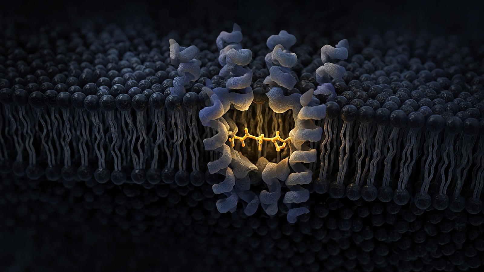
51 min · long read · Peptide research
Ipamorelin: the ghrelin receptor without the circus
A pentapeptide GHS-R1a agonist that raises GH with minimal ACTH, cortisol or prolactin. Raun 1998 is still the paper. Selectivity is the entire product.
More in this desk
65 min · long read · The living cell
The living cell is a city, and you are 36 trillion of them
A 70 kg adult is on the order of 36 trillion cells, most of them red blood cells with no nucleus. A typical nucleated cell holds about ten billion proteins and two metres of DNA. The body recycles 40–60 kg of ATP a day. Those figures are published; this essay is what they mean.
64 min · long read · The living cell
From gene to protein: how a cell actually reads itself
DNA is transcribed, spliced, exported, translated and folded. A research peptide is the ligand at the end of that pipeline, already made. This essay is every machine in between — polymerases, spliceosome, ribosome — with the actual rates attached.
64 min · long read · The living cell
Mitochondria: the bacterium you kept, the genome it kept, the peptides it writes
You turn over 40–60 kg of ATP a day using a 16,569-base genome that still uses a bacterial genetic code. NAD+ is the hydride carrier Complex I spends. MOTS-c is a 16-mer translated from mitochondrial 12S rRNA — Lee, Kim, Cohen, 2015. That last sentence is real, and it is surprising.
57 min · long read · The living cell
Proteostasis: the cell that eats its own mistakes
Ten billion proteins, a 76-residue tag, a 2.5-megadalton proteasome, and autophagy for whole organelles. Hershko, Ciechanover and Rose, 2004. Ohsumi, 2016. DSIP is a sleep-isolation nonapeptide, not an autophagy ligand.
Essays describe published research. They are not medical advice and they do not authorise human use of any catalogue item.