

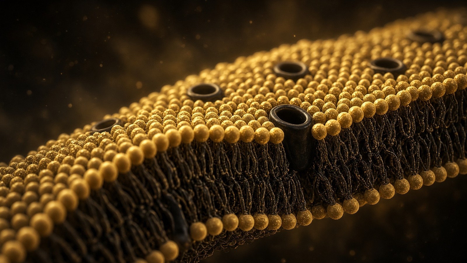
The living cell · 51 min · 11,138 words
Membranes: a five-nanometre wall
Every cell, every organelle, every synapse sits on a lipid bilayer about five nanometres thick. Singer and Nicolson, Hodgkin and Huxley, a billion lipids, and why a peptide ligand at a GPCR does not need to enter the cell.
What this essay actually tells you
- A plasma membrane is ~5 nm of lipid bilayer holding 10⁸–10⁹ lipids. Singer and Nicolson, 1972: fluid mosaic, not a sandwich. Flippases keep phosphatidylserine inside; scramblases expose it as eat-me.
- Hodgkin and Huxley (1952) wrote the action potential as sodium then potassium conductances across that wall. A GPCR is seven helices in the same object: occupancy outside, G protein inside. The ligand need not enter.
- Ipamorelin, PT-141 and retatrutide are plasma-membrane GPCR ligands. KPV can cross via PepT1 (SLC15A1). NAD+ is a cofactor, not a membrane ligand.
What this actually means
A cell is a city because it has a wall. The wall is a lipid bilayer about five nanometres thick — two leaflets of amphipathic lipids with proteins floating in them, as Singer and Nicolson said in 1972. A typical animal cell carries on the order of 10⁸–10⁹ lipid molecules in that plasma membrane, plus more in the organelles. The wall is fluid, asymmetric (flippases spend ATP to keep phosphatidylserine inside), locally organised (rafts, caveolae), and electrically alive (a voltage of tens of millivolts across five nanometres is an enormous field). Channels let selected ions through. Pumps spend ATP to put them back. Hodgkin and Huxley wrote the action potential as a change in those conductances. GPCRs are seven helices in the same wall: a peptide occupies the outside, the helices rearrange, a G protein spends GTP on the inside. The ligand did not enter. That is the ordinary catalogue story for ipamorelin, PT-141 and retatrutide. KPV is an exception because PepT1 is a transporter, not a GPCR. NAD+ is not a membrane ligand at all; it is a cofactor. The occupancy essay next door is the handshake. This essay is the wall the handshake sits in. Research use only. Not a protocol.
- Bilayer thickness
- ~5 nm
- Lipids in a plasma membrane
- 10⁸–10⁹
- Area per lipid
- ~0.7 nm²
- Resting voltage
- −40 to −90 mV
- GPCR helices in the wall
- 7
- Human GPCR genes
- ~800
Hydrophobic core plus headgroups. The entire wall of the city.
A typical animal cell. Organelle membranes add more.
Two leaflets. Geometry, not a metaphor.
Across five nanometres that is an enormous electric field.
The information that crosses is shape. The ligand stays outside.
About half are olfactory. The catalogue lives in a handful of the rest.
A civilisation, if you're being careful with the word, is a set of rooms that keep their insides different from their outsides, plus a post that carries messages through the walls without knocking the walls down. That isn't a metaphor borrowed from politics. That's a plasma membrane. Every cell you have — 36 trillion of them, Sender, Fuchs and Milo — is a room of a few picolitres whose interior chemistry isn't the plasma around it. The wall that makes that possible is about five nanometres thick. Two leaflets of amphipathic lipid, proteins floating and fenced in the fluid, a voltage of tens of millivolts dropped across a distance you could hide under a small protein. Civilisation is a strong word. It's still the right one. Everything we sell as a ligand is, in the ordinary case, a message addressed to a protein in that wall. Not a visitor. Not a repair crew. A message.
In short. A cell is a tiny room because a five-nanometre fat wall keeps inside different from outside. Most research peptides are messages for proteins in that wall, not passengers through it.
The occupancy essay next door is the handshake: a peptide occupies a GPCR, a G protein spends GTP, a second messenger floods, arrestin eventually says enough. We're looking at the wall that handshake sits in. Thickness, census, fluidity, asymmetry, voltage, channels, the seven helices, the one honest transporter exception, and the cofactor that isn't a membrane ligand no matter how often a forum parks it next to a receptor. If you have already read how peptides talk to cells, you have the lock. Here's the door. If you have not, you can still start here: a door is a physical object with a published thickness, and a peptide that can't name the object is a sequence looking for a mood. Thickness, census, fluidity, asymmetry, voltage, channels, the seven helices: that's the furniture of this wall. GPCRs — G protein-coupled receptors — live in it. PepT1 is the transporter exception. NAD+ is a cofactor, not a membrane ligand, and it helps to keep those jobs in different sentences.
In short. The other essay is how a peptide talks to a receptor. This one is the membrane the receptor lives in, and why most peptides never need to cross it.
The ligand does not have to enter. The wall is how information crosses without tourism. That is the only reason a polar peptide can run a cell.
Five nanometres is a civilisation
Start with the length, because every later argument hangs on it. A phospholipid molecule is roughly two nanometres from headgroup to tail-tip. Two of them, tails in, make a bilayer whose hydrophobic core sits near three nanometres and whose whole sandwich, headgroups included, sits near four to five. Neutron scattering, X-ray diffraction on stacked bilayers, freeze-fracture electron microscopy, and the old Gorter–Grendel erythrocyte extraction (1925: enough lipid from red cells to cover twice the surface, hence two leaflets) all converge on that scale. Alberts prints ~5 nm and we'll print ~5 nm. A BPC-157 chain is about the same length as the wall is thick. A globular protein domain is thicker. A water molecule is twenty times smaller. An ion is a dot. The wall isn't a line on a textbook cartoon. It's a physical object with a thickness comparable to the ligands the catalogue sells, which is already a hint that slipping through it isn't how a 20-residue polar peptide usually works.
In short. The membrane is about five nanometres thick — the same size as a small peptide. A charged chain doesn't usually slip through the fat.
Do the area arithmetic once, the way Milo and Phillips taught us to. A lipid occupies on the order of 0.6–0.7 square nanometres in a fluid bilayer. A spherical cell 20 µm across has a surface of about 1,250 µm², which is 1.25 × 10⁹ nm²; two leaflets, so a few 10⁹ lipids if the membrane were a smooth sphere with no proteins. Real cells aren't smooth spheres: microvilli, caveolae, folds, and a protein occupancy that takes up a substantial fraction of the area (the ‘mosaic’ half of fluid mosaic). A typical animal-cell plasma membrane is therefore quoted at 10⁸–10⁹ lipid molecules, depending on cell type, and that is the number we'll keep using. A hepatocyte with a generous canalicular and sinusoidal surface sits toward the top. A lymphocyte sits lower. An erythrocyte, which is a membrane with a job and almost no interior industry, was Gorter and Grendel’s original counting object and still a clean one. Organelle membranes — ER especially — can exceed the plasma membrane in area, which is why a cell’s total lipid census is larger than its plasma-membrane census. When this desk says 10⁸–10⁹, it means the outer wall unless it says otherwise.
In short. A typical animal cell has about a hundred million to a billion fat molecules in its outer membrane. Internal membranes add extra walls inside.
Five nanometres is also an electrical fact. Drop 70 millivolts across 5 nm and you have a field of 14 million volts per metre. That isn't a curiosity for biophysicists. That's why voltage-gated channels can feel the field with a handful of charged residues (the S4 helix of a Kv or Nav channel), why a membrane can store charge like a capacitor at about 1 microfarad per square centimetre, and why a neuron can send a binary message down an axon by briefly letting sodium in. The civilisation is electrical as well as chemical. Hodgkin and Huxley are later in this essay. The field is already here, in the thickness. Hodgkin and Huxley come later, when we watch a squid axon fire. The field is already here, in the thickness, which is why a handful of charged residues on an S4 helix can feel voltage at all. I find that quietly thrilling: a neuron's binary message is a five-nanometre capacitor discharging.
In short. Seventy millivolts across five nanometres is a huge electric field. That's how nerve channels feel voltage, and how a neuron can fire.
What a lipid actually is
A membrane lipid is an amphipath: a hydrophilic head that wants water and a hydrophobic tail that does not. Phosphatidylcholine (PC) is the abundant outer-leaflet citizen in animal cells — a choline head, a glycerol, two fatty-acyl tails, a phosphate. Phosphatidylethanolamine (PE) is smaller-headed, cone-shaped, a curvature specialist, enriched on the inside. Phosphatidylserine (PS) is negatively charged and kept cytoplasmic until a scramblase or a dying cell says otherwise; macrophages read exposed PS as eat-me. Phosphatidylinositol (PI) and its phosphorylated children (PI4P, PIP2, PIP3) are rare by mass and enormous by politics: PIP2 is the substrate phospholipase C spends when a Gq-coupled GPCR fires, and PIP3 is the PI3K badge that Akt reads. Sphingomyelin carries a sphingosine backbone rather than glycerol and prefers the outer leaflet, packed with cholesterol. Cholesterol itself is a small, rigid, mostly hydrophobic steroid with a single hydroxyl; it sits between the tails, buffers fluidity, and is the reason animal plasma membranes aren't a floppy soap at body temperature. That's the cast. If a caption says ‘lipids’ as if they were one object, it hasn't met the cast.
In short. Membrane fats have a water-loving head and a water-hating tail. Cholesterol stiffens the mix so the wall isn't a soap bubble.
The tails matter as much as the heads. Saturated tails pack; unsaturated tails kink at the cis double bond and keep the bilayer fluid at temperatures that would freeze a butter. Palmitoyl and stearoyl versus oleoyl and arachidonoyl is a fluidity decision, and also a signalling decision (arachidonate is the precursor phospholipase A2 releases toward eicosanoids). Chain length sets thickness: longer tails, thicker hydrophobic core, a matching problem for transmembrane helices that have a hydrophobic stretch of a given length. Mismatch isn't decorative. It's how some proteins prefer thicker, cholesterol-rich patches. The cell writes its membrane composition the way a city writes building codes, and the codes differ by organelle: the ER is cholesterol-poor and thin; the plasma membrane is cholesterol-rich and thicker; mitochondrial inner membrane is cardiolipin country, a four-tailed bacterial memory. A peptide assay in a dish whose cells have an odd lipid diet is a slightly different wall. Nobody prints that on a vial. It's still true.
In short. Straight fat tails pack tight; kinked ones keep the membrane runny. The ER, the cell surface and the mitochondrion each run a different fat recipe.
Self-assembly is the cheap miracle. Put enough amphipaths in water and they bury their tails against each other because water would rather hydrogen-bond with itself than order around a hydrocarbon — the hydrophobic effect, not a mysterious lipid glue. Micelles, bilayers, hexagonal phases: the shape of the molecule (Israelachvili’s packing parameter) decides which. Cylindrical lipids (PC) prefer bilayers. Conical lipids (PE, cardiolipin) prefer curvature and will bend a leaflet if they cluster. The wall of the cell is therefore not a covalently stitched bag. It's an equilibrium structure, held by the solvent’s opinions about oil, patchable by adding more lipid, sealable after a puncture if the edges can find each other, and catastrophically leaky if a detergent or a pore-forming peptide says so. That last sentence is why cell-penetrating peptides and antimicrobial peptides are a real literature, and why they aren't the catalogue’s GPCR story. Different objects. Different assays.
In short. Fat molecules stack into a bilayer because water hates their tails, not because they are stitched. The wall can reseal. Detergents can wreck it.
The bilayer as a physical object, not a cartoon line
Permeability is the first job. A pure lipid bilayer is an excellent insulator for charged things and a poor one for small nonpolar things. Water crosses slowly on its own and quickly through aquaporins (Agre, Nobel 2003, with MacKinnon’s channels). Gases cross. Urea crosses badly. Glucose does not, which is why GLUT transporters exist. Sodium, potassium, calcium, chloride: no. A naked bilayer’s electrical resistance is enormous; a cell’s is lower because the cell installed proteins on purpose. A typical 10–40 residue peptide is charged, hydrogen-bonded to water, and has no more business in the hydrocarbon core than a sodium ion has, unless it is a designed cell-penetrating sequence (arginine-rich, amphipathic helix, a literature of its own), a very small unusually behaved fragment, or a substrate of a transporter. That's the physical reason GPCRs exist. The cell declined to let the message in. It built a protein that can feel the message from outside and speak inside.
In short. Fat walls block ions and most peptides. Water and gases sneak through; sugar and salt need protein doors. That's why GPCRs exist.
Fluidity is the second job. Lipids diffuse laterally at something like 1 µm²/s in a dilute bilayer; slower in a crowded plasma membrane, faster in a protein-poor liposome. Frye and Edidin, 1970, fused a human cell to a mouse cell and watched fluorescent antibodies against each species’ surface antigens mix in tens of minutes. That experiment retired the idea of a rigid mosaic. Rotation around the lipid’s long axis is fast. Flip-flop from one leaflet to the other, uncatalysed, is slow — hours to days for a typical PC — which is why flippases have a job and why asymmetry can be maintained. The gel-to-fluid transition temperature depends on tail saturation and cholesterol; cholesterol smears the transition and keeps animal plasma membranes in a liquid-ordered-ish state rather than a frozen gel or a floppy liquid-disordered soap. A ‘fluid mosaic’ isn't a metaphor for flexibility in your life. It's a diffusion coefficient and a phase.
In short. Lipids slide past each other in the plane of the membrane, but they almost never spontaneously swap faces. Enzymes have to do the swapping.
Capacitance is the third job, and the one the electrical civilisation spends. A bilayer is two conductors (the aqueous insides and outsides) separated by a thin insulator (the hydrocarbon). That's a capacitor. Specific capacitance of biological membranes clusters around 1 µF/cm², a number Hodgkin and Huxley used as a parameter and every electrophysiologist still uses as a sanity check: if your ‘cell’ has a weird capacitance, you have a weird area or a bad seal. Charge the capacitor and you have a voltage. Discharge it through open channels and the voltage moves on a time constant τ = RmCm. The length constant of an axon is a cable-theory cousin. None of this requires a single named peptide. All of it is why a Gq-coupled receptor that opens a calcium channel, or a Gs-coupled receptor that modulates one, is talking to an electrical object, not just a chemical one. The occupancy essay’s second messengers land on this capacitor.
In short. The membrane stores electric charge like a tiny capacitor. Open channels drain it; that's how voltage changes.
Singer and Nicolson, 1972: the mosaic that still stands
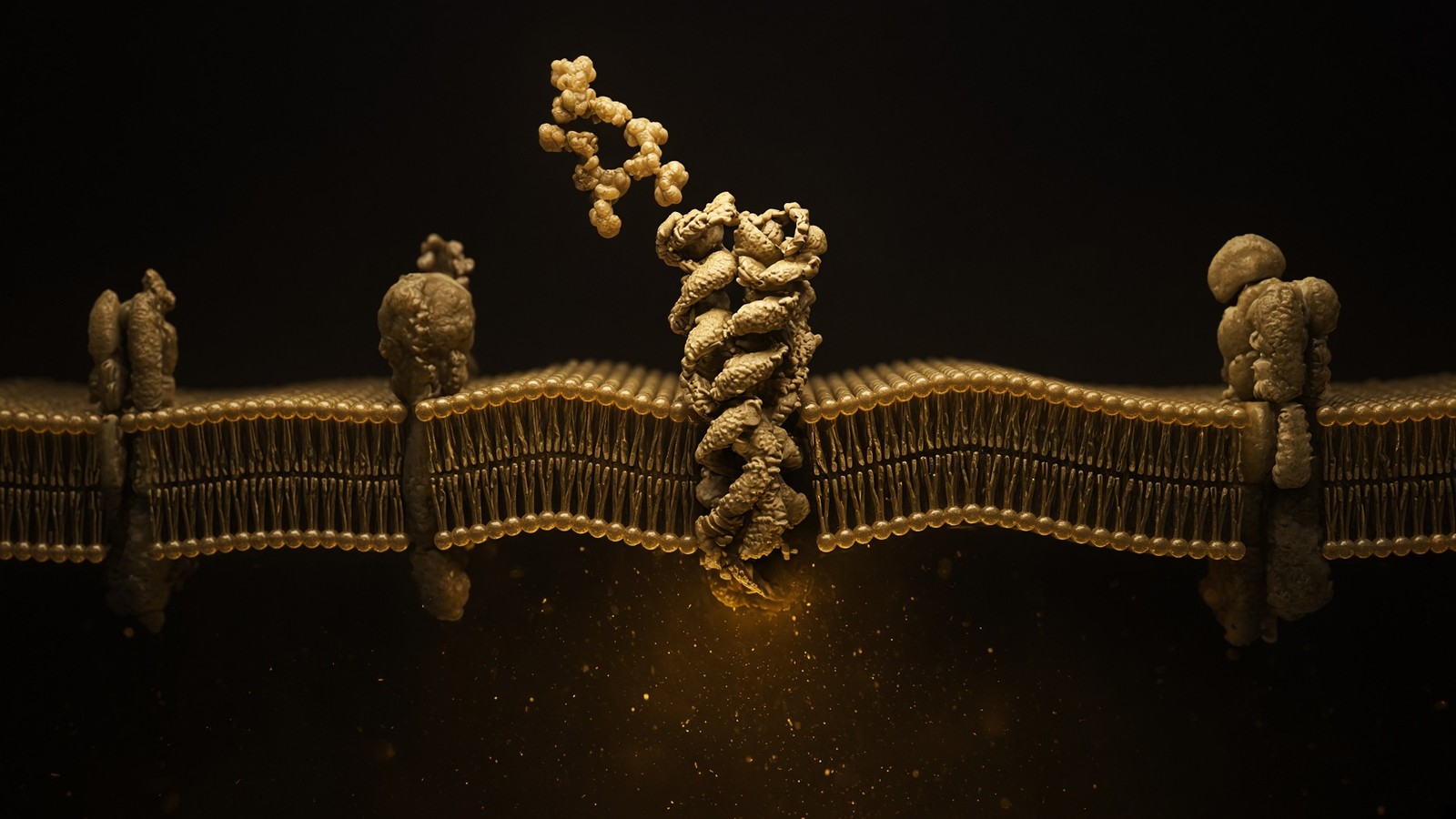
Before 1972 the textbook wall was often a sandwich: protein, then lipid, then protein, the Davson–Danielli–Robertson unit membrane, an interpretation of the railroad-track electron micrograph that made the bilayer look coated. It wasn't a stupid model. It was the wrong one. S. J. Singer and G. L. Nicolson, Science 175: 720–731, 18 February 1972, replaced it with a fluid mosaic: a lipid bilayer as a two-dimensional solution, globular proteins dissolved in it, some spanning, some stuck to one face, free to diffuse in the plane unless something stops them. Integral proteins had hydrophobic surfaces that matched the hydrocarbon; peripheral proteins could be washed off without dissolving the wall. The model explained freeze-fracture particles, the Frye–Edidin mixing, the fact that phospholipases could attack lipids without first chewing a protein coat, and the emerging amino-acid sequences of membrane proteins that looked like they had transmembrane stretches. Fifty years on, the mosaic is still the picture. The footnotes are rafts, cytoskeletal picket fences (Kusumi), hop diffusion, and the admission that ‘free to diffuse’ was always more true for some proteins than others.
In short. In 1972 Singer and Nicolson said the membrane is a fluid sea of fats with proteins floating in it, not a stiff sandwich. That picture still stands.
Unwin and Henderson, 1975, put seven helices in that mosaic: bacteriorhodopsin from Halobacterium, a light-driven proton pump, the first membrane protein whose structure (at low resolution, from purple-membrane 2D crystals) showed a bundle of transmembrane α-helices. GPCRs aren't bacteriorhodopsin, but they're the same architectural family in the broad sense — seven helices in Singer and Nicolson’s solvent — and when Palczewski’s group solved rhodopsin in 2000 and Kobilka’s group solved β2-adrenergic receptor a few years later, the occupancy essay’s object became a set of coordinates. The wall had been waiting. The ligand-binding pocket faces the aqueous outside or, for retinal, a pocket in the bundle. The G-protein face is the aqueous inside. The hydrocarbon never had to admit a peptide tourist. Structure made the tourism theory optional, then embarrassing. The ligand-binding pocket faces water on the outside, or, for retinal, a pocket in the bundle. The G-protein face is water on the inside. The hydrocarbon never had to admit a peptide tourist. Structure made that optional, then obvious, which is a lovely way for a theory to retire.
In short. The first seven-helix membrane protein seen in 3D was a bacterial light pump. GPCRs turned out to be the same kind of object.
What Singer and Nicolson underplayed, because the data were thinner, is that the mosaic isn't an ideal 2D gas. The inner leaflet talks to the actin cortex. Transmembrane proteins picket the fence. Some lipids prefer some proteins (annular lipids; PIP2 as a local charge cloud). Cholesterol and sphingomyelin cluster on scales that may or may not deserve the word raft. Palmitoylation, myristoylation, GPI anchors: covalent lipid handles that park proteins on one leaflet or the other. The nuclear envelope is a double mosaic continuous with ER. Mitochondrial inner membrane is a mosaic with cristae and a bacterial lipid. The model was a liberation from the sandwich, not a claim that all membranes are one membrane. We'll keep using ‘the wall’ in the singular because the physics is shared. The recipes are not. Palmitoylation, myristoylation, GPI anchors: covalent lipid handles that park proteins on one leaflet or the other. The nuclear envelope is a double mosaic continuous with ER. Mitochondrial inner membrane is a mosaic with cristae and cardiolipin. We'll keep saying the wall in the singular because the physics is shared. The recipes aren't.
In short. The fluid mosaic isn't a perfect open sea. The cell's skeleton fences proteins in, and different organelles run different recipes.
Asymmetry is a job: flippases, floppases, scramblases
The two leaflets aren't copies of each other. In an animal plasma membrane the outer leaflet is rich in phosphatidylcholine and sphingomyelin; the inner leaflet is rich in phosphatidylethanolamine and phosphatidylserine, with the phosphoinositides as a charged inner-leaflet minority. That asymmetry isn't a birth defect of vesicle traffic. It's maintained. Uncatalysed flip-flop is slow, but it isn't zero, and without a pump the leaflets would equilibrate on a timescale of the cell’s life. The pumps are P4-ATPases, the flippases: ATP8A1, ATP8A2, ATP11A, ATP11C and their cousins, each with a CDC50/TMEM30 β-subunit, spending ATP to move PS and PE toward the cytoplasm. Mutations prove the job. ATP11C lesions mess with erythrocyte PS and B-cell development; ATP8A2 with neurological disease. A flippase isn't a personality. It's how the inner leaflet stays negative and how a macrophage isn't invited to eat a healthy cell.
In short. The inside face of the membrane has different fats from the outside. Flippases spend ATP to keep the eat-me fat on the inside.
Floppases push the other way. They're ABC transporters — ABCA1 the cholesterol-to-apoA-I specialist of reverse cholesterol transport, ABCG1, ABCB1 (P-glycoprotein) as a lipid and drug flopper, a family that also includes the peptide and xenobiotic exporters that ruin chemotherapy. The names are ugly. The direction is the point: ATP out, lipid toward the extracellular leaflet or an acceptor particle. Tangier disease (ABCA1) is what happens when cholesterol can't flop onto HDL: orange tonsils, vanishing HDL, a textbook photograph. A research peptide isn't a floppase ligand. A research peptide that can't tell a floppase from a GPCR hasn't yet named a lock. The names are ugly. The direction is the point: ATP out, lipid toward the extracellular leaflet or an acceptor particle. Tangier disease, ABCA1 lost, is what happens when cholesterol can't flop onto HDL — orange tonsils, vanishing HDL, a textbook photograph. A research peptide isn't a floppase ligand. Naming the lock still matters.
In short. Floppases spend ATP to push fats the other way, toward the outside. ABCA1 loading cholesterol onto HDL is the famous one.
Scramblases are the emergency doors. They don't spend ATP. They allow lipids to equilibrate between leaflets when a signal says the asymmetry is now a message. TMEM16F (ANO6) is a calcium-activated scramblase; loss-of-function is Scott syndrome, a bleeding phenotype, because platelets can't expose PS as a procoagulant surface. Xkr8 is a caspase-activated scramblase; apoptosis turns it on, PS appears on the outer leaflet, and macrophages recognise the corpse. The same lipid that flippases spent ATP to hide becomes, in minutes, a flag. That's why ‘phosphatidylserine’ is both a leaflet citizen and an immunology word. Collapsing annexin-V staining into ‘the cell was inflamed’ has skipped the scramblase. Inflammation can be involved. The mechanism is a lipid moving house. That's why phosphatidylserine is both a leaflet citizen and an immunology word. Annexin-V staining reads exposed PS; it doesn't, by itself, tell you the cell was inflamed. Inflammation can be involved. The mechanism is a lipid moving house, and scramblases are how it moves without spending ATP.
In short. Scramblases let fats mix between the two faces when calcium or cell-death enzymes say so. That's how a dying cell raises an eat-me flag.
The ATP cost of asymmetry isn't a rounding error. A living cell is always spending to keep PS in. At death the spending stops, caspases cut Xkr8 into its on-form, and the flag goes up even if TMEM16F never saw a calcium spike. Lipid traffic from the ER — where most phospholipid is made on the cytoplasmic face — to the plasma membrane is a second asymmetry problem: how do newly made lipids reach the luminal leaflet of the ER, and how do they reach the outer leaflet of the cell? ER scramblases (TMEM41B, VMP1, and a still-moving literature) equilibrate ER leaflets so the luminal face can grow. Non-vesicular transfer proteins (OSBP, CERT, phosphatidylinositol transfer proteins at membrane contact sites) hand lipids between organelles without a vesicle. The wall isn't a closed bag made once in the Golgi. It's a budget, a set of contact sites, and a set of enzymes that care which leaflet a headgroup sits on. A peptide protocol that never mentions a leaflet is still allowed to be a peptide protocol, because we don't print those. A mechanism that never mentions a leaflet is incomplete if the story is a membrane story.
In short. Keeping the two faces different costs energy for the cell's whole life. Fats are made in the ER and handed around at contact sites.
Rafts, and the honesty about them
Kai Simons and Elina Ikonen, Nature 1997: cholesterol- and sphingolipid-rich microdomains in the outer leaflet, packed in a liquid-ordered state, proposed as platforms that sort proteins in the Golgi and cluster signalling molecules at the plasma membrane. GPI-anchored proteins, some Src-family kinases, some receptors, influenza haemagglutinin — a guest list that made the idea irresistible. The biochemical proxy was the detergent-resistant membrane, the DRM, a pellet that survived cold Triton. The proxy oversold the biology. Cold detergent doesn't equal a living nanodomain, and a generation of papers treated a pellet as a place. The modern argument, still live, is about size, lifetime and function: nanometres to tens of nanometres, microseconds to seconds, enrichment rather than a sealed raft you could step onto. Kusumi’s picket-fence and hop-diffusion work, super-resolution imaging, and the careful cholesterol-depletion sceptics all belong in the footnote. We won't adjudicate a twenty-year fight in a catalogue essay. We'll say: composition isn't uniform, cholesterol and sphingomyelin cluster, some proteins prefer those clusters, and ‘raft’ is a word that needs a method attached.
In short. Rafts are proposed cholesterol-rich patches that cluster some proteins. They are smaller and more short-lived than the first cartoons.
Caveolae are the morphologically honest cousins. Invaginations of 50–80 nm, caveolin-1 (or -3 in muscle) plus cavins, cholesterol-dependent, visible in electron micrographs rather than only in a detergent pellet. They buffer membrane tension, traffic some cargoes, and host a signalling literature that is real and also over-claimed. Palmitoylation can park a protein near these neighbourhoods. None of this makes a research peptide a ‘raft modulator’. If a paper has a cholesterol depletion, a caveolin blot and a named receptor, it has a membrane story. If a forum has the word raft and a stack, it has an adjective. Palmitoylation can park a protein near these neighbourhoods. A paper with a cholesterol depletion, a caveolin blot and a named receptor has a membrane story you can follow. Caveolae buffer tension and traffic some cargoes; the signalling literature is real and sometimes over-claimed. They're pits you can photograph, which is already more honest than a detergent pellet.
In short. Caveolae are little pits in the membrane you can actually photograph. They're the respectable cousins of rafts.
The protein inventory of a wall
A plasma membrane isn't mostly lipid by mass once you count the proteins, and not mostly protein by area once you count the lipids. Both sentences are true in different units, which is how membrane arguments go bad at dinner. Integral proteins span or are deeply embedded: α-helical bundles (GPCRs, channels, transporters, pumps), β-barrels (mitochondrial outer membrane, Gram-negative outer membrane). Peripheral proteins bind the surface electrostatically or via a lipid anchor and can be washed off. GPI anchors pin a protein to the outer leaflet with a glycolipid handle. Prenyl and acyl chains pin others to the inner leaflet. Roughly a quarter of human genes encode membrane proteins, a census that makes the wall the densest pharmacological neighbourhood in the proteome — which is why a third of small-molecule drugs have historically hit GPCRs, and why a peptide house that can't name a membrane protein is in the wrong business.
In short. Lots of proteins live in the membrane: receivers, doors, pumps, anchors. About a quarter of human genes encode them.
The jobs split cleanly if you insist. Channels: passive, downhill, often ion-selective, gated by voltage, ligand, stretch or temperature. Transporters (carriers): bind, conformational-cycle, can be uniport, symport or antiport, can run uphill if coupled. Pumps: ATPases that write the gradients the channels spend — Na⁺/K⁺-ATPase, SERCA, PMCA, the V-ATPase on endosomes and lysosomes, F-type ATP synthase (a pump run backward) on mitochondria. Receptors: bind an extracellular message and speak intracellularly without the message necessarily entering — GPCRs, receptor tyrosine kinases, cytokine receptors, ligand-gated ion channels which are both receptors and channels. Adhesion: cadherins, integrins, a mechanical conversation with the matrix and the neighbour. The occupancy essay is the receptor paragraph. We're looking at why that paragraph has a place to sit, and why a transporter (PepT1) is a different sentence from a receptor (GHSR) even though both are integral membrane proteins.
In short. Channels leak selected ions downhill. Carriers shuttle. Pumps spend ATP. Receptors hear a message outside and talk inside.
- Channels: downhill, gated, selective. Nav, Kv, Cav, ligand-gated nicotinic and iGluR, GIRK, CFTR as the honorary ion-channel ABC.
- Carriers: GLUT, PepT1 (SLC15A1), neurotransmitter sodium symporters. Bind and flip. PepT1 is the KPV exception.
- Pumps: Na⁺/K⁺-ATPase (3Na⁺/2K⁺), SERCA, PMCA, H⁺/K⁺-ATPase, V-ATPase, F-type ATP synthase.
- Receptors: GPCR 7TM (ipamorelin, PT-141, retatrutide), RTK (IGF1R), cytokine (GHR), ligand-gated ion channel.
- Not membrane ligands: NAD+ (cofactor), TB-500’s LKKTETQ (actin), GHK-Cu (copper ligand), Epithalon’s nuclear claims.
Channels are holes with opinions
A channel isn't a pore you could pour coffee through. Selectivity filters are a few angstroms of backbone carbonyls or carefully placed charges that dehydrate an ion just enough, and not the wrong ion. MacKinnon’s potassium-channel structures (KcsA, then KvAP, then the eukaryotic Kv; Nobel 2003 with Agre) showed why K⁺ beats Na⁺ despite sodium being smaller: the filter mimics the hydration shell of potassium, not of sodium. Sodium channels have a different filter (the DEKA locus in Nav). Calcium channels another. Chloride channels (CLC) are a different architecture again, and CFTR is an ABC transporter that evolved into a chloride channel and fails in cystic fibrosis when it misfolds or misgates. The point for this desk isn't to teach a channel course. It's to say that the wall’s holes are chemistry, that Hodgkin and Huxley’s conductances had molecular names waiting, and that a peptide which opens a channel (a ligand-gated receptor) is still not entering. It's occupying an extracellular face. Information crossed as a conformation. Ions crossed because that was the conformation’s job.
In short. Ion channels are choosy holes. A peptide that opens a channel still sits outside. Ions go through; the peptide doesn't have to.
Gating is the opinion. Voltage-gated channels feel the field with charged S4 helices and open or inactivate on millisecond clocks — the Hodgkin–Huxley sodium inactivation that makes an axon able to fire again, the potassium delayed rectifier that repolarises. Ligand-gated channels (nicotinic AChR, GABAA, glycine, iGluRs) bind transmitter and open a pore; some are pentamers, some tetramers, none of them GPCRs, a distinction the internet collapses at every opportunity. Stretch-gated channels (Piezo1/2) feel membrane tension, a direct mechanical job of the wall. Temperature-gated TRP channels are why capsaicin is hot and menthol is cold. Inwardly rectifying potassium channels, including GIRK, can be opened by Gβγ from a Gi-coupled GPCR — which is how many inhibitory synapses actually quiet a neuron, and how a peptide GPCR can be an electrical object without being an ion channel. The occupancy essay named Gβγ in passing. The wall is where Gβγ finds the channel.
In short. Channels open and close on purpose: voltage, a transmitter, stretch, temperature, or a G-protein fragment from a receptor.
Patch clamp made the opinions audible. Neher and Sakmann, Nobel 1991, a glass pipette on a patch of membrane, a single channel’s picoampere flickering as a square wave. Before that, Hodgkin and Huxley had macroscopic currents. After that, a graduate student could watch one protein in Singer and Nicolson’s mosaic open and close. The method is why we believe gating is discrete, why we know a channel has substates, and why a binding-assay EC50 isn't the same object as a single-channel open probability. We don't sell a patch-clamp kit. It sells ligands some of which, in someone else’s laboratory, will change an open probability two proteins downstream. That's a long sentence. It's still shorter than ‘it enters the cell and repairs’. Before patch clamp, Hodgkin and Huxley had macroscopic currents. After it, a graduate student could watch one protein in Singer and Nicolson's mosaic open and close. The method is why we believe gating is discrete, why we know a channel has substates, and why a binding-assay EC50 isn't the same object as a single-channel open probability.
In short. Patch clamp lets you hear one channel protein open and shut. That's how we know gating is a real on/off, not a smear.
Voltage is a membrane fact, not a personality
A typical neuron rests near −70 mV, interior negative. A typical skeletal myocyte a little more negative. A typical epithelial cell less so. A typical pancreatic β-cell sits near −70 mV when glucose is low and depolarises when ATP-sensitive potassium channels close — which is the electrical sentence under first-phase insulin, and a neighbour of the incretin GPCR story, not a substitute for it. The voltage is a weighted average of the equilibrium potentials of the ions the membrane is permeable to at that moment: Goldman–Hodgkin–Katz, a cousin of Nernst. Potassium wants the interior negative (EK around −90 mV). Sodium wants it positive (ENa around +60 mV). At rest, potassium leak wins. During a spike, sodium channels open, the average swings toward ENa, the interior goes positive for a millisecond, potassium delayed rectifiers and sodium inactivation swing it back. Chloride and calcium have their own Nernst potentials and their own fans. None of this is a metaphor for mood. It's millivolts, millimolar gradients, and a wall.
In short. Nerve cells sit about seventy millivolts negative inside because they leak potassium. A spike is sodium in, then potassium out.
The gradients are written by pumps. Na⁺/K⁺-ATPase, Skou’s enzyme, Nobel 1997: three sodium out, two potassium in, one ATP, a net positive charge leaving, a small electrogenic contribution to the resting potential and a large contribution to the gradients everyone else spends. A neuron can spend half its ATP budget on this pump after a busy afternoon of spikes. SERCA puts calcium back into the ER. PMCA puts it out of the cell. The mitochondrial calcium uniporter swallows the overflow because it can. Secondary transporters ride the sodium gradient: SGLT, neurotransmitter sodium symporters, the sodium–calcium exchanger NCX running in whichever direction the gradients dictate. The wall is a set of gradients that channels spend and pumps restore. A peptide that modulates a pump or a channel is a membrane story. A peptide that occupies a GPCR which then modulates a channel is also a membrane story, one floor up. A peptide advertised as ‘for energy’ because membranes use ATP is a category error we won't print.
In short. Pumps spend ATP to keep sodium out and potassium in. After a busy burst of firing, a lot of a neuron's energy bill is restoring that.
Ten thousand-fold is the calcium number worth memorising. Resting cytosol ~100 nM. Extracellular and ER lumen ~1 mM. The wall — plasma membrane or ER membrane — holds that gradient. IP3 receptors and ryanodine receptors are channels in the ER wall. Voltage-gated calcium channels are channels in the plasma wall. Gq-coupled GPCRs (GHSR among them) talk to the ER wall via IP3. Gs-coupled GPCRs talk to the plasma wall’s calcium channels via PKA in tissues that set it up that way. The occupancy essay’s calcium paragraph is this wall’s paragraph, seen from the ligand. Seen from the lipid, it is a 10,000-fold gradient across five nanometres, a cocked gun, and a set of proteins that open on purpose. Ipamorelin’s Gq lean on a somatotroph is an IP3 tap on the ER gun, which is why Bowers’ synergy with a Gs-coupled GHRH analogue is two taps on two walls that share a cytosol.
In short. Calcium is ten thousand times more concentrated outside the cytosol than in it. Gq receptors tap an ER calcium door via IP3.
Hodgkin and Huxley, because the wall fires
Plymouth, a squid, an axon half a millimetre thick because squid motor fibres are built to be voltage-clampable by mid-century instruments. Hodgkin, Huxley, and Katz: a space clamp, a feedback circuit, currents separated by ionic substitution. Four Journal of Physiology papers in 1952. Sodium conductance activates and inactivates. Potassium conductance activates more slowly and doesn't inactivate on that clock. Leak is the rest. They wrote differential equations for m, h and n gates — hypothetical particles with voltage-dependent rate constants — and reconstructed the action potential, the refractory period, the conduction speed. They didn't have Nav1.x or Kv2. They had the wall’s currents. The molecular biology of the next forty years found the proteins and, to first order, believed them. That's the best kind of biophysics: a prediction that survived the gene. They wrote differential equations for m, h and n gates — hypothetical particles with voltage-dependent rate constants — and reconstructed the action potential, the refractory period, the conduction speed. They didn't have Nav1.x or Kv2. They had the wall's currents. The molecular biology of the next forty years found the proteins and, to first order, believed them.
In short. In 1952 Hodgkin and Huxley measured currents in a giant squid nerve and wrote equations that still describe a spike.
Why a peptide essay cares: because a subset of catalogue ligands change excitability two or three steps downstream of occupancy, and because a subset of the internet talks as if a peptide were a current. Semax and Selank sit on BDNF and GABA literatures that are electrical at the tissue end; they have their own queued essay. GHSR on vagal afferents is an appetite wire. GLP-1R in the brainstem is an aversive and satiety wire. MC4R in the paraventricular hypothalamus is an energy and sexual-function wire. Those are GPCRs on electrically interesting cells. The spike is still Hodgkin–Huxley plus whatever neuromodulators shifted the conductances. Occupying the GPCR isn't injecting current. It's changing the wall’s opinions about which channels should be more open, on a slower clock than a spike, in a particular cell type. If you can't name the cell type, you're holding a sequence. If you name the cell type and skip the voltage, you have a cAMP kit and not a neuron.
In short. Some peptide receptors sit on nerve cells and change how easily those cells fire, but the peptide isn't itself an electric current.
Myelin, nodes of Ranvier, saltatory conduction: a later specialisation of the same wall. Schwann cells and oligodendrocytes wrap the axon in stacked bilayers, drop the capacitance, raise the transverse resistance, and force the spike to jump from node to node where Nav is clustered. Multiple sclerosis is, among other things, a wall problem. A peptide catalogue has no business claiming a remyelination protocol. It has a duty to know that stacked bilayers are how a two-metre axon stays fast, and that the five-nanometre object, multiplied and wrapped, is white matter. Civilisation again. The same physics, a different architecture, a disease that proves the architecture wasn't optional. Multiple sclerosis is, among other things, a wall problem. A peptide catalogue has no business claiming a remyelination protocol. It has a duty to know that stacked bilayers are how a two-metre axon stays fast, and that the five-nanometre object, multiplied and wrapped, is white matter. Same physics, a different architecture.
In short. Myelin is many membrane wraps that make a nerve impulse jump faster. That's still the five-nanometre wall, stacked.
Seven helices: why the ligand need not enter
Diagram
Outside
Peptide ligand
Named sequence in the nM–µM pocket. Shape complementarity, not vibes. A 15-mer and a 4-mer do not fit the same hole.
Membrane
7-TM receptor
Helices rearrange. The cytoplasmic face becomes a GEF for a heterotrimeric G protein (Gs, Gi, Gq, G12/13).
Inside
Second messengers
cAMP, IP₃, Ca²⁺, β-arrestin. One occupied receptor can spawn thousands of messenger molecules. That is amplification.
~800 GPCRs in the human genome. Seven transmembrane helices, an extracellular ligand pocket, an intracellular G-protein handshake. Catalogue neighbours: ipamorelin at GHSR, PT-141/MT2 at melanocortin receptors, retatrutide at GLP-1R/GIPR/GCGR.
A G protein-coupled receptor is seven transmembrane α-helices in Singer and Nicolson’s solvent, an extracellular face that binds ligand, an intracellular face that becomes a guanine-nucleotide exchange factor for a heterotrimeric G protein. About 800 in the human genome, half of them olfactory. Lefkowitz and Kobilka, chemistry Nobel 2012. The occupancy essay is the full handshake: pocket, TM6 outward swing, GDP out, GTP in, Gs/Gi/Gq/G12-13, arrestin, amplification. This paragraph is the wall’s version of the same story. The helices match the hydrophobic thickness. The loops and termini live in water. The ligand — if it is a peptide hormone or a research analogue of one — meets water-facing residues and a pocket, not the hydrocarbon core. Occupancy rearranges the bundle. The rearrangement is the message. The peptide doesn't have to follow the message through the wall. That's the entire physical reason a polar 5-mer, 7-mer or 39-mer can run a cell it never entered.
In short. A GPCR is seven helices through the fat wall, pocket outside, G-protein switch inside. The peptide can stay outside.
Walk the three related peptides this essay was assigned and the sentence holds. Ipamorelin, Aib-His-D-2-Nal-D-Phe-Lys-NH2, occupies GHSR, a class-A GPCR, Gq-leaning, on somatotrophs and elsewhere. The pentapeptide isn't a cell-penetrating sequence. It doesn't need to be. GHSR’s pocket faces the world; PLC, IP3 and calcium are the interior sentence; Raun 1998 is the selectivity paper. PT-141, the free-acid cyclic heptapeptide neighbour of licensed bremelanotide, occupies melanocortin receptors, class A, Gs, cAMP. The ring is a Hadley/Molinoff trick against proteases, not a ticket through the bilayer. Retatrutide, a 39-residue fatty-acylated triple agonist, occupies GLP-1R, GIPR and GCGR, class B, Gs, cAMP; the fatty acid is an albumin half-life handle, not a membrane-crossing handle. Three ligands. Three (or five) receptors. One wall. Zero requirement for the peptide to sit in the cytosol as the message. The occupancy essay named the locks. The wall is why the locks are enough.
In short. Ipamorelin, PT-141 and retatrutide all bind pockets on the outside of GPCRs. None of them needs to enter the cell to do that job.
Class B receptors make the ‘need not enter’ sentence almost architectural. A large N-terminal extracellular domain catches the C-terminal half of the peptide hormone in solution, raising the local concentration of the peptide’s N-terminus, which then inserts into the transmembrane bundle from the outside and does the activating work. Two steps, both aqueous-facing, both on the extracellular side of the five-nanometre wall. GHRH, GLP-1, GIP, glucagon: this is their geometry. Truncating the N-terminus is how you make an antagonist, because you keep the ECD catch and lose the insertion. A 39-mer triple agonist can occupy three related class-B pockets with one chain because the pockets are related and the chain is long, not because the chain is a submarine. If you need retatrutide in the cytosol to work, you haven't looked at a class-B structure. Truncating the N-terminus is how you make an antagonist, because you keep the ECD catch and lose the insertion. A 39-mer triple agonist can occupy three related class-B pockets with one chain because the pockets are related and the chain is long, not because the chain is a submarine. Class-B geometry is why retatrutide can stay outside.
In short. Hormone receptors like GLP-1's catch the peptide in a large outer domain, then dip the front end into the helix bundle from outside.
Second messengers live on the inside of the wall
Diagram
× 1
Ligand
One peptide in one pocket. nM–µM. Shape, not a mood.
× 10–10²
G proteins
The occupied GPCR is a GEF. Each Gα is a catalyst.
× 10³–10⁴
cAMP / IP₃ / Ca²⁺
Adenylyl cyclase and PLC do not make one molecule. They make a cloud.
× 10⁴–10⁶
PKA / PKC / CaMK
Kinases phosphorylate many substrates per messenger.
× tissue
Secretion, transcription, motility
The organism-level readout. Still not a protocol.
This is the only magic, and it is not magic. A nanomolar ligand can move a micromolar messenger because enzymes sit between them. Desensitisation (GRK, β-arrestin, endocytosis) is how the cell refuses to let ‘more ligand’ mean ‘more signal’ forever.
The wall’s inner leaflet is a factory floor. Adenylyl cyclase is a transmembrane enzyme; Gs holds it on, Gi holds it down; ATP becomes cAMP on the cytoplasmic face. Phospholipase C-β is recruited to inner-leaflet PIP2; Gq holds it on; PIP2 becomes IP3 (aqueous) and DAG (still in the leaflet). PI3K writes PIP3 on the same leaflet; PH-domain proteins (Akt) land. Small GTPases of the Ras, Rho and Arf families are inner-leaflet citizens via lipid anchors. The second-messenger flood the occupancy essay quantified — nanomolar ligand, micromolar cAMP — is an inner-leaflet and cytosolic event triggered by an outer-leaflet occupancy. AKAPs nail PKA to particular neighbourhoods of the inner face, which is why a cAMP nanodomain next to a calcium channel isn't a cAMP nanodomain next to a transcription factor. The mosaic, internally, is a set of postcodes. Global cAMP is a city-wide average of those postcodes.
In short. The inner face of the membrane is where cyclases and lipases run after a receptor is occupied. The peptide is still outside.
PIP2 is worth a dedicated sentence because it is a lipid and a signal at once. Phosphatidylinositol 4,5-bisphosphate is a rare inner-leaflet species, the substrate of PLC, the badge that some channels and ER–plasma-membrane tethers read, the parent of PIP3, and a molecule whose local depletion during a Gq stimulus can close KCNQ channels (the M-current) and change excitability without a single cAMP being made. A Gq-coupled peptide receptor is therefore a lipid-metabolising device as well as a calcium device. GHSR on a somatotroph is PLC, IP3, ER calcium; GHSR on a neuron can also be an M-current story depending on the neighbours. The wall is the substrate. The occupancy is the trigger. Mixing those with ‘the peptide entered’ is how you fail the oral exam this desk keeps setting. A Gq-coupled peptide receptor is therefore a lipid-metabolising device as well as a calcium device. GHSR on a somatotroph is PLC, IP3, ER calcium; GHSR on a neuron can also be an M-current story depending on the neighbours. The wall is the substrate. The occupancy is the trigger. The peptide is still outside.
In short. PIP2 is a rare inner-face fat that receptors can cut or decorate. Cutting it makes the calcium signal. The peptide didn't walk in.
Three ligands that never needed to enter
Diagram
| Node | Catalogue | Conversation |
|---|---|---|
| GPCR | Ipamorelin, MT2, PT-141, retatrutide, CJC | Second messengers, secretion, appetite, pigment |
| RTK / IGF1R | IGF-1 LR3 | IRS–PI3K–Akt–mTOR and Shc–ERK |
| Cytokine receptor | Somatropin (HGH) | GHR–JAK2–STAT5b, hepatic IGF-1 |
| Cofactor | NAD+ | Sirtuins, PARPs, CD38, redox |
| Actin buffer | TB-500 / Tβ4 motif | G-actin sequestration, motility |
| Growth-factor-like | BPC-157 | VEGFR2 / FAK / eNOS neighbourhood |
| Copper ligand | GHK-Cu | Transcriptome shift in fibroblasts |
| MC fragment | KPV | NF-κB, PepT1, no pigment |
| Nuclear / pineal | Epithalon (AEDG) | TERT and melatonin literatures |
| mtORF peptide | MOTS-c | AMPK, folate–methionine cycle |
Each row is a different kind of molecular conversation. The catalogue peptides bind at these nodes; they are not interchangeable, and stacking them because a forum did mixes unrelated literatures.
Ipamorelin — GHSR, Gq, the wall of a somatotroph
GHSR is a class-A GPCR in the plasma membrane of pituitary somatotrophs, hypothalamic neurons, vagal afferents and a list of peripheral sites that keep expanding. Endogenous ligand ghrelin, 28 residues, octanoylated on Ser3 by GOAT, a stomach peptide that had to be found after the receptor was already being occupied by chemists (Howard 1996; Kojima 1999). Ipamorelin is a pentapeptide chosen by Raun, Hansen, Johansen and the Novo Nordisk group (Eur J Endocrinol 1998) for GH release with a flat ACTH and prolactin line relative to GHRP-6. The lock is the same. The bias is the point. The lock faces the extracellular space. Occupancy is a rearrangement of seven helices in a five-nanometre wall. PLC, IP3, calcium from ER stores, a somatotroph granule that was waiting, synergy with Gs-coupled GHRHR as Bowers measured in men in 1990. None of those clauses require the pentapeptide in the cytosol. Research use only. The sequence is Aib-His-D-2-Nal-D-Phe-Lys-NH2. The wall is the one in this essay.
In short. Ipamorelin fills the ghrelin receptor on the outside of growth-hormone cells. Calcium rises inside. The pentapeptide doesn't need to go in.
PT-141 — melanocortin receptors, Gs, a cyclic peptide still outside
MC1R, MC3R, MC4R, MC5R are class-A GPCRs, Gs, cAMP, POMC fragments as endogenous ligands, agouti and AgRP as endogenous inverse agonists. They sit in plasma membranes of melanocytes, hypothalamic neurons, exocrine cells, and other tissues the family actually uses. PT-141 is the free-acid cyclic heptapeptide neighbour of bremelanotide (Vyleesi), a licensed medicine in some jurisdictions for hypoactive sexual desire disorder. We don't sell that medicine. We name the neighbour so the receptor story isn't a secret. The cyclic lactam bridge is a protease trick, not a bilayer ticket. Occupancy at MC3/MC4 is a Gs–cAMP sentence in the neurons that have those receptors. MC1 pigment is the sentence PT-141 was developed away from relative to melanotan II. The wall is still five nanometres. The ligand is still outside. A tan, a libido endpoint in a licensed product, and a research cyclic peptide are three objects. Leaflet physics doesn't care which object you confuse. Pharmacology does.
In short. PT-141 binds melanocortin receptors on the cell surface and raises cAMP inside. The ring is to survive enzymes, not to cross the fat.
Retatrutide — three class-B pockets, one chain, still a wall story
GLP-1R, GIPR and GCGR are class-B GPCRs in the plasma membranes of β-cells, brainstem neurons, hepatocytes, adipocytes and the rest of the incretin–glucagon map. Retatrutide (LY3437943) is a unimolecular 39-residue triple agonist with a fatty-acid handle for albumin. Jastreboff et al., New England Journal of Medicine 2023, is a Phase 2 obesity trial of an investigational medicine; 24.2% mean weight loss at the 12 mg dose at 48 weeks is a floor-8 number from a floor-3 occupancy. We sell the published research structure as a characterised ligand, labelled for research use only. It isn't Mounjaro, not Zepbound, not Eli Lilly’s pen. Occupancy is still extracellular. Class-B two-step binding is still aqueous-facing. cAMP is still an inner-leaflet and cytosolic flood. The fatty acid is still a half-life trick, not a membrane-crossing trick. A research vial of a 39-mer is a defined ligand for a defined assay. The assay happens at the wall. The person who notices a glucose curve in a medicine trial is eight floors up, and someone else’s profession.
In short. Retatrutide occupies three hormone receptors on the cell surface. The fatty tail helps it stick to albumin so it lasts.
The exceptions, named honestly
PepT1, SLC15A1, is a proton-coupled di- and tripeptide transporter in the SLC15 family. It sits in the apical plasma membrane of small-intestinal enterocytes and in some immune cells, and its physiological job is to absorb dietary di- and tripeptides using the proton-motive force the brush-border NHE3 and the cell’s metabolism maintain. That's a carrier, not a GPCR. Bind, conformational cycle, cargo released on the other side. KPV — Lys-Pro-Val, the C-terminal tripeptide of α-MSH — is a legal-size cargo. Dalmasso, Merlin and colleagues published uptake and anti-inflammatory (NF-κB) literature on this transporter in colitis models. That's a real exception to ‘the peptide stays outside’. It isn't a smear you get to paint across ipamorelin. A tripeptide on a peptide transporter is cargo. A pentapeptide on GHSR is occupancy. If you have only one sentence for both, you don't yet have a mechanism.
In short. KPV is small enough for the gut's peptide transporter PepT1 to carry it inside some cells. That's a real exception, not a smear across the others.
Cell-penetrating peptides are a second exception, and they're mostly not in the catalogue. Tat, penetratin, polyarginine, some amphipathic helices: sequences that interact with the bilayer and with endocytic machinery hard enough to get cargo in, with a literature that is real, messy, concentration-dependent, and full of false positives from cells that were more damaged than transfected. Antimicrobial peptides that pore the wall are a third, older literature (magainin, defensins). Neither is how a GPCR ligand works. Citing them to justify a ‘peptides enter cells’ caption on a GHSR agonist is a category error with a bibliography. We'll name CPP and AMP literatures so nobody thinks we haven't heard of them. We won't launder them into the catalogue’s occupancy row. Antimicrobial peptides that pore the wall are a third, older literature — magainin, defensins. Neither is how a GPCR ligand works. Citing them to justify a peptides-enter-cells caption on a GHSR agonist is a category error with a bibliography. We'll name CPP and AMP literatures so the exception is visible. We won't launder them into the occupancy row.
In short. Some special peptides are designed to cross membranes or punch holes. That's a different research field from a ghrelin-receptor ligand.
NAD+ isn't a membrane ligand. It's a 663-dalton dinucleotide, the oxidised hydride coin of dehydrogenases, a co-substrate of sirtuins and PARPs, a molecule whose salvage (NAMPT, NMNAT) and consumption (CD38, PARP1 after DNA breaks) are a budget, not a receptor occupancy. CD38 is an ecto-NADase on some plasma membranes; that makes NAD+ a substrate of a membrane enzyme, which is a different sentence from ‘NAD+ binds a GPCR’. Connexin hemichannels and other release routes have a literature. ENTs and CNTs move nucleosides. Whether NMN has a dedicated transporter (the Slc12a8 claim is disputed) isn't a problem we'll fake a settlement of. The 1000 mg listing in the catalogue is lyophilised β-NAD+ for the bench, HPLC-characterised, research use only. eLIVEate’s intramuscular NAD+ appointment is a different product on a different till; Patriot takes no commission on the booking. Neither object is ipamorelin. Parking NAD+ next to a membrane-receptor peptide because both sit in a catalogue is how a journal becomes a caption.
In short. NAD+ is a rechargeable chemical coin inside metabolism, not a key for a membrane receptor. Different floor, different vial, different job.
Organelle membranes are still walls
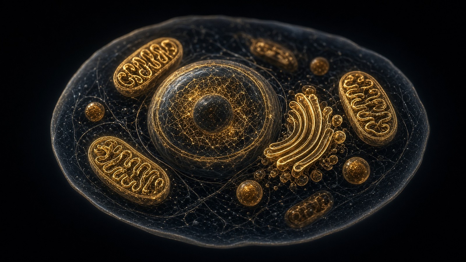
The plasma membrane is the outer wall. The city has interior walls. Rough ER is a bilayer continuous with the nuclear envelope, studded with ribosomes, the factory that writes secreted and membrane proteins into the lumen or the membrane itself — which is how a GPCR is born, helix by helix, at the Sec61 translocon, not in the cytosol as a finished tourist. Smooth ER is calcium store and lipid workshop. Golgi cisternae are a stack of walls that glycosylate and sort. Endosomes and lysosomes are walls whose interior is acid because a V-ATPase pumped it that way; that acid is why some occupied GPCRs drop ligand in the endosome and why LDL drops cholesterol. Mitochondrial outer membrane is a β-barrel porin wall, leaky to small solutes. Mitochondrial inner membrane is the five-nanometre object Mitchell needed for chemiosmosis: cardiolipin, cristae, Complexes I–V, ~150 mV, the ATP essay next door. Same physics as the plasma membrane. Different recipe. Different voltage. Different peptide (MOTS-c is written inside that inner city, not addressed to its outer wall).
In short. Inside the cell there are more membranes: ER, Golgi, nucleus, mitochondria, lysosomes. Same kind of wall, different job.
The nuclear envelope is a double bilayer, two walls, periplasmic-ish lumen continuous with ER, studded with nuclear pore complexes that aren't holes in a simple sense but selective phases of FG-repeat nucleoporins. Small proteins wander. Everything else needs a transport receptor and a Ran gradient. That's why Epithalon’s nuclear claims, whatever you think of them, are a different essay: a tetrapeptide accused of a transcriptional conversation has to have a route to chromatin or a receptor that talks to chromatin, and neither route is ‘it crossed one plasma membrane and therefore arrived’. Two walls, a pore with opinions, a lamina, a nucleolus. The nucleus essay holds that object. This paragraph exists so nobody treats ‘membrane’ as only the plasma membrane. A civilisation has interior doors. Small proteins wander. Everything else needs a transport receptor and a Ran gradient. That's why Epithalon's nuclear claims, whatever you think of them, are a different essay: a tetrapeptide accused of a transcriptional conversation has to have a route to chromatin or a receptor that talks to chromatin. Two walls, a pore with opinions, a lamina, a nucleolus.
In short. The nucleus has two membranes and guarded pores. Getting a message to DNA isn't the same as binding a receptor on the cell surface.
Traffic: the wall that moves
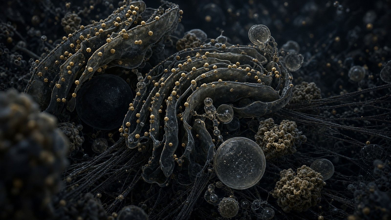
A membrane isn't a static bag. Vesicles bud and fuse continuously. Coat proteins (clathrin, COPI, COPII) bend the wall on purpose; BAR-domain proteins help; dynamin pinches. SNARE proteins on vesicle and target zip, membranes fuse, contents spill or proteins are delivered. Rothman, Schekman, Südhof, Nobel 2013. Occupied GPCRs are cargo in this traffic: GRK phosphorylation, β-arrestin, clathrin, endosome, recycle or lysosome, the desensitisation paragraph of the occupancy essay rewritten as a vesicle paragraph. Secretory granules are the opposite direction: a somatotroph’s GH, a β-cell’s insulin, a neuron’s peptide transmitter, fusion triggered by calcium that entered or was released through channels in a wall. The ligand on the outside that raised the calcium didn't have to enter. The granule on the inside had to fuse. Two uses of the same physics in one secretory event. Occupied GPCRs are cargo in this traffic: GRK phosphorylation, β-arrestin, clathrin, endosome, recycle or lysosome. Secretory granules go the other way: a somatotroph's GH, a β-cell's insulin, fusion triggered by calcium that entered or was released through channels in a wall. The ligand on the outside that raised the calcium didn't have to enter. The granule on the inside had to fuse.
In short. Membranes constantly pinch off as vesicles and fuse again. That's how occupied receptors get pulled inside, and how hormone granules dump.
Exocytosis of a granule is a membrane-area event as well as a cargo event. Add membrane, then retrieve it by endocytosis, or the cell grows. Synapses do this on a millisecond-to-second clock that would look like science fiction if it weren't measured. Palade saw the secretory pathway in the pancreas. A peptide house that sells ligands for secretory cells is, whether it admits it or not, in the vesicle business two steps downstream. We'll admit it. We won't pretend the lyophilised cake is a vesicle. HPLC characterises a chain. Fusion characterises a SNARE assembly in a living wall. Different assays, different floors. Synapses do this on a millisecond-to-second clock that would look like science fiction if it weren't measured. Palade saw the secretory pathway in the pancreas. A peptide house that sells ligands for secretory cells is, whether it admits it or not, in the vesicle business two steps downstream. HPLC characterises a chain. Fusion characterises a SNARE assembly in a living wall.
In short. When a cell dumps a hormone granule, it also adds a patch of membrane that it must later take back. The vial is still a chain on a chromatogram.
Sheets of wall: epithelia, endothelia, and a brief honesty about barriers
Stack the five-nanometre object into a sheet of cells, glue the cells with tight junctions — claudins, occludin, ZO proteins — and you have an epithelium: a barrier with opinions about what may cross paracellularly versus transcellularly. The intestinal epithelium is why PepT1 sits on an apical membrane and why a tripeptide has a dietary job. The blood–brain barrier is an endothelium with tight junctions, astrocyte endfeet, a transporter set that keeps most peptides out and lets a few through on purpose — LAT1, some SLC and ABC exporters. That's a real pharmacokinetic wall, and it's why a polar research peptide in a vial isn't, by default, a CNS ligand. Semax has an intranasal literature that is an attempt to negotiate this wall; the negotiation is a paper, not a caption. We don't print a crosses-the-BBB badge without a method. GHSR, melanocortin receptors and incretin receptors exist on the blood side of many interesting neurons — circumventricular organs, vagal afferents, brainstem with leakier bits — as well as on peripheral cells. Occupancy can be peripheral and still be neural.
In short. Sheets of cells sealed by tight junctions make the gut lining and the blood–brain barrier. Most peptides don't wander through those seals.
Skin is a wall of a different architecture: stratum corneum as dead, keratinised, lipid-mortared bricks, a barrier that topical cosmetic peptides mostly fail, which is why a GHK-Cu cream and a GHK-Cu research vial aren't the same object even when the tripeptide is. Endothelium is a wall that can fenestrate (liver, marrow) or be continuous (muscle, brain). Fenestration is why a hepatocyte sees more of the plasma’s contents than a cortical neuron does. Receptor occupancy is therefore a tissue-access story as well as a pocket story. The occupancy essay said tissue is a forgotten word. The wall is why. A ligand can't occupy a GPCR it can't reach, and reaching is a problem in sheets of this five-nanometre object plus the junctions between the cells that hold it. Endothelium is a wall that can fenestrate — liver, marrow — or be continuous — muscle, brain. Fenestration is why a hepatocyte sees more of the plasma's contents than a cortical neuron does. Receptor occupancy is therefore a tissue-access story as well as a pocket story. A ligand can't occupy a GPCR it can't reach, and reaching is a problem in sheets of this five-nanometre object.
In short. Skin, liver sinusoids and brain capillaries are different kinds of wall. A peptide can only occupy a receptor it can reach.
A dish at ten nanomolar is still a wall, and still not a person
A cultured cell has a plasma membrane with the same physics and a different composition, a different protein census, often a different voltage, and an unstirred layer the size of your technique. Ten nanomolar ligand in 50 millilitres is Avogadro's number of molecules against a few thousand receptors per cell against a million cells, which is still a large excess, which is how binding assays are designed. The wall is there. The GPCR is there, if you picked a cell that actually expresses it — HEK-CRISPR, CHO, a primary somatotroph: name it. Proteases in the medium, plastic adsorption, oxidation, the fact that a fatty-acylated peptide will stick to albumin if you added serum and to the dish if you didn't: all of that is the wall's neighbourhood in vitro. None of it is a person. A person is 36 trillion walls, a liver that does first-pass, peptidases such as DPP-4 for incretins, plasma binding, a blood–brain barrier, a kidney. We characterise ligands so a bench can make a concentration. The kit reconstitutes a cake. It doesn't authorise a body.
In short. A cell in a dish still has a real membrane. A human is 36 trillion membranes plus liver, kidney and enzymes. The vial is for the dish.
HPLC is how we know which chain we're talking about. Mass spectrometry asks whether the main peak has the right mass. ≥98% is a specification about a reagent, not a clinical claim. Solid-phase synthesis (Merrifield 1963; Nobel 1984) built the chain on a resin; TFA cleaved it; the cake is the dried object in the vial. Reconstitution is chemistry, not a rite. The occupancy happens when that solution meets a protein in a wall, in an assay you designed, with a readout you can defend: cAMP, calcium, internalisation, a binding isotherm, a secretion ELISA. If the readout is a forum, you have left this desk. If the readout is a person, you have left we’s label and entered someone else’s profession. The five-nanometre wall doesn't care. The Medicines and Healthcare products Regulatory Agency does. The occupancy happens when that solution meets a protein in a wall, in an assay you designed, with a readout you can defend: cAMP, calcium, internalisation, a binding isotherm, a secretion ELISA. Purity is a chromatogram. Identity is a mass. Occupancy is a wall with a pocket. Research use only sits at the close, as the legal class of the reagent.
In short. We prove what is in the vial with a chromatogram and a mass. You prove occupancy with an assay on a membrane protein.
A map of the wall, not a protocol
The map, then, in one paragraph you could tape above a bench. Thickness ~5 nm. Census 10⁸–10⁹ lipids in a typical plasma membrane. Singer and Nicolson 1972, fluid mosaic, still standing. Flippases spend ATP to keep PS inside; scramblases expose it on purpose. Rafts are a method-dependent word; caveolae you can photograph. Voltage is tens of millivolts across those five nanometres; Hodgkin and Huxley wrote the spike as sodium then potassium. Channels are holes with opinions; pumps write the gradients; GPCRs are seven helices that carry a message without carrying the messenger. Ipamorelin occupies GHSR in this wall. PT-141 occupies melanocortin receptors in this wall. Retatrutide occupies GLP-1R, GIPR and GCGR in this wall. KPV can be cargo on PepT1, a different protein in this wall. NAD+ isn't a ligand of this wall. The occupancy essay is the handshake. The scale essay is the city. We're looking at the five-nanometre reason a polar peptide can run a civilisation it never entered.
In short. Five nanometres, a billion fats, a fluid mosaic, pumps, channels, voltage, seven-helix receptors. Three catalogue peptides bind the outside.
If you can't name the wall, you don't yet have a place for a lock. If you name the wall and skip the leaflet, you have a cartoon. If you name the leaflet and skip the protein, you have a lipid and not a pharmacology. If you name the protein and then write a protocol for a person, you have left we’s label. The membrane essay stops at the map. The chromatogram is the receipt. The papers are public — Singer and Nicolson, Hodgkin and Huxley, Raun, Jastreboff, Dalmasso and Merlin, Lefkowitz and Kobilka’s lectures. The vial is a reagent. Research use only. That's the whole arrangement, and it is enough. Five nanometres is a civilisation. We sell messages for one class of door in it. We don't sell the city. The chromatogram is the receipt. The papers are public — Singer and Nicolson, Hodgkin and Huxley, Raun, Jastreboff, Dalmasso and Merlin, Lefkowitz and Kobilka's lectures. The vial is a reagent. Research use only. That's the whole arrangement, and it's enough. Five nanometres is a civilisation. We sell messages for one class of door in it. We don't sell the city.
In short. Name the membrane, the protein, and the assay. Don't turn a research vial into a human protocol. The cake is a reagent.
A peptide ligand never has to enter if the GPCR is doing its job. The wall is why. Tourism is a different literature. NAD+ is a different floor.
Questions the essay actually answers
- How thick is a cell membrane?
- About five nanometres. Two phospholipid leaflets, headgroups out, hydrocarbon tails in. Gorter and Grendel measured the bilayer idea on erythrocytes in 1925; electron microscopy and later X-ray and neutron scattering put the hydrophobic core near 3 nm and the whole sandwich near 4–5 nm. A research peptide a few nanometres long is the same size as the wall, which is why tourism through the hydrocarbon core is not the default plan.
- How many lipids are in a cell?
- On the order of 10⁸–10⁹ lipid molecules in the plasma membrane of a typical animal cell, more if you count the ER, Golgi, mitochondria and endosomes. Alberts’ textbook figure and the BioNumbers compilation agree on that order. An E. coli is an order of magnitude leaner. The number is a census, not a mood.
- What is the fluid mosaic model?
- Singer and Nicolson, Science, 1972: a lipid bilayer as a two-dimensional fluid, globular proteins embedded in it and free to diffuse laterally, not a rigid protein–lipid–protein sandwich. Frye and Edidin’s 1970 heterokaryon mixing experiment was the kinetic hint. Rafts, cytoskeletal fences and the picket-fence picture are later footnotes. The mosaic still stands.
- What does a flippase do?
- It spends ATP to move a specific lipid from one leaflet to the other, against the easy direction. P4-ATPases (ATP8, ATP11 families, with a CDC50 subunit) flip phosphatidylserine and phosphatidylethanolamine to the cytoplasmic leaflet. Floppases (ABC transporters) push lipids the other way. Scramblases (TMEM16F, Xkr8) equilibrate the two leaflets when calcium or caspase says so — which is how dying cells expose PS as an eat-me signal.
- Do peptide ligands need to enter the cell?
- Most GPCR ligands do not. They occupy an extracellular pocket. The seven helices rearrange. A G protein on the inside spends GTP. Information crossed; the peptide, in the boring and correct case, is still outside. Ipamorelin at GHSR, PT-141 at melanocortin receptors, retatrutide at GLP-1R/GIPR/GCGR: occupancy, not tourism. KPV can cross via PepT1, which is a transporter. NAD+ is not a membrane ligand.
- What did Hodgkin and Huxley actually do?
- They voltage-clamped the squid giant axon and wrote the action potential as time- and voltage-dependent sodium and potassium conductances, in four Journal of Physiology papers in 1952. Nobel Prize 1963, with Eccles. They did not have the channel proteins; they had the currents. The proteins (Nav, Kv) were cloned decades later. The equations still run every serious neuroscience course.
- What is PepT1, and why does KPV care?
- PepT1 is SLC15A1, a proton-coupled di- and tripeptide transporter on the apical membrane of enterocytes and some immune cells. KPV (Lys-Pro-Val), the C-terminal tripeptide of α-MSH, has uptake literature on this transporter and an NF-κB literature downstream. That is a cargo story, not a melanocortin tan. Dalmasso, Merlin and colleagues are the papers.
- Is NAD+ a membrane ligand?
- No. NAD+ is a 663-dalton redox cofactor and a co-substrate for sirtuins and PARPs. It is not a GPCR ligand and it is not a channel ligand. CD38 on some plasma membranes is an NAD-consuming ectoenzyme; that makes NAD+ a substrate of a membrane enzyme, not a ligand of a receptor. The 1000 mg vial in the catalogue is lyophilised β-NAD+ for the bench. Different floor from ipamorelin.
- What is a lipid raft?
- A cholesterol- and sphingolipid-enriched nanodomain in the outer leaflet, proposed by Simons and Ikonen in 1997 as a sorting and signalling platform. Detergent-resistant membranes oversold their stability. The modern picture is smaller, more transient, still argued. Caveolae (caveolin-1, cavin) are the morphologically honest cousins. Not a protocol and not a product claim.
- What is a GPCR doing in the membrane?
- Seven transmembrane helices, an extracellular face that binds ligand, an intracellular face that acts as a guanine-nucleotide exchange factor for a heterotrimeric G protein. About 800 in the human genome. The wall is why the ligand does not have to enter. Lefkowitz and Kobilka, chemistry Nobel 2012. The occupancy essay next door is the handshake in full.
Hypothetical research reconstitution
How these vials are typically mixed
Hypothetical research reconstitution for the named catalogue vial. Not a protocol, not medical advice, not a use instruction. These amounts sit in published and commonly cited laboratory ranges. The vial is labelled for research use only — not for human or veterinary administration.
Ipamorelin
10mg
Mix with 2 ml bacteriostatic water → 5 mg/ml · 5,000 mcg/ml
- Hypothetical aliquot
- 200–300 mcg
- 0.04–0.06 ml · 4–6 units on a U-100 syringe
- How often
- Once or twice daily (morning and/or evening)
- 8–12 weeks
Bench steps
- Let the vial sit until it is no longer cold to the touch.
- Wipe the stopper with 70% isopropyl alcohol. Let it dry.
- Draw 2 ml bacteriostatic water (0.9% benzyl alcohol).
- Run the water slowly down the inside glass — do not blast the cake.
- Roll between finger and thumb until the cake is gone. Do not shake.
- Label the date. Store the solution at 2–8 °C. Do not freeze. Use within 30 days unless the note below says otherwise.
GHS-R1a hexapeptide. The 200 mcg mark is the usual starting aliquot. Stacks with CJC-1295 no DAC in the papers that run both.
PT-141
10mg
Mix with 2 ml bacteriostatic water → 5 mg/ml
- Hypothetical aliquot
- 0.5–1.0 mg
- 0.10–0.20 ml · 10–20 units on a U-100 syringe
- How often
- As required in the bremelanotide literature; not a daily molecule
- Per experiment, not a calendar cycle
Bench steps
- Let the vial sit until it is no longer cold to the touch.
- Wipe the stopper with 70% isopropyl alcohol. Let it dry.
- Draw 2 ml bacteriostatic water (0.9% benzyl alcohol).
- Run the water slowly down the inside glass — do not blast the cake.
- Roll between finger and thumb until the cake is gone. Do not shake.
- Label the date. Store the solution at 2–8 °C. Do not freeze. Use within 30 days unless the note below says otherwise.
Free-acid analogue of MT-II. Palatin’s research dose around 1.75 mg is a licensed-medicine figure, not a shop instruction. 0.5 mg is where most bench notes start.
Retatrutide
30mg
Mix with 3 ml bacteriostatic water → 10 mg/ml
- Hypothetical aliquot
- 1–2 mg to start; published trial arms ran higher by week
- 0.10–0.20 ml · 10–20 units on a U-100 syringe (at 1–2 mg)
- How often
- Once weekly
- The Jastreboff NEJM 2023 arms ran 48 weeks. That is a trial, not a shop protocol.
Bench steps
- Let the vial sit until it is no longer cold to the touch.
- Wipe the stopper with 70% isopropyl alcohol. Let it dry.
- Draw 3 ml bacteriostatic water (0.9% benzyl alcohol).
- Run the water slowly down the inside glass — do not blast the cake.
- Roll between finger and thumb until the cake is gone. Do not shake.
- Label the date. Store the solution at 2–8 °C. Do not freeze. Use within 30 days unless the note below says otherwise.
LY3437943 architecture. Weekly, not daily. Those milligram figures are what the papers used on the investigational medicine — they are not a use instruction for this reagent.
Bacteriostatic water and sterile syringes ship with peptide orders over £75. Kit details · 10 ml bacteriostatic water
The vials this essay sits on
Named sequences the essay maps — Ipamorelin, PT-141, Retatrutide. Hypothetical research neighbourhood, not a protocol, not a medicine. One press puts every in-stock vial in the bag.
 Research only
Growth axis
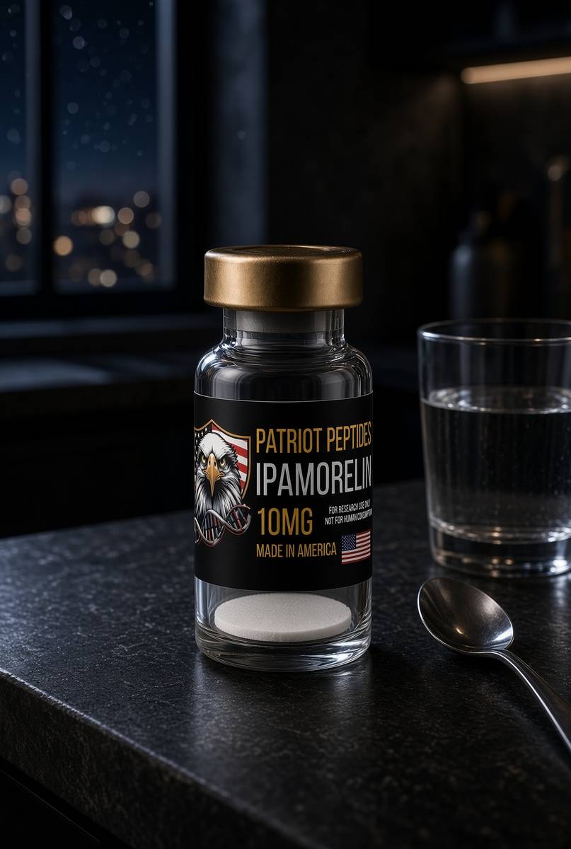
Ipamorelin
10 mg ipamorelin. The clean ghrelin-receptor pentapeptide.
4.7(457)
35 browsing this now · 4 purchased in the last 24 hours
10mg · In stock
£30.00
 Made in USA
 Made in USAOut of stock
Incretin
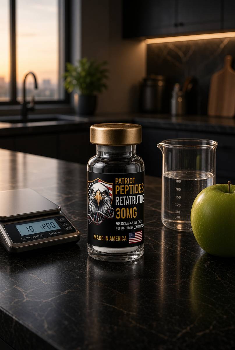
Retatrutide
US-made retatrutide 30mg — the published structure LY3437943, HPLC-MS verified.
4.6(609)
60 browsing this now · 5 purchased in the last 24 hours
30mg
£120.00
Research use only. Not a combined-use instruction.
Read next
70 min · long read · The living cell
How peptides talk to cells: occupancy, amplification, arrestin
A peptide is a ligand. Most of the catalogue binds a GPCR on the cell surface: one occupancy, then enzymes make thousands of second messengers. That amplification is real, and it is not magic. Desensitisation is why more ligand is not more signal forever.
65 min · long read · The living cell
The living cell is a city, and you are 36 trillion of them
A 70 kg adult is on the order of 36 trillion cells, most of them red blood cells with no nucleus. A typical nucleated cell holds about ten billion proteins and two metres of DNA. The body recycles 40–60 kg of ATP a day. Those figures are published; this essay is what they mean.
More in this desk
82 min · long read · The living cell
Pathophysiology from the genome to a person who notices
Disease is a stack: genome, transcriptome, proteome, metabolome, organelle, cell fate, tissue, organism. A peptide binds one node. The rest of the stack keeps running. CFTR, type 2 diabetes and a tendon as worked examples.
64 min · long read · The living cell
From gene to protein: how a cell actually reads itself
DNA is transcribed, spliced, exported, translated and folded. A research peptide is the ligand at the end of that pipeline, already made. This essay is every machine in between — polymerases, spliceosome, ribosome — with the actual rates attached.
64 min · long read · The living cell
Mitochondria: the bacterium you kept, the genome it kept, the peptides it writes
You turn over 40–60 kg of ATP a day using a 16,569-base genome that still uses a bacterial genetic code. NAD+ is the hydride carrier Complex I spends. MOTS-c is a 16-mer translated from mitochondrial 12S rRNA — Lee, Kim, Cohen, 2015. That last sentence is real, and it is surprising.
57 min · long read · The living cell
Proteostasis: the cell that eats its own mistakes
Ten billion proteins, a 76-residue tag, a 2.5-megadalton proteasome, and autophagy for whole organelles. Hershko, Ciechanover and Rose, 2004. Ohsumi, 2016. DSIP is a sleep-isolation nonapeptide, not an autophagy ligand.
Essays describe published research. They are not medical advice and they do not authorise human use of any catalogue item.