

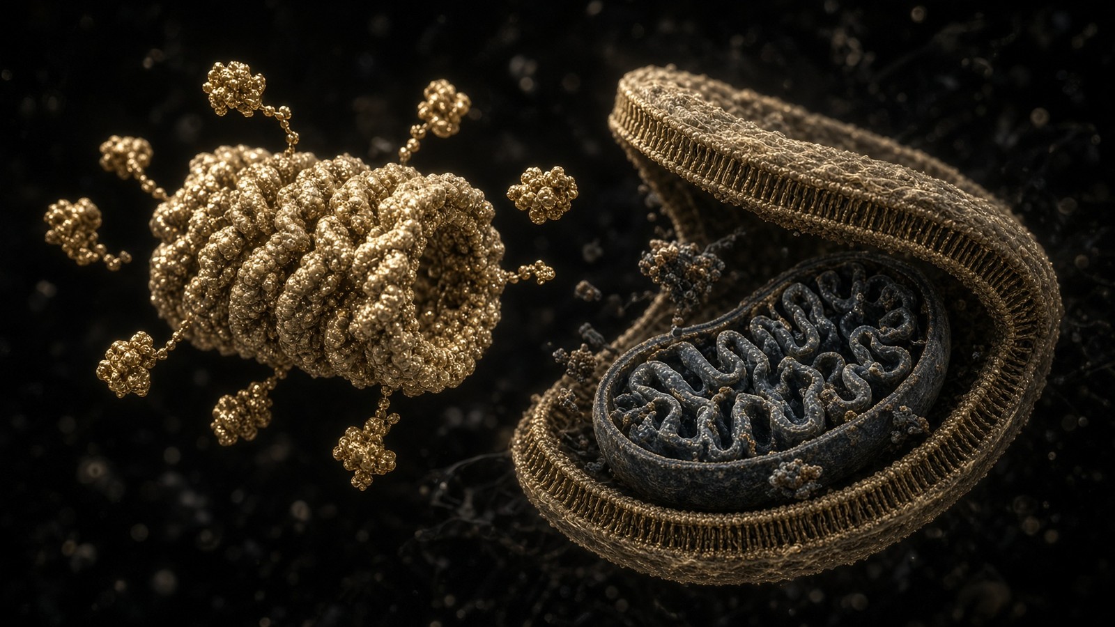
The living cell · 57 min · 12,495 words
Proteostasis: the cell that eats its own mistakes
Ten billion proteins, a 76-residue tag, a 2.5-megadalton proteasome, and autophagy for whole organelles. Hershko, Ciechanover and Rose, 2004. Ohsumi, 2016. DSIP is a sleep-isolation nonapeptide, not an autophagy ligand.
What this essay actually tells you
- A growing mammalian cell holds ~10¹⁰ protein molecules. Translation errs at ~10⁻⁴ per residue. Proteostasis is chaperones, a 76-residue ubiquitin tag, the 26S proteasome, and autophagy. Balch, Morimoto, Dillin, Kelly, Science 2008.
- K48-linked ubiquitin chains sentence a client to the 26S. K63-linked chains are more often a signal. Hershko, Ciechanover and Rose, Chemistry Nobel 2004. Ohsumi, Medicine Nobel 2016, for the ATG genes.
- mTORC1 at the lysosome says build; AMPK says inventory. DSIP is WAGGDASGE from a sleeping rabbit brain — not an autophagy ligand. NAD+ and MOTS-c sit on nearby nodes. HPLC-characterised sequences, labelled for the bench.
What this actually means
A growing mammalian cell holds on the order of ten billion protein molecules. They are not furniture. A few percent turn over every hour because synthesis is a draft: translation errs at about one residue in ten thousand, folding is a kinetic bet, oxidative damage never clocks off, and a protein that has finished its job is a liability if it stays. Proteostasis is the network that keeps that warehouse honest — chaperones that fold, a 76-residue tag called ubiquitin that sentences, a 26S proteasome that shreds tagged clients into peptides, and autophagy that swallows what the proteasome cannot, including whole mitochondria. K48-linked ubiquitin chains are the classic degradative sentence. K63-linked chains are more often a signal. mTORC1, sitting on the lysosome, says build; AMPK, listening to AMP/ATP, says inventory. ULK1 is the kinase both of them phosphorylate, on different residues, with opposite intent. PINK1 and Parkin are how a depolarised mitochondrion is tagged for mitophagy. Hershko, Ciechanover and Rose got the 2004 Chemistry Nobel for ubiquitin-mediated degradation. Ohsumi got the 2016 Medicine Nobel for autophagy. DSIP is WAGGDASGE, a nonapeptide isolated from rabbit cerebral venous blood during delta-wave sleep; it is not an autophagy ligand. NAD+ is the hydride coin sirtuins spend when they deacetylate autophagy machinery. MOTS-c is a 16-mer from mitochondrial 12S rRNA that sits on AMPK. Three occupancies. One warehouse. The vials are reagents. Research use only.
Diagram
A growing mammalian cell turns over a few percent of its proteome per hour. 10 billion proteins is a warehouse with a shredder on the floor, not a museum. Ageing is partly what happens when the shredder slows and the warehouse fills with unfolded inventory.
Hershko, Ciechanover, Rose — Nobel 2004. E1 activates, E2 conjugates, E3 selects the victim. K48-linked chains go to the 26S proteasome; K63 is more often a signal. Autophagy handles the organelles the proteasome cannot swallow. mTOR vs AMPK is the appetite switch.
A growing mammalian cell holds on the order of ten billion protein molecules. Milo and Phillips put the figure there; a HeLa cell and a hepatocyte sit near it; a red blood cell, which has ejected its nucleus and most of its proteome, does not. Those ten billion aren't a collection. They're a flux. Eden, Alon and colleagues, watching living human cells with a fluorescent timer, saw proteome half-lives that run from tens of minutes to days. Schwanhäusser and Selbach, doing ribosome profiling and mass spectrometry in mouse fibroblasts, put copy numbers across six orders of magnitude and synthesis rates that would fill a warehouse every generation if nothing left. Something has to leave. A few percent of the protein mass of a growing cell is degraded every hour. That isn't entropy winning. That's a programme. The cell eats its own mistakes, and its finished jobs, and its organelles when those organelles have failed a voltage test, and it does this with named machines, named tags, named kinases, and two Nobels. If you file the whole of that under detox, you haven't visited the floor.
In short. A typical cell holds about ten billion proteins, and a few percent of them are destroyed every hour on purpose. That turnover is a programme with named machines, not a cleanse.
We're looking at that programme in some detail. Proteostasis is the word Balch, Morimoto, Dillin and Kelly put on the network in Science in 2008: folding, holding, tagging, shredding, swallowing. Upstream sits translation, which errs at about 10⁻⁴ per residue, and folding, which Anfinsen showed is written in the sequence and which the cytoplasm, crowded at 200–400 mg/ml, doesn't always honour on the first try. Midstream sits ubiquitin, 76 residues, a C-terminal glycine, seven lysines that can be chained in different topologies, and an E1–E2–E3 cascade that Hershko, Ciechanover and Rose took apart in reticulocyte lysates until the field had to stop calling ATP-dependent proteolysis a curiosity. Downstream sit two disposal routes that aren't synonyms: the 26S proteasome, a 2.5-megadalton barrel that unfolds a tagged client and cuts it into peptides, and autophagy, a double-membrane appetite that can take a protein aggregate, a bacterium, or a mitochondrion that has lost its membrane potential. mTORC1 and AMPK set the appetite by phosphorylating ULK1 on opposite residues. PINK1 and Parkin are how a former bacterium is sentenced when the voltage fails. The neighbouring essays are the living cell at scale, the mitochondrion as second genome, and fasting as the organism-level switch.
In short. Folding, ubiquitin tagging, the proteasome and autophagy are one network. Two Nobels named the machines.
- Proteins per growing cell
- ~10¹⁰
- Translation error
- ~10⁻⁴ / residue
- Ubiquitin
- 76 residues
- Human E3 ligases
- ~600+
- 26S proteasome
- ~2.5 MDa
- ATG genes (yeast)
- 18+ core
- mTORC1 vs AMPK on ULK1
- Ser757 vs Ser317/777
- MOTS-c
- 16 residues
- DSIP
- 9 residues
Milo, Phillips, BioNumbers. A flux, not a museum.
One wrong amino acid per ten thousand. Proteins turn over. DNA does not.
8.6 kDa. C-terminal Gly. Seven lysines plus Met1. The tag.
RING, HECT, RBR. Specificity lives here, not in the barrel.
19S + 20S. Threonine proteases in a gated barrel. Peptides out, Ub recycled.
Ohsumi’s screens. ULK1, VPS34–Beclin-1, LC3 lipidation in mammals.
Opposite phosphorylations. Build versus inventory. Kim 2011; Egan 2011.
From 12S rRNA. AMPK neighbourhood. Not an autophagy drug.
WAGGDASGE. Sleep-isolation story, 1977. Not ULK1, not LC3.
A warehouse, not a museum
The temptation, looking at a stained section, is to treat the proteome as architecture. Actin looks like architecture. Histones look like architecture. Haemoglobin, in a red cell, is architecture that has fired the architect. Most of the rest is stock. Enzymes of glycolysis are present in millions of copies and still have half-lives measured in hours to a day. Transcription factors may be present in tens or hundreds of copies and last minutes. Cyclins are built to be destroyed; that is the whole point of a cell cycle, which is the neighbouring desk. MHC class I peptides are proteasome products, which is why a proteasome inhibitor is an immunology drug as well as a myeloma drug, and why Rock, Goldberg and Palombella had to teach a generation of immunologists that antigen presentation is a degradation story. A protein that has done its job and remains isn't a keepsake. It's a surface that can oxidise, a domain that can unfold, a partner that can stick to the wrong neighbour in a cytoplasm that is already 200–400 mg/ml of macromolecule. Ellis called that crowding; Zimmerman and Trach measured it. Weak interactions become chemistry at those concentrations.
In short. Most proteins are stock with a half-life, not architecture. Crowding turns a misfolded molecule into a nucleation event.
Half-lives aren't a moral quality. They're a design. Bachmair, Finley and Varshavsky, Science, 1986, showed that the in vivo half-life of a β-galactosidase reporter in yeast is a function of its N-terminal residue: some amino acids sentence a protein to minutes, others to hours. That's the N-end rule, a ubiquitin-dependent pathway, and it is still one of the cleanest demonstrations that degradation is encoded, not accidental. Ornithine decarboxylase is a famous exception that proves the aesthetic: it can be degraded by the proteasome without ubiquitin, via antizyme, because polyamine homoeostasis couldn't wait for a cascade. Hypoxia-inducible factor α is hydroxylated when oxygen is available, captured by the von Hippel–Lindau E3, and shredded, so that HIF is a proteasome story as much as a transcription story. NF-κB is held in the cytoplasm by IκB; a cytokine signal leads to IκB ubiquitination and proteasomal destruction, and the transcription factor walks into the nucleus. Palombella, Rando, Goldberg and Maniatis, Cell, 1994. A generation of ‘inflammatory tone’ language is downstream of a tagged inhibitor being fed into a barrel. If you can't name the tag and the barrel you're decorating a caption.
In short. Half-life is encoded. The N-end rule, HIF and NF-κB are ubiquitin-and-proteasome stories, not moods That's the take-home, and it's enough to walk with.
Ageing, in this vocabulary, is partly what happens when the warehouse fills. López-Otín, Blasco, Partridge, Serrano and Kroemer put ‘loss of proteostasis’ among the hallmarks of ageing in Cell in 2013, and they weren't reaching: inclusion bodies, lipofuscin, a slower proteasome, a blunter autophagic response to starvation, chaperones that have been asked to hold too much for too long. Ben-Zvi, Miller and Morimoto watched proteostasis collapse as an early event in C. elegans, before the organism looked old. That's a worm. It's also a warning about captions. A human neuron isn't a worm, and a lyophilised peptide isn't a restoration of a network that took a billion years to assemble. The honest move is to name the machines, name the assays, and leave the restoration claims to people who have both. We have the machines. We have three catalogue occupancies that sit near the network without being it. We don't have a proteostasis therapy on the till, and we won't pretend the chromatogram is one.
In short. Ageing is partly a warehouse that fills as the shredder and the appetite slow. That's a named hallmark, not a product category.
The draft is allowed to be wrong. The archive is not.
Diagram
- DNA replication + MMR10⁻⁹ to 10⁻¹⁰A genome of 6 Gbp (diploid) accumulates a handful of mutations per division.
- Transcription~10⁻⁵RNA is disposable. The cell can afford a wrong letter in a message that lasts hours.
- Translation~10⁻⁴One wrong amino acid per ten thousand. Proteins turn over. DNA does not.
- mtDNA10–100× nuclearNo histones, ROS next door, weaker repair. The second genome ages faster.
The genome is sacred, the message is cheap, the protein is cheaper. Ageing is partly what happens when the sacred copy still drifts — and when mitochondria, which never got the nuclear repair budget, drift faster.
The neighbouring dogma essay made the gradient of care explicit. DNA replication, after proofreading and mismatch repair, is allowed about one error in a billion to ten billion bases, because a mutation is an archive error and the archive is copied into every descendant. Transcription is sloppier by four or five orders of magnitude because an mRNA is disposable. Translation, at about 10⁻⁴ per residue, is sloppier again. A 400-residue protein, printed on a eukaryotic ribosome at five or six amino acids a second, is statistically likely to carry a wrong residue somewhere if you make enough copies. Many of those substitutions are silent in the fold. Some are not. Some stall the ribosome. Stalled ribosomes are themselves a proteostasis problem: the ribosome-associated quality-control pathway, with Listerin (Ltn1) as an E3 on the split 60S, ubiquitinates the aborted nascent chain so the proteasome can finish a job the ribosome could not. Bengtson and Joazeiro, Nature, 2010. Brandman, Hegde, the RQC complex. A cell that couldn't eat a stalled translation product would fill with stubs. The stubs aren't a physiology. They're a substrate.
In short. Translation is allowed to be wrong at one residue in ten thousand because proteins turn over. Stalled ribosomes are tagged and shredded.
Mitochondrial translation is worse, and the neighbouring mitochondria essay is why. The organelle prints thirteen hydrophobic respiratory subunits on bacterial-ish ribosomes, next to a chain that leaks superoxide, with a code that already drifted, and with quality control that is real but thinner than the cytosolic apparatus. Unassembled respiratory subunits are a proteostasis problem inside the matrix (LON, ClpXP) and at the inner membrane (i-AAA, m-AAA). A failed assembly is how a complex I deficiency begins even when the mtDNA letters are fine. The cytosolic warehouse and the mitochondrial warehouse share a cell and don't share a shredder. When the voltage fails, the whole organelle can be offered to autophagy. That's later in this essay. The point of putting error rates this high in a proteostasis piece is to stop treating degradation as a pathology. Degradation is the reason a 10⁻⁴ process is compatible with a 36-trillion-cell organism. The genome is sacred. The proteome is a draft. Ageing is partly what happens when the draft starts to be treated as an archive because the editors have slowed.
In short. Mitochondria print thirteen subunits next to a furnace, so their quality control is harsher and thinner. Degradation is why a sloppy printer can still run a body.
Folding is written in the sequence, and the cytoplasm still cheats
Christian Anfinsen took reduced, denatured ribonuclease and watched it find its disulphides and its activity again. Science, 1973, summing work that had been running since the early 1960s: the native fold of a small protein is thermodynamically written in the amino-acid sequence, given the solvent. Levinthal had already pointed out that a random search of conformational space would take longer than the age of the universe; the sequence must encode a pathway, not a phone book. That's the in-vitro story, and it is true, and it isn't a cell. A cell is a crowded, ATP-burning, membrane-partitioned solvent in which a nascent chain is coming off a ribosome at five residues a second with its N-terminus already trying to fold while the C-terminus is still in the exit tunnel. Hartl, Hayer-Hartl, Bukau, Horwich: the chaperone field is the admission that Anfinsen’s thermodynamic sentence has a kinetic problem in vivo. Hsp70s, with their J-domain partners, bind hydrophobic stretches that shouldn't be exposed, using ATP to time the hold. Hsp90 takes a more specialised clientele — kinases, steroid receptors — and is itself an ATP-burning dimer.
In short. Anfinsen showed the fold is in the sequence. Chaperones exist because a crowded cell isn't a dilute tube.
The heat-shock response is how the warehouse shouts. Ritossa saw the puffs on Drosophila chromosomes in 1962; Tissières named the proteins; Morimoto, Wu, Lindquist, Sistonen spent careers on HSF1, the transcription factor that, when released from Hsp90 and Hsp70, writes the chaperone genes. A cell that is already holding too much unfolded protein will, if it can, make more holders. That's a transcriptional programme, floor 2 of the pathophysiology stack, in the service of floor 3. It's also saturable. Gidalevitz and Morimoto showed that a polyglutamine expansion in C. elegans can break folding of metastable proteins that had nothing to do with the expansion, because the chaperone budget is finite. That sentence is the deepest one in proteostasis: there is a budget. You can spend it on a huntingtin exon, or on the endogenous metastable proteome, or on a heat shock, and you can't spend it three times. Ageing spends it slowly. A catalogue peptide doesn't top it up. Saying otherwise is selling a budget as a feeling.
In short. Heat shock writes more chaperones because the holding budget is finite. A polyglutamine protein can steal that budget from innocent clients.
The endoplasmic reticulum has its own panic, and the pipeline has a whole essay queued for it. Briefly, because the proteasome and autophagy both take ER clients: a secretory or membrane protein that fails to fold is held by BiP, calnexin, calreticulin, the oligosaccharide code Helenius mapped, and then retrotranslocated, ubiquitinated by ER-resident E3s (Hrd1, gp78, MARCH6, RNF5, depending on the lesion), and shredded by the cytosolic proteasome. That's ER-associated degradation, ERAD. If the load is high, IRE1 splices XBP1 mRNA, PERK phosphorylates eIF2α and shuts down most translation while ATF4 is still made, and ATF6 is cut in the Golgi and becomes a transcription factor. Walter, Ron, Mori, Harding. The unfolded protein response is a proteostasis programme for one organelle. Chronic UPR is a cell-fate programme: CHOP, apoptosis. CFTR ΔF508, which the pathophysiology essay already used as a worked example, is a folding-and-ERAD story before it is a chloride-channel story. Riordan named the gene in 1989. The lung is six floors up. The proteasome ate the almost-right channel on floor 3. A peptide doesn't ‘fix CF’. A corrector that chaperones CFTR toward the membrane is a different literature, and it isn't this catalogue.
In short. Misfolded ER proteins are sent back and shredded — ERAD. If the load is high, the unfolded protein response panics.
Seventy-six residues that sentence a protein
Ubiquitin is 76 amino acids, about 8.6 kilodaltons, with a C-terminal glycine that is the business end and a fold so conserved that yeast ubiquitin works in a human cell and the sequence differences are a rounding error. Gideon Goldstein isolated it in 1975 as a polypeptide that seemed to be everywhere — hence the name, which has aged into a joke the molecule has earned. The proteolytic identity came later, from a different bench. Avram Hershko and Aaron Ciechanover, in Haifa, fractionating rabbit reticulocyte lysates that degraded abnormal proteins in an ATP-dependent way, found a heat-stable factor they called APF-1. Ciechanover, Hod, Hershko, Biochemical and Biophysical Research Communications, 1978. Irwin Rose, at Fox Chase, put the ATP chemistry on a proper footing: ubiquitin is adenylated, then held as a thioester. Hershko, Ciechanover, Heller, Haas and Rose, Proceedings of the National Academy of Sciences, 1980, proposed that proteins were being conjugated to multiple chains of the polypeptide. Wilkinson, Urban and Haas, in 1980, showed that APF-1 was Goldstein’s ubiquitin. A tag from an immunology paper had become the tag in ATP-dependent proteolysis.
In short. Ubiquitin is a 76-residue tag. Hershko, Ciechanover and Rose showed it marks proteins for ATP-dependent destruction.
The conjugation is an isopeptide bond: the C-terminal carboxyl of ubiquitin, usually to the ε-amino of a lysine on the substrate. Sometimes the N-terminal methionine of ubiquitin itself is used (M1, linear chains, LUBAC). Sometimes a serine or threonine, rarely. The chain is built on one of ubiquitin’s own lysines. There are seven: K6, K11, K27, K29, K33, K48, K63. Homotypic K48 chains are the textbook proteasome-targeting signal. Chau, Tobias, Bachmair, Varshavsky, Science, 1989: a multiubiquitin chain is required for efficient degradation. Thrower, Hoffman, Rechsteiner and Pickart, EMBO Journal, 2000: four K48-linked ubiquitins is about the threshold the proteasome wants. K11 chains, especially those written by the anaphase-promoting complex, are also degradative; Jin, Rape and colleagues made that a cell-cycle fact. K63 chains are the other famous topology: they scaffold, they recruit, they don't usually feed the barrel. DNA-damage response, endocytosis, innate-immunity signalling, the handles that autophagy receptors hold. Komander and Rape called it the ubiquitin code, and they weren't being cute. Mixed chains, branched chains, phosphorylated ubiquitin (PINK1 puts phosphate on Ser65 — later), acetylated ubiquitin: the code got denser after the Nobel, which is what codes do when a field is allowed to look.
In short. Ubiquitin is usually glued to a lysine. K48 chains of about four units send a protein to the proteasome.
E1 activates, E2 conjugates, E3 selects the victim
Three enzymes, a bucket brigade, ATP spent at the first step. UBA1, the principal human E1, binds ubiquitin, ATP and magnesium, forms ubiquitin-adenylate, and then a thioester between the C-terminus of ubiquitin and a cysteine on E1. A second ubiquitin is adenylated while the first waits as a thioester; the loaded E1 is a two-ubiquitin enzyme. Haas and Rose put numbers on that chemistry. The thioester is transferred to a cysteine on an E2. Humans encode something on the order of forty E2s; they aren't interchangeable. Some prefer to build K48 chains, some K63, some monoubiquitin, some need an E3 to dictate the linkage. The E3 is the selector. There are more than six hundred in the human genome, which is a number that should make anyone who wants a single ‘ubiquitin pathway’ sit down. RING-domain E3s — the majority — don't take the ubiquitin themselves. They hold the E2 and the substrate in a geometry that lets the E2 fire. c-Cbl, MDM2, the SCF complexes, the anaphase-promoting complex, Parkin in its RING–between–RING family: different RINGs, different victims.
In short. E1 spends ATP to activate ubiquitin. E2 carries it That's the take-home, and it's enough to walk with.
MDM2 is the E3 the neighbouring cell-cycle essay already named: it ubiquitinates p53, the proteasome eats p53, and a cell that hasn't seen damage keeps a guardian at a trickle. Nutlins occupy the MDM2–p53 interface; they aren't this catalogue. Parkin is the E3 we'll spend pages on, because a depolarised mitochondrion is one of the warehouse’s largest mistakes and Parkin is how the mistake is tagged. BRCA1/BARD1 is a RING heterodimer that ubiquitinates at DNA-damage sites, mostly K6 and K63, a signalling story that is also a tumour-suppressor story. The SCF family — Skp1, Cullin, F-box — swaps F-box proteins to change substrate, and the F-box is often reading a phosphodegron, which is how a kinase cascade becomes a degradation cascade. β-TrCP reading phosphorylated IκB. Fbw7 reading phosphorylated cyclin E and Myc. The anaphase-promoting complex, an enormous RING-based E3, reading D-boxes and KEN-boxes so that securin and cyclin B die on schedule and anaphase can happen. Hershko’s later work, and Nurse’s, and Hunt’s, and the whole 2001 cell-cycle Nobel, sit on this E3. Proteostasis isn't a side-quest of the cell cycle. The cell cycle is a proteostasis programme with a spindle.
In short. MDM2 eats p53, Parkin tags failed mitochondria, APC/C destroys cyclin B so anaphase can happen. The cell cycle is a proteostasis programme with a spindle.
A research peptide isn't an E3, and it isn't a ubiquitin. The 76-residue protein is too large and too conserved to be a catalogue ligand in we, and the E3s are enzymes, not pockets waiting for a 9-mer. The honest occupancy question, when a peptide sits next to this essay, is whether the ligand moves a kinase that the network already listens to, or a cofactor the deacetylases spend, or a night-time literature that search bars keep filing under autophagy because the internet can't tell ULK1 from a sleep-isolation story. Those three questions have names in the catalogue: MOTS-c, NAD+, DSIP. We'll answer them after the machines are on the table. Answering them first is selling the gap. MOTS-c sits on AMPK, a fuel-gauge kinase the autophagy switch already listens to. NAD+ is the coin sirtuins spend when they deacetylate autophagy machinery. DSIP is WAGGDASGE, a sleep-isolation nonapeptide, and it doesn't occupy ULK1 or LC3. We'll put those three back on their own nodes after the machines are on the table, which is the kinder order.
In short. This catalogue doesn't sell ubiquitin or an E3. MOTS-c, NAD+ and DSIP sit near the network as occupancies of other nodes.
K48 is a sentence. K63 is often a memo.
The proteasome’s 19S cap has receptors (Rpn1, Rpn10, Rpn13) that prefer chains, and a geometry that has been measured: a tetraubiquitin K48 chain binds well enough to commit a client. K63 tetraubiquitin binds more poorly to those same receptors and is edited more readily by proteasome-associated deubiquitinases, which is one reason K63-tagged proteins are not, as a class, proteasome substrates. That's biochemistry, not a personality. Exceptions exist; the code was always a bit leaky. K11 chains from APC/C are a cell-cycle degradative signal that Pickart didn't have in the 1989 picture. M1 linear chains, written by LUBAC (HOIP, HOIL-1L, SHARPIN), are an NF-κB signalling device; Kirisako, Tokunaga, Iwai. K6 has a DNA-repair and mitophagy flavour. K27, K29, K33 are the under-studied remainder, which is a polite way of saying the code still has unread letters. Xu, Peng, Gygi and colleagues, using quantitative proteomics, showed that unconventional chains aren't folklore; they're on real substrates in real cells. A Western blot with ‘anti-ubiquitin’ is a start. A linkage-specific antibody or a middle-down mass spectrum is a sentence. Most peptide papers that mention ubiquitination have the blot. Almost none have the linkage. That's the gap, and it isn't mysterious. Linkage-specific work is harder.
In short. K48 tetraubiquitin is what the proteasome wants. K63 is usually a signal other proteins read.
Deubiquitinases, DUBs, take the tag off. There are roughly a hundred in humans, in families (USP, UCH, OTU, Josephin, JAMM/MPN) that are proteases of different catalytic flavours. Some are highly specific for a linkage: CYLD and A20 trim K63 in NF-κB neighbourhoods; OTUB1 is fussy about K48; the proteasome’s own Rpn11 (a JAMM metalloprotease) cleaves at the base of a chain as the substrate is committed to the pore, which is how ubiquitin is recycled rather than shredded with the client. Verma, Deshaies, 2002; Yao and Cohen, Nature, 2002, independently. USP14 and UCH37/UCH-L5 sit on the 19S and edit chains before that commitment, which is one of the ways a tagged protein can still be spared. A DUB isn't the opposite of proteostasis. It's how the sentence gets proofread. Ubiquitin itself would run out in minutes if the tags were disposable. They're not. The 76-residue protein is recycled with a seriousness the proteome as a whole isn't granted. That's the gradient of care, applied to a tag.
In short. Deubiquitinases take the tag off, and the proteasome’s own Rpn11 recycles ubiquitin as the client is committed. The tag is reused.
The 26S proteasome is a shredder with a vestibule
The 20S core is four stacked rings, α7β7β7α7, a barrel you could almost love if you forgot what it does. Lowe, Stock, Huber, Baumeister, Science, 1995: the archaeal 20S from Thermoplasma acidophilum at atomic resolution, a threonine protease with the nucleophile at the N-terminus of the β-subunit, a new catalytic class. Seemüller, Baumeister, the same year: N-terminal threonine, not serine, not cysteine. Groll, Huber, Nature, 1997: the yeast 20S, now with three distinct catalytic β-subunits — β1 caspase-like, β2 trypsin-like, β5 chymotrypsin-like — so that a polypeptide fed into the chamber is cut with a mixture of specificities into peptides of a few to a couple of dozen residues. The α-rings gate the chamber. Groll again, 2000: a gated channel, N-termini of α-subunits sealing the pore until a regulatory particle or PA28 opens it. An open 20S is a hazard; random proteolysis in the cytoplasm isn't proteostasis. The 19S regulatory particle is how the hazard is licensed. Six AAA+ ATPases, Rpt1 through Rpt6, form a hexameric unfoldase that sits on the α-ring, opens the gate, and pulls the substrate through as a taut chain. Receptors on the 19S (Rpn1, Rpn10, Rpn13) catch the ubiquitin chain. Rpn11 cuts the chain.
In short. The 20S is a gated barrel of threonine proteases. The 19S cap recognises ubiquitin, unfolds the victim with ATP, and feeds it in.
There's more than one cap. PA28 (11S) is an ATP-independent activator induced by interferon-γ, relevant to antigen presentation; the immunoproteasome swaps catalytic β-subunits (β1i/LMP2, β2i/MECL-1, β5i/LMP7) so that the peptide products fit MHC class I a little better. Goldberg and Rock, Nature, 1992; Rock, Goldberg, Palombella, Cell, 1994, with the inhibitors that blocked both bulk proteolysis and peptide presentation. Bortezomib occupies the β5 threonine; Richardson, NEJM, 2005, in myeloma, which is a plasma-cell tumour that lives on proteasome capacity because a factory making antibodies is a proteostasis stress even before you poison the shredder. That's a medicine. We don't stock it. The sentence is here so that ‘proteasome’ on a journal page is allowed to be a myeloma drug, an antigen-presentation machine, a cyclin destructor, and a misfolding bin at once. It's all of those. A peptide paper that says it ‘supports proteasome function’ without an occupancy, an assay, and a subunit hasn't supported anything. It has adjective’d a barrel.
In short. Interferon swaps subunits so the proteasome makes better MHC peptides. Bortezomib occupies the β5 threonine in myeloma.
Inhibitors taught the physiology. Lactacystin, MG132, epoxomicin, then the boronic acids. Shut the barrel and cyclin B stays, NF-κB stays in the cytoplasm because IκB stays, misfolded species stay, and the cell eventually dies, often by apoptosis, because a warehouse that can't shred won't be a warehouse for long. That's why proteasome inhibitors are cytotoxic and why they're more cytotoxic to cells that were already using the barrel as a crutch. It's also why a chronic, mild proteasome deficit is an ageing and neurodegeneration conversation rather than an oncology one: the neuron has to live for decades with a shredder that is slightly less good, in a cytoplasm that is already crowded, with clients (tau, α-synuclein, huntingtin exon 1, TDP-43) that are themselves shredder-unfriendly. Bence, Sampat and Kopito, Science, 2001: protein aggregation impairs the ubiquitin–proteasome system. The warehouse fills, the shredder slows, the warehouse fills faster. Feedback of the unkind sort. Myeku and colleagues, Nature Medicine, 2016, could move tau-driven proteasome impairment in a mouse with a cAMP–PKA lever. A mouse. A lever that isn't a research peptide in the catalogue. The lesson is the feedback, not a shopping list.
In short. Block the proteasome and the warehouse fills; aggregates then slow the proteasome further. That feedback is an ageing and neurodegeneration fact.
The N-end rule, and the proteins that never needed a chain
Varshavsky’s N-end rule remains the pedagogical gift. An N-terminal arginine, lysine, histidine, phenylalanine, leucine, tryptophan, isoleucine, in the right organism and the right processing context, is a degron. Primary, secondary, tertiary destabilising residues, arginyl-tRNA-protein transferases, specific E3s (Ubr1 in yeast; UBR1–UBR7 in mammals) reading the N-terminus the way an MHC molecule reads a peptide. Bachmair, Finley, Varshavsky, Science, 1986. A protein’s first residue is allowed to be a fate. Most proteins hide a stabilising residue after the initiator methionine is processed. Some, on purpose, do not. The pathway isn't the majority of proteasomal traffic. It's the existence proof that degradation can be as genetically encoded as a nuclear localisation signal. Ornithine decarboxylase, as already noted, can skip ubiquitin entirely: antizyme binds it and the 26S takes it, a reminder that the barrel can be aimed by a protein–protein interface instead of a chain. The point of the exceptions isn't to dethrone ubiquitin. It's to stop treating the cascade as a religion. Most regulated cytosolic degradation is ubiquitin-dependent. Not all. Honesty is a minority catalytic mechanism plus a majority cascade, stated as such.
In short. The N-end rule makes a protein’s first residue a fate. A few clients, like ornithine decarboxylase, skip ubiquitin and still enter the barrel.
When the barrel cannot swallow the mistake
A single tagged enzyme is a proteasome substrate. A 200-nanometre amyloid, a depolarised mitochondrion, a cluster of misfolded chains that have already bound each other with the enthusiasm crowding encourages — those are not. The 20S pore is a nanometre-scale mouth. It will take an unfolded polypeptide. It won't take a fibril, an organelle, or a piece of ER. Johnston, Ward and Kopito, Journal of Cell Biology, 1998, named the aggresome: a microtubule-dependent pericentriolar pile of misfolded, often ubiquitinated protein, HDAC6 as a dynein adaptor, a cell’s attempt to put the unspreadable in one place. Kawaguchi, Yao, Cell, 2003. An aggresome isn't a solution. It's a sorting decision: one inclusion instead of a thousand nucleation sites. Autophagy can take an aggresome. The proteasome cannot. p62/SQSTM1 binds ubiquitin and LC3 and is how a tagged aggregate is offered to the phagophore; Pankiv, Johansen, Journal of Biological Chemistry, 2007. K63 chains are, here, a degradative signal after all — just not a proteasomal one. The two disposal routes share a tag and split the cargo by size and topology. If you treat the ubiquitin–proteasome system and autophagy as rival brands, you haven't watched p62 work.
In short. The proteasome can take an unfolded chain, not a fibril or an organelle. Aggresomes pile what can't be shredded.
Spatial quality control is a field. Kaganovich, Kopito and Frydman, Nature, 2008, split cytoplasmic misfolded species into JUNQ (a juxtanuclear quality-control compartment, still exchanging with proteasomes) and IPOD (an insoluble protein deposit, more autophagy’s problem). BAG3, with Hsp70 and the small heat-shock protein HspB8, can reroute clients from a proteasome fate toward autophagy; Gamerdinger, Carra. Ageing tissues seem to lean more on that BAG3 route as the barrel slows. The details are still being argued. The geometry is not: a cell doesn't let its mistakes diffuse forever. It holds, tags, piles, and then either unfolds-and-shreds or wraps-and-swallows. The neighbouring senescence essay, when you want it, is what happens when a cell gives up on both and starts shouting at its neighbours instead. Campisi’s SASP is a fate. Proteostasis failure is one of the roads to it. A peptide doesn't close that road by being nearby on a webpage.
In short. Misfolded proteins are sorted in space: some still feed the proteasome, some are piled for autophagy. Ageing leans on the pile.
A 20S pore is a nanometre. A mitochondrion is a micrometre. One machine does not do both jobs — the cargo tells you which room you are in.
Autophagy: a double membrane around a mistake, then a lysosome
The word is older than the genetics. Christian de Duve named the lysosome and, watching glucagon-treated hepatocytes, named autophagy: self-eating, a portion of cytoplasm wrapped and delivered to the lytic compartment. Ashford and Porter, 1962, had the electron micrographs. Deter and de Duve, 1967, had the glucagon. Mortimore and Schworer, Nature, 1977, had amino-acid deprivation inducing the process in perfused rat liver. Seglen’s 3-methyladenine, 1982, could stop it. For thirty years it was a morphological phenomenon with a metabolic flavour and no gene. Then Yoshinori Ohsumi, using a proteinase-deficient yeast so that autophagic bodies piled up in the vacuole and could be seen, and then a genetic screen for mutants that couldn't pile them, named the APG genes — later ATG, a unified nomenclature Klionsky brokered in 2003. Takeshige, Baba, Tsuboi, Noda, Ohsumi, Journal of Cell Biology, 1992. Tsukada and Ohsumi, FEBS Letters, 1993.
In short. De Duve named self-eating as morphology. Ohsumi’s yeast mutants named the genes That's the take-home, and it's enough to walk with.
Three flavours, because the internet uses one word for all of them. Macroautophagy is the double-membrane pathway: a phagophore nucleates, grows, closes around cargo to make an autophagosome, fuses with a lysosome, becomes an autolysosome, and hydrolases do the rest. Microautophagy is invagination of the lysosomal (or late-endosomal) membrane itself, a gulp rather than a wrap. Chaperone-mediated autophagy, CMA, is neither wrap nor gulp: a KFERQ-like motif is recognised by Hsc70, delivered to LAMP2A at the lysosomal membrane, unfolded, and translocated into the lumen. Cuervo and Dice, Science, 1996. Dice had the peptide sequences in 1990. CMA takes one protein at a time, which makes it closer in spirit to the proteasome than to macroautophagy, except the destination is a lysosome, not a barrel. All three rise when nutrients fall. All three are ‘autophagy’ in a review and not in a blot. LC3-II is a macroautophagy blot. A CMA paper that only shows LC3 hasn't done CMA. A wellness caption that says autophagy as if it were one lever hasn't done any of them.
In short. Macroautophagy wraps cargo in a double membrane. Microautophagy is a gulp at the lysosome That's the take-home, and it's enough to walk with.
ULK1 starts it, PI3P marks it, LC3 dresses it
In yeast the initiating kinase is Atg1, in a complex with Atg13 and Atg17. In mammals it is ULK1 (with ULK2 as a cousin), in a complex with ATG13, FIP200 (RB1CC1) and ATG101. Hara, Mizushima, FIP200, Journal of Cell Biology, 2008. Hosokawa, Mizushima; Jung, Kim, Guan; Ganley, Jiang: three papers in 2009 that put mTORC1 on that complex. When nutrients and growth factors are on, mTORC1 phosphorylates ULK1 at Ser757 and the complex is quiet. When mTORC1 lets go, ULK1 phosphorylates itself, ATG13, FIP200, and — Russell, Guan, Nature Cell Biology, 2013 — Beclin-1, which is how the next complex is woken. AMPK, independently, phosphorylates ULK1 at different serines (317 and 777 in the Egan/Shaw numbering; the map has grown) and starts the same kinase. Opposite inputs, one output: a phagophore. The phagophore in mammals often nucleates at an omegasome, a PI3P-rich ER subdomain Axe, Ktistakis, Tooze described in 2008, with DFCP1 as the marker. Mitochondria–ER contact sites, plasma membrane, Golgi, recycling endosomes have all been nominated as membrane donors. Tooze and Yoshimori wrote the honest review: origins unknown, biogenesis complex.
In short. ULK1 is the starting kinase, held off by mTORC1 and encouraged by AMPK. It wakes a PI3P-making complex so a phagophore can grow.
The PI3P-making complex is VPS34, a class III PI 3-kinase, with Beclin-1, VPS15 and, depending on the job, ATG14L (autophagosome nucleation) or UVRAG (later maturation, and endosomal work). Kihara, Ohsumi; Itakura, Mizushima. Beclin-1 is the mammalian homologue of yeast Atg6, cloned as a Bcl-2-interacting protein by Liang and Levine, and then, Nature, 1999, as an autophagy gene that also suppressed tumorigenesis. Yue, Levine, 2003: haploinsufficient tumour suppressor. Qu, Levine, 2003: the same lesson in a different mouse. Bcl-2 binding keeps Beclin quiet; starvation and a set of phosphorylations and BH3-only proteins loosen that hold. Pattingre, Levine, Cell, 2005. Wortmannin and 3-methyladenine inhibit VPS34 (and, inconveniently, other PI3Ks), which is why they block autophagosome formation and why they're blunt. The PI3P that VPS34 makes recruits WIPI proteins and the next conjugation system. Two ubiquitin-like conjugations, because Ohsumi’s genetics found both. ATG12 is conjugated to ATG5 by ATG7 (E1-like) and ATG10 (E2-like); the ATG12~ATG5 conjugate binds ATG16L1 and the trimer is an E3-like enzyme for the second conjugation. Mizushima, Ohsumi, Nature, 1998. Hanada, Ohsumi, 2007: that E3-like activity is how LC3 finds the right membrane.
In short. VPS34 and Beclin-1 make PI3P at the phagophore. Two ubiquitin-like conjugations then follow: ATG12 onto ATG5, and LC3 onto a lipid.
LC3 is the mammalian Atg8. Kabeya, Mizushima, Yoshimori, EMBO Journal, 2000: LC3 is processed and then lipidated onto autophagosome membranes. ATG4 cleaves the C-terminus to expose a glycine (LC3-I). ATG7 (E1-like again) and ATG3 (E2-like) conjugate that glycine to phosphatidylethanolamine. LC3-II is the lipidated species. It runs faster on a gel, which is a gift and a trap. Mizushima and Yoshimori, Autophagy, 2007: how to interpret the blot. LC3-II accumulates if you make more autophagosomes, and also if you block their degradation. A rise in LC3-II without a flux assay (bafilomycin A1, chloroquine, tandem mRFP-GFP-LC3 as Kimura, Yoshimori designed) isn't autophagy induction. It might be a traffic jam. Klionsky’s guidelines — 2008, 2012, 2016, 2021 — exist because the field was drowning in traffic-jam papers. GABARAP and GATE-16 are the other Atg8-family members; Weidberg, Elazar argued both subfamilies are required for proper sealing. The LIR motif — W/F/Y-x-x-L/I/V — is how cargo receptors and a long list of regulators bind Atg8 proteins. Noda, Ohsumi, Inagaki; Johansen, Lamark. A peptide with a LIR is a research tool. A peptide in the catalogue doesn't have one, and we won't invent it.
In short. LC3 is cut and then glued to a lipid on the autophagosome. LC3-II on a blot isn't autophagy by itself; you have to show flux.
Kuma, Mizushima, Ohsumi, Nature, 2004: Atg5-deficient mice make it through embryogenesis and then die in the neonatal starvation window, because a mammal that has just been cut from placental nutrition needs autophagy to buffer the gap. Komatsu, Tanaka, 2005: Atg7-deficient mice, a similar lesson, plus a liver that fills with ubiquitin-positive aggregates. Hara, Mizushima, Nature, 2006, and Komatsu, Tanaka, Nature, 2006: neural-specific deletion of Atg5 or Atg7 causes neurodegeneration, inclusion bodies, motor deficits. Basal autophagy isn't a fasting optional. It's how a neuron, which won't divide and won't be replaced, keeps the warehouse from filling for decades. A week of time-restricted eating is a different experiment, at organism scale, sitting next door on the metabolism desk. This desk is the neuron that needed ATG5 when nobody was fasting. Confusing those two experiments is how a Nobel becomes a brand. The neonatal death is the teaching case: a mammal cut from the placenta needs autophagy to buffer the gap, and without Atg5 or Atg7 it doesn't get one. Neural-specific deletions then fill the brain with ubiquitin-positive inclusions. That's why Ohsumi's genes are not a fasting brand. They're how a neuron stays a neuron between meals.
In short. Mice lacking core autophagy genes die as neonates or, if the deletion is in brain, degenerate with inclusions. Basal autophagy isn't optional in a neuron.
Selective autophagy: a receptor, a LIR, a tagged cargo
Non-selective autophagy, the starvation gulp, takes cytoplasm in bulk. Selective autophagy takes a chosen object: a ubiquitin-coated aggregate (aggrephagy), a mitochondrion (mitophagy), ER (ER-phagy), a peroxisome, a pathogen (xenophagy), a piece of nucleus. The receptors share a habit. They bind the cargo — often via ubiquitin, sometimes via a resident protein on the organelle — and they bind LC3 or GABARAP via a LIR. p62/SQSTM1 is the textbook: a UBA domain for ubiquitin, a LIR for LC3, a PB1 domain that oligomerises so the cargo can be clustered. Pankiv 2007; Bjørkøy, Johansen, 2005; Komatsu 2007, p62 levels as a readout because the receptor is itself degraded. NBR1 is a cousin. NDP52 and optineurin are the mitophagy and xenophagy specialists, phosphorylated by TBK1 to bind chains harder. Wild, Randow; Thurston; Heo, Harper; Lazarou, Youle, Nature, 2015: PINK1 recruits NDP52 and OPTN, and p62 is dispensable for Parkin mitophagy in that reconstitution. OPTN mutations are an ALS gene (Maruyama, 2010); SQSTM1 mutations likewise (Fecto, 2011); TBK1 haploinsufficiency too (Freischmidt, 2015). Selective autophagy isn't a side-quest of neurodegeneration. It's one of the ways neurodegeneration is a proteostasis disease. A receptor that can't find LC3 is an inclusion waiting to happen.
In short. Selective autophagy uses receptors such as p62, NDP52 and optineurin that hold cargo in one hand and LC3 in the other. Several are ALS genes.
ER-phagy has its own receptors — FAM134B, RTN3, CCPG1, Sec62, TEX264 — because the ER is a vast, folding, calcium-storing organelle that must be trimmed as well as panicked over. Khaminets, Dikic, Nature, 2015, FAM134B. Smith, Wilkinson, CCPG1, 2018. Xenophagy coats an escaped cytosolic bacterium with ubiquitin (Parkin can help; so can LRSAM1, LUBAC) and then the same receptors. Randow, Youle. The ubiquitin code, again: K63 and M1 chains as handles, not as proteasome tickets. A cell that can tag a Salmonella and tag a depolarised mitochondrion with related machinery is a cell that treated both as cargo. That isn't a metaphor. It's OPTN. A cell that can tag an organelle, a bacterium and a stretch of ER with the same 76-residue verb is using one vocabulary for three mouths. K63 and M1 chains are handles here, not proteasome tickets. Named receptors make the cargo specific. Without the receptor, you're watching bulk appetite, which is a different experiment.
In short. ER-phagy trims the endoplasmic reticulum with its own receptors. Xenophagy uses similar ubiquitin handles on escaped bacteria.
Chaperone-mediated autophagy is one protein, one pore, no LC3
A KFERQ-like motif, Hsc70, LAMP2A. The motif isn't rare; something like a third of cytosolic proteins can present a version of it, especially after a post-translational modification exposes it. Hsc70, with cochaperones, delivers the client to LAMP2A at the lysosomal membrane. LAMP2A oligomerises, the client unfolds, and a lysosomal Hsc70 pulls it in. Cuervo, Dice, Science, 1996; the age-related decline, Journal of Biological Chemistry, 2000; restoration of LAMP2A in ageing mouse liver, Zhang and Cuervo, Nature Medicine, 2008, improved hepatic function. α-Synuclein is a CMA client in the wild-type form and a CMA inhibitor in some pathogenic forms; Cuervo, Sulzer, Science, 2004. That paper is why a Parkinson conversation that only knows Parkin hasn't finished the disposal routes. CMA doesn't want LC3. It doesn't want a phagophore. It wants a motif, a chaperone, and a lysosomal receptor that ageing lowers. Assays: a gap, a proteolytic fragment, a LAMP2A level, not an LC3-II band. The number of papers that have shown the wrong blot for the claimed flavour would fill a lysosome.
In short. CMA takes one protein at a time through LAMP2A, guided by a KFERQ motif and Hsc70. No LC3.
mTORC1 builds. AMPK inventories. ULK1 is the argument.
Target of rapamycin was a yeast gene before it was a kinase you could buy an antibody against. Heitman, Movva, Hall, Science, 1991: rapamycin, via FKBP12, arrests the cell cycle at a protein that looked like a PI 3-kinase and was not. Sabatini, Snyder, Cell, 1994; Brown, Schreiber, Nature, 1994: the mammalian protein, mTOR. Two complexes. mTORC1, with Raptor, is rapamycin-sensitive, sits on the lysosomal surface when it is on, and tells the cell to translate, to make lipids, to suppress autophagy. mTORC2, with Rictor, is mostly rapamycin-resistant, and is a different essay (Akt, cytoskeleton). Hara, Yonezawa; Kim, Sabatini: Raptor, 2002. Loewith, Hall: two complexes in yeast, 2002. Sarbassov, Sabatini: Rictor, 2004. mTORC1 phosphorylates S6K1 and 4E-BP1, which is how a ribosome gets the message to work harder, and phosphorylates ULK1 at Ser757, which is how a phagophore gets the message not to start. Ma and Blenis wrote the translation half. Hosokawa, Jung, Ganley wrote the ULK1 half. One kinase, two jobs, same nutritional yes.
In short. mTORC1 sits on the lysosome when amino acids and growth factors are on, pushes translation, and phosphorylates ULK1 so autophagy stays down. Rapamycin occupies this complex.
The lysosomal surface is the amino-acid office. Rag GTPases, in a heterodimer, bind Raptor when the nucleotide state is right and deliver mTORC1 to Rheb, which lives on the lysosome and, when GTP-bound, turns mTORC1 on. Sancak, Sabatini, Science, 2008; Kim, Guan, 2008. Ragulator (LAMTOR) is the scaffold and a GEF; Sancak, Cell, 2010; Bar-Peled, 2012. The vacuolar H+-ATPase is required for the sensing, an inside-out mechanism Zoncu, Sabatini, Science, 2011. SLC38A9 is an arginine-regulated transporter in the membrane that talks to Ragulator; Wang, Sabatini; Rebsamen, Superti-Furga, 2015. Cytosolic sensors: Sestrin2 binds leucine and, when leucine is absent, inhibits GATOR2, which otherwise holds GATOR1 in check; GATOR1 is a GAP for RagA/B. Wolfson, Sabatini, Science, 2016; Saxton, the structure, the same year. CASTOR1 binds arginine and does a similar job; Chantranupong, Sabatini, Cell, 2016. SAMTOR binds S-adenosylmethionine. Gu, Sabatini, Science, 2017. The office is ridiculous in the best way: a kinase, a GTPase heterodimer, a scaffold, a proton pump, a transporter, and three metabolite-binding proteins, all to decide whether a lysosome has seen lunch. Growth-factor input arrives separately, via PI3K–Akt, phosphorylation of TSC2, and Rheb. Inoki, Guan. Insulin and amino acids have to agree.
In short. Amino acids are sensed at the lysosome by Rags, Ragulator, a proton pump, SLC38A9, Sestrin2 (leucine) and CASTOR1 (arginine). Growth factors arrive via Akt and TSC2.
AMPK is the other inbox. A heterotrimeric kinase, αβγ, that binds AMP and ADP at the γ-subunit and is phosphorylated at Thr172 on the α-subunit by LKB1 (the tumour suppressor; Shaw, Alessi, Hardie, 2003–2004) or, in calcium-rich conditions, by CaMKK2 (Hawley, Hardie; Woods, Carling, 2005). Hardie spent a career on this enzyme as an energy sensor. When AMP/ATP rises — exercise, ischaemia, a genuine fast, a mitochondrial insult — AMPK phosphorylates TSC2 (Inoki, Guan, Cell, 2003) and Raptor (Gwinn, Shaw, Molecular Cell, 2008), which quiets mTORC1, and phosphorylates ULK1 at the activating serines (Egan, Shaw, Science, 2011; Kim, Guan, Nature Cell Biology, 2011). Two papers, 2011, opposite residues from mTORC1’s Ser757, a switch you can draw. AMPK also phosphorylates acetyl-CoA carboxylase, turns on glucose uptake, and, over a longer window, turns on mitochondrial biogenesis via PGC-1α. Herzig and Shaw called it a guardian of metabolism and mitochondrial homoeostasis. It's allowed to be both the kinase that starts autophagy and the kinase that builds more mitochondria. Those aren't contradictory. A cell that has eaten its damaged organelles and still needs ATP will want more of the good ones. MOTS-c, later, sits on this kinase.
In short. AMPK turns on when AMP rises, quiets mTORC1, and phosphorylates ULK1 on different serines so autophagy starts. Same kinase also encourages mitochondrial biogenesis.
Rapamycin extends lifespan in genetically heterogeneous mice even when started late; Harrison, Miller, Nadon, Nature, 2009, the NIA Interventions Testing Program. That's a rodent, a licensed (in other indications) mTORC1 occupier, and a lifespan number, and it isn't a reason to take rapamycin, and it isn't a reason to confuse a research peptide with rapamycin. Torin 1, an ATP-competitive mTOR inhibitor, blocks functions rapamycin misses; Thoreen, Sabatini. Bafilomycin A1 blocks the vacuolar ATPase, stops lysosomal acidification, and jams autophagic flux; Yoshimori, 1991; Yamamoto, 1998. Chloroquine does a cruder version of the same jam. These are tools. A tool isn't a protocol. The metabolism-desk essay ‘Fasting, autophagy and the mTOR switch’ is the organism-level account: eat, and mTORC1 builds; fast long enough, and AMPK and ULK1 recycle. Resistance training still needs the build side. A week that only clocks one of them is a week with a missing shift. This Cell-desk essay won't repeat that week. It will insist that the week, if you run one, is running these phosphorylations, in named tissues, with a flux assay if you intend to say autophagy, and that a nonapeptide isolated during delta sleep isn't a member of the cascade.
In short. Rapamycin extends mouse lifespan and isn't a catalogue peptide. Fasting is the organism switch next door.
Mitophagy: when the former bacterium fails a voltage test
A mitochondrion that has lost its membrane potential is a superoxide factory and an apoptosome risk. The cell can fuse it (mitofusins, OPA1), hoping complementation will rescue the genome; or fission it (DRP1), hoping to segregate the damaged fragment; or wrap it. Lemasters coined mitophagy in 2005. Youle made it a pathway. On a healthy organelle, PINK1 is imported through TOM and TIM, cleaved by PARL in the inner membrane, and the truncated form is released to the cytosol and degraded, in part by the N-end rule. Yamano, Youle; Jin, Youle, Journal of Cell Biology, 2010. On a depolarised organelle the import stall leaves full-length PINK1 on the outer membrane, where it dimerises, autophosphorylates, and phosphorylates ubiquitin at Ser65. Koyano, Matsuda, Nature, 2014; Kane, Youle, Journal of Cell Biology, 2014. Phospho-ubiquitin is both a Parkin recruiter and a Parkin activator. Parkin, an RBR E3 encoded by PRKN, is the gene Kitada, Shimizu, Mizuno, Nature, 1998, found in autosomal-recessive juvenile parkinsonism. Shimura, Mizuno, 2000: Parkin is an E3. Narendra, Youle, Journal of Cell Biology, 2008: Parkin is recruited selectively to impaired mitochondria and promotes their autophagy. Narendra, Youle, PLoS Biology, 2010: PINK1 is the upstream stabiliser.
In short. PINK1 is imported and destroyed on a healthy mitochondrion. On a depolarised one it stays, phosphorylates ubiquitin, and recruits Parkin.
Parkin, once activated, ubiquitinates outer-membrane proteins in bulk: mitofusins (so the damaged fragment can't fuse back), VDAC, Miro (so the organelle stops being trafficked), a landscape Sarraf, Harper mapped in 2013. The chains are mixed; K63 and K6 are prominent; phospho-ubiquitin is in them because PINK1 keeps phosphorylating. A feed-forward loop: more ubiquitin, more substrate for PINK1, more Parkin activity, more chains. Ordureau, Harper. The 26S proteasome, with p97/VCP, can extract some of those outer-membrane proteins — mitofusins especially — so that the organelle is both tagged for autophagy and stripped of fusion machinery; Tanaka, Youle, 2010. OPTN and NDP52 read the chains, TBK1 phosphorylates the receptors, LC3 is recruited, and a phagophore grows around a micrometre of former bacterium. Lazarou, Youle, Nature, 2015: in cells reconstituted without the five known receptors, NDP52 and OPTN are the ones PINK1 actually needs; p62 is dispensable for this particular cargo. Wong and Holzbaur had already put OPTN on Parkin mitophagy in 2014. A neuron that can't do this accumulates mitochondria that should have been eaten. Whether the rare-disease genetics (PINK1, PRKN) scale to idiopathic Parkinson’s is a live argument. The pathway is not.
In short. Parkin ubiquitinates outer-membrane proteins, mitofusins are stripped, OPTN and NDP52 read the chains, and a phagophore grows around the organelle. Rare parkinsonism named the genes.
Receptor-mediated mitophagy exists without PINK1 and Parkin, because a developing reticulocyte has to dump its mitochondria on a schedule, not on a voltage failure. NIX/BNIP3L is the receptor; Schweers, Ney, 2007; Sandoval, 2008; Novak, Dikic, 2010. FUNDC1 does a hypoxia version; Liu, Chen, Nature Cell Biology, 2012. BNIP3, a BH3-only relative, sits in the same family. Prohibitin 2, on the inner membrane, can become a receptor once the outer membrane is opened; Wei, Levine, Cell, 2017. Yeast used Atg32. Okamoto, Ohsumi; Kanki, Klionsky, Developmental Cell, 2009, a pair. The PINK1/Parkin pathway is the damage-response pathway that a human neuron cares about. It isn't the only mitophagy. Assays: mt-Keima (Katayama; Sun, Finkel), mito-QC (McWilliams, Ganley), Parkin recruitment movies, PINK1 accumulation, pUb-Ser65 antibodies. An LC3 blot without a mitochondrial cargo marker isn't mitophagy. A peptide paper that says mitochondrial quality control without one of those assays is a caption. MOTS-c is a mitochondrial 16-mer with an AMPK literature. AMPK can start autophagy and can, in some settings, encourage mitophagy. That's a neighbourhood, not a demonstration. The demonstration would be pUb, a receptor, a flux, a tissue, a species.
In short. Reticulocytes dump mitochondria via NIX, not PINK1. Hypoxia can use FUNDC1 That's the take-home, and it's enough to walk with.
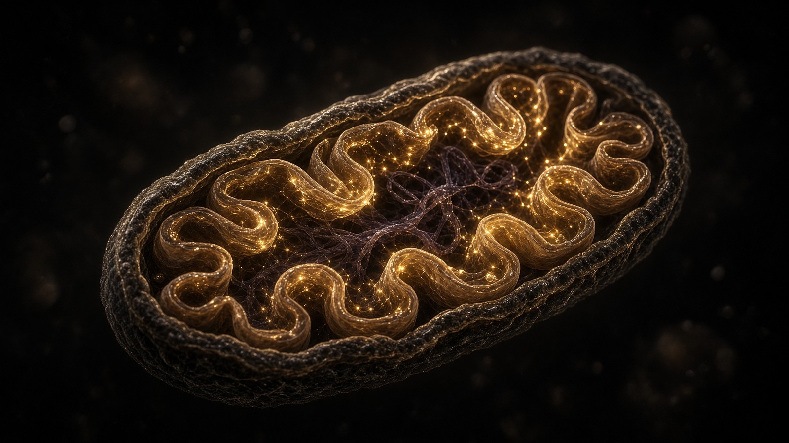
Diagram
Matrix
- TCA cycle · β-oxidation · mtDNA nucleoids
- NADH produced here. Complex I spends it.
- MOTS-c (MRWQEMGYIFYPRKLR) from 12S rRNA.
Inner membrane
- I → II → III → IV → V (ATP synthase)
- ~150 mV proton-motive force
- ~40–60 kg of ATP turned over per human day
mtDNA is 16,569 bp, 37 genes, 13 proteins of the respiratory chain. Nuclear DNA encodes the other ~1,200 mitochondrial proteins. NAD+ is the hydride carrier between dehydrogenases and Complex I. MOTS-c is a 16-mer translated from 12S rRNA — a peptide the mitochondrion wrote itself.
The lysosome is the destination, and also the office
De Duve’s organelle isn't a bin. It's an acidic chamber (pH ~4.5–5, vacuolar ATPase) with hydrolases that were delivered through the mannose-6-phosphate pathway, membrane proteins (LAMPs) that survive the interior, and a surface that has become the amino-acid office we already named. Fusion of an autophagosome with a lysosome needs SNAREs (STX17, SNAP29, VAMP8), HOPS, and a lysosome that is actually acidic. Bafilomycin, as noted, breaks the last assumption and is why flux assays use it. If lysosomes fail, autophagosomes pile up, and the picture looks like ‘more autophagy’ to a careless blot. Nixon’s Alzheimer work: autophagic vesicles accumulating in dystrophic neurites, a traffic jam, not an induction. Lee, Nixon, Cell, 2010: presenilin 1 mutations, which are familial-Alzheimer alleles, impair lysosomal acidification. The amyloid hypothesis and the proteostasis hypothesis are allowed to occupy the same neuron. They aren't allowed to be the same sentence.
In short. The lysosome is an acidic hydrolase chamber and the surface mTORC1 uses as an office. If it fails, autophagosomes pile up and a blot can lie.
TFEB is the transcription factor that writes lysosomal and autophagy genes together — the CLEAR network, Sardiello, Ballabio, Science, 2009. Settembre, Ballabio, Science, 2011: TFEB links autophagy to lysosomal biogenesis. When mTORC1 is on at the lysosome it phosphorylates TFEB and the factor stays cytoplasmic; when mTORC1 is off, TFEB goes to the nucleus and writes more of the destination and more of the wrapping machinery. Martina; Roczniak-Ferguson, Zoncu; Settembre, 2012, the same lesson from three directions. Calcineurin, when lysosomal calcium is released, can dephosphorylate TFEB even against mTORC1; Medina, Ballabio, 2015. A starving cell doesn't only start ULK1. It transcribes more lysosomes, because a programme that wraps cargo without a destination is a pile-up. Ballabio and Bonifacino, and Lawrence and Zoncu, wrote the reviews that made the lysosome a hub rather than a bin. The pipeline has a whole essay queued on this organelle. This paragraph is the hub as it bears on proteostasis: you can't eat your mistakes if the stomach is closed, and the stomach is also the restaurant’s front desk.
In short. TFEB writes lysosome and autophagy genes together and is held in the cytoplasm by mTORC1. Starvation sends it to the nucleus.
Ageing is partly a warehouse that no longer keeps up
López-Otín’s hallmarks, 2013, and the 2023 expansion, keep loss of proteostasis on the list because the evidence is boringly consistent. Proteasome capacity declines in ageing tissues, not uniformly, not in every subunit, but enough that a neuron notices. Autophagic flux to starvation blunts. CMA’s LAMP2A falls; Cuervo, Dice, 2000; Zhang, Cuervo, 2008, the restoration experiment in liver. Chaperone inducibility via HSF1 flattens; Morimoto’s organismal work. Inclusion bodies — tau tangles, Lewy bodies, huntingtin aggregates, TDP-43 inclusions, lipofuscin — are the morphology of a warehouse that has been losing the argument with its own stock. David, Morimoto, 2010: widespread protein aggregation as an inherent part of ageing in C. elegans. Walther, Mann, 2015: proteome remodelling and aggregation in the same worm. Hipp, Kasturi, Hartl, Nature Reviews Molecular Cell Biology, 2019: the network and its decline. A hallmark isn't a product category. A hallmark is a description of what fails, with a literature, in named organisms, with named machines. The leap from a worm’s aggregated proteome to a 10 mg vial is the leap we don't make.
In short. Proteasome capacity, autophagic flux, CMA and heat-shock inducibility all blunt with age. Inclusions are the morphology.
Senescence is one fate a proteostasis failure can buy. A cell that has too much unfolded protein can arrest, via p53 and p21 or via p16, and then stay, secreting. Campisi named the SASP. Baker, van Deursen, the INK-ATTAC mouse, showed that clearing p16-positive cells delayed several ageing phenotypes — in a mouse. The neighbouring cell-cycle essay is the decision; the neighbouring pathophysiology essay is the floor. This essay’s contribution is the road: a warehouse that can't shred or swallow will eventually stop dividing or die, and if it stops dividing and refuses to die it will talk too much. Ubiquitin sits on that road because p53 itself is an MDM2-and-proteasome story, and because a senescent cell’s secretome includes proteases and cytokines that are themselves proteostasis clients in the neighbours who have to hear them. Feedback, again, of the unkind sort. A research peptide that claims to ‘clear senescent cells’ without a p16 reporter, a SASP panel, and a species is claiming a fate it hasn't stained.
In short. A warehouse that can't shred or swallow may senesce or die. p53 is a proteasome client via MDM2.
Named diseases are named cargo
Alzheimer’s disease is, among other things, a proteostasis failure in a long-lived neuron: amyloid-β from APP (Glenner and Wong, 1984; Kang, 1987; Goate, 1991), tau in paired helical filaments (Grundke-Iqbal; Goedert), autophagic vesicles piling in neurites (Nixon), proteasome impairment by the aggregates themselves (Keller; Keck; Myeku). Familial alleles in APP, PSEN1, PSEN2 sit on production. MAPT sits on the cargo. The sporadic majority is a warehouse plus a lifetime. Parkinson’s disease: α-synuclein in Lewy bodies (Spillantini, 1997), SNCA mutations (Polymeropoulos, 1997), Parkin as an E3 (Kitada, 1998), PINK1 as the upstream kinase (Valente, 2004), UCH-L1 as a DUB (Leroy, 1998), CMA clogged by pathogenic α-synuclein (Cuervo, 2004), proteasome impairment in substantia nigra (McNaught). Huntington’s: a polyglutamine expansion in huntingtin (HD Collaborative Research Group, Cell, 1993), nuclear inclusions (DiFiglia, 1997), a cargo-recognition problem for autophagy (Martinez-Vicente, 2010), and a rapamycin-sensitive rescue in fly and mouse models (Ravikumar, Rubinsztein, Nature Genetics, 2004) that is a tool-and-model fact, not a prescription. ALS and FTD: TDP-43 in ubiquitin-positive inclusions (Neumann, 2006; Arai, 2006), OPTN, SQSTM1, TBK1, C9ORF72 (which sits on ULK1 via SMCR8/WDR41). Ciechanover and Brundin, Neuron, 2003: the ubiquitin–proteasome system in neurodegeneration, sometimes the chicken, sometimes the egg.
In short. Alzheimer, Parkinson, Huntington and ALS pile named cargo — amyloid, tau, α-synuclein, huntingtin, TDP-43 — and each has a disposal-route literature. Rare genes named the machines.
CFTR ΔF508 remains the pedagogical folding disease that isn't a neurodegeneration. A phenylalanine missing, a channel that would work if it arrived, ERAD eating it instead, a lung six floors up. Correctors and potentiators that chaperone or open CFTR are a licensed literature this catalogue doesn't carry. The lesson for a peptide till is geometric: occupancy of a GPCR, a cofactor pocket, or a poorly defined sleep literature won't reroute a specific ERAD client. Named cargo wants a named intervention. Brochure language wants a warehouse mood. We stock sequences. We don't stock the mood. Named cargo wants a named intervention. Occupancy of a GPCR, a cofactor pocket, or a sleep literature will not reroute a specific ERAD client. Correctors and potentiators that chaperone or open CFTR are a licensed literature this catalogue doesn't carry. The geometry still teaches: fold first, arrive second, chloride third, lung last.
In short. CFTR ΔF508 is a folding error the proteasome ate, and the lung is six floors up. A catalogue peptide doesn't reroute that client.
Diagram
01 Genome
Variant, CNV, methylation, telomere length
The script. Most of it never becomes a phenotype you can bill for.
02 Transcriptome
Which genes are on, splice isoforms, noncoding RNA
The script being read this hour. A cell type is a transcriptome.
03 Proteome
Abundance, PTMs, localisation, complexes
The machines. Phosphorylation can flip a pathway without new DNA.
04 Metabolome
NAD+/NADH, ATP/AMP, acetyl-CoA, ROS
The fuel gauges. They feed back onto the genome through sirtuins and chromatin.
05 Organelle
Mitochondria, ER stress, lysosome, nucleus
Compartments fail as units. A tired mitochondrion is a tired cell.
06 Cell fate
Proliferation, senescence, apoptosis, identity
Hayflick, SASP, p53. The cell decides whether to keep being a citizen.
07 Tissue
Inflammation, fibrosis, barrier, innervation
Where a person actually hurts. Collagen, endothelium, synapses.
08 Organism
Glucose curve, VO₂, sleep, fertility, lifespan
The readout. Everything above is allowed to be invisible until it isn’t.
Pathophysiology is this stack, not a single molecule. A research peptide occupies one node — a receptor, a cofactor, a cytoskeletal motif — and the rest of the stack is still running. That is why ‘what does it do?’ is a bad question and ‘where does it bind?’ is a good one.
The stack is the neighbouring pathophysiology essay, applied to one network. Floor 1: a polyglutamine codon, a PSEN1 allele, a PRKN deletion, or nothing, because most of the burden isn't a spelling error. Floor 2: HSF1, TFEB, XBP1s, ATF4, p53 — the transcription factors that write the holders and the shredders and the appetite. Floor 3: the proteome as client and as machine, ubiquitin, E3s, LC3, LAMP2A. Floor 4: AMP/ATP, NAD+/NADH, amino acids, acetyl-CoA — the metabolites the sensors bind. Floor 5: the mitochondrion that failed a voltage test, the ER that panicked, the lysosome that was also the office. Floor 6: divide, senesce, die, stay. Floor 7: a substantia nigra, a hippocampus, a liver, a tendon. Floor 8: a person who notices a tremor, a memory, a glucose curve. A research peptide almost always occupies floor 3 or floor 4. MOTS-c: floor 3 as a 16-mer, floor 4 as an AMPK neighbourhood. NAD+: floor 4 as a cofactor, with sirtuin consequences on floors 2 and 3. DSIP: a night-time literature that doesn't have a named floor on this stack, which is the most important sentence in the catalogue half of this essay.
In short. Pathophysiology is a stack. Ubiquitin and LC3 live on the proteome floor That's the take-home, and it's enough to walk with.
Three catalogue occupancies, none of them an autophagy drug
The catalogue holds three sequences that search bars keep sliding onto this essay, and only two of them have a biochemical right to be in the neighbourhood. The third is a filing error we can name. Occupancy, assay, species, legal class. The same four nouns the rest of the Cell desk uses. A lyophilised cake is a reagent. An intramuscular appointment at eLIVEate is a different product on a different till. A fasting schedule is a behaviour, not a vial. Mixing those jobs is how a 2016 Nobel becomes a night-time nonapeptide in a cart. A lyophilised cake is a reagent. An intramuscular appointment at eLIVEate is a different product on a different till. A fasting schedule is a behaviour, not a vial. Mixing those jobs is how a 2016 Nobel becomes a shopping list. Occupancy, assay, species, legal class: the same four nouns the rest of the Cell desk uses.
In short. Three sequences sit near this essay on a till. Two have a biochemical neighbourhood That's the take-home, and it's enough to walk with.
DSIP is a sleep-isolation story. It is not ULK1.
Delta sleep-inducing peptide is Trp-Ala-Gly-Gly-Asp-Ala-Ser-Gly-Glu. Schoenenberger and Monnier, Proceedings of the National Academy of Sciences, 1977: a nonapeptide from rabbit cerebral venous blood, sampled during thalamic stimulation that produced delta-wave sleep. They named it for the finding. Subsequent animal literature sits on sleep architecture and on HPA-axis markers, mixed, as that field is. There's no named occupancy of ULK1, ATG proteins, LC3 lipidation, mTORC1, AMPK, a proteasome subunit, or an autophagy receptor. There's no flux assay in which WAGGDASGE is the independent variable and LC3-II, in the presence and absence of bafilomycin, is the dependent one that the field would accept. The neighbouring peptide-desk essay is ‘Sleep architecture, delta waves and the two peptides people file under insomnia’. The neighbouring metabolism-desk essay is ‘Fasting, autophagy and the mTOR switch’. This Cell-desk essay is the machines. DSIP sits on the first of those three, not the second, not this. The search bar that typed ‘autophagy peptide’ and landed on a delta-sleep nonapeptide has done a string match. String match isn't pathophysiology.
In short. DSIP is nine residues isolated during delta sleep in 1977. Its papers sit on sleep architecture and stress-axis markers.
The 10 mg listing is that named nonapeptide, US-made, ≥98% HPLC, lyophilised, CAS 62568-57-4, 848.8 g·mol⁻¹. It isn't zopiclone, not melatonin, not CBT-I, not a dark room, not bafilomycin, not rapamycin, and not an autophagy inducer. A bench that wants to ask whether WAGGDASGE does anything to a sleep-architecture readout in a species, with a vehicle control and a chromatogram, can buy the sequence. A cart that wants to stack it next to a fasting protocol because both words appeared on a blog is doing the opposite of this journal. Research use only. Not a sleeping tablet. Not a protocol. A bench that wants to ask whether WAGGDASGE does anything to a sleep-architecture readout in a species, with a vehicle control and a chromatogram, can buy the sequence. A cart that wants to stack it next to a fasting protocol because both words appeared on a blog is stacking two different objects. Night is a physiology. It isn't a product category.
In short. The 10 mg vial is WAGGDASGE, HPLC, lyophilised, for a tube. Not a hypnotic, not rapamycin, not an autophagy inducer.
NAD+ is the coin sirtuins spend. The 1000 mg cake is the coin as a reagent.
SIRT1 is an NAD+-dependent deacetylase. Imai, Guarente, Nature, 2000, in yeast Sir2; the mammalian family followed. Lee, Finkel, Proceedings of the National Academy of Sciences, 2008: SIRT1 deacetylates autophagy machinery and a role for the enzyme in autophagy regulation is the claim. Huang, 2015: deacetylation of nuclear LC3 helps autophagy initiate under starvation. Hariharan: FoxO deacetylation and starvation-induced autophagy in cardiac myocytes. Cantó, Auwerx, 2009: AMPK regulates energy expenditure by modulating NAD+ metabolism and SIRT1. The loops braid. AMPK, NAD+, SIRT1, autophagy proteins, PGC-1α: a metabolic neighbourhood in which the hydride coin is a substrate, not a vitamin caption. PARP1 also spends NAD+, on DNA repair, and a PARP-hyperactive cell can drain the pool that sirtuins wanted; Bai, Auwerx; Fang, Bohr. CD38 rises with age and is an NADase; Camacho-Pereira, Chini, 2016. The pool falls. The budget tightens. That's why a cofactor has a literature in ageing biology, and why we stock it at 1000 mg, and why none of that is an autophagy protocol. Occupying a cofactor pocket in a tube is a redox experiment. Writing LC3-II in a mouse is a different experiment. Injecting NAD+ into a deltoid at a clinic is a third. Three objects.
In short. SIRT1 spends NAD+ and can deacetylate autophagy machinery. The pool falls with age as CD38 rises.
The listing is lyophilised β-nicotinamide adenine dinucleotide, C21H27N7O14P2, 663.43 g·mol⁻¹, CAS 53-84-9, ≥98% HPLC, a 1000 mg aliquot. It's the oxidised hydride coin Complex I wants, the co-substrate sirtuins and PARPs consume, a reagent for a tube that can declare a dinucleotide. eLIVEate’s intramuscular NAD+ appointment is a clinic product on JP’s diary; Patriot takes no commission on the booking. NR and NMN are salvage precursors with human pharmacokinetic data (Trammell; Airhart; Martens) and a mixed endpoint literature we'll not launder into a proteostasis claim. The neighbouring NAD+ journal piece is the budget as budget. This piece is the budget as it touches a deacetylase that can touch a phagophore. The touching is real in papers. The vial is a reagent. Research use only on the solid. It isn't 40 kg of ATP, a sirtuin activator as a product claim, or a mitophagy drug.
In short. 1000 mg β-NAD+, HPLC, lyophilised, for a tube. The clinic shot is a different product.
MOTS-c is a 16-mer the mitochondrion wrote. It sits on AMPK. Sitting is occupancy.
MOTS-c is Met-Arg-Trp-Gln-Glu-Met-Gly-Tyr-Ile-Phe-Tyr-Pro-Arg-Lys-Leu-Arg, translated from an open reading frame in mitochondrial 12S rRNA. Lee, Kim, Cohen, Cell Metabolism, 2015: the peptide promotes metabolic homoeostasis in mice, with AMPK and the folate–methionine cycle as the named neighbourhood. Kim, Lee, Cell Metabolism, 2018: under metabolic stress the peptide can translocate to the nucleus and regulate nuclear gene expression. Reynolds, 2021: exercise-induced, age-dependent physiology in a Nature Communications paper. The neighbouring mitochondria essay is the organelle as organelle, and the 16-mer as the sentence that shouldn't exist. This essay’s interest is the AMPK occupancy. AMPK phosphorylates ULK1. AMPK phosphorylates TSC2 and Raptor. A ligand that sits on AMPK sits one phosphorylation away from a phagophore. That's a neighbourhood. It isn't a demonstration of LC3 flux, not a mitophagy assay, not a TFEB nuclear-translocation series, and not a reason to file MOTS-c as an autophagy peptide. The 2015 paper is a metabolic-homoeostasis paper. Honour it as that. The vial is the named 16-mer, 40 mg, US-made, ≥98% HPLC, 2174.6 g·mol⁻¹, CAS 1627580-64-6, lyophilised, for a tube that can declare a sequence. Not an exercise mimetic as a product claim. Not a medicine. Research use only.
In short. MOTS-c is a 16-mer from mitochondrial 12S rRNA with an AMPK literature. AMPK is one kinase of the autophagy switch.
Retatrutide, which the mitochondria essay already had to unmix from this organelle, occupies GLP-1R, GIPR and GCGR. Organism-level fuel demand then changes, and AMP/ATP, NAD+/NADH, and autophagic tone in liver and adipose will follow because that is what flux does when the animal eats less. Occupancy is three class-B GPCRs. Different floor from a 16-mer written in 12S rRNA, different floor from a dinucleotide, different floor from a sleep nonapeptide. Don't confuse two different molecules. The Cell desk’s only product claim, across all three of these ligands and every other, is occupancy of a named node with a chromatogram. The network in this page is the reason that claim has to be small. Occupancy is three class-B GPCRs. Different floor from a 16-mer written in 12S rRNA, different floor from a dinucleotide, different floor from a sleep nonapeptide. Don't confuse two different molecules. The organelle notices later, the way a power station notices a city using less electricity. Fuel demand moved. The warehouse followed.
In short. Retatrutide occupies three GPCRs and may change fuel demand; autophagy will follow as flux follows. That isn't MOTS-c, not NAD+, not DSIP.
A warehouse, two Nobels, three reagents
Retrace once, without the caption. A growing cell holds ~10¹⁰ proteins and turns over a few percent an hour because translation errs at 10⁻⁴ and folding is a kinetic bet in a crowded solvent. Chaperones hold. Ubiquitin, 76 residues, is conjugated by E1, E2 and E3, in chains whose linkage is a code: K48 to the 26S, K63 more often to a signal or an autophagy receptor. The 26S is a 19S cap on a 20S threonine-protease barrel. DUBs proofread and recycle the tag. Cargo the barrel can't swallow is piled, then wrapped. Ohsumi’s ATG genes build a phagophore; ULK1 starts it; VPS34–Beclin-1 marks it with PI3P; LC3-II dresses it; a lysosome finishes it. mTORC1 at the lysosomal surface, amino acids and growth factors on, phosphorylates ULK1 at Ser757 and says no. AMPK, AMP on, phosphorylates ULK1 at other serines and says yes. PINK1 and Parkin tag a depolarised mitochondrion. CMA threads KFERQ clients through LAMP2A. Ageing blunts the network. Named diseases pile named cargo. Hershko, Ciechanover, Rose, 2004. Ohsumi, 2016. DSIP is WAGGDASGE from a sleeping rabbit brain and isn't a member of this cascade. NAD+ is the coin SIRT1 spends. MOTS-c is a 16-mer on AMPK. Three occupancies.
In short. Chaperones, ubiquitin, the 26S, autophagy, mTORC1 versus AMPK, PINK1/Parkin. Two Nobels That's the take-home, and it's enough to walk with.
The vials are reagents. DSIP 10 mg is WAGGDASGE, HPLC, lyophilised, for a tube that can ask a sleep-architecture question. NAD+ 1000 mg is β-NAD+, HPLC, lyophilised, for a tube that can declare a dinucleotide. MOTS-c 40 mg is MRWQEMGYIFYPRKLR, HPLC, lyophilised, for a tube that can ask an AMPK question. Peptide orders over £75 include the sterile kit. Next-day UK postage is £5. eLIVEate’s intramuscular NAD+ is a clinic appointment on a different till, no commission. None of these is rapamycin. None of them is bafilomycin. None of them is a 26S. None of them is a phagophore. None of them is a protocol. Research use only on the solids. The cell will still eat its own mistakes in the morning, with the machines this essay named, whether or not a cart was filled. That's the point of a programme that is older than a catalogue.
In short. Three HPLC-characterised sequences, labelled for the bench, kit over £75, next-day UK £5. Not medicines.
Ten billion proteins, a 76-residue tag, a 2.5-megadalton barrel, a double membrane around a former bacterium. The cell eats its own mistakes because the draft is the product. We sell sequences. We do not sell the appetite.
Questions the essay actually answers
- What is proteostasis, precisely?
- Protein homeostasis: the network that folds, holds, tags and degrades the proteome so that a cell of ~10¹⁰ protein molecules does not fill with unfolded inventory. Chaperones, the ubiquitin–proteasome system, and autophagy are the three main shops. Balch, Morimoto, Dillin and Kelly named the network in Science, 2008. It is not a wellness word, and it is not a protocol.
- What is ubiquitin?
- A 76-residue protein, one of the most conserved in eukaryotes, whose C-terminal glycine is conjugated to lysine (usually) on a substrate. Goldstein isolated it in 1975 as a ‘ubiquitous’ polypeptide. Hershko, Ciechanover and Rose showed it was the tag in ATP-dependent proteolysis. Nobel Prize in Chemistry, 2004.
- What do E1, E2 and E3 actually do?
- A cascade. E1 (UBA1 in humans) uses ATP to adenylate ubiquitin and then holds it as a thioester. E2 is the conjugating enzyme that receives that thioester. E3 is the ligase that selects the substrate — RING E3s scaffold E2 against the victim; HECT and RBR E3s take a thioester themselves. About 600 human E3s. Specificity lives there.
- What is the difference between K48 and K63 chains?
- Both are polyubiquitin, linked through different lysines of ubiquitin itself. K48-linked chains (and K11) are the classic proteasome-targeting signal — Chau, Varshavsky, Pickart. K63-linked chains are more often a non-degradative signal: DNA repair, endocytosis, NF-κB, and the handles that autophagy receptors grab. The chain is a code, not a stamp.
- What is the 26S proteasome?
- A ~2.5 MDa machine: a 20S barrel of four stacked heptameric rings (α7β7β7α7) capped by one or two 19S regulatory particles. The 19S recognises the chain, unfolds the client with six AAA+ ATPases, and feeds it through a gated channel to threonine proteases in the β-ring. Peptides come out. Ubiquitin is recycled. It is a shredder with a vestibule, not a bin.
- What is autophagy, and what are ULK1 and LC3?
- Macroautophagy: a double membrane (phagophore) grows around cargo, closes as an autophagosome, fuses with a lysosome. ULK1 is the initiating kinase, held off by mTORC1 and encouraged by AMPK. LC3 (ATG8 family) is conjugated to phosphatidylethanolamine; LC3-II is the blot everyone runs. Ohsumi’s yeast genetics named the ATG genes. Nobel, 2016.
- How do mTORC1 and AMPK set the appetite?
- Opposite phosphorylations of ULK1. mTORC1, amino-acid- and growth-factor-on at the lysosomal surface, phosphorylates ULK1 at Ser757 and keeps autophagy down. AMPK, AMP/ATP-on, phosphorylates ULK1 at other serines (classically 317 and 777) and starts it, and also leans on TSC2 and Raptor to quiet mTORC1. Kim, Guan, 2011; Egan, Shaw, 2011. The fasting essay next door is the organism-level switch. This essay is the kinases.
- What is mitophagy, and what do PINK1 and Parkin do?
- Selective autophagy of mitochondria. On a healthy organelle, PINK1 is imported and cleaved. On a depolarised one it accumulates on the outer membrane, phosphorylates ubiquitin at Ser65, and recruits Parkin, an RBR E3. Parkin builds ubiquitin chains on outer-membrane proteins. OPTN and NDP52 read those chains and recruit LC3. Kitada 1998 (parkin); Valente 2004 (PINK1); Narendra and Youle 2008–2010; Lazarou 2015.
- Is DSIP an autophagy drug?
- No. DSIP is Trp-Ala-Gly-Gly-Asp-Ala-Ser-Gly-Glu, isolated by Schoenenberger and Monnier in 1977 from rabbit cerebral venous blood during delta-wave sleep. The literature sits on sleep architecture and HPA tone. It does not occupy ULK1, LC3, mTORC1 or AMPK as a named ligand. The fasting–autophagy essay is the neighbouring metabolism piece. This vial is a characterised nonapeptide. Research use only.
- Where do NAD+ and MOTS-c sit on this network?
- Occupancies, not autophagy drugs. SIRT1 is an NAD+-dependent deacetylase that can deacetylate autophagy machinery (Lee, Finkel, 2008); the 1000 mg listing is lyophilised β-NAD+ for the bench, and the clinic injection is a different till. MOTS-c is MRWQEMGYIFYPRKLR from mitochondrial 12S rRNA (Lee, Cohen, 2015) and sits on AMPK, which is one kinase of the appetite switch. HPLC research sequences. Not a protocol.
Hypothetical research reconstitution
How these vials are typically mixed
Hypothetical research reconstitution for the named catalogue vial. Not a protocol, not medical advice, not a use instruction. These amounts sit in published and commonly cited laboratory ranges. The vial is labelled for research use only — not for human or veterinary administration.
DSIP
10mg
Mix with 2 ml bacteriostatic water → 5 mg/ml · 5,000 mcg/ml
- Hypothetical aliquot
- 100–300 mcg
- 0.02–0.06 ml · 2–6 units on a U-100 syringe
- How often
- Once daily, evening
- 7–14 nights, then a pause
Bench steps
- Let the vial sit until it is no longer cold to the touch.
- Wipe the stopper with 70% isopropyl alcohol. Let it dry.
- Draw 2 ml bacteriostatic water (0.9% benzyl alcohol).
- Run the water slowly down the inside glass — do not blast the cake.
- Roll between finger and thumb until the cake is gone. Do not shake.
- Label the date. Store the solution at 2–8 °C. Do not freeze. Use within 30 days unless the note below says otherwise.
Delta-sleep peptide. Night-time aliquot in the papers that bother with a clock. Fridge. Short runs.
NAD+
1000mg
Mix with 10 ml bacteriostatic water → 100 mg/ml
- Hypothetical aliquot
- 50–100 mg
- 0.50–1.00 ml · 50–100 units on a U-100 syringe
- How often
- Two or three times per week in published infusion and assay notes
- 4–8 weeks, then a pause
Bench steps
- Let the vial sit until it is no longer cold to the touch.
- Wipe the stopper with 70% isopropyl alcohol. Let it dry.
- Draw 10 ml bacteriostatic water (0.9% benzyl alcohol).
- Run the water slowly down the inside glass — do not blast the cake.
- Roll between finger and thumb until the cake is gone. Do not shake.
- Label the date. Store the solution at 2–8 °C. Do not freeze. Use within 30 days unless the note below says otherwise.
A 1000mg cake wants 10 ml. Protect from light. Solution yellows as it oxidises — that is the cofactor dying, not a flavour. Use promptly.
MOTS-c
40mg
Mix with 2 ml bacteriostatic water → 20 mg/ml
- Hypothetical aliquot
- 5–10 mg
- 0.25–0.50 ml · 25–50 units on a U-100 syringe
- How often
- Two or three times per week
- 4–8 weeks
Bench steps
- Let the vial sit until it is no longer cold to the touch.
- Wipe the stopper with 70% isopropyl alcohol. Let it dry.
- Draw 2 ml bacteriostatic water (0.9% benzyl alcohol).
- Run the water slowly down the inside glass — do not blast the cake.
- Roll between finger and thumb until the cake is gone. Do not shake.
- Label the date. Store the solution at 2–8 °C. Do not freeze. Use within 30 days unless the note below says otherwise.
Mitochondrial 16-mer. Fridge. Do not freeze. The 5 mg mark is where most bench notes start.
Bacteriostatic water and sterile syringes ship with peptide orders over £75. Kit details · 10 ml bacteriostatic water
The vials this essay sits on
Named sequences the essay maps — DSIP, NAD+, MOTS-C. Hypothetical research neighbourhood, not a protocol, not a medicine. One press puts every in-stock vial in the bag.
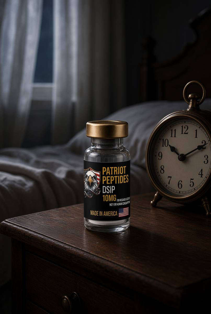 Made in USAOut of stock
Neuropeptide
DSIP
10 mg DSIP — the nonapeptide isolated during slow-wave sleep.
4.7(521)
15 browsing this now · 1 purchased in the last 24 hours
10mg
£25.00
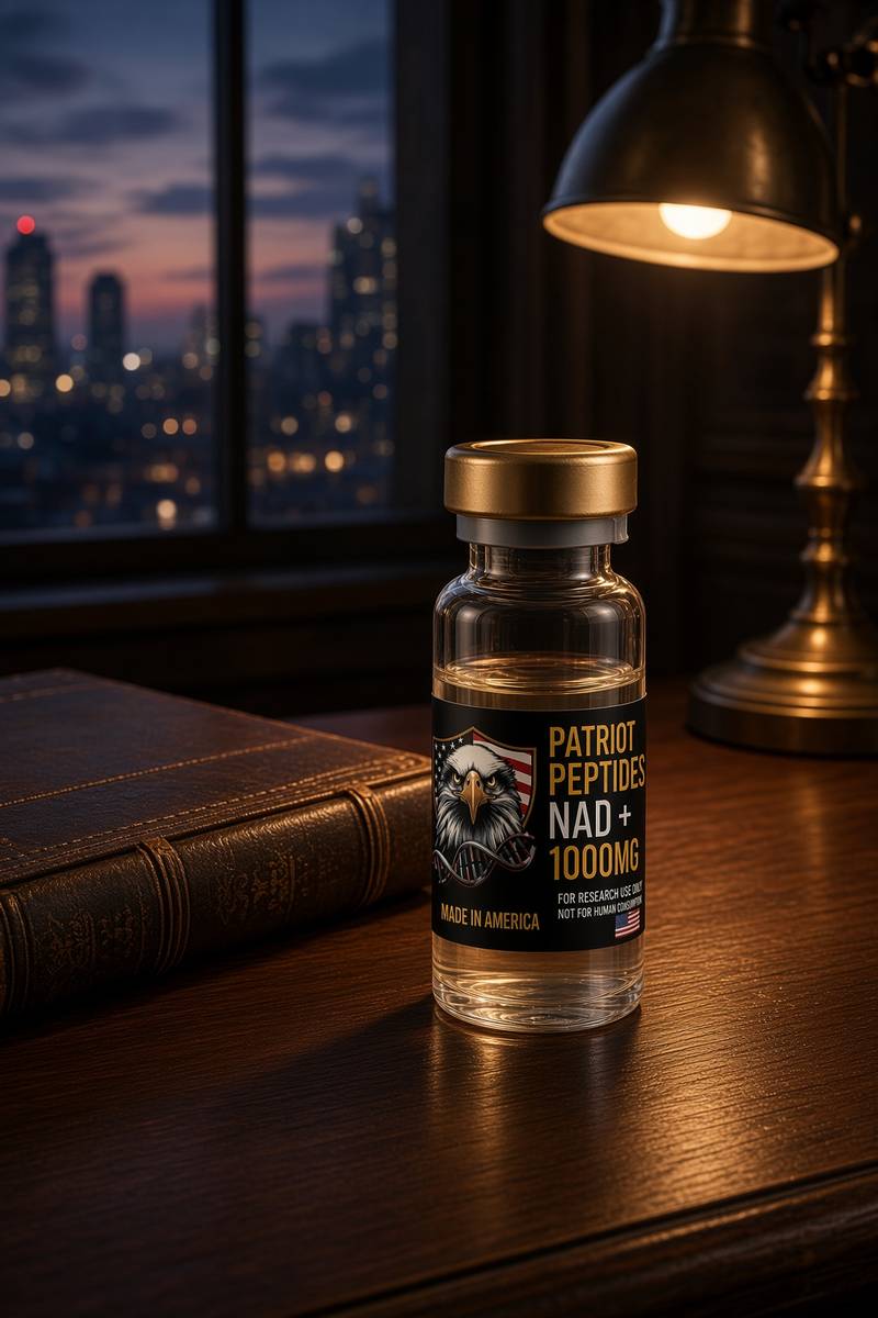 Research only
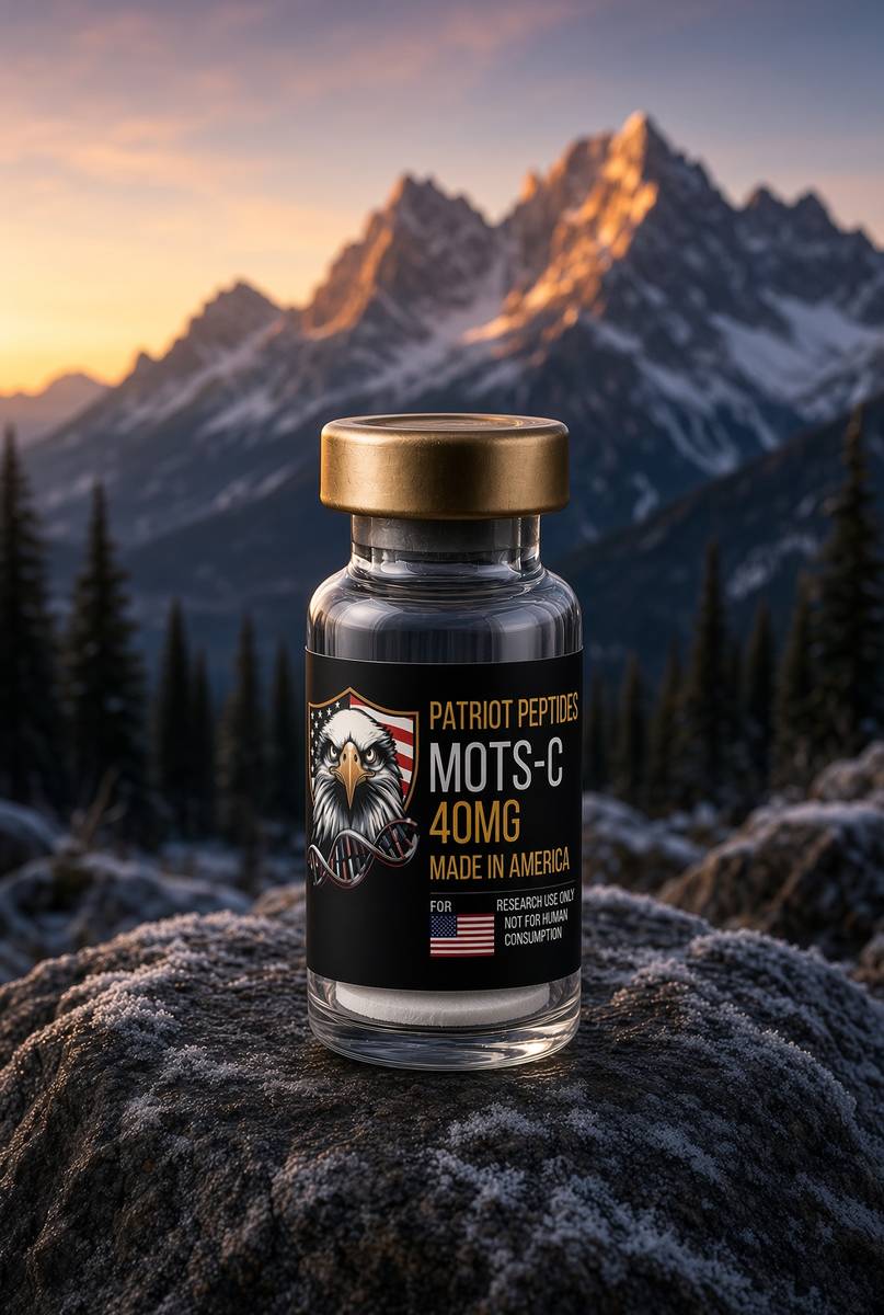 Made in USA
Research use only. Not a combined-use instruction.
Read next
65 min · long read · The living cell
The living cell is a city, and you are 36 trillion of them
A 70 kg adult is on the order of 36 trillion cells, most of them red blood cells with no nucleus. A typical nucleated cell holds about ten billion proteins and two metres of DNA. The body recycles 40–60 kg of ATP a day. Those figures are published; this essay is what they mean.
64 min · long read · The living cell
Mitochondria: the bacterium you kept, the genome it kept, the peptides it writes
You turn over 40–60 kg of ATP a day using a 16,569-base genome that still uses a bacterial genetic code. NAD+ is the hydride carrier Complex I spends. MOTS-c is a 16-mer translated from mitochondrial 12S rRNA — Lee, Kim, Cohen, 2015. That last sentence is real, and it is surprising.
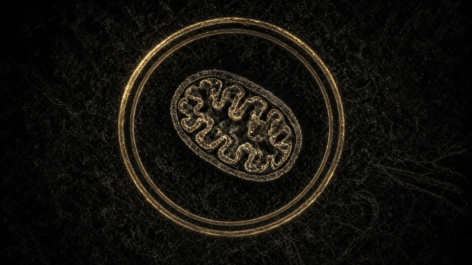
50 min · long read · Metabolism
Fasting, autophagy and the mTOR switch
Eat, and mTOR builds. Fast, and AMPK and ULK1 start recycling the cell. The molecular story is a switch, not a personality.
More in this desk
82 min · long read · The living cell
Pathophysiology from the genome to a person who notices
Disease is a stack: genome, transcriptome, proteome, metabolome, organelle, cell fate, tissue, organism. A peptide binds one node. The rest of the stack keeps running. CFTR, type 2 diabetes and a tendon as worked examples.
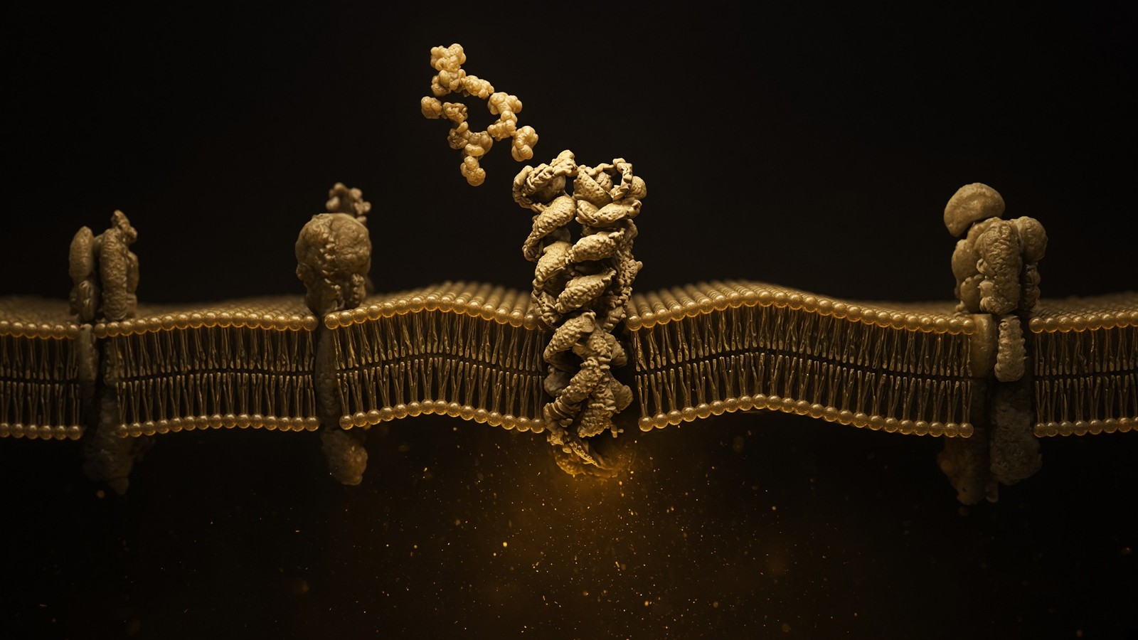
70 min · long read · The living cell
How peptides talk to cells: occupancy, amplification, arrestin
A peptide is a ligand. Most of the catalogue binds a GPCR on the cell surface: one occupancy, then enzymes make thousands of second messengers. That amplification is real, and it is not magic. Desensitisation is why more ligand is not more signal forever.
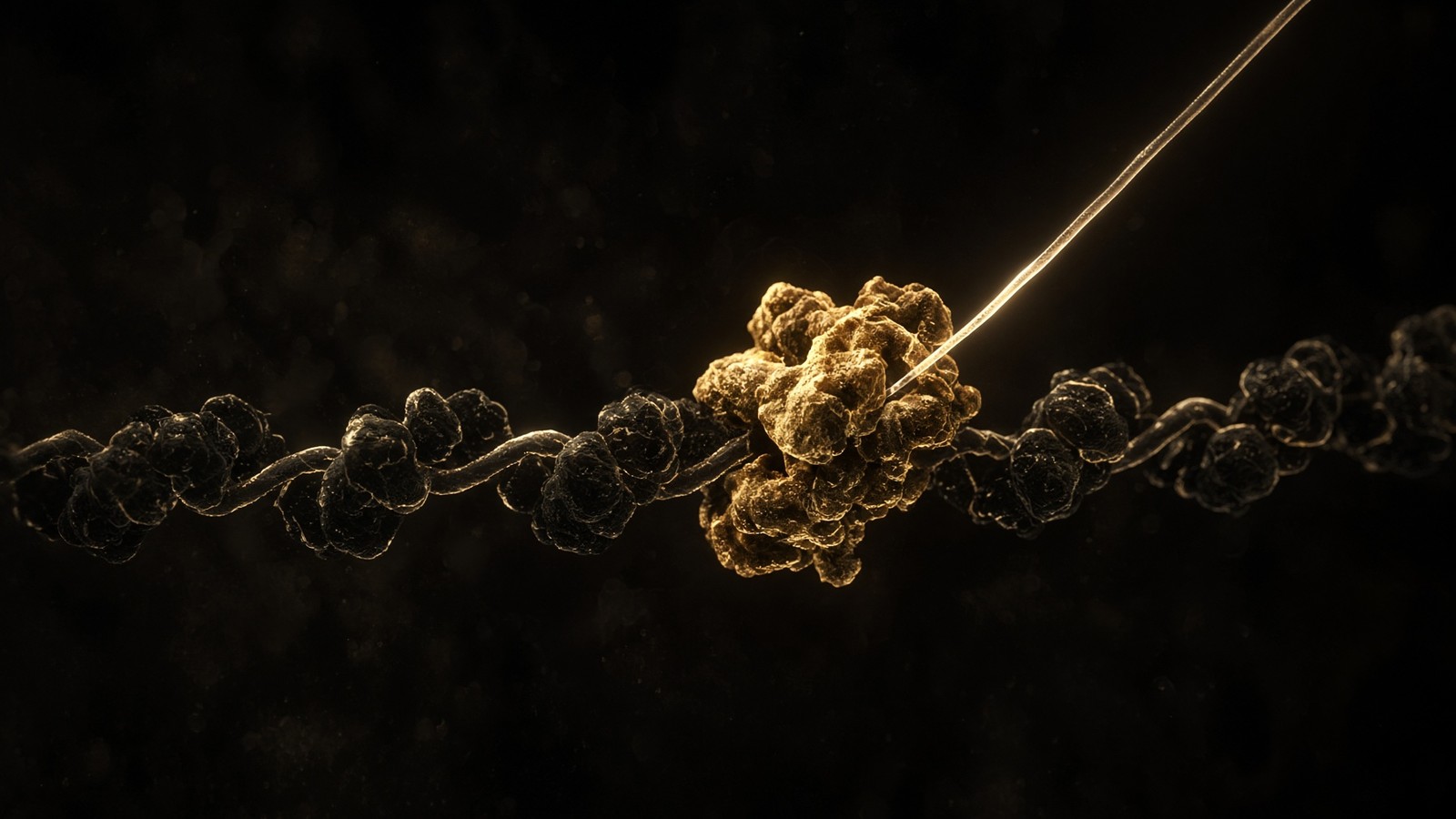
64 min · long read · The living cell
From gene to protein: how a cell actually reads itself
DNA is transcribed, spliced, exported, translated and folded. A research peptide is the ligand at the end of that pipeline, already made. This essay is every machine in between — polymerases, spliceosome, ribosome — with the actual rates attached.
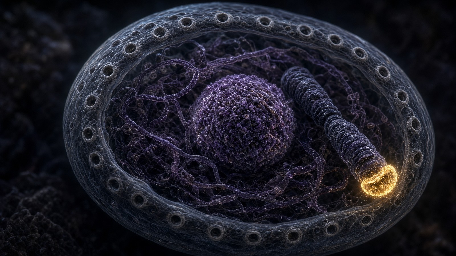
54 min · long read · The living cell
The nucleus, telomeres, and the four residues of Epithalon
Two metres of DNA folded into a nucleus a few micrometres across. TERT is off in most somatic cells on purpose. Epithalon is four residues, Ala-Glu-Asp-Gly, with a TERT and pineal literature. The machines are real. A large Western trial of telomere length in adults is not.
Essays describe published research. They are not medical advice and they do not authorise human use of any catalogue item.