

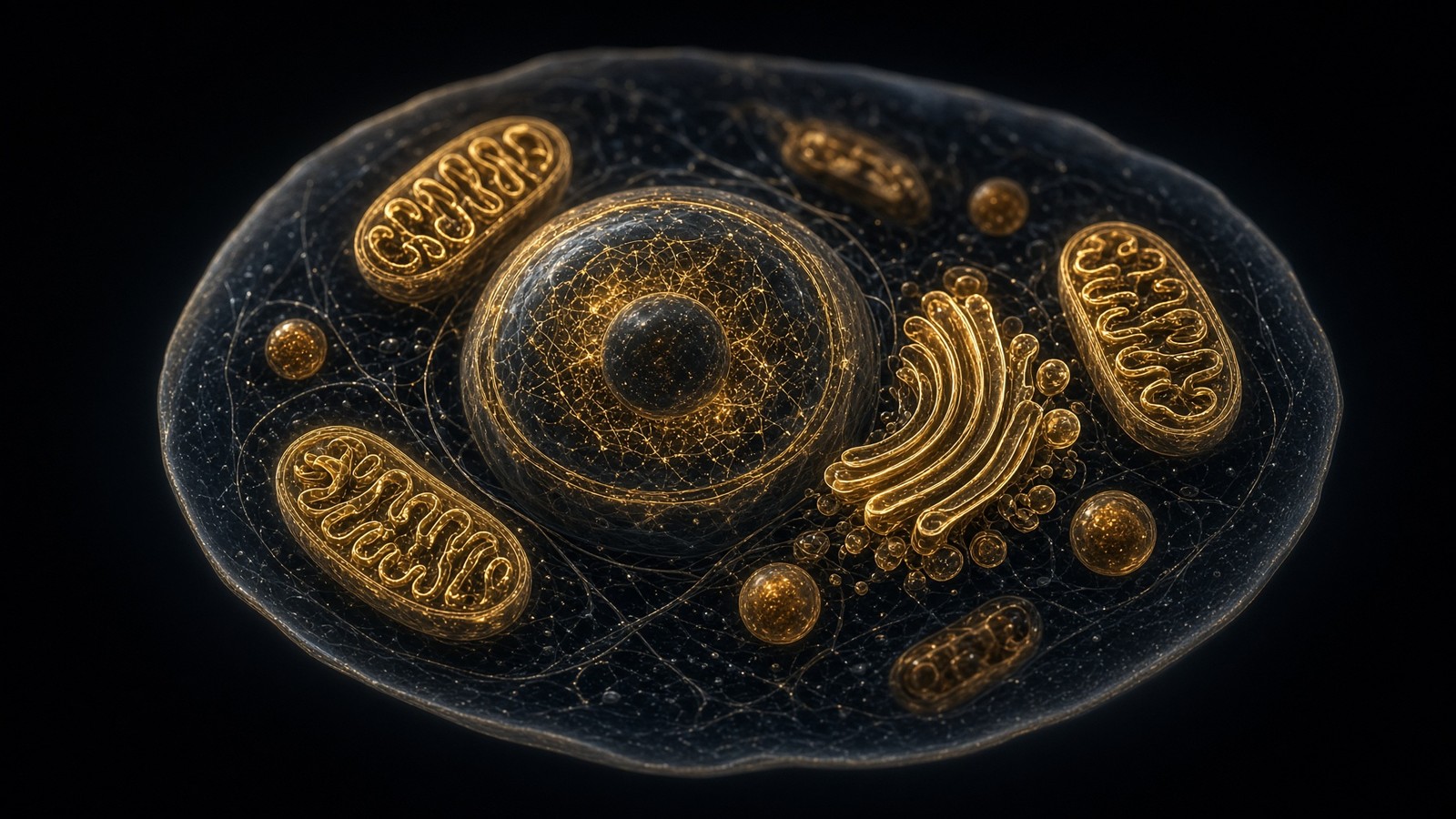
The living cell · 65 min · 14,220 words
The living cell is a city, and you are 36 trillion of them
A 70 kg adult is on the order of 36 trillion cells, most of them red blood cells with no nucleus. A typical nucleated cell holds about ten billion proteins and two metres of DNA. The body recycles 40–60 kg of ATP a day. Those figures are published; this essay is what they mean.
What this essay actually tells you
- Sender, Fuchs and Milo: ~36 trillion human cells, ~84% of them erythrocytes that have no nucleus. Count yourself and you are looking at red blood cells. Weigh yourself and you are looking at fat and muscle.
- Peter Rich, 2003: 40–60 kg of ATP turned over per day. The standing pool is ~50 g. Every molecule is recycled hundreds of times. That is ~10²¹ hydrolyses a second in a body.
- A typical mammalian cell holds on the order of 10¹⁰ proteins in a few picolitres, crowded at 200–400 mg/ml. A research peptide is a few nanometres of ligand in that interior — one protein, not a physiology.
What this actually means
If you could walk around inside a liver cell you wouldn't find a bag of water with a nucleus in the middle. You'd find a packed interior: on the order of ten billion protein molecules, a few million ribosomes, a thousand or more mitochondria that began as bacteria, and two metres of DNA folded into a nucleus a few micrometres across. Multiply by 36 trillion, remember that most of those cells are red blood cells that have thrown their nucleus away, and you have something like a person. A research peptide is a ligand — a small binding partner — for one protein in that interior. It is a few nanometres long. The numbers in this essay exist so that size stays honest.
- Human cells
- 3.6 × 10¹³
- DNA in one nucleus
- 2 metres
- Proteins per cell
- ~10¹⁰
- ATP turned over / day
- 40–60 kg
- ATP made in a body / second
- ~10²¹
- Water in a cell
- ~70%
- Macromolecular crowding
- 200–400 mg/ml
- Gut bacteria
- ~3.8 × 10¹³
Thirty-six trillion. Sender, Fuchs, Milo — not a round number we made up.
Folded into a sphere you could hide under a grain of salt.
Ten billion machines in a hepatocyte. Most of them are copies of the abundant few.
Recycled, not stored. The standing pool is a coffee-cup, not a warehouse.
A billion trillion hydrolysis events while you read this sentence.
By mass. The rest is protein, lipid, nucleic acid, metabolite, ion.
Ellis; Zimmerman and Trach. Proteins occupy 20–30% of the volume.
Roughly 1:1 with human cells by number. The 10:1 factoid is retired.
If you could shrink yourself and walk into a living cell, you wouldn't find the empty oval-with-a-dot from a school textbook. You'd find a packed interior a few picolitres across — a cube about a hundredth of a millimetre on a side — and it is busy in a way water alone never is. Water is still the solvent, about 70% of the mass, but macromolecules occupy 20–30% of the intracellular volume. That density, 200–400 milligrams per millilitre, is crowding, and it isn't a defect to be designed out. At that packing a protein cannot diffuse far without colliding, and those collisions are how the chemistry actually works. Excluded volume — the space already taken by other macromolecules — raises the effective concentration of whatever is left, so a weak, sticky interaction that looks optional in a dilute tube can become obligatory in cytoplasm. A millimolar metabolite becomes a signal. A peptide present at nanomolar concentration can still find a receptor if the shape is right. This is the room everything else in the essay happens in.
In short. Inside a cell the proteins are packed so tightly they keep bumping into each other. That crowding is how the chemistry actually works.
Ron Milo and Rob Phillips spent a decade pulling numbers out of papers and putting them in BioNumbers so the rest of us would stop waving our hands. A rapidly growing E. coli is roughly three million proteins, 20,000–50,000 ribosomes, a 1 µm × 2 µm cylinder, and a genome of 4.6 million base pairs that it copies in 20 minutes by firing overlapping rounds of replication. A HeLa cell is about a thousand times the volume, with on the order of ten billion proteins and several million ribosomes, a 6-billion-base-pair diploid genome, and a cell cycle measured in hours rather than minutes. You are not E. coli. You are also not a vague 'body'. You are a census: Sender, Fuchs and Milo put a 70 kg adult near 36 trillion human cells, most of them red blood cells with no nucleus. Once you have counted the parts, a milligram of peptide has a size, a receptor has a copy number, and a physiology is something you have to earn with an assay rather than assume from a vial.
In short. The parts have been counted. A bacterium has a few million protein molecules; one of your cells, about ten billion. A person is a census, not a blur.
Diagram
- 0.1 nmHydrogen atomA proton and an electron. Chemistry starts here.
- 0.3 nmWater molecule70% of a cell by mass. The solvent life is.
- 1 nmAmino acidTwenty kinds. Peptide bonds string them.
- 2–4 nmResearch peptideA named chain. BPC-157 is 1.4 kDa, 15 residues.
- 4–10 nmGlobular proteinHaemoglobin, a GPCR’s extracellular face.
- 25 nmRibosomeThe factory that reads mRNA into protein.
- 5 nmMembraneA lipid bilayer. Every compartment starts here.
- 0.5–1 µmMitochondrionA bacterium the cell swallowed and kept.
- 6–10 µmNucleusTwo metres of DNA folded into a sphere.
- 10–30 µmTypical cellA city. 10¹⁰ proteins. One genome.
- 1 mmTissue grainA thousand cells talking across ECM.
- 1.7 mYou~36 trillion human cells. Most of them are red blood cells.
Lengths are characteristic, not exact. A research peptide is closer in size to a water molecule than to the cell that assays it — which is why a 15-mer can occupy a receptor pocket a small-molecule drug also wants.
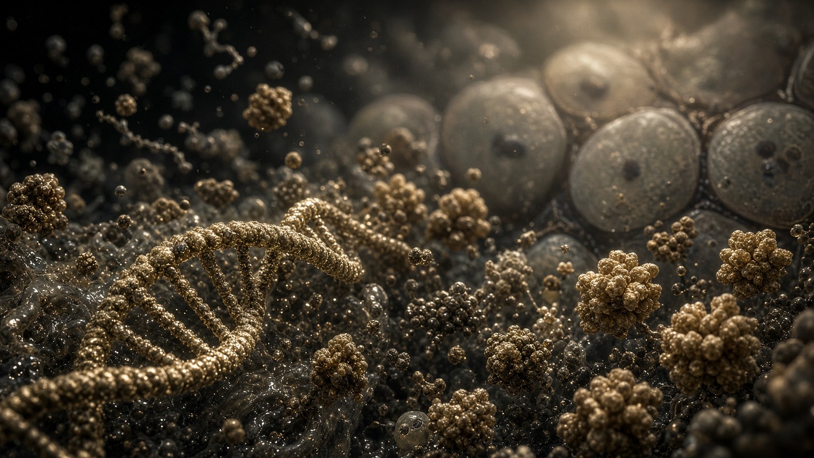
The reason to put numbers on a living cell is so that a milligram of lyophilised peptide is not asked to stand in for a physiology. A cell has a published census. A peptide is a ligand — a small binding partner — that fits one protein. The rest of this essay is that census: the ions, the filaments, the organelles, the 36 trillion cells, the ATP budget, with the papers attached. Alberts already wrote the textbook. Milo and Phillips already wrote the appendix of numbers. Sender, Fuchs and Milo already counted the cells. What we are doing here is reading those books out loud, at the scale of a picolitre, before anyone looks at a vial. A 10–50 residue peptide is a few nanometres long, closer in size to a water molecule than to the cell that assays it. That gap is a factor of ten thousand in length and about a trillion in volume. Keep the exponents attached and the claim stays the right size: occupancy of one pocket, not a renovation of a city.
In short. A milligram of peptide binds one protein. It does not stand in for a cell, let alone a person. The numbers that follow keep that claim the right size.
What a picolitre actually holds
A picolitre is 10⁻¹² litres: a cube ten micrometres on a side, or a sphere a little over twelve micrometres across. That is not a metaphor for a cell. It is the cell. A typical mammalian somatic cell occupies 2,000–4,000 cubic micrometres, which is two to four picolitres. A circulating lymphocyte is smaller, a few hundred cubic micrometres. A hepatocyte is larger, often 3,000–6,000 µm³ depending on ploidy and nutritional state. A mature adipocyte, which is mostly one lipid droplet, can exceed 100,000 µm³ and still be one cell. 'The average cell' is already a fib at the level of volume, before anyone has mentioned a nucleus. HeLa, the workhorse of cell biology, sits near 3,000 µm³ in suspension and spreads to more on a dish. When BioNumbers says a growing mammalian cell holds 10¹⁰ proteins, it is talking about a picolitre-scale object, not a philosophy. Hold that volume in your head: everything we count later — ions, ribosomes, mitochondria, two metres of DNA — has to fit in it.
In short. A typical human cell is two to four picolitres — a cube about a hundredth of a millimetre on a side. Fat cells are larger and lymphocytes smaller: there is no average cell.
Do the mass arithmetic once, because it is the only way the later numbers become inevitable rather than decorative. Three picolitres of material at roughly the density of water is three nanograms. Water is about 70% of that mass, so a little over two nanograms of H₂O and the rest a packed slurry of protein, RNA, lipid, metabolite and ion. If protein is a quarter of the mass, you are looking at about 0.75 nanograms of polypeptide in one cell. An 'average' protein of 50 kDa has a molar mass of 50,000 g·mol⁻¹. Avogadro's number does the rest: 0.75 × 10⁻⁹ g divided by 50,000 g·mol⁻¹ is 1.5 × 10⁻¹⁴ moles, times 6.022 × 10²³, which is on the order of 10¹⁰ molecules. That is not a coincidence Milo and Phillips discovered. It is what you get when you refuse to leave the units off. The cell is a few nanograms. The proteome is ten billion copies. Both sentences are the same sentence, and once you have said it you can feel why a handful of a transcription factor and tens of millions of actin molecules can share the same tiny room.
In short. One cell weighs a few billionths of a gram; keep the units on and you get about ten billion protein molecules. Both sentences are the same sentence.
Crowding is the physics, not the mess
R. John Ellis spent a career pointing out that biochemists assay enzymes at a milligram per millilitre and then wonder why the cell, which runs the same enzymes at two to four hundred milligrams per millilitre, does not always agree. Macromolecular crowding — the phrase is his, and the reviews in Trends in Biochemical Sciences are still the ones to read — means that 20–30% of the intracellular volume is occupied by macromolecules. Zimmerman and Trach, in E. coli, measured cytoplasmic concentrations in the region of 300–400 mg/ml of protein plus RNA. Eukaryotic cytoplasm is a little more polite, commonly quoted at 200–300 mg/ml, still a density at which a protein cannot execute a random walk of any length without colliding. The textbook illustration of a cell as a bag of dilute water with a few floating organelles is a drawing of a cuvette, not a hepatocyte. Once you have seen that number, a lot of 'mysterious' cellular chemistry stops being mysterious: the tube was too empty, and the cell never was.
In short. Biochemists assay enzymes in dilute soup; the cell runs the same enzymes at two to four hundred milligrams per millilitre. A bag of water with floating organelles is a drawing of a test tube.
Crowding is not clutter. Excluded volume raises the effective concentration of the remaining solution, so a micromolar association that looks optional in a dilute assay can become obligatory in the cytoplasm. Weak, sticky interactions that a biochemist would dismiss as non-specific start to matter. Phase-separated condensates — nucleoli, stress granules, P-bodies, the Cajal bodies that process snRNPs — are what you get when a crowded polymer solution is allowed to have opinions about solubility. Alberts's Molecular Biology of the Cell has, in recent editions, had to add this physics because the cartoon of freely mixing cytoplasm did not survive the microscope. A research peptide arriving at a membrane from the outside is not entering a swimming pool. It is knocking on the wall of a packed factory. That is a hopeful sentence as well as a careful one: the factory is so crowded that the right shape, at the right pocket, still finds its partner among ten billion other proteins, which is one of the quietly astonishing things a cell does before breakfast.
In short. Crowding is not clutter. Packing raises effective concentrations, so weak sticky contacts start to matter, and a peptide from outside is knocking on a packed factory, not diving into a pool.
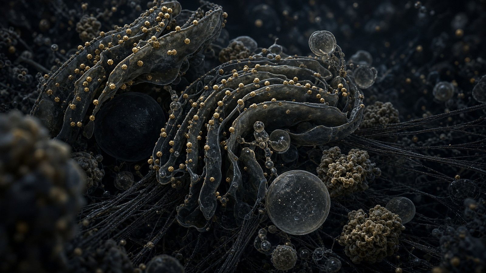
GFP as a traffic report
Green fluorescent protein, 27 kDa, a barrel you can see, has a diffusion coefficient of about 87 µm²/s in water. In mammalian cytoplasm the same protein typically manages 10–20 µm²/s, sometimes a little more in a generous cell, sometimes less if the compartment is packed with filaments. That is a five- to eight-fold slowing, which sounds modest until you remember that time to diffuse scales with distance squared over six D. Across a 10 µm cell at 20 µm²/s, a GFP-sized protein still crosses in under a second. Across a 100 µm neuronal process, the same protein would take on the order of a minute if it had to diffuse, which is why neurons invented motors. A 50 nm vesicle, meanwhile, barely diffuses at all on cellular timescales. Logistics in a picolitre are optional for a small protein and mandatory for everything larger than a ribosome. The city has roads because Brownian motion does not deliver mitochondria to synapses. When you watch a fluorescent protein wander in a living cell, you are watching that traffic report in real time, and it is one of the loveliest measurements in the field.
In short. Green fluorescent protein crosses water fast and cytoplasm five- to eight-fold slower. A small protein still crosses a cell in under a second; a vesicle does not, which is why motors exist.
A millimolar metabolite in that same picolitre is not a vague abundance. One millimolar is 6.022 × 10²⁰ molecules per litre. Three picolitres of 1 mM solution therefore hold about 1.8 million molecules. ATP, typically several millimolar, is a few tens of millions of molecules — comparable, as it happens, to the protein census itself. Free calcium at rest, ~100 nM, is only about 180,000 ions in the same volume. That is why calcium can be a signal: the denominator is small enough that a pulse of a few hundred thousand ions, released from the ER, is a ten- to hundred-fold change rather than a rounding error on a warehouse. Potassium at 140 mM is ~2.5 × 10¹¹ ions, a crowd so large it is the solvent's second ingredient. The cell speaks chemistry in at least three registers at once: the rare ion, the millimolar metabolite, and the macromolecular machine. Mixing the registers is how a concentration becomes a pile, and a pile is not what a receptor sees. Occupancy is a concentration story at a surface.
In short. A millimole of fuel is millions of molecules in one cell; resting calcium is only a few hundred thousand ions. That is why calcium can be a signal and potassium is furniture.
Ions as the cell's first language
Before there were peptides, before there were receptors, before there was a nucleus, there were ion gradients. A typical mammalian cytoplasm holds potassium at about 140 mM and sodium at about 10 mM; the extracellular fluid inverts that, sodium ~145 mM, potassium ~4–5 mM. The Na+/K+ ATPase spends a substantial fraction of a resting cell's ATP budget maintaining the inversion — often quoted near a third in neurons, less in a hepatocyte, more in a thick ascending limb. The membrane potential that results, typically −40 to −80 mV depending on the cell, is not a trivia fact for a physiology viva. It is the reason a channel opening for a millisecond is a decision, the reason a mitochondrion can hang 150 mV across 5 nm of inner membrane and call it a proton-motive force, and the reason the word 'excitable' means anything. Alberts lists the ions in a table. The table is the oldest signalling system you have, and you spend ATP on it every second of your life, including the second you are reading this.
In short. Before peptides or nuclei there were salt gradients. Potassium in, sodium out, a membrane voltage as a result: the oldest signalling system you have, and it spends a lot of ATP.
Calcium, the rare ion that means something happened
Free cytosolic calcium sits near 100 nM at rest and spikes to 1–10 µM when a channel or a receptor says so. Total calcium in the cell is much higher; the ER and the mitochondrial matrix keep stores, and a zoo of buffers (calmodulin, parvalbumin, calbindin, the C2 domains of a hundred other proteins) soak up what the stores release so that the free number can return to 100 nM in tens of milliseconds. Fold-change, not absolute abundance, is the language. A synaptic bouton converting an action potential into vesicle fusion is a calcium story at a specialised active zone. A T cell deciding it has seen antigen is a calcium story through IP3 and store-operated channels. A fertilised oocyte blocking polyspermy is a calcium wave. The same ion, the same 100 nM baseline, wildly different cities. That is why a label that says 'calcium support' misses the interesting number: it is the nanomolar fold-change, in a particular room, on a particular clock, that means something happened.
In short. Free calcium sits at a trace at rest and soars when a channel opens. Fold-change is the language, and that nanomolar pulse is how a cell records that something happened.
ATP, magnesium, and a pH of about 7.2
ATP in a healthy cytoplasm is typically 1–10 mM, most often quoted around 3–5 mM in mammalian cells, with the ATP/ADP ratio held high so that hydrolysis remains strongly downhill. Almost all of it is complexed with magnesium; free Mg2+ is about 0.5–1 mM, total magnesium an order of magnitude higher because so much is bound to ATP, to RNA, to the phosphates of membranes. A kinase that 'uses ATP' is usually using Mg-ATP. The distinction is not pedantry. It is why a magnesium number out of range is a kinetic number, and why the cell treats ATP as both currency and ligand: AMPK is reading the AMP/ATP ratio, not a mood. Cytoplasmic pH sits near 7.2, a shade more alkaline than the 7.4 of blood, with the lysosome at 4.5–5, the mitochondrial matrix nearer 7.8 when the chain is running, and the secretory pathway progressively acidifying from ER to granule. Every one of those compartments is a different solvent. A hydrolase that is a citizen of the lysosome is a vandal in the cytosol. Compartments exist because chemistry is not one room.
In short. ATP is both currency and ligand, usually glued to magnesium. Each compartment keeps a different pH, because chemistry is not one room.
The Debye length in cytoplasm is about one nanometre. That is the distance over which electrostatics are screened by the millimolar soup of small ions. Beyond a nanometre, two charges barely know about each other unless the protein has gone to the trouble of making a pocket that excludes the soup. This is why binding sites are pockets, why a peptide ligand of a few nanometres can occupy one with geometric prejudice, and why charge–charge attraction at long range is a sentence that belongs in a vacuum, not in 140 mM KCl. Physical Biology of the Cell (Phillips, Kondev, Theriot, Garcia) is the book that made this boring and therefore useful. A millimolar metabolite, to say it again because the arithmetic is the whole point, is ~6 × 10²⁰ molecules per litre. In the picolitre of a cell that is a few million copies. In a litre of extracellular fluid it is a warehouse. Concentrations are not interchangeable with amounts. Occupancy is a concentration story at a surface, which is why a vial-count without a volume is not yet an experiment.
In short. Beyond a nanometre, charges barely notice each other in salty cytoplasm. Binding happens in pockets, not across the room, and a concentration is not the same as a pile of molecules.
- K+ ~140 mM inside, ~4–5 mM outside: the bulk cation, the reason the resting potential is negative.
- Na+ ~10 mM inside, ~145 mM outside: the inverted partner, the ion the Na+/K+ ATPase keeps throwing out.
- Free Ca2+ ~100 nM at rest, 1–10 µM in a spike: a few hundred thousand ions, which is why it can be a signal.
- ATP 1–10 mM, mostly as Mg-ATP: currency, substrate, and the number AMPK is watching.
- Free Mg2+ ~0.5–1 mM; pH ~7.2 in cytoplasm, ~4.5–5 in lysosomes, ~7.8 in a working matrix.
- Debye length ~1 nm: electrostatics are a contact sport. A peptide occupies a pocket, not a field.
The cytoskeleton as logistics
A picolitre without roads is a warehouse that cannot ship. The cytoskeleton is the logistics network: actin filaments of about 7–8 nm, microtubules of 25 nm outer diameter built from 13 protofilaments of α/β-tubulin, intermediate filaments of about 10 nm that take the mechanical load so the other two can be dynamic. In motile cells actin can be 5–20% of total protein; in a skeletal myofibre it is the job. G-actin polymerises to F-actin, ATP-loaded, and a zoo of nucleators (Arp2/3 for branched networks, formins for unbranched cables), capping proteins, severing proteins (cofilin, gelsolin) and motors (myosins) turn that polymer into cortex, filopodium, contractile ring, or the stress fibres a fibroblast uses to pull on collagen. TB-500's literature sits on an actin-binding motif. That is a logistics claim: it has to be argued with polymerisation assays and cell-motility numbers, not with adjectives. Name the filament, name the assay, and the peptide becomes a question you can actually ask of a dish.
In short. Without filaments a cell is a warehouse that cannot ship. Actin, microtubules and intermediate filaments are the roads, and a peptide that binds actin is a logistics claim.
Microtubules, kinesin, dynein, and a metre of axon
Microtubules are polar. Plus ends usually point out; minus ends usually point at the centrosome, except in an axon, where they all point the same way, plus-end distal, for tens of centimetres to a metre. Kinesin-1 walks toward the plus end, 8 nm per ATP, on the order of a hundred steps a second, which is about 0.8 µm/s or 70 mm per day if it never fell off. Cytoplasmic dynein walks toward the minus end and is the reason a vesicle at a synapse can ever get home. Fast axonal transport, the textbook 400 mm/day, is this. Slow axonal transport is millimetres per day and is how cytoskeletal polymers themselves crawl out. A hepatocyte is 20–30 µm across; a kinesin crossing it is a job of tens of seconds. A sensory axon in a sciatic nerve can be a metre; the same kinesin, if it stayed on, would take on the order of two weeks. That is why a neuron is not a round cell with a long bit, and why neuropathies of motors and of microtubule-associated proteins are logistics failures before they are clinical signs.
In short. Kinesin walks one way along microtubules, dynein the other: seconds to cross a liver cell, weeks to cross a metre of nerve. A neuron is not a round cell with a long bit.
Dynamic instability — microtubules growing, catastrophically shrinking, growing again — is how a cell searches space without a map. GTP-tubulin adds; hydrolysis lags; a GDP-tubulin lattice is unstable; a rescue or a catastrophe follows. Taxanes freeze the lattice; vinca alkaloids pick it apart; both are chemotherapeutic because a mitotic spindle is a microtubule machine that has to get the geometry right in minutes. None of that is a research-peptide story. It is the reason the word 'cytoskeleton' is a poor metaphor. Skeletons do not treadmill at both ends. This one does, continuously, and the ATP and GTP bill is not optional. Intermediate filaments (keratins in epithelia, vimentin in mesenchyme, neurofilaments in axons, lamins at the nuclear envelope) are the least glamorous and the most honest: they take the strain so the dynamic polymers can afford to be dynamic. A laminopathy is what you get when the nuclear 'skeleton' is the thing that fails. Scale, again: a lamin filament is 10 nm; the nucleus it lines is 6–10 µm; the tissue that tears is centimetres. The failure starts at the nanometre, which is a humbling and useful way to look at a rare disease.
In short. Microtubules grow and collapse as a way of searching space. Intermediate filaments take the strain so the others can fidget — and failure of a ten-nanometre fibre can tear a tissue.
Cell crawling is actin plus adhesion plus a decision about where the front is. A neutrophil chasing a bacterium in a dish is the demonstration that a 10 µm city can polarise, polymerise a leading edge, retract a tail, and arrive in minutes. A fibroblast closing a scratch wound is slower and more adhesive. A metastatic cell is the same toolkit used without permission. Catalogue peptides that claim to support repair are, if they have a mechanism at all, usually sitting on one of these logistics nodes: an actin motif, a growth-factor receptor that turns on motility genes, a copper-binding tripeptide whose microarray literature includes matrix metalloproteinases. Name the node. Name the assay. The scratch wound is not a person, and a person is not a scratch wound, but the dish is still a start: it is where a motility claim becomes a number rather than a hope. Logistics is the layer that makes the difference visible in culture, which is why the dish is a beginning and not a finish.
In short. Crawling is actin, grip, and a decision about the front. Name the node and the assay: a scratch in a dish is not a person, and a person is not a scratch.
Organelle census of a hepatocyte
If you have to pick one cell as the industrial city, pick the hepatocyte. It is large, polygonal, often binucleate, often polyploid, and it runs more concurrent metabolisms than any other mammalian cell that is not a steroidogenic factory. Weibel's morphometry, and the tables that Alberts still reprints, give you a census rather than a cartoon. A hepatocyte holds on the order of 1,000–2,000 mitochondria, occupying a substantial fraction of cytoplasmic volume — commonly a fifth or more, depending on the metabolic state you caught it in. The endoplasmic reticulum, rough and smooth, is the majority of the cell's membrane surface, ten to twenty-five times the plasma membrane by area; the liver as an organ is, among other jobs, a square-metre-scale endomembrane. The Golgi is a smaller, polarised stack through which a third of the proteome is processed for secretion or the plasma membrane. Lysosomes are numbered in the hundreds, acidic, hydrolase-packed. Peroxisomes, a few hundred, handle very-long-chain fatty acids. The nucleus, one or two, is 6–10 µm, with about 3,000 nuclear pores. That is one city. You have about 10¹¹ hepatocytes.
In short. If you pick one industrial city, pick the liver cell: a thousand-odd mitochondria, vast internal membrane, hundreds of lysosomes, a nucleus with three thousand doors. You have about a hundred billion of them.
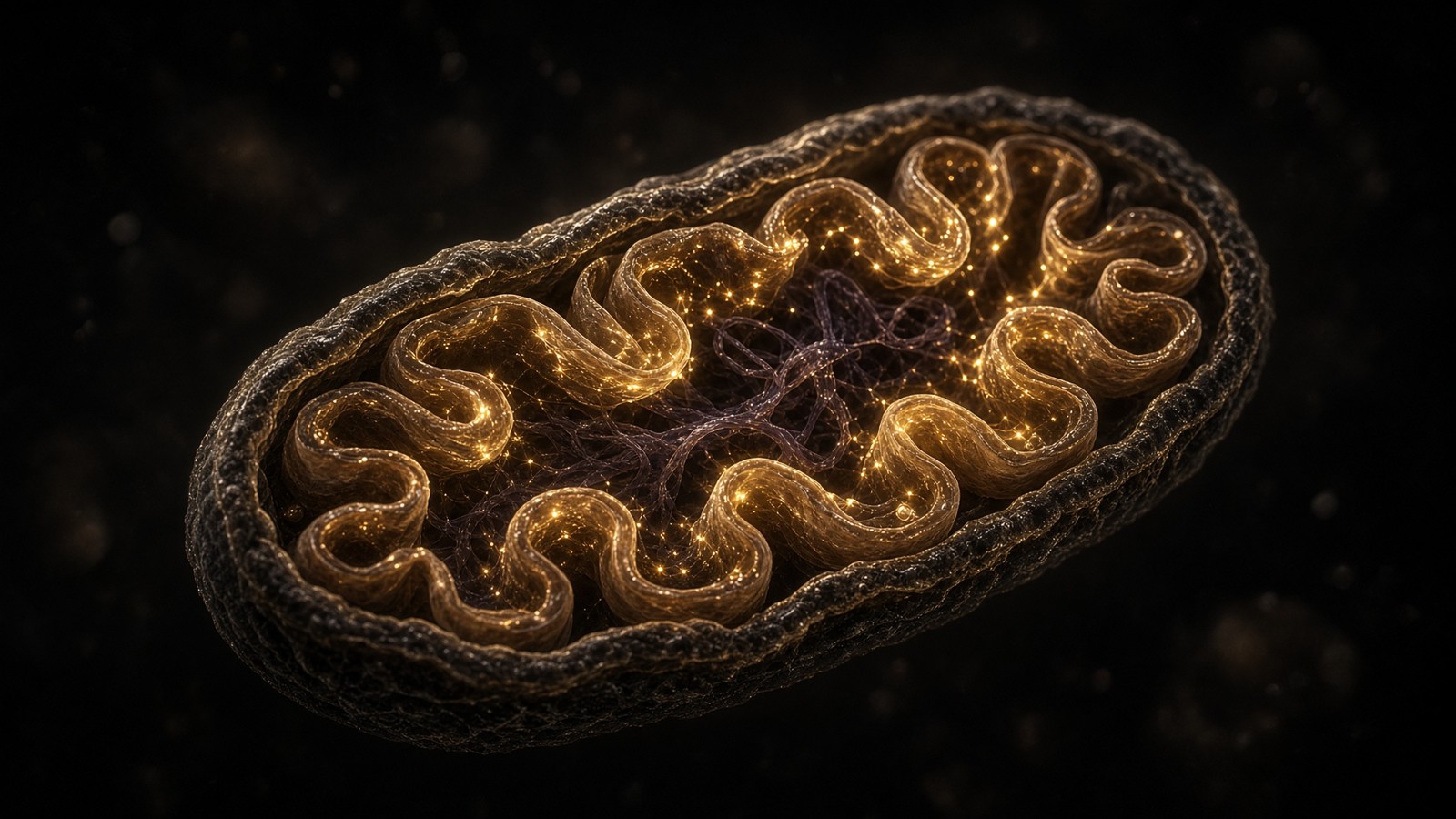
Diagram
Matrix
- TCA cycle · β-oxidation · mtDNA nucleoids
- NADH produced here. Complex I spends it.
- MOTS-c (MRWQEMGYIFYPRKLR) from 12S rRNA.
Inner membrane
- I → II → III → IV → V (ATP synthase)
- ~150 mV proton-motive force
- ~40–60 kg of ATP turned over per human day
mtDNA is 16,569 bp, 37 genes, 13 proteins of the respiratory chain. Nuclear DNA encodes the other ~1,200 mitochondrial proteins. NAD+ is the hydride carrier between dehydrogenases and Complex I. MOTS-c is a 16-mer translated from 12S rRNA — a peptide the mitochondrion wrote itself.
The endomembrane is most of the surface
Students are taught the plasma membrane as 'the' membrane and then spend a decade being surprised that most of the lipid bilayer in a secretory cell is inside. Rough ER is ribosome-studded and makes everything destined for secretion, membrane, or lysosome. Smooth ER in a hepatocyte is a cytochrome P450 forest, a glucose-6-phosphatase station, a lipid-synthesis bench, and a calcium store. The Golgi adds and edits glycans, sorts cargo, and is the reason a plasma-membrane protein and a lysosomal hydrolase, both born into the ER lumen, do not end up in the same place. COPII vesicles leave the ER; COPI vesicles come back; clathrin coats the routes to and from the plasma membrane and the endosomes. This is not trivia. It is why a peptide synthesised on a bench, lyophilised, and added to a dish from the outside is a completely different object from a peptide hormone the ER–Golgi route would have folded, glycosylated, and stored in a granule. Insulin is a secretory-pathway citizen. BPC-157 in a vial is a chemical. Same class of bond. Different biography. Both can occupy a pocket. Only one of them was born into the lumen.
In short. Most of a secretory cell's membrane is inside, not on the surface. Insulin is a citizen of that route; a peptide in a vial is a chemical with the same bond and a different biography.
Three thousand doors in the nuclear envelope
The nuclear pore complex is a 100–120 MDa machine of about thirty nucleoporins, eight-fold symmetry, a central channel that can pass a 40 nm cargo if the cargo has the right transportins attached, and FG-repeat unstructured domains that make the channel a selective hydrogel rather than a hole. A typical nucleus keeps about 3,000 of them. Macromolecular traffic through the lot is enormous: ribosomal subunits outwards, transcription factors inwards, mRNA outwards as packed RNPs, histones inwards, snRNPs on a round trip. Importins read nuclear localisation signals; exportins read nuclear export signals; the Ran-GTP gradient, high in the nucleus because RCC1 sits on chromatin, gives the transport direction for free. A transcription factor that cannot get in is not a transcription factor today. A ribosome that cannot get out is a nucleolar argument, not a cytoplasmic printer. Scale: the pore is ~100 nm in overall diameter; the envelope it punctures is a double 5 nm bilayer with a 20–40 nm lumen; the nucleus is 6–10 µm. Finding the door is not the hard part. Being the right shape, with the right export factors, is.
In short. About three thousand pores puncture the nuclear envelope. A transcription factor stuck on the wrong side is not a transcription factor today; being the right shape, with the right escorts, is the hard part.
The nucleolus is not an organelle with a membrane. It is a phase-separated condensate, classically three zones (fibrillar centre, dense fibrillar component, granular component), where RNA polymerase I transcribes hundreds of rDNA repeats into 45S pre-rRNA, and where the small and large subunits are assembled from that RNA plus ~80 ribosomal proteins imported from the cytoplasm. A growing cell's nucleolus is huge because the ribosome census has to be rebuilt every cycle. A quiescent cell's nucleolus shrinks. Nucleolar size is one of the oldest pathologists' observations and one of the least mysterious once you have counted ribosomes. Cajal bodies, nuclear speckles, PML bodies — more condensates, more specialised. The nucleus is a packed, moving, transcribed gel with rooms that have no walls, and the rooms still manage not to mix their chemistry. That sentence is why a circle labelled nucleus in a textbook is a drawing of a door, not of the room. Walk in and it is a factory, a library, and a liquid that has opinions.
In short. The nucleolus has no membrane: it is a liquid droplet where ribosomes are built, large when the cell is growing. A textbook circle labelled nucleus is only the door to that room.
- Mitochondria: ~1,000–2,000 per hepatocyte, ~0.5–1 µm, two membranes, their own 16.6 kb genome.
- Endoplasmic reticulum: most of the membrane surface, rough for secretion, smooth for P450 and calcium.
- Golgi: polarised stack, glycan editing, the sorting office for a third of the proteome.
- Lysosomes: hundreds, pH 4.5–5, hydrolases, mTORC1’s amino-acid radar on the cytosolic face.
- Peroxisomes: hundreds, very-long-chain fatty acids, a private catalase budget.
- Nuclear pores: ~3,000 per nucleus, ~110 MDa each, the only legal door in a double envelope.
- Nucleolus: no membrane, Pol I, rRNA, the factory that keeps the 10⁶–10⁷ ribosomes coming.
Thirty-six trillion, and most of them cannot read a gene
Sender, Fuchs and Milo, 2016, Cell: a 70 kg adult holds about 3.0 × 10¹³ human cells by their first pass, later revised toward 3.6 × 10¹³. The headline that broke the internet was not the total. It was the composition. Erythrocytes are ~84% of the count and ~4% of the mass. Fat and muscle are most of the mass and a rounding error of the count. Glia and neurons together are ~1% of cells and a fifth of the energy budget. If you weigh yourself you are looking at adipocytes and myocytes. If you count yourself you are looking at red blood cells that ejected their nuclei before they ever saw a capillary. That double census is one of the most useful corrections modern cell biology has given the rest of us. It means that 'what cells want' is already a broken sentence: the cell you are statistically most of cannot read a gene, and the cell you are by mass is not the cell you are by number. Hold both rankings. We will need them.
In short. A 70 kg adult holds about 36 trillion human cells, most of them red blood cells with no nucleus. Weigh yourself and you are fat and muscle; count yourself and you are haemoglobin bags.
The census by number, the census by mass, the census by power
The 2016 paper did something that popular science had refused to do, which was to add. Take a reference 70 kg man. Estimate the number of cells in each tissue from volume, cellularity, and the literature's best morphometry. Sum. The sum is not 100 trillion, which was a round number looking for a home, and it is not 37.2 trillion, which was a later press-release rounding. It is about 30 trillion on the first pass, revised toward 36 trillion as adipose cellularity and a few other under-counts were repaired, including in the 2023 updates. About 25 trillion of those are erythrocytes: 84% of the count, 4% of the mass, no nucleus, no mitochondrion, a bag of haemoglobin and a membrane with a 120-day appointment. Platelets are next by number, anucleate fragments of megakaryocytes, 1–2 × 10¹². Neutrophils follow among the nucleated majority. The cells that occupy the public imagination — hepatocytes, cardiomyocytes, neurons, skeletal myofibres — are, by number, scarce. They dominate mass, or energy, or the idea of a person. They do not dominate the tally, and that is a gift of a fact once you have it.
In short. The old round numbers — a hundred trillion, 37.2 trillion — were looking for a home. The real tally is about 36 trillion, and 25 trillion of those are red cells that ejected their nuclei.
Mass is a different ranking. Skeletal muscle and adipose tissue are most of a 70 kg body. A myofibre is a centimetre-scale multinucleate cell and there are not that many of them; an adipocyte is a lipid droplet with a rim of cytoplasm and there are tens of billions at most, not trillions. The liver is a kilogram and ~10¹¹ hepatocytes. The brain is 1.3–1.4 kg and ~86 billion neurons plus a comparable number of non-neuronal cells. The heart is 300 g and a few billion cardiomyocytes, postmitotic, irreplaceable on any useful timescale, each one a mitochondrial forest. Energy is a third ranking again. The brain is ~2% of body mass and ~20% of resting metabolic rate. The heart never stops. Brown adipose tissue, when you have some and when it is on, is a small mass with a large UCP1-shaped hole in the proton-motive force. Sender and Milo's later turnover paper makes the rankings even less interchangeable: the cells you make fastest are not the cells you are by mass, and the cells you are by mass turn over slowly. A body is three different cities depending on whether you count, weigh, or meter the power.
In short. Count, weigh, or meter the power: three different cities. Muscle and fat are the mass, red cells the tally, the brain two percent of mass and a fifth of the bill.
Two million red cells a second, for decades
Bacteria in the gut are a comparable census, ~3–4 × 10¹³, which is why the 'ten-to-one bacterial cells' factoid had to be retired. It was a 1970s back-of-envelope that assumed a litre of faeces was a person. The current ratio is roughly 1:1 by number, with the bacterial biomass still only a few hundred grams. You are not a walking microbiome with a human attached. You are a human who rents a fermenter, and it is a rather wonderful fermenter: almost all of it lives in the colon, it weighs as much as a pineapple, and it has its own literature on a different desk. Research peptides are not a prebiotic, a probiotic, or a microbiome intervention. They are ligands. The 1:1 ratio is the useful sentence. The older 10:1 ratio was a litre of stool pretending to be a body, and once you have seen the arithmetic you will not go back.
In short. Gut bacteria number about as many as your own cells, not ten times more, and weigh a few hundred grams. You rent a fermenter; you are not one.
Red blood cells live about 120 days. To hold 25 trillion of them you have to make roughly two million per second, every second, for decades, from haematopoietic stem cells in marrow that you will never see. Two million is a small number next to the ATP figure, which is the next one that should make you pause, but it is still a production line that would humble any factory on Earth. Each of those cells is a descendant of a haematopoietic stem cell via burst-forming then colony-forming erythroid progenitors, a shrinking nucleus, a dumping of mitochondria, a loading of haemoglobin to ~330 mg/ml, and a squeeze into a 7–8 µm biconcave disc that has to deform through capillaries narrower than itself. At 120 days the disc is stiff, the membrane is tagged, and a macrophage in spleen or liver eats it. Iron is recycled with a stinginess the rest of metabolism can only envy. Nothing in that sentence is a peptide story. It is a stem-cell story and a logistics story about a cell that threw away the nucleus in order to be a bag of oxygen-binding protein.
In short. Red cells last about 120 days. To keep 25 trillion of them you make roughly two million a second, every second, for decades.
The production arithmetic is worth writing in longhand because it is the cleanest illustration of turnover as a way of being alive. 2.5 × 10¹³ erythrocytes divided by 120 days is 2.1 × 10¹¹ cells per day, which is 2.4 × 10⁶ per second. Each of those cells dumps its nucleus and its mitochondria, loads haemoglobin, and becomes a disc. When a catalogue talks about cellular health as if there were one cell, this is the cell it is statistically talking about, and it is the cell least able to read a gene. Transcription factors, nuclear-targeted peptides, anything that needs a promoter — this city fired the government. GATA1, the EPO receptor, HIF when the oxygen number drops: those are the conversations that happen upstream, in the progenitors, not in the circulating disc. Two million a second, for decades, from a stem-cell pool you will never meet. If that does not fill you with a certain respect for marrow, we have not yet counted high enough.
In short. Each red cell dumps its nucleus and mitochondria, loads haemoglobin, squeezes through capillaries, and is eaten at four months. Statistically this is you, and it is the cell least able to read a gene.
Eighty-six billion neurons, not a hundred, and not ten glia each
The other fossil number is the brain. A hundred billion neurons and ten glia per neuron is a sentence that escaped from mid-century estimates and refused to die. Suzana Herculano-Houzel's isotropic fractionator, applied to human brains with Azevedo and colleagues in the Journal of Comparative Neurology in 2009, found about 86 billion neurons and about 85 billion non-neuronal cells in the whole brain: roughly 1:1, not 10:1. The distribution is the second surprise. The cerebellum, 10% of brain mass, holds about 69 billion neurons, almost all granule cells, tiny. The cerebral cortex holds about 16 billion neurons and a larger non-neuronal (mostly glial) population, which is why a cortical histologist sees more glia than neurons and why the 10:1 myth felt locally true. Glia are not a homogeneous support staff; astrocytes, oligodendrocytes, microglia and the NG2 cells are different cities. The energy budget is the third surprise: 86 billion neurons, 2% of body mass, 20% of resting ATP turnover, paid largely at synapses. A research peptide that supports brain health without naming a receptor, a cell type, and an assay has not yet entered this census. Occupancy is still the only legal move.
In short. The brain has 86 billion neurons, not 100 billion, and about as many other cells. A peptide that cannot name a receptor, a cell type and an assay has not entered this census.
Gut bacteria, to finish the retired factoid properly, were estimated by Sender, Fuchs and Milo at about 3.8 × 10¹³ in a reference colon, against their human-cell total of the same order. The 10:1 ratio came from taking the older, inflated human-cell number as 10 trillion and the colonic contents as a litre at 10¹¹ bacteria per millilitre, and not noticing that most of a person is not a colon. Bacterial biomass is a few hundred grams. Viral particles in the gut outnumber the bacteria, as they do almost everywhere, and are not in this essay because this essay is already long and because a bacteriophage is not a cell. The useful sentence is smaller than the myth: you carry about as many bacterial cells as human cells, they weigh as much as a pineapple, they are almost all in the colon, and they are a fermenter you rent, not a second self. Research peptides are ligands, not a microbiome intervention. The fermenter has its own literature, on a different desk, and it is a good literature once you let the 10:1 story go.
In short. You carry about as many bacterial cells as human cells; they weigh as much as a pineapple and live almost entirely in the colon. Research peptides are ligands, not a microbiome intervention.
A body-weight of ATP, every day, and you store a coffee cup
Peter Rich, 2003, Biochemical Society Transactions: resting ATP turnover in an adult is on the order of 40 kg per day. Athletes and fever push it higher, toward 60 kg. The standing pool of ATP in the whole body is about 50 g — a shot-glass. The only way those two numbers can both be true is if every molecule is recycled hundreds of times a day. At the mitochondrion that looks like a proton-motive force of ~150 mV across a 5 nm membrane, Complex V spinning, ADP in, ATP out. At the organism it looks like breathing. That pairing — a coffee-cup of stock, a body-weight of flux — is one of the deepest facts in physiology, and it is still under-taught. You do not store ATP. You remake it, continuously, from the food you oxidise and the air you take in. A 1000 mg vial of NAD+ on a shelf is the hydride coin that feeds Complex I so this machine has something to oxidise. It is not 40 kg of ATP. It is the coin the enzyme spends, lyophilised, for the bench.
In short. You turn over about a body-weight of ATP each day and store a shot-glass. Every molecule is recycled hundreds of times; at the organism that looks like breathing.
Do the arithmetic once and you will not forget it. 50 kg of ATP is 50,000 g. ATP's molar mass is 507 g·mol⁻¹, so ~99 moles. Avogadro's number is 6.022 × 10²³. That is ~6 × 10²⁵ molecules per day, or ~7 × 10²⁰ per second — call it 10²¹ if you like round numbers and a more active day. A billion trillion hydrolyses while you finish this paragraph. NAD+ is nicotinamide adenine dinucleotide — a small molecule that shuttles electrons. One carbon on a vitamin-B3 ring does the whole job. The 1000 mg vial on the shelf is that cofactor, lyophilised. It is ~1.5 millimoles, ~9 × 10²⁰ molecules. Whole-body ATP turnover is ~10²¹ per second. The vial is, coincidentally, about a second of that flux as a count of molecules, and not a second of that flux as a physiology, because the vial is not in the matrix, not being reduced, not being reoxidised, and not a person. The coincidence is a teaching toy. It is not an argument.
In short. Fifty kilograms of ATP is on the order of 10²¹ hydrolyses a second. The NAD+ vial on the shelf is the coin the enzyme spends, not the 40 kilograms.
Diagram
- I. Pumps H⁺. ~45 subunits. The NADH coin is spent here.
- II. TCA entry. No proton pump. FADH₂ neighbourhood.
- Q. Lipid-soluble shuttle in the inner membrane.
- III. Q-cycle. Pumps H⁺. Superoxide leak site.
- c. Intermembrane space. The shuttle everyone has heard of.
- IV. O₂ → H₂O. The reason you breathe.
- V. F₁Fₒ rotary. Protons in, ATP out. ~10²¹ times a second in you.
Mitchell’s chemiosmotic theory (Nobel 1978): the inner membrane is a battery of ~150 mV. NAD+ is the hydride carrier that feeds Complex I. MOTS-c is a 16-mer the mitochondrion translated from 12S rRNA — a different object on the same campus.
The brain is two percent of the mass and a fifth of the bill
A 1.3 kg brain in a 70 kg body is a little under 2% of mass. Resting cerebral metabolic rate is about 20% of whole-body oxygen consumption, a figure so often repeated it has become furniture, and still true. The ATP is spent on Na+/K+ pumping after excitatory postsynaptic currents, on filling vesicles, on cycling glutamate and GABA, on the housekeeping of 86 billion neurons and their glia. Grey matter costs more than white; cortex costs more than cerebellum per gram, even though cerebellum holds more neurons, because a granule cell is a cheap neuron and a cortical pyramidal cell is not. Anaesthesia drops the bill. Seizure multiplies it. Sleep does not turn it off; it rearranges it. None of this is optional, which is why the brain is greedy when the rest of the body is fasting, and why a few minutes of ischaemia is a disaster in cortex and a shrug in a resting myocyte. The mitochondrion of a neuron is not a different machine from the mitochondrion of a hepatocyte. The duty cycle is different. Getting a mitochondrion to a bouton, a metre from the soma, is the cytoskeleton section again.
In short. The brain is two percent of your mass and about a fifth of resting oxygen use. It is greedy when the rest of you is fasting, because synapses are expensive pumps.
The heart is the other organ that does not get to have a quiet shift. A 300 g pump, a few billion cardiomyocytes, each one packed with mitochondria to a volume fraction that textbooks put near 30–40% in the ventricle, beating 100,000 times a day, turning over its own ATP pool every few seconds. Phosphocreatine is the local buffer so that the pump at the sarcolemma and the myosin ATPase at the myofilament do not have to wait for a phosphate to diffuse from a mitochondrion. Ischaemia of minutes is an infarct because this city has no savings account, only a current account and a very small overdraft. Skeletal muscle, by contrast, can rest, can go glycolytic, can tolerate a phosphocreatine crash that would kill a myocyte. Brown adipose tissue is the optional furnace: UCP1 opens a proton leak, the chain runs without making ATP, the energy becomes heat. Infants have it. Adults have a little, more if they are cold often. It is a rounding error of mass and, when it is on, a detectable rounding of the energy budget.
In short. The heart has no savings account, only a current account that overdraws in minutes of ischaemia. Skeletal muscle can rest; brown fat, when it is on, turns fuel into heat on purpose.
NAD+ is the hydride coin, not the mint
NAD+ (nicotinamide adenine dinucleotide, 663.43 g·mol⁻¹) accepts a hydride at Complex I's flavin and at a dozen dehydrogenases, and is the obligatory co-substrate of sirtuins and PARPs, which consume it rather than redox-cycle it. The cellular NAD+ pool is millimolar in some compartments and lower in others; the mitochondrial pool is not in free equilibrium with the cytosolic pool because the cofactor does not cross the inner membrane as such. NAMPT salvages nicotinamide back to NMN then NAD+; CD38 and related hydrolases spend the pool; DNA damage spends it through PARP1; age, in many tissues, correlates with a smaller pool, which is a measurement, not a marketing claim. The 1000 mg vial in the catalogue is β-NAD+, lyophilised, HPLC-characterised, a reagent. It is ~1.5 millimoles, ~9 × 10²⁰ molecules. Whole-body ATP turnover is ~10²¹ per second. MOTS-c, the 16-residue peptide translated from mitochondrial 12S rRNA, is a different object from the same organelle: a claimed AMPK-and-folate–methionine signal, a nuclear translocation under stress. Two catalogue lines, one power station, and no need to confuse the coin with the mint.
In short. NAD+ carries hydrides into the mitochondrion and is spent by sirtuins and PARPs. The vial is a characterised reagent, not a second of your ATP flux as a physiology.
Two metres of DNA in a six-micrometre sphere
A haploid human genome is about 3.1 billion base pairs (GRCh38; T2T-CHM13 pushed the dark corners). Diploid, in G1, that is ~6.2 Gbp. Each base pair is 0.34 nm along the axis of B-DNA, so 6.2 × 10⁹ × 0.34 × 10⁻⁹ m ≈ 2.1 metres. The nucleus of a typical somatic cell is 6–10 µm in diameter. Packing two metres of a 2 nm fibre into that sphere, with the nucleosomes, loops, compartments and lamina that actually exist, is the most violent origami on Earth. It happens in every nucleated cell you have, and you have about 6 trillion of those once you subtract the erythrocytes. Pause on that. Two metres, into a room you could hide under a grain of salt, six trillion times, and most of those rooms are reading a different chapter. Gene regulation is what you invent when you have done that packing and still need to find the right thousand base pairs among 3.1 billion in a minute. The next essay on this desk is that search. This one is only here to make the packing arithmetic unforgettable.
In short. Diploid DNA, stretched, is about two metres. It is folded into a nucleus six to ten micrometres across, in every nucleated cell you have — about six trillion of them.
If you uncoiled the nuclear DNA from those 6 trillion nucleated cells you would have on the order of 10¹³ metres — about 70 AU, twice the distance from the Sun to Pluto at aphelion, a number we are only writing down because it is true and because people still talk about the genome as if it were a book on a shelf. It is a library stuffed into a room the size of a bacterium, 6 trillion times, and most of the rooms are reading a different chapter. A hepatocyte is reading albumin and P450s and urea-cycle enzymes. A neuron is reading a splice-isoform programme the hepatocyte skips. A mature erythrocyte is reading nothing, because it fired the library. The same letters, wildly different cities. That is why chromatin, methylation, and the three-dimensional genome are not decorations on a linear string. They are how one archive becomes a liver rather than a cortex without changing a base. Wonder is allowed here. Two metres in a micrometre-scale room, times six trillion, is a fact that still makes this teacher grin.
In short. Uncoil the DNA from those six trillion nuclei and you get a length that reaches past Pluto. Most of those rooms are reading a different chapter.
Thirty million nucleosomes, and the fibre is not a solenoid any more
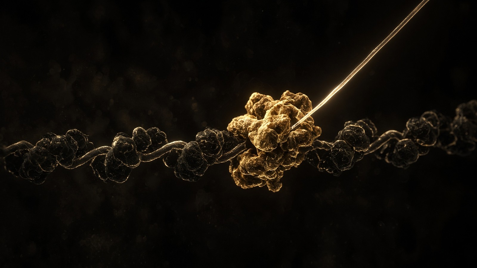
The packing unit is the nucleosome: 147 base pairs of DNA wrapped 1.65 turns around a histone octamer (two each of H2A, H2B, H3, H4), with histone H1 at the entry/exit and a linker of variable length that brings the repeat to roughly 200 bp in much of the human genome. Six billion base pairs divided by 200 is about 30 million nucleosomes per diploid G1 nucleus. Each octamer is ~11 nm in diameter; the DNA is the ribbon. The old textbook 30 nm fibre, a solenoid of nucleosomes, has had a difficult century: in vivo, much of chromatin looks more like an irregular 10 nm beads-on-a-string, folded into loops by cohesin and CTCF, compacted by macromolecular crowding and by the lamina at the edge. Hi-C, from Lieberman-Aiden and colleagues in 2009 and the industry that followed, sees A compartments (open, gene-rich) and B compartments (closed, gene-poor), topologically associating domains, and loops that put an enhancer in physical contact with a promoter a million base pairs away on the sequence and a few hundred nanometres away in the room. Gene regulation is geometry plus chemistry.
In short. The packing unit is the nucleosome: DNA wrapped around a histone spool, about 30 million of them per nucleus. Finding the right thousand base pairs among billions, without unpacking the rest, is the search problem.
Diagram
- 2 nmB-DNA0.34 nm/bp. Diploid G1 is ~2 metres of this.
- 11 nmNucleosome147 bp around a histone octamer. ~30 million per nucleus.
- loopsCTCF / cohesinEnhancers meet promoters by folding, not by sliding.
- µmA/B compartmentsHi-C: open A, closed B, territories at the lamina.
- 6–10 µmNucleusThe room. The search problem is the entire point of gene regulation.
Packing is not storage. It is the first regulatory decision: a promoter buried in H3K27me3 is not a promoter, it is furniture. Transcription starts when this origami opens the right 1,000 base pairs among 3.1 billion.
Gene regulation is what you invent when you have done that packing and still need to find the right 1,000 base pairs among 3.1 billion in a minute. Promoters, enhancers, CTCF loops, A/B compartments, histone marks, DNA methylation, the Mediator complex, RNA polymerase II pausing. Epithalon's literature sits on TERT — the reverse transcriptase that extends telomeres — and on pineal melatonin amplitude. That is two promoters in a genome of twenty thousand protein-coding genes and a much larger noncoding census. The peptide is four residues. The search problem is the genome. We stock the tetrapeptide as a characterised ligand. We do not stock a compression algorithm for two metres of DNA. The nucleus essay and the transcription essay on this desk are where that argument belongs; this one is only here to make the packing arithmetic sit in your head so that four residues next to the word telomere still look like four residues. Geometry first. Then the papers. Then the gap, named honestly, as a gap.
In short. Gene regulation is what you invent after packing two metres into a speck and still needing the right stretch in a minute. Four residues sit on one promoter among twenty thousand genes.
Diagram
Out → in
Proteins, TF, histones
Importins + Ran-GTP cycle. A transcription factor that cannot clear the pore is not a transcription factor. It is a cytosolic rumour.
The mesh
NPC · FG nups
Passive cutoff a few nanometres. A ribosomal subunit is assembled in the nucleolus and exported as cargo, not as a wanderer.
In → out
mRNA, assembled ribosomes
TREX, NXF1/NXT1. Unspliced RNA is retained on purpose. Export is a licence, not a leak.
~3,000 pores per nucleus. ~30 nucleoporins. FG-repeat mesh that lets small molecules through and makes macromolecules show a passport (NLS, NES, NXF1 for mRNA). The nucleus is not a bag. It is a gated compartment.
A preview, because this essay is the city and the next ones are the buildings. DNA does not make protein. DNA is transcribed by RNA polymerase II at typically 20–40 nucleotides per second, with long pauses; spliced on a 3-megadalton spliceosome; exported through one of those 3,000 pores; translated at 5–6 amino acids per second. The central dogma is still the spine. The clocks are why a 20 kb gene is minutes and dystrophin is hours. Chromatin state is why a hepatocyte and a neuron, with almost the same DNA, are not the same city. Epithalon's claim, if you take the St Petersburg literature at face value, is a conversation with one promoter (TERT) and one pineal enzyme (AANAT) in this mess. Four residues. A 6–10 µm room. Thirty million nucleosomes. The gap between those numbers is the entire intellectual problem of writing a peptide next to the word telomere. We stock the tetrapeptide as a characterised ligand. We do not stock a compression algorithm for the genome. The clocks on every arrow are the next lesson, and they are as real as the packing.
In short. DNA does not make protein: it is copied into RNA, shipped through a pore, and printed at five amino acids a second. A tetrapeptide next to telomere is still four residues among 30 million nucleosomes.
Diagram
A lyophilised research peptide skips every step after “protein”. It is the ligand already. That is the entire point of the catalogue, and the reason it is not a gene therapy.
Crick’s flow is still right. The numbers are the part textbooks skip: a mammalian polymerase is slow, splicing is a machine the size of a ribosome, and translation errors run about one in 10⁴ amino acids.
A ribosome is a 4-megadalton computer that prints protein
The eukaryotic ribosome is ~4 MDa of RNA and protein, 80S, assembled in the nucleolus from 18S, 5.8S, 28S and 5S rRNA plus ~80 ribosomal proteins. Bacterial 70S is smaller and faster. In a growing mammalian cell you keep several million of these machines. Each one adds amino acids at about 5–6 per second in eukaryotes (yeast is a bit quicker; bacteria run 12–21). A 400-residue protein is a one-minute print job on one ribosome, except that initiation, not elongation, is usually rate-limiting, and the mRNA is being used by a polysome of many ribosomes at once. Pause on the machine for a moment. Four megadaltons, mostly RNA, an active site that is a ribozyme, printing the proteome at a walking pace. You have millions of them in a growing cell. Five million ribosomes elongating at 5 amino acids per second is 2.5 × 10⁷ amino acids per second, which at 400 residues per typical protein is about 60,000 proteins finished per second from one cell — if every ribosome is busy, which they are not, because initiation is gated. Protein synthesis is a budget the cell can turn down.
In short. A ribosome is a four-megadalton printer; a growing human cell keeps millions, each adding about five amino acids a second. A typical protein is a one-minute job if initiation would get out of the way.
Error rate in translation is about 10⁻⁴ per amino acid. A 400-residue protein has a few percent chance of a substitution. The cell lives with that because proteins turn over. DNA replication, after proofreading and mismatch repair, runs at 10⁻⁹ to 10⁻¹⁰. The genome is sacred. The proteome is a draft. Ageing is partly what happens when the sacred copy still drifts, and when mitochondria — which replicate their 16.6 kb circles in a ROS-rich matrix with a thinner repair budget — drift faster. This gradient of care is one of the deepest facts in molecular biology. A research peptide, synthesised chemically, has a different error structure: deletion sequences from missed couplings, incomplete deprotections. HPLC is the filter; mass spec is the identity check. The cell and the chemist both make amide bonds. They fail differently. They are checked differently. Neither of them is a reason to confuse a vial with a nucleus. DNA is copied as if it mattered forever. RNA and protein are allowed to be sloppy because they are disposable. The mitochondrion never quite got the memo.
In short. Translation gets about one amino acid in ten thousand wrong; DNA replication, after repair, is a million times more careful. The genome is sacred; the proteome is a draft.
Diagram
- DNA replication + MMR10⁻⁹ to 10⁻¹⁰A genome of 6 Gbp (diploid) accumulates a handful of mutations per division.
- Transcription~10⁻⁵RNA is disposable. The cell can afford a wrong letter in a message that lasts hours.
- Translation~10⁻⁴One wrong amino acid per ten thousand. Proteins turn over. DNA does not.
- mtDNA10–100× nuclearNo histones, ROS next door, weaker repair. The second genome ages faster.
The genome is sacred, the message is cheap, the protein is cheaper. Ageing is partly what happens when the sacred copy still drifts — and when mitochondria, which never got the nuclear repair budget, drift faster.
Ten billion proteins, a Zipf’s law, a dynamic range of ten million
A growing mammalian cell's proteome is on the order of 10¹⁰ protein molecules. That number is not evenly spread across 20,000 gene products. A typical cell expresses about 10,000 protein species at a time; the copy numbers run from a handful of a transcription factor or a signalling GTPase to tens of millions of actin, tubulin, histones, and the glycolytic enzymes (GAPDH, enolase, aldolase, pyruvate kinase) that have to process millimolar glucose. Mass spectrometry of HeLa and of tissues has made this boring in the best way: the abundant proteins are the abundant proteins, across cell types, and the interesting proteins are often the rare ones. Dynamic range of protein abundance in a single cell is about 1 to 10⁷ copies. That is why a Western blot against actin is a loading control and a Western blot against a transcription factor is an act of faith in the antibody. It is also why the proteome is not a list of names. It is a list of names with exponents. Look at the exponents and you know what city you are in.
In short. Ten billion proteins are not spread evenly: a few species run to tens of millions of copies, some transcription factors to handfuls. The proteome is a list of names with exponents.
Ribosome census follows demand. Growing cells: 10⁶–10⁷ ribosomes. Quiescent cells: fewer. A rapidly growing HeLa cell, several million; a hepatocyte, similar order, with a large fraction docked on rough ER because so much of the liver's proteome is secretory. Five million ribosomes elongating at 5 amino acids per second is 2.5 × 10⁷ amino acids per second — if every ribosome is busy, which they are not, because initiation is gated by eIF2 phosphorylation, by mTORC1's reading of amino acids at the lysosome, by eIF4E availability. Protein synthesis is a budget the cell can turn down when the energy number or the amino-acid number goes out of range. That is pathophysiology at this floor: not a mutation, a rate. Half-lives run from minutes (some cyclins, some transcription factors) to days and weeks (cytoskeletal proteins, histones in a non-dividing nucleus, haemoglobin in an erythrocyte that can no longer make any more). Proteostasis is the joint operation of chaperones (Hsp70, Hsp90, TRiC), the ubiquitin–proteasome system, and autophagy. A 10¹⁰-molecule city that could not throw things away would fill with errors at 10⁻⁴ per residue in an afternoon.
In short. Protein synthesis is a budget the cell can turn down. Half-lives run from minutes to weeks, and a city of ten billion that could not throw things away would fill with errors in an afternoon.
The most abundant proteins are a tour of what a cell actually spends itself on. Actin and the tubulins are the logistics budget. Histones are the packing budget: 30 million nucleosomes times eight core histones is 2.4 × 10⁸ histone molecules, a few percent of the proteome in a growing cell and a larger percent in a small one. The glycolytic enzymes are the anaerobic-and-always energy budget, present at hundreds of thousands to millions of copies because millimolar substrate needs millimolar-scale catalytic capacity, not a handful of perfect proteins. Haemoglobin in an erythrocyte is the reductio: one protein, ~270 million copies per cell, the reason the cell exists. In a hepatocyte the abundant list includes serum albumin in the secretory pathway, carbamoyl phosphate synthetase in the urea cycle, the P450s in smooth ER. In a cardiomyocyte, myosin, actin, tropomyosin, the ATP synthase itself. Look at a cell's top twenty proteins and you know what city you are in. Look at a catalogue peptide's target and you are usually looking at a protein that is not in the top twenty: a receptor at 10³–10⁵ copies, a rare enzyme, a transcription-factor neighbourhood.
In short. Look at a cell's top twenty proteins and you know which city you are in. Catalogue peptides usually occupy a rare machine at thousands of copies: a real event, not a renovation.
Avogadro’s number is why milligrams matter
A 10 mg vial of BPC-157 is 10 × 10⁻³ g of a 1419.5 g·mol⁻¹ peptide, so ~7 micromoles, so ~4 × 10¹⁸ molecules. That is four million trillion copies of GEPPPGKPADDAGLV. A receptor on a cell might number 10³–10⁵ copies. A culture dish might hold 10⁶ cells. You are not short of ligand. You are short of the right pocket, the right membrane, the right assay, and the honesty to admit that a lyophilised solid is not a physiology. Avogadro's number is why milligrams matter: it turns a mass on a label into a count of molecules, and the count is almost always a huge excess over the binding sites in a well. That is how binding assays are designed. It is also why the experiment is never 'do I have enough peptide'. The experiment is whether the pocket is the one you think it is, in the cell type you think it is, with the downstream assay you are actually running, at a concentration you have measured rather than hoped. Kd is the concentration at which half the pockets are full. A nanomolar Kd and a nanomolar concentration is a half-occupied receptor.
In short. A 10 mg vial of BPC-157 holds about 4 × 10¹⁸ molecules. You are not short of ligand; you are short of the right pocket, and a powder is not a physiology.
NAD+ at 1000 mg is 1.5 millimoles, ~9 × 10²⁰ molecules — coincidentally a tenth of a second of whole-body ATP turnover, and not a coincidence we would hang an argument on. The vial is a reagent. The body is a flux. Avogadro's number is the adult in the conversation when a mass on a label starts being treated as a physiology: it turns milligrams into a count, and the count is a count of cofactor molecules, not a count of hydrolyses in a matrix. The 1000 mg cake is β-NAD+, lyophilised, HPLC-characterised, for the bench. It is the same carbon skeleton sirtuins and PARPs consume. It is not 40 kg of ATP, not a second of your flux as a physiology, and not a person. MOTS-c is a different object from the same organelle. Two lines, one power station. Keep them separate and both numbers stay interesting. Confuse them and you have a mass pretending to be a flux, which is the one arithmetic error this essay exists to retire.
In short. A gram of NAD+ is about 10²⁰ molecules — coincidentally a tenth of a second of whole-body ATP turnover, and not an argument. The vial is a reagent; the body is a flux.
Occupancy is a number at a surface, not a physiology
The arithmetic is worth doing on a few more catalogue lines, not because the milligrams are a dose — they are not a dose; they are a mass on a label of a research reagent — but because Avogadro is how you keep a vial from becoming a physiology by accident. GHK-Cu, 340 Da as the tripeptide plus copper, at 50 mg is on the order of 10¹⁹ molecules. MOTS-c, 16 residues, ~2.2 kDa, at 10 mg is a few 10¹⁸. A GPCR at 10,000 copies on 10⁶ cells in a well is 10¹⁰ binding sites. Ligand molecules in a 10 mg vial exceed those sites by eight orders of magnitude. The experiment is never whether you have enough peptide. The experiment is whether the pocket is the one you think it is, in the cell type you think it is, with the downstream assay you are actually running, at a concentration you have measured rather than hoped. Kd is the concentration at which half the pockets are full. A nanomolar Kd and a nanomolar concentration is a half-occupied receptor. Mixing a vial-count with a receptor-count without a volume is how a number becomes a feeling.
In short. A 10 mg vial outnumbers the receptors in a well by eight orders of magnitude. The experiment is never whether you have enough peptide; it is whether the pocket is the one you think.
Diagram
| Node | Catalogue | Conversation |
|---|---|---|
| GPCR | Ipamorelin, MT2, PT-141, retatrutide, CJC | Second messengers, secretion, appetite, pigment |
| RTK / IGF1R | IGF-1 LR3 | IRS–PI3K–Akt–mTOR and Shc–ERK |
| Cytokine receptor | Somatropin (HGH) | GHR–JAK2–STAT5b, hepatic IGF-1 |
| Cofactor | NAD+ | Sirtuins, PARPs, CD38, redox |
| Actin buffer | TB-500 / Tβ4 motif | G-actin sequestration, motility |
| Growth-factor-like | BPC-157 | VEGFR2 / FAK / eNOS neighbourhood |
| Copper ligand | GHK-Cu | Transcriptome shift in fibroblasts |
| MC fragment | KPV | NF-κB, PepT1, no pigment |
| Nuclear / pineal | Epithalon (AEDG) | TERT and melatonin literatures |
| mtORF peptide | MOTS-c | AMPK, folate–methionine cycle |
Each row is a different kind of molecular conversation. The catalogue peptides bind at these nodes; they are not interchangeable, and stacking them because a forum did mixes unrelated literatures.
A 50 ml tube of culture medium at 10 nM peptide holds 50 × 10⁻³ L × 10 × 10⁻⁹ mol·L⁻¹ × 6.022 × 10²³ ≈ 3 × 10¹⁴ molecules. Against 10⁶ cells with 10⁴ receptors each, that is still 30,000 ligands per receptor, or a large excess, which is how binding assays are designed. Against 36 trillion cells in a person, the same 10 mg of BPC-157, if it were evenly distributed in 42 litres of body water and if it survived peptidases, which it would not, would be a nanomolar-or-less argument and a pharmacokinetic fantasy we are not going to entertain, because we do not dose people. The sentence exists only to kill a different sentence, the one that treats a research vial as a body-supply. We characterise ligands. We publish the milligrams so a bench scientist can make a concentration. We do not do arithmetic that turns a lyophilised solid into a treatment plan. Avogadro is a chemist's number. It stays on this side of the till, which is the only adult place for it.
In short. A well at nanomolar peptide is still a large excess of ligand per receptor. The same arithmetic on a person is a fantasy we will not entertain: we do not dose people.
How cell types are different cities
The average cell is a fib, and by this point in the essay it should be an obvious one. An erythrocyte is 7–8 µm, biconcave, anucleate, without mitochondria, a 120-day bag of haemoglobin at 330 mg/ml. A hepatocyte is 20–30 µm, often polyploid, 1,000–2,000 mitochondria, an ER that is a chemical plant, a nucleus that is reading the genes for albumin and P450s and urea-cycle enzymes, a life measured in months, a regeneration trick the liver still has and the heart does not. A cardiomyocyte is postmitotic, a mitochondrial volume fraction that would look pathological in a lymphocyte, sarcomeres at 2 µm repeat, a current account of ATP with no overdraft, a life measured in decades. A neuron may have a soma of 10–50 µm and an axon a metre long; it does not divide; it ships mitochondria by kinesin; it is one of 86 billion, not 100 billion. A neutrophil is hours: a multilobed nucleus so it can squeeze, granules of hydrolases, production of ~10¹¹ per day from marrow. Naming the cell as if those five objects were one object is popular biology's original mix-up, and the census is the cure.
In short. An average cell is a fib: red cell, liver cell, heart cell, neuron, neutrophil are five cities. Naming them as one object is how popular biology loses the plot.
An erythrocyte is a city that fired the government
No nucleus means no transcription. No mitochondrion means no oxidative phosphorylation; the energy budget is glycolysis, which is why glucose-6-phosphate dehydrogenase deficiency is a haemolytic disease and why erythrocytes care about the pentose-phosphate pathway's NADPH for glutathione. The membrane is a spectrin–actin–ankyrin cortex, a logistics network that has become a structural network, because a bag of haemoglobin that cannot hold its shape is a spherocyte and then a corpse. Band 3 exchanges bicarbonate for chloride so that CO2 transport works. The 120-day clock is oxidative damage, membrane loss, and a macrophage with a preference for stiff discs. Nothing a nuclear-targeted peptide could say to this cell would be heard, because there is no nucleus to hear it. It is 84% of you by number. Any pharmacology that begins 'cells want' has already failed the census: the cell you are statistically most of fired the government, loaded haemoglobin, and went to work as a disc. That is not a criticism of the disc. It is a compliment. It is also why the interesting peptides in a catalogue are usually talking to the rare cities, not this one.
In short. No nucleus means no transcription; no mitochondrion means no oxidative power. This bag of haemoglobin is 84 percent of you by number, and nothing a nuclear-targeted peptide could say would be heard.
A cardiomyocyte is a city that cannot be rebuilt on a useful timescale
Adult human ventricular myocytes are postmitotic. They can be binucleate; they can be polyploid; they do not complete cytokinesis in any number that would repair an infarct. Bergmann's carbon-14 dating put adult human cardiomyocyte turnover at about 1% per year at age 20, declining thereafter — a rate that is real and that will not replace a wall of dead cells. The city is therefore a conservation project: thousands of mitochondria, a sarcoplasmic reticulum that is a calcium store on a millisecond clock, T-tubules that bring the action potential within nanometres of that store, contractile filaments that are the point of the cell. A research literature that claims to support heart cells without saying whether it means a receptor on a myocyte, an endothelial cell, a fibroblast, or a circulating leukocyte has not named the city. The myocyte is the one that cannot be replaced. The fibroblast is the one that will replace it with collagen if you let it. Different occupancy, different outcome, same organ. Name the cell type and the assay starts being possible.
In short. Adult heart muscle cells barely divide: about one percent a year in youth and falling, which will not replace an infarct. The myocyte cannot be rebuilt; the fibroblast will fill it with collagen.
A neuron is a city with a one-metre motorway
The longest human axons, in the sciatic nerve to the foot, run about a metre. Fast axonal transport at ~1 µm/s is still a week or two for a mitochondrion or a vesicle to make the trip. Slow transport is months. The soma has the nucleus, the ribosomes, the Golgi; the axon has what was shipped; the bouton has what the motors delivered and what local translation, in the cases where it exists, can patch. A sensory neuron in a dorsal-root ganglion is a slightly different geometry (the axon is a T, the soma off to the side) and the same logistics problem. Demyelination is a speed problem (saltatory conduction dies; the ATP bill at the node goes up). Motor-protein mutations are a delivery problem. Protein-aggregation diseases are, among other things, a traffic-jam problem. None of those is a hepatocyte problem. A peptide assayed in a cortical culture is not a peptide assayed in a liver slice, and neither is a person. The metre-scale axon is the most extreme illustration we have that the cell is not 20 µm across. Sometimes it is 20 µm across and a metre long, and the logistics section of this essay was not optional.
In short. The longest axons run about a metre; shipping a mitochondrion down that road takes a week or two. Sometimes the cell is 20 micrometres across and a metre long, and the logistics were not optional.
A neutrophil lives about a day in circulation, less in a fight, and is produced at a rate that makes erythropoiesis look leisurely. The nucleus is multilobed so the cell can crawl through endothelium. The granules are weapons. The death is often NETosis or a quieter apoptosis that a macrophage tidies away. A gut epithelial cell on a villus lives four or five days and is replaced from a crypt stem cell; the entire inner surface of the small intestine is a conveyor belt. A memory T cell can live years. An oocyte can live decades, arrested in meiosis I from foetal life until ovulation. An adipocyte can live a decade; the fat in it turns over faster than the cell. Listing these lifetimes next to each other is the only known cure for the sentence 'cells renew every seven years', which is a magazine number looking for a tissue. Sender and Milo's turnover census is the paper. The peptide catalogue does not get to average them. Hours, days, years, decades: five clocks, five cities, and no seven-year mean that would make a person simple.
In short. A neutrophil lives about a day, a gut lining cell about five, a memory T cell years, an oocyte decades. That list is the cure for the magazine claim that cells renew every seven years.
- Erythrocyte: no nucleus, no mitochondrion, 120 days, 25 trillion of them, two million made per second.
- Hepatocyte: 20–30 µm, 1,000–2,000 mitochondria, months, a chemical plant, ~10¹¹ of them, can regenerate.
- Cardiomyocyte: postmitotic, mitochondrial volume fraction ~30–40%, decades, a few billion, cannot usefully regenerate.
- Neuron: 86 billion, soma 10–50 µm, axon up to a metre, postmitotic, 20% of resting ATP, logistics or death.
- Neutrophil: hours to a day, ~10¹¹ made per day, a crawling weapons platform, not a liver.
The city has a government, and it is chemistry
Nothing in the cell votes. Gradients vote. ATP/AMP via AMPK. NAD+/NADH via sirtuins and dehydrogenases. Acetyl-CoA via histone acetyltransferases. Calcium via calmodulin. Amino acids via mTORC1 at the lysosome. The decision to grow, to recycle, to fire an action potential, to present antigen, is a set of enzymes looking at metabolite concentrations that the last ten minutes of flux produced. That is pathophysiology at the bottom: a number went out of range and a machine noticed. The cell is not wise. It is instrumented, and the instruments are beautiful. AMPK is a nucleotide sensor, not a lifestyle coach. mTORC1 is an amino-acid and growth-factor committee sitting on a lysosome. Sirtuins spend NAD+ to deacetylate. Calcium is the fast vote. Unfolded proteins in the ER vote through IRE1, PERK and ATF6. DNA double-strand breaks vote through ATM and p53. Once you have named the gauges, 'support' can be retired as a verb. What remains is occupancy of a machine that sits on one of those numbers.
In short. Nothing in the cell votes: enzymes read ATP, NAD+, calcium and amino acids. A decision to grow or recycle is a number going out of range.
Diagram
01 Genome
Variant, CNV, methylation, telomere length
The script. Most of it never becomes a phenotype you can bill for.
02 Transcriptome
Which genes are on, splice isoforms, noncoding RNA
The script being read this hour. A cell type is a transcriptome.
03 Proteome
Abundance, PTMs, localisation, complexes
The machines. Phosphorylation can flip a pathway without new DNA.
04 Metabolome
NAD+/NADH, ATP/AMP, acetyl-CoA, ROS
The fuel gauges. They feed back onto the genome through sirtuins and chromatin.
05 Organelle
Mitochondria, ER stress, lysosome, nucleus
Compartments fail as units. A tired mitochondrion is a tired cell.
06 Cell fate
Proliferation, senescence, apoptosis, identity
Hayflick, SASP, p53. The cell decides whether to keep being a citizen.
07 Tissue
Inflammation, fibrosis, barrier, innervation
Where a person actually hurts. Collagen, endothelium, synapses.
08 Organism
Glucose curve, VO₂, sleep, fertility, lifespan
The readout. Everything above is allowed to be invisible until it isn’t.
Pathophysiology is this stack, not a single molecule. A research peptide occupies one node — a receptor, a cofactor, a cytoskeletal motif — and the rest of the stack is still running. That is why ‘what does it do?’ is a bad question and ‘where does it bind?’ is a good one.
A number out of range, a machine that notices
AMPK is a heterotrimeric kinase that reads AMP (and ADP) against ATP. When the ratio shifts because the pump is spending faster than the mitochondrion is making, AMPK phosphorylates a guest list of substrates that turn on catabolism and turn down expensive anabolism: less ACC, less mTORC1, more autophagy, more glucose uptake in the tissues that have that lever. It is not a physiology sensor. It is a nucleotide sensor. mTORC1, sitting on the lysosome with Rag GTPases and a nutrient-and-growth-factor logic that took twenty years to draw, is the opposite machine: amino acids in, growth-factor licences in, translation and lipid synthesis up, autophagy down. A cell cannot fully run both. The antagonism is the government. Sirtuins (SIRT1 in the nucleus and cytosol, SIRT3 in the matrix, the rest with their own addresses) deacetylate using NAD+ as co-substrate, which is why a redox cofactor is a chromatin cofactor and why the NAD+ pool is a vote in the same election as the ATP pool. Calcium, already treated, is the fast vote. The cell is not wise. It is instrumented, and instrumentation is a kind of wonder once you see it.
In short. AMPK reads a low-energy nucleotide ratio and turns down expensive building; mTORC1 reads amino acids and growth-factor licences and does the opposite. The cell is not wise — it is instrumented.
Pathophysiology, at this scale, is a number leaving the range the instruments were calibrated for. Ischaemia: ATP down, AMPK up, calcium up because the pumps stopped, mitochondria swelling, cytochrome c in the cytosol if the outer membrane failed, apoptosis or necrosis depending on the remaining ATP. Insulin resistance: the growth-factor licence to mTORC1 and to glucose uptake is noisy; the liver keeps making glucose; the β-cell keeps making insulin until it cannot. Heart failure: the current account of the cardiomyocyte is overdrawn; calcium handling slows; the fibroblast lays down collagen; the census of myocytes does not recover. Neurodegeneration: a rare protein becomes a traffic jam; the axon cannot ship; the synapse is expensive and is pruned. Senescence: a cell that should have died or divided instead sits and shouts (SASP), and the tissue pays. None of those stories starts with a peptide. They start with a number. The peptide, if it has a literature, occupies one machine that sits on one of those numbers — a GPCR, a cofactor pocket, an actin motif, a copper-binding site. The pathophysiology essay on this desk is the stack from genome to a person who notices.
In short. Disease here is a number leaving the range the instruments were calibrated for. Peptides, if they have a literature, occupy one machine on one of those numbers.
Peptides enter this government as ligands. A GPCR occupancy is a vote in the second-messenger assembly. An IGF-1 analogue is a vote at a tyrosine kinase. A copper tripeptide is a vote in a fibroblast's transcriptome. A mitochondrial 16-mer is a vote the organelle cast in its own 12S rRNA. None of those votes is a person getting well. They are nodes. The next essay on this desk is the path from a gene to a protein, because until you have watched transcription and translation happen at their actual speeds, 'how does a peptide work?' is a category mix-up: the peptide is the finished ligand, and the cell takes minutes to hours to make the endogenous version, when there is one. Occupancy, once you have the ligand, can be milliseconds to seconds. That gap in clocks is the entire commercial fact of a research peptide. We sell the last object. We do not sell the hours. HPLC-characterised, mass-checked, lyophilised, labelled for laboratory use only: that is the whole of the honest product description. The living cell is the rest of the description, and it was not for sale.
In short. Peptides enter this government as ligands: a vote at one receptor, one kinase, one copper site. None of those votes is a person getting well; they are nodes.
A cell is the only building that is also the builder, the blueprint, the power station and the waste-disposal system, and it runs on the order of 10 million reactions a second.
A ligand is not a city
We have now walked the picolitre, the ion soup, the roads, the organelle census, the 36 trillion, the 40 kilograms of ATP, the two metres of DNA, the ten billion proteins, the Avogadro arithmetic on a 10 mg vial, the five cities that are not one city, and the government that is a set of enzymes reading numbers. The catalogue occupies a few of the machines in a few of the cities. BPC-157 is a 15-residue gastric sequence with a receptor-neighbourhood literature. TB-500 is an actin-motif peptide. GHK-Cu is a copper tripeptide with a microarray literature in fibroblasts. NAD+ is the hydride coin. MOTS-c is a 16-mer the mitochondrion translated from an rRNA. Epithalon is four residues next to a TERT claim. Retatrutide, when we stock the research sequence, is a triple agonist at three class-B GPCRs. Each of those sentences names a pocket. None of them names a person. HPLC-characterised, mass-checked, lyophilised, labelled for laboratory use only: that is the whole of the honest product description. The living cell is the rest of the description, and it was not for sale.
In short. We have walked the city; each catalogue line names a pocket, not a person. HPLC-characterised, lyophilised, labelled for laboratory use only — that is the honest product.
A 40-minute walk around a city does not make you the mayor, and a 10 mg vial does not make you a physiology. The useful attitude, if you work at a bench, is to keep the exponents attached. 10¹⁰ proteins, 10³–10⁵ receptors, 10¹⁸ ligands in the vial, 10²¹ ATP per second in the body, 3.6 × 10¹³ cells, 86 × 10⁹ neurons, 25 × 10¹² erythrocytes, 3,000 pores, 30 million nucleosomes, 5 amino acids per second, 10–20 µm²/s, 140 mM K+, 100 nM Ca2+, 200–400 mg/ml crowding, 40–60 kg of ATP a day, 50 g in the pool. Those are published numbers, from Sender, Fuchs, Milo; from Rich; from Ellis; from Zimmerman and Trach; from Azevedo and Herculano-Houzel; from Alberts; from Milo and Phillips's Cell Biology by the Numbers and the BioNumbers database that made waving hands gauche. They are still hard to hold in your head. They are the reason a research-peptide house can sell a ligand without pretending to sell a physiology. The next essays on this desk take the city apart. This one was only the walk through the gate, with the scale bar on.
In short. Keep the exponents attached. The numbers are published, they are large, and they are why a milligram of peptide stays a milligram of peptide.
Questions the essay actually answers
- How many cells are in a human body?
- On the order of 36 trillion (3.6 × 10¹³) human cells in a 70 kg adult. About 84% of those by number are red blood cells, which have no nucleus and no mitochondria. Sender, Fuchs and Milo’s 2016 Cell census, revised toward 3.6 × 10¹³ in later work including PNAS 2023, is the paper to read. The round 37 trillion that still circulates is a press-release rounding of the same work.
- How long is the DNA in one cell?
- Diploid nuclear DNA, stretched, is about two metres. It is folded into a nucleus six to ten micrometres across, as ~30 million nucleosomes of 147 bp each. That packing ratio is the entire problem of gene regulation, which is the next essay on this desk.
- How much ATP does a person turn over in a day?
- On the order of 40–60 kilograms — roughly a body-weight of ATP recycled, not stored (Rich, Biochemical Society Transactions, 2003). The standing pool itself is only about 50 grams at any instant. That implies ~10²¹ hydrolysis events per second in a living adult.
- Where do research peptides sit at this scale?
- A 10–50 residue peptide is a few nanometres long. It is closer in size to a water molecule than to the cell. A 10 mg vial of BPC-157 holds ~4 × 10¹⁸ molecules; a typical receptor is 10³–10⁵ copies per cell. That is occupancy of one pocket, not a physiology, and the vial is labelled for laboratory use only.
- How many proteins are in a typical human cell?
- On the order of 10¹⁰ protein molecules in a growing mammalian cell, spanning a dynamic range from a few copies of a transcription factor to tens of millions of copies of actin, tubulin, histones and glycolytic enzymes (Milo and Phillips, Cell Biology by the Numbers). A HeLa cell and a hepatocyte sit near that 10-billion figure; a red blood cell, which has ejected its nucleus and most of its proteome, does not.
- How many mitochondria are in a liver cell?
- A hepatocyte typically holds about 1,000–2,000 mitochondria, which occupy a substantial fraction of cytoplasmic volume (Weibel morphometry; Alberts, Molecular Biology of the Cell). A cardiomyocyte can pack thousands more, a substantial fraction of cell volume. A mature erythrocyte has zero. 'The mitochondrion' is not a standard-issue organelle counted the same way in every city.
- Is it true we have ten times more bacterial cells than human cells?
- No. That 10:1 ratio was a 1970s back-of-envelope that treated a litre of faeces as a person. Sender, Fuchs and Milo put gut bacteria at ~3.8 × 10¹³, against ~3.6 × 10¹³ human cells, a ratio of roughly 1:1 by number and a bacterial biomass of only a few hundred grams. You rent a fermenter. You are not one.
- How many neurons are in the human brain?
- About 86 billion, not 100 billion, and not accompanied by ten glia per neuron. Azevedo, Herculano-Houzel and colleagues (Journal of Comparative Neurology, 2009) counted isotropic fractions and found roughly equal numbers of neuronal and non-neuronal cells in the whole brain, with the cerebellum holding most of the neurons. The older 100-billion / 10:1 pair came from mid-century estimates that didn't survive a proper count.
- What is molecular crowding inside a cell?
- Macromolecules occupy 20–30% of intracellular volume at 200–400 mg/ml (Ellis; Zimmerman and Trach). That is not a dilute solution. GFP, which diffuses at ~87 µm²/s in water, manages only ~10–20 µm²/s in cytoplasm. Crowding is how weak interactions become chemistry, and why a cuvette at 1 mg/ml is not a cell.
- How fast do ribosomes make protein?
- Eukaryotic ribosomes elongate at about 5–6 amino acids per second; bacterial ones run 12–21. A growing mammalian cell keeps 10⁶–10⁷ ribosomes. A 400-residue protein is a one-minute print job on one ribosome, except that initiation is usually rate-limiting and the mRNA is being read by a polysome of many ribosomes at once.
Hypothetical research reconstitution
How these vials are typically mixed
Hypothetical research reconstitution for the named catalogue vial. Not a protocol, not medical advice, not a use instruction. These amounts sit in published and commonly cited laboratory ranges. The vial is labelled for research use only — not for human or veterinary administration.
NAD+
1000mg
Mix with 10 ml bacteriostatic water → 100 mg/ml
- Hypothetical aliquot
- 50–100 mg
- 0.50–1.00 ml · 50–100 units on a U-100 syringe
- How often
- Two or three times per week in published infusion and assay notes
- 4–8 weeks, then a pause
Bench steps
- Let the vial sit until it is no longer cold to the touch.
- Wipe the stopper with 70% isopropyl alcohol. Let it dry.
- Draw 10 ml bacteriostatic water (0.9% benzyl alcohol).
- Run the water slowly down the inside glass — do not blast the cake.
- Roll between finger and thumb until the cake is gone. Do not shake.
- Label the date. Store the solution at 2–8 °C. Do not freeze. Use within 30 days unless the note below says otherwise.
A 1000mg cake wants 10 ml. Protect from light. Solution yellows as it oxidises — that is the cofactor dying, not a flavour. Use promptly.
MOTS-c
40mg
Mix with 2 ml bacteriostatic water → 20 mg/ml
- Hypothetical aliquot
- 5–10 mg
- 0.25–0.50 ml · 25–50 units on a U-100 syringe
- How often
- Two or three times per week
- 4–8 weeks
Bench steps
- Let the vial sit until it is no longer cold to the touch.
- Wipe the stopper with 70% isopropyl alcohol. Let it dry.
- Draw 2 ml bacteriostatic water (0.9% benzyl alcohol).
- Run the water slowly down the inside glass — do not blast the cake.
- Roll between finger and thumb until the cake is gone. Do not shake.
- Label the date. Store the solution at 2–8 °C. Do not freeze. Use within 30 days unless the note below says otherwise.
Mitochondrial 16-mer. Fridge. Do not freeze. The 5 mg mark is where most bench notes start.
GHK-Cu
100mg
Mix with 5 ml bacteriostatic water → 20 mg/ml
- Hypothetical aliquot
- 1–2 mg
- 0.05–0.10 ml · 5–10 units on a U-100 syringe
- How often
- Once daily
- 4–8 weeks
Bench steps
- Let the vial sit until it is no longer cold to the touch.
- Wipe the stopper with 70% isopropyl alcohol. Let it dry.
- Draw 5 ml bacteriostatic water (0.9% benzyl alcohol).
- Run the water slowly down the inside glass — do not blast the cake.
- Roll between finger and thumb until the cake is gone. Do not shake.
- Label the date. Store the solution at 2–8 °C. Do not freeze. Use within 30 days unless the note below says otherwise.
The solution is blue if the copper is on the peptide. Grey or colourless is the wrong cake. 100mg wants 5 ml or the syringe marks get silly.
Bacteriostatic water and sterile syringes ship with peptide orders over £75. Kit details · 10 ml bacteriostatic water
The vials this essay sits on
Named sequences the essay maps — NAD+, MOTS-C, GHK-Cu. Hypothetical research neighbourhood, not a protocol, not a medicine. One press puts every in-stock vial in the bag.
 Research only
 Made in USA
 Research onlyOut of stock
Copper complex
GHK-Cu
100 mg GHK-Cu. Pickart’s copper tripeptide, lyophilised.
4.9(590)
110 browsing this now · 5 purchased in the last 24 hours
100mg
£35.00
Research use only. Not a combined-use instruction.
Read next
64 min · long read · The living cell
From gene to protein: how a cell actually reads itself
DNA is transcribed, spliced, exported, translated and folded. A research peptide is the ligand at the end of that pipeline, already made. This essay is every machine in between — polymerases, spliceosome, ribosome — with the actual rates attached.
82 min · long read · The living cell
Pathophysiology from the genome to a person who notices
Disease is a stack: genome, transcriptome, proteome, metabolome, organelle, cell fate, tissue, organism. A peptide binds one node. The rest of the stack keeps running. CFTR, type 2 diabetes and a tendon as worked examples.
70 min · long read · The living cell
How peptides talk to cells: occupancy, amplification, arrestin
A peptide is a ligand. Most of the catalogue binds a GPCR on the cell surface: one occupancy, then enzymes make thousands of second messengers. That amplification is real, and it is not magic. Desensitisation is why more ligand is not more signal forever.
64 min · long read · The living cell
Mitochondria: the bacterium you kept, the genome it kept, the peptides it writes
You turn over 40–60 kg of ATP a day using a 16,569-base genome that still uses a bacterial genetic code. NAD+ is the hydride carrier Complex I spends. MOTS-c is a 16-mer translated from mitochondrial 12S rRNA — Lee, Kim, Cohen, 2015. That last sentence is real, and it is surprising.
More in this desk
57 min · long read · The living cell
Proteostasis: the cell that eats its own mistakes
Ten billion proteins, a 76-residue tag, a 2.5-megadalton proteasome, and autophagy for whole organelles. Hershko, Ciechanover and Rose, 2004. Ohsumi, 2016. DSIP is a sleep-isolation nonapeptide, not an autophagy ligand.
54 min · long read · The living cell
The nucleus, telomeres, and the four residues of Epithalon
Two metres of DNA folded into a nucleus a few micrometres across. TERT is off in most somatic cells on purpose. Epithalon is four residues, Ala-Glu-Asp-Gly, with a TERT and pineal literature. The machines are real. A large Western trial of telomere length in adults is not.
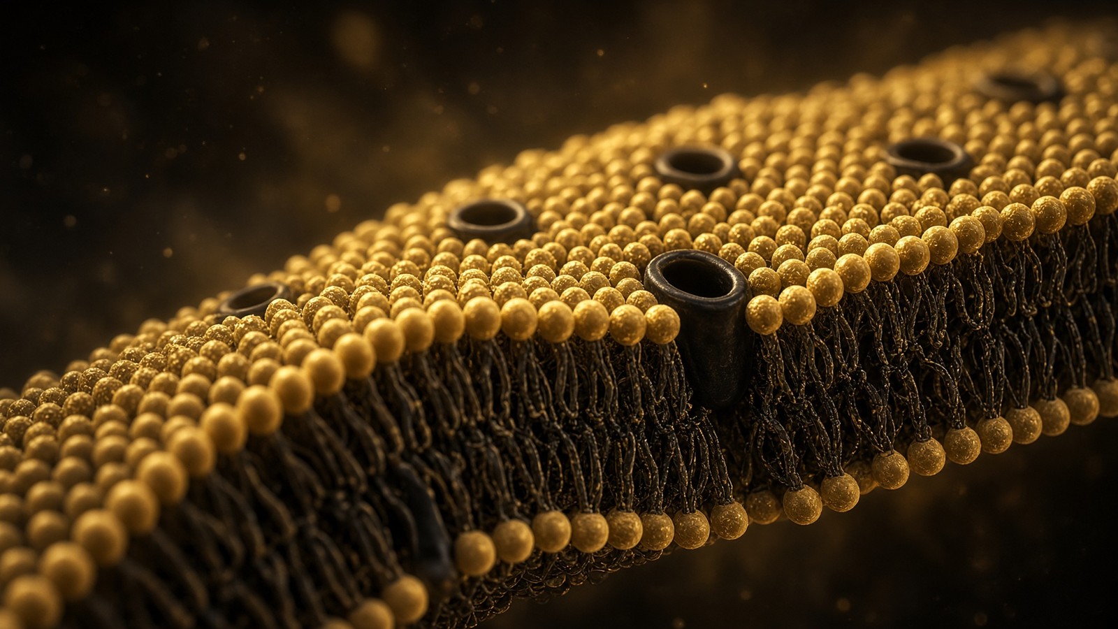
51 min · long read · The living cell
Membranes: a five-nanometre wall
Every cell, every organelle, every synapse sits on a lipid bilayer about five nanometres thick. Singer and Nicolson, Hodgkin and Huxley, a billion lipids, and why a peptide ligand at a GPCR does not need to enter the cell.
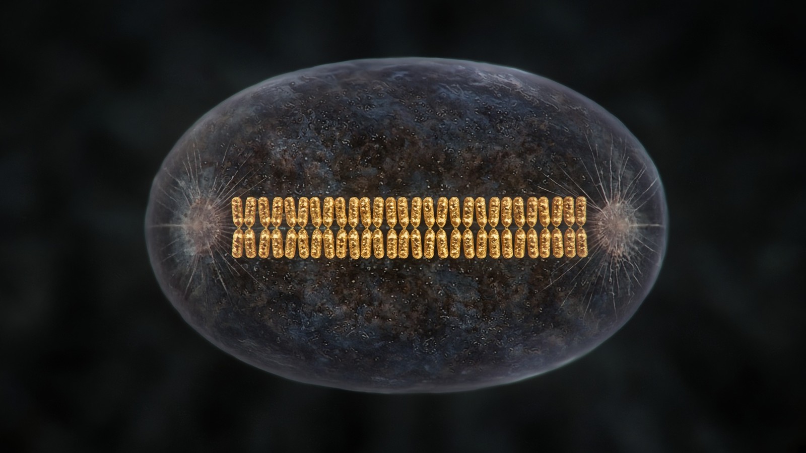
47 min · long read · The living cell
The cell cycle, p53, and the decision to divide
G1, S, G2, M. Cyclins license kinases. Checkpoints can say no. p53 reads damage and writes p21, or apoptosis, or a senescence that will not reverse. Hayflick counted the doublings. A repair-peptide paper that cannot name which of those fates moved is still only a description.
Essays describe published research. They are not medical advice and they do not authorise human use of any catalogue item.