

The living cell · 47 min · 10,330 words
The cell cycle, p53, and the decision to divide
G1, S, G2, M. Cyclins license kinases. Checkpoints can say no. p53 reads damage and writes p21, or apoptosis, or a senescence that will not reverse. Hayflick counted the doublings. A repair-peptide paper that cannot name which of those fates moved is still only a description.
What this essay actually tells you
- G1, S, G2, M. Cyclin D–CDK4/6 then cyclin E–CDK2 take the restriction point. Cyclin B–CDK1 takes mitosis, held by Wee1 until Cdc25, held again by Mad2 if a kinetochore is late. Hartwell, Hunt, Nurse — Nobel 2001.
- p53 is a transcription factor MDM2 usually eats. Damage lets it stay; it writes p21 (pause) or PUMA/NOXA (apoptosis). Hayflick counted ~40–60 doublings; the counter is the telomere. Senescence is not apoptosis and not G0.
- A repair-peptide paper that cannot name a fate — Ki-67, TUNEL, p16, a vessel — has not yet named a result. GHK-Cu is a transcriptome. BPC-157 is an endothelial animal neighbourhood. Epithalon is a TERT claim, not a cell-cycle intervention.
What this actually means
A cell that is going to make two cells has to copy a genome without wrecking it, then pull the copies apart, then cut the cytoplasm. That is G1, S, G2 and M — growth, synthesis, a second gap, mitosis — run by cyclins that license cyclin-dependent kinases and by checkpoints that can stop the kinases if DNA is broken, a fork has stalled, or a kinetochore is unattached. p53 is the damage transcription factor: MDM2 usually keeps it unstable; nicks, uncapped telomeres and oncogene stress let it stay; p21 pauses the cycle; PUMA and NOXA can kill the cell; a durable arrest is senescence. Hayflick counted ~40–60 doublings in human fibroblasts before that arrest. Senescence is not apoptosis and not quiescence and not mitosis. Catalogue peptides sit next to this decision, they do not run it. GHK-Cu has a fibroblast transcriptome. BPC-157 has an endothelial and angiogenic animal literature. Epithalon has a TERT and pineal literature — a gene-level claim, not a cell-cycle protocol. A paper that says repair without naming proliferation, senescence or apoptosis is still only a description. Research use only. No invented human trial.
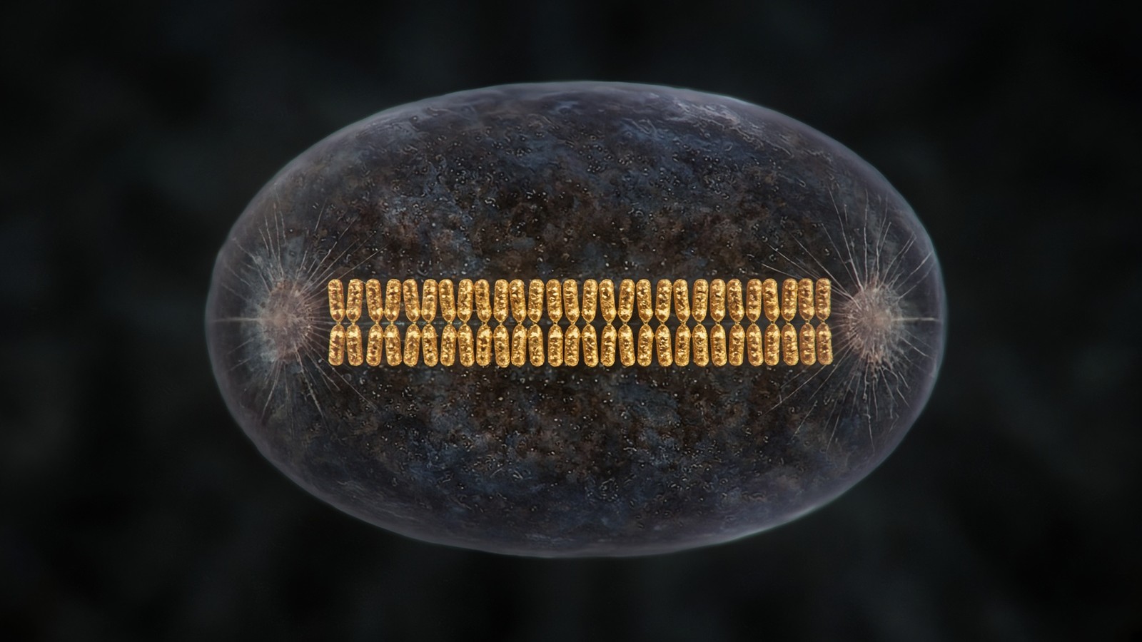
A cell doesn't drift into two cells. It decides. Somewhere in G1 a restriction point is crossed — that's Arthur Pardee's latch, the place after which the cell no longer needs a mitogen to finish the job. Rb, retinoblastoma protein, lets go of E2F transcription factors. Cyclin E licenses CDK2, a cyclin-dependent kinase. Origins that were licensed in the previous gap are allowed to fire. A genome of six billion base pairs is copied at an error rate that only mismatch repair and proofreading make tolerable, and then a second gap asks whether the copy is finished and whether the DNA is intact. Only then does cyclin B–CDK1 — mitosis-promoting factor — pull the nuclear envelope apart, condense the chromosomes, and line them up on a spindle so that anaphase can be a geometry instead of a smash. p53 sits on that sequence as a transcription factor with a veto. Hayflick sat on it as a census: human fibroblasts don't do this forever. The decision has a biochemistry, a Nobel and a pathology, and we're going to walk all three.
In short. A cell decides to divide. G1 commits, S copies the genome, G2 checks the copy, M pulls the sisters apart. p53 can stop the sequence. Hayflick showed it doesn't run forever.
We're going to sit with that decision. Four phases, the cyclin–CDK engines — cyclins are the licenses, cyclin-dependent kinases are the engines — the checkpoints that can say no, p53 and its partners p21 and MDM2, the Hayflick limit, and then the three fates a dividing tissue actually has: mitosis, senescence, apoptosis, with quiescence as the reversible cousin people keep mixing in. After the machinery, three catalogue sequences that sit near this story without running it. GHK-Cu is a copper tripeptide with a fibroblast transcriptome. BPC-157 is a gastric 15-mer with an endothelial neighbourhood in animals. Epithalon is four residues with a TERT literature. None of them is a cyclin. None of them is palbociclib, the licensed CDK4/6 inhibitor. A paper that can't name which fate moved — proliferate, senesce, die — hasn't finished the biology. Research use only belongs once, at the close, as the legal class of the reagent.
In short. We'll walk the phases and checkpoints, then three research peptides that sit nearby. They aren't cyclins. A repair paper still has to say whether cells divided, stopped or died.
Diagram
01 Genome
Variant, CNV, methylation, telomere length
The script. Most of it never becomes a phenotype you can bill for.
02 Transcriptome
Which genes are on, splice isoforms, noncoding RNA
The script being read this hour. A cell type is a transcriptome.
03 Proteome
Abundance, PTMs, localisation, complexes
The machines. Phosphorylation can flip a pathway without new DNA.
04 Metabolome
NAD+/NADH, ATP/AMP, acetyl-CoA, ROS
The fuel gauges. They feed back onto the genome through sirtuins and chromatin.
05 Organelle
Mitochondria, ER stress, lysosome, nucleus
Compartments fail as units. A tired mitochondrion is a tired cell.
06 Cell fate
Proliferation, senescence, apoptosis, identity
Hayflick, SASP, p53. The cell decides whether to keep being a citizen.
07 Tissue
Inflammation, fibrosis, barrier, innervation
Where a person actually hurts. Collagen, endothelium, synapses.
08 Organism
Glucose curve, VO₂, sleep, fertility, lifespan
The readout. Everything above is allowed to be invisible until it isn’t.
Pathophysiology is this stack, not a single molecule. A research peptide occupies one node — a receptor, a cofactor, a cytoskeletal motif — and the rest of the stack is still running. That is why ‘what does it do?’ is a bad question and ‘where does it bind?’ is a good one.
The neighbouring pathophysiology essay put cell fate on floor 6 of a stack that starts at the genome and ends at a person who notices. Stay here for the machinery of that floor. Divide, senesce, die, or stay. Those aren't synonyms, and once you can see why, a lot of captions get easier. Mitosis is a spindle and a cyclin-B wave. Senescence is a durable CDK blockade plus, often, a secretome Judith Campisi named SASP — the senescence-associated secretory phenotype. Apoptosis is a mitochondrial pore and a caspase cascade. Stay, if you're being precise, includes quiescence, which is reversible, and terminal differentiation, a transcriptional identity that doesn't come back because you asked nicely. Peptide papers that claim repair are almost always claiming one of those without saying which. The kinases are here at this length so that when a 15-mer or a tripeptide or a tetrapeptide is placed next to a wound, you can ask the useful question: which fate, which cell type, which assay, which species.
In short. Divide, senesce, die, or stay are different fates. Repair claims are usually one of those unspoken. Name the fate, the cell, the assay and the species.
A cell that copies a genome and then fails to decide what to do with the copy is not a healing cell. It is a karyotype waiting to happen.
Four phases, not a circle someone coloured in
Textbooks draw a wheel. The wheel is a kindness to first-year students, and it hides how different tissues actually live. A proliferating human cell in culture, given serum and space, might run a cycle in about twenty-four hours, of which S is six to eight, M is about one, G2 is three or four, and G1 takes whatever is left and can take much more. A hepatocyte in a quiet liver isn't on that wheel at all. A keratinocyte in a crypt-like basal layer is. A neuron in your cortex hasn't been on it since you were a fetus, with exceptions the field still argues about. The phases are a vocabulary for a cell that has committed to duplicate. They're not a personality, and they're not a product cycle. Cellular regeneration, used without a phase, is using the wheel as a picture instead of a clock. Once you can name G1 or S or M, you can ask whether anything actually copied DNA.
In short. G1, S, G2 and M are phases of a cell that's committed to copy itself. Timing varies by tissue. Most of your neurons aren't on that wheel at all.
- G1
- hours, variable
- S
- ~6–8 h
- G2
- ~3–4 h
- M
- ~1 h
- G0
- reversible exit
- Hayflick
- ~40–60 doublings
Growth, cyclin D, restriction point. Mitogen still matters. After Rb lets go, it mostly does not.
Two metres of DNA copied. Proofreading plus mismatch repair to ~10⁻⁹–10⁻¹⁰ errors per base.
Cyclin B accumulates. Wee1 holds CDK1 off. Cdc25 is the flip. DNA-damage checkpoint lives here too.
Prophase, prometaphase, metaphase, anaphase, telophase, cytokinesis. The pretty part. The shortest part.
Quiescence. A hepatocyte can come back. Senescence cannot, not by the same door.
Human diploid fibroblasts, 1961. The counter was the telomere. The stop is senescence.
G1 is the argument, and the restriction point is where the argument ends
G1 is when a cell is a cell rather than a copying machine. Mass is added. Organelles duplicate on their own clocks — mitochondria by fission, centrosomes by a quieter cycle that will matter in M. Transcription programmes that were appropriate to a newborn daughter are rewritten toward S. Mitogens — serum in a dish, growth-factor occupancy of receptor tyrosine kinases and GPCRs in a tissue — raise cyclin D. Cyclin D with CDK4 and CDK6 phosphorylates Rb. Rb, while hypophosphorylated, holds E2F transcription factors and recruits chromatin machines that keep S-phase genes off. As Rb is phosphorylated it lets go. E2F writes cyclin E, cyclin A, DNA-polymerase subunits, the MCM genes, dihydrofolate reductase — the list that makes S possible. Cyclin E–CDK2 then finishes the Rb phosphorylation, which is why the restriction point is a latch, not a dimmer. Arthur Pardee, 1974, in a paper that still earns its keep: after that point, withdrawing serum doesn't send the cell back. Before it, it can still choose G0.
In short. G1 is growth plus a latch. Mitogens raise cyclin D, Rb lets go of E2F, and after Pardee's restriction point the cell is committed unless a checkpoint fires.
p16INK4a is the INK4-family inhibitor that binds CDK4/6 and keeps cyclin D from completing the handshake. Leave it on and it's a senescence marker — a cell that's parked itself out of the cycle. Delete it and it's a tumour suppressor gone, which is why 9p21 deletions are so common in cancer. It's also why CDK4/6 inhibitors such as palbociclib exist as licensed medicines in hormone-receptor-positive breast cancer. That last sentence is a medicine-class fact, not a catalogue neighbour. We don't stock a CDK4/6 inhibitor. We stock ligands that occupy other proteins, on other floors. The reason to name palbociclib here is scale: when a chemist actually wants to stop G1, they occupy the kinase, with a dose and a neutrophil count in the same conversation. A research peptide sitting next to a wound is a different object. Feelings don't phosphorylate Rb. Kinases do, and p16 is how a cell can refuse them.
In short. p16 blocks the G1 kinases CDK4 and CDK6. Palbociclib is a licensed drug that occupies the same pair. A research peptide isn't that brake.
Myc is the rude oncogene on this stretch of road. Mitogens raise it. It feeds cyclin D and E2F targets and ribosomal biogenesis, which is why a Myc-driven cell is a nucleolar cell as well as a cycling one — it is building ribosomes as fast as it is building a genome. Too much Myc, in a cell that still has p53 and ARF, can trip oncogene-induced senescence or apoptosis rather than a clean S phase. That's a safety catch. Lose ARF or p53 and the catch is gone. TGF-β is the anti-mitogen the textbooks under-teach: it raises p15INK4b and p21 in epithelial cells and can park them in G1, which is why a wound that stays in a TGF-β neighbourhood too long is a fibrotic wound rather than a cycling one. GHK-Cu's arrays sometimes move TGF-β neighbourhood genes in fibroblasts. That's a transcriptome observation — many messages shifting on a chip — not a restriction-point protocol. Hold the distinction and the spreadsheet stays a spreadsheet.
In short. Myc pushes G1; too much of it can trip p53. TGF-β can park epithelial cells in G1. A fibroblast array that touches this neighbourhood is still an array.
S phase is a copying problem with a re-licensing ban
DNA replication in a eukaryotic cell is licensed in G1 and fired in S, and those two verbs must not swap. In G1, when CDK activity is low, origin recognition complexes sit on origins, Cdc6 and Cdt1 load MCM helicases, and a pre-replication complex is assembled that isn't yet unwinding anything. When cyclin E– and then cyclin A–CDK2 activity rises, origins fire, the helicase is activated, polymerases are recruited, and the same CDK activity — plus geminin, which parks Cdt1 — prevents a second load. The cell copies once. Endoreduplication, re-replication, a gene-amplified tumour: those are failures of that ban. A diploid G1 nucleus has about 6.2 billion base pairs. S turns it into a G2 nucleus with twice the DNA content, still in one nucleus, waiting for mitosis to make the geometry match the arithmetic. That's a quietly thrilling number once you sit with it: two metres of DNA, copied once, on a clock of six to eight hours, with a re-licensing ban as the whole point.
In short. Origins are licensed in G1 and fired in S, once. Kinases and geminin stop a second copy. S doubles the DNA; mitosis is what makes two nuclei out of it.
The fork is a machine that can stall. Nucleotide shortage, a bulky adduct, a collision with a still-sitting RNA polymerase, a difficult repeat: the replisome stops, single-stranded DNA accumulates, RPA coats it, ATR and its partner ATRIP are recruited, Chk1 is phosphorylated, late origins are held, the G2/M transition is delayed, and fork-protection and restart pathways try to keep the newly written DNA from becoming a double-strand break. BRCA1, BRCA2, RAD51 — homologous recombination is available in S and G2 because a sister chromatid is there to copy from. In G1 it isn't, which is why non-homologous end joining, sloppier, is the G1 default for a break. The pathophysiology essay put BRCA on the genome floor. Here it is a cell-cycle-phase fact: the same break is a different repair because of the phase. A caption that says DNA repair without a phase hasn't yet said which machine, in which window, on which template.
In short. Stalled forks call ATR and Chk1. Homologous recombination needs a sister chromatid, so it lives in S and G2. G1 uses a sloppier join. Phase is part of the mechanism.
Diagram
- DNA replication + MMR10⁻⁹ to 10⁻¹⁰A genome of 6 Gbp (diploid) accumulates a handful of mutations per division.
- Transcription~10⁻⁵RNA is disposable. The cell can afford a wrong letter in a message that lasts hours.
- Translation~10⁻⁴One wrong amino acid per ten thousand. Proteins turn over. DNA does not.
- mtDNA10–100× nuclearNo histones, ROS next door, weaker repair. The second genome ages faster.
The genome is sacred, the message is cheap, the protein is cheaper. Ageing is partly what happens when the sacred copy still drifts — and when mitochondria, which never got the nuclear repair budget, drift faster.
The error-rate diagram is the moral of S. DNA polymerase proofreading plus mismatch repair brings the archival error rate down to something like one in a billion to ten billion bases. A diploid genome of six billion bases therefore accumulates a handful of mutations per division if the mismatch system is intact — not zero, a handful. Lynch syndrome is mismatch repair lost in a tissue that divides, and the phenotype is tumours. Proofreading-mutant polymerases are rarer and nastier. The cell can afford a wrong amino acid in a protein that will be gone by Thursday. It can't afford a wrong base in a daughter that will still be here in a decade. That's why S has checkpoints, and why G1 and G2 ask questions before and after. A research ligand that supports DNA, in a caption, hasn't yet named polymerase, mismatch, nucleotide-excision repair, base-excision repair, or homologous recombination. Support isn't an enzyme. The enzymes have names, and S is where they earn them.
In short. DNA is copied almost sacredly; RNA and protein are allowed to be sloppier. A handful of mutations per division is the intact rate. Lynch syndrome is mismatch repair gone.
G2 is a quality-control gap, not a rest
G2 is the gap in which cyclin B is transcribed and translated and parked in a complex with CDK1 that isn't yet a mitosis-promoting factor. Wee1 and Myt1 have phosphorylated CDK1 on threonine 14 and tyrosine 15; those phosphates keep the kinase off even though the cyclin is bound. Cdc25 phosphatases, when the cell is satisfied, remove them. The satisfaction criteria include: replication finished, no persistent ATR signal, no unrepaired double-strand breaks shouting at ATM. Polo-like kinase 1 and Aurora A sit in the same neighbourhood, priming centrosomes and helping the Cdc25/Wee1 flip become bistable, which is why mitosis, once it starts, doesn't dribble — it switches. A G2 checkpoint arrest is Wee1 still on, Cdc25 still off, cyclin B–CDK1 still a loaded gun with the safety catch in. p21 can contribute here as well as at G1. A cell that enters mitosis with unrepaired DNA does mitotic catastrophe, which isn't a metaphor: shattered chromosomes, lagging fragments, micronuclei, a daughter that p53 will have opinions about if p53 is still there.
In short. G2 loads cyclin B–CDK1 and keeps it off with Wee1 until Cdc25 flips the safety. Unrepaired DNA shouldn't enter mitosis. If it does, chromosomes shatter.
M is geometry, then a cut
Mitosis is five named stages and a cytokinesis, and the names still earn their keep. Prophase: chromosomes condense — condensins, phosphorylation of histones and condensins by CDK1 and Aurora B — transcription of most genes shuts down, centrosomes that duplicated in S/G2 separate. Prometaphase: nuclear envelope breakdown, microtubules searching for kinetochores, a chaos that looks, in a still image, like a mess and is, in time, a search. Metaphase: every sister-kinetochore pair occupied by microtubules from opposite poles, chromosomes on the equator, the spindle assembly checkpoint satisfied. Anaphase: APC/C–Cdc20 ubiquitinates securin; separase is free; cohesin rings that held sisters open; sisters walk toward poles, and the poles themselves move apart. Telophase: envelope reforms, chromosomes decondense, a nucleus is a nucleus again, twice. Cytokinesis: an actomyosin ring at the equator, a midbody, two cells. TB-500's actin literature is a motility and G-actin-buffer literature, not a cytokinesis protocol. Two actin sentences. Don't blend them just because both say actin.
In short. Prophase condenses, prometaphase searches, metaphase aligns, anaphase splits sisters, telophase rebuilds nuclei, cytokinesis cuts. Actin in a furrow isn't TB-500's G-actin story.
The spindle assembly checkpoint is the last grown-up veto. Unattached kinetochores catalyse a Mad2-dependent inhibitor of APC/C–Cdc20. One unattached kinetochore is enough, in a healthy cell, to hold anaphase. Aurora B at the inner centromere also senses lack of tension and can detach incorrect attachments so the search can try again. Taxanes and vinca alkaloids, as medicines, poison microtubules and freeze this checkpoint until the cell dies or slips through. That's chemotherapy, a different desk, a different law. The reason to name it here is the same reason to name palbociclib: when a pharmacologist wants to occupy mitosis, they occupy tubulin or a checkpoint kinase, with karyotypes and dose-limiting toxicity in the same sentence. A lyophilised research peptide that hasn't produced a mitotic index, a Mad2 blot, or a chromosome count isn't in this sentence. It can still be a perfectly good ligand for a different protein. Mitosis is just a high bar, and it should stay one.
In short. One unattached kinetochore can hold anaphase. Microtubule poisons are real mitotic drugs. A research peptide without a mitotic index isn't occupying this checkpoint.
Cyclins license kinases; destruction is the other half of the clock
Leland Hartwell sat in yeast and found cdc mutants that stopped at particular points. Paul Nurse sat in fission yeast and found wee mutants that divided too small, and then found that the same gene, cdc2, was a kinase conserved in humans as CDK1. Tim Hunt sat over sea-urchin eggs and watched a protein appear and vanish in time with cleavage, and named it cyclin. Nobel Prize in Physiology or Medicine, 2001. The sentence that survived is almost domestic: oscillating subunits license a family of kinases; ubiquitin-mediated destruction of the subunits is the tick of the clock. Synthesis isn't enough. If cyclin B isn't destroyed, anaphase doesn't finish and the cell doesn't exit mitosis. If cyclin E hangs around, G1/S is a mess. The cell cycle is as much a proteasome story as a kinase story, which is why the proteostasis desk is next in the pipeline and why a caption that only says signalling has already lost the destruction half. The clock ticks because something is built, and because something is eaten.
In short. Hartwell, Nurse and Hunt found CDKs, wee mutants and cyclins. Oscillating cyclins license kinases; the proteasome destroys the cyclins to tick the clock. Both halves matter.
Mammalian somatic cells run several CDKs, not one. CDK1 is the mitotic kinase and, in a pinch, can do more of the cycle than the textbooks first claimed — mouse genetics from the Barbacid school showed that other CDKs are more dispensable than anyone was comfortable with. CDK2 takes S. CDK4 and CDK6 take G1 in most tissues that care about Rb. CDK7 is part of CAK, the CDK-activating kinase, and of TFIIH, which is why a transcription essay and a cell-cycle essay share a kinase. CDK9 is P-TEFb, pause-release of Pol II, next door in the dogma essay. The numbering is historical, not a moral ranking. What matters for this page is the pairing: D with 4/6, E with 2, A with 2 then 1, B with 1. Those pairs phosphorylate different overlapping substrate sets — Rb, replication proteins, lamins, microtubule motors, APC/C coactivators — and the overlapping is the biochemistry, not a failure of naming. Once you can see the pairs, a lot of drug names and peptide captions get easier to place.
In short. CDK4/6 with cyclin D in G1, CDK2 with E and A around S, CDK1 with A and B toward mitosis. CDK7 and CDK9 also touch transcription. The pairs are the point.
- Cyclin D–CDK4/6: G1, Rb, mitogen-coupled. p16INK4a is the dedicated brake. Palbociclib occupies this pair as a medicine.
- Cyclin E–CDK2: restriction-point latch, G1/S, origin firing. p21 and p27 can sit on it.
- Cyclin A–CDK2 then CDK1: S and G2. Prevents re-licensing. Lives until early mitosis.
- Cyclin B–CDK1: MPF. Nuclear envelope, condensins, spindle. Destroyed by APC/C so mitosis can end.
- INK4 CKIs: p16, p15, p18, p19. Bind CDK4/6. p16 is the senescence tattoo.
- Cip/Kip CKIs: p21 (CDKN1A, p53 target), p27, p57. Broader CDK inhibition. p21 is the pause p53 actually writes.
APC/C and SCF: the ubiquitin clocks
Two E3 ligase systems do most of the destroying. SCF complexes — Skp1–Cullin–F-box — operate through G1 and S, using F-box proteins such as Skp2 to recognise phosphorylated CDK inhibitors and cyclins. APC/C, the anaphase-promoting complex/cyclosome, is the mitotic specialist: with Cdc20 it takes securin and cyclin B at the metaphase–anaphase transition; with Cdh1 it keeps mitotic cyclins down through G1 so a new cycle can't skip back into M. Activation of APC/C–Cdc20 is what the spindle checkpoint withholds. That's a direct sentence from kinetochore to ubiquitin. The proteasome then chews what was tagged. A cell-cycle essay that never says ubiquitin is a kinase cartoon. The pathophysiology stack already put ubiquitin on the proteome floor. Here it is time-keeping. I find that quietly thrilling: the same 76-residue tag that sentences a misfolded enzyme also opens anaphase. One vocabulary, two jobs, a clock you can actually draw.
In short. SCF destroys G1/S brakes and cyclins. APC/C–Cdc20 opens anaphase; APC/C–Cdh1 keeps G1 from sliding back into mitosis. The spindle checkpoint is a ubiquitin veto.
p27 is the CDK inhibitor that often parks a quiescent cell, and Skp2 is how a cell that has decided to re-enter G1 gets rid of it. p21 is the CDK inhibitor p53 writes after damage, and it can also, in some contexts, sit in cyclin D complexes in a way that isn't purely inhibitory — biochemistry is ruder than captions. p57 is imprinted, developmental, a named disease when the imprint or the gene is wrong (Beckwith–Wiedemann, some IMAGe). The INK4/ARF locus on 9p21 is the other named geography: CDKN2A encodes p16INK4a and, in a different reading frame, ARF, which inhibits MDM2 and therefore stabilises p53. One locus, two tumour suppressors, a cell-cycle brake and a p53 input. Deletion of 9p21 is a common cancer event because evolution noticed the bargain. A peptide doesn't restore 9p21. That's not a scold. It's a chromosome. Ligands occupy proteins. Loci are DNA. Keeping those two objects apart is how you stay in the right conversation.
In short. p27 parks G0. p21 is p53's brake. CDKN2A writes p16 and ARF, so one deletion hits CDK4/6 and p53 at once. A peptide doesn't put a locus back.
Checkpoints are how a cycle says no
Hartwell again, with Weinert: checkpoints aren't the engines. They're the surveillance that stops the engines when an earlier job is unfinished. DNA damage in G1 shouldn't enter S. A stalled fork in S shouldn't ignore late origins and shouldn't enter G2 as if nothing happened. Unrepaired breaks in G2 shouldn't enter M. Unattached kinetochores shouldn't enter anaphase. The sensors are kinases: ATM for double-strand breaks, ATR for RPA-coated single-stranded DNA, DNA-PKcs in the NHEJ neighbourhood. The transducers include Chk2, more ATM, and Chk1, more ATR. The effectors are Cdc25 — inhibited, so CDK stays phosphorylated and off — p53, stabilised, so p21 is written, and the Mad2 system at kinetochores. A checkpoint is a pathway with a named sensor, a named kinase, and a named CDK consequence. The cell was stressed is not a checkpoint. Stress is a feeling. ATM phosphorylating Chk2 is a mechanism, and we can be glad the cell bothered to install it.
In short. Checkpoints stop the cycle when an earlier job failed. ATM and ATR sense damage, Chk1 and Chk2 transmit, Cdc25, p53 and Mad2 execute. Stress isn't a pathway.
ATM is a large PI3K-related kinase recruited to double-strand breaks by the MRN complex — Mre11, Rad50, Nbs1. It phosphorylates histone H2AX, the γH2AX focus that immunofluorescence lives on, phosphorylates Chk2, phosphorylates p53, phosphorylates MDM2, and a long list of repair proteins. Ataxia-telangiectasia is the named disease of ATM: radiosensitivity, cerebellar ataxia, immune failure, cancer risk. ATR is essential in a way ATM isn't; complete ATR loss is early-lethal because replication stress is a fact of every S phase, not an accident. Seckel syndrome and related ATR/ATRIP hypomorphs are the rare human glimpse. These are genome-maintenance diseases that read out as cell-cycle checkpoint diseases. They aren't peptide neighbourhoods. They're here so that DNA damage in a GHK-Cu array discussion has a kinase to point at, or doesn't get to use the words. Named sensors make the conversation smaller and better. That's the opposite of a scold. It's a gift.
In short. ATM answers breaks; ATR answers replication stress. Ataxia-telangiectasia is ATM lost. ATR is essential because every S phase is a little stressed. These are the named sensors.
G1/S damage checkpoint: ATM/Chk2/p53/p21, CDK2 off, Rb stays on E2F, origins don't fire. G2/M: ATR/Chk1 and ATM, Cdc25 off, Wee1 on, CDK1 off, cyclin B waiting. Intra-S: a slower fork, late origins held, a delay rather than a full stop. Spindle: Mad2, BubR1, Bub3, the mitotic checkpoint complex on Cdc20. Four vetoes, four biochemistries. A paper that reports a G2/M arrest has a FACS profile to show — 4N DNA content, a phospho-histone H3 question, a cyclin B question — or it has a caption. Ki-67 is a marker of cells that aren't in G0; it doesn't tell you which phase, and it doesn't tell you whether the cell will finish. PCNA and BrdU/EdU tell you S. Phospho-H3 tells you mitosis. p21 and p16 start the senescence argument. Pick the stain that matches the claim. That's not pedantry. That's how you know whether the wheel actually moved.
In short. G1/S, intra-S, G2/M, spindle: four vetoes. A G2/M arrest is a FACS profile. Ki-67 isn't a phase. Match the stain to the sentence.
p53 is a transcription factor with a veto, not a mascot
TP53 encodes a 393-residue protein that binds DNA as a tetramer and writes a programme. David Lane's 1992 sentence — guardian of the genome — was earned, and has since been asked to do more brand work than a transcription factor should. The biochemistry is still specific. In an unstressed cell, MDM2, an E3 ubiquitin ligase and itself a p53 target gene, keeps p53 unstable; the half-life is minutes. MDMX, also called MDM4, helps. Stress — double-strand breaks, UV, stalled forks, ribosomal stress from a nucleolus that isn't happy, oncogene signalling via ARF, telomere uncapping — leads to phosphorylation by ATM, ATR, Chk1, Chk2, DNA-PKcs, acetylation, a pause in the MDM2 handshake, and a protein that now stays long enough to occupy promoters. The promoters include CDKN1A (p21), GADD45, several DNA-repair genes, MDM2, the feedback, and, when the vote is death, BBC3 (PUMA), PMAIP1 (NOXA), BAX. Pause, repair, senesce, or die. The cell type, the dose of damage, the duration, and which BH3-only proteins the cell already expresses decide which of those four is written. That's a transcriptome decision. p53 is floor 2 talking to floor 6.
In short. p53 is unstable until damage or oncogene stress parks MDM2. Then it writes p21 to pause, repair genes, or PUMA and NOXA to kill. Which fate depends on cell type and dose.
Diagram
Closed chromatin (H3K27me3, DNA methylation) hides the promoter. Pioneer factors and histone acetyltransferases open it.
PIC: TFIID, TFIIH, Mediator, Pol II. Ser5 phosphorylation of the CTD lets the polymerase leave the promoter.
Elongation ~20–40 nt/s. Capping, splicing, cleavage and polyadenylation happen on the still-growing RNA.
Human genes are islands in 3.1 billion base pairs of mostly noncoding sequence. Promoter, enhancers, chromatin state and the Mediator complex decide whether Pol II is allowed to fire. Epithalon’s literature sits on TERT and pineal clocks — two of the rare promoters anyone bothers to name in a peptide essay.
MDM2 isn't a villain. It's the reset. A p53 that can't be destroyed is a p53 that will park or kill cells that didn't need parking or killing; Li-Fraumeni is p53 lost, but there are also MDM2-amplified tumours that lose p53 function with the gene still wild-type, which is why nutlins and other MDM2–p53 disruptors exist as an oncology idea. The feedback is tight: p53 writes MDM2, MDM2 eats p53. ARF, from that 9p21 locus, binds MDM2 and is how too much Myc can stabilise p53 without a broken chromosome. Ribosomal proteins RPL5 and RPL11, released from a sick nucleolus, do a similar MDM2 block, which is why a nucleolus essay and a p53 essay are the same building. The nucleus desk already told you most transcription by mass is rRNA. Here is the political consequence: a factory that can't make ribosomes correctly will veto the cycle through p53. A peptide that claims to activate p53 has to say whether it broke DNA, blocked MDM2, stressed the nucleolus, or wrote a caption.
In short. MDM2 resets p53. ARF and sick ribosomes block MDM2. Nutlins occupy that handshake as an oncology idea. Activate p53 still needs a how.
Li-Fraumeni syndrome is germline TP53, a devastating tumour predisposition, named so that p53 can't be a wellness word. Somatic TP53 mutation is among the commonest events in human cancer — not always the fifty percent in every cohort that everyone quotes, but common enough that a cell-cycle essay that doesn't mention it is missing the plot. Loss of p53 is how a cell ignores the G1/S damage veto, enters S with breaks, ignores the later apoptotic vote, and keeps a karyotype that should have been a corpse. Gain-of-function mutant p53 proteins are a ruder story still: they don't just go quiet, some of them acquire new occupancies. The point for this page is narrower. We don't run human trials of ligands against p53. We don't claim a tetrapeptide restored a guardian. We describe a node so that a TERT-on claim can be seen in the same building as a karyotype, which is the building it actually occupies. That's the grown-up neighbourhood for a reverse-transcriptase story.
In short. Li-Fraumeni is germline TP53. Somatic p53 loss is how tumours ignore checkpoints and apoptosis. A TERT-on claim belongs in the same building as a karyotype.
p21 is the CDK inhibitor everyone can spell and still misuse. It binds cyclin–CDK complexes, especially CDK2, and can arrest G1 or G2. It is a p53 target, so a p21 induction after damage is often a p53 signature — not always, because p53-independent routes to p21 exist, including some TGF-β neighbourhoods. A cell with p21 on and p16 off may still reverse; a cell with p16 on is closer to the senescence that doesn't reverse by the same door. SA-β-gal, the blue stain, is a lysosomal β-galactosidase activity at pH 6, useful and imperfect. 53BP1 foci and γH2AX foci are damage marks that, when they persist at telomeres, are the Hayflick checkpoint in a picture. Put p21, p16, SA-β-gal, a SASP cytokine, and a lack of Ki-67 on the same figure and you may say senescence. Put anti-ageing on a peptide without those stains and you may not. The stains are how the noun becomes a measurement. I like that kind of honesty. It's cheaper than a new word.
In short. p21 pauses CDKs and is often a p53 signature. p16 is the harder senescence mark. SA-β-gal is useful and imperfect. Anti-ageing without those stains is still a caption.
The apoptotic vote is a mitochondrial pore, not a mood
When p53 writes PUMA and NOXA, those BH3-only proteins occupy Bcl-2-family guardians — Bcl-2, Bcl-xL, Mcl-1 — and help Bax and Bak oligomerise in the outer mitochondrial membrane. Cytochrome c leaves the intermembrane space, which the mitochondria essay already told you is in fairly free conversation with cytosol once the outer membrane is porous. APAF1, cytochrome c and dATP assemble the apoptosome; caspase-9 is activated; caspase-3 and -7 execute. Phosphatidylserine flips. A neighbour phagocytoses. Inflammation is limited. That's apoptosis, floor 5 becoming floor 6 becoming, if enough cells do it, a tissue fact. Necroptosis — RIPK3, MLKL — and pyroptosis — gasdermin, inflammasome, IL-1β — are louder deaths. Ferroptosis is lipid peroxidation. A paper that says a peptide prevented cell death without annexin, cleaved caspase-3, TUNEL, or a named alternative death hasn't measured a fate. A paper that says anti-apoptotic in a tumour cell is a different moral object from the same words in a neuron. We won't blur them, because the stains are different and the ethics are too.
In short. p53 can write PUMA and NOXA; Bax and Bak pore the mitochondrion; cytochrome c plus APAF1 is the apoptosome. Name the death. Prevented cell death without a stain isn't data.
Hayflick counted. The counter was the telomere.
Leonard Hayflick and Paul Moorhead, Experimental Cell Research, 1961: human diploid fibroblasts in culture don't divide indefinitely. They run through a number of doublings — later textbooks say about forty to sixty, depending on the donor age, the tissue, and how you count — and then the population stops. Carrel had claimed immortality in culture and had, Hayflick argued, been feeding his flasks living cells with the medium. The 1961 paper is a census, a contamination argument, and the beginning of a field. They didn't yet know what was being counted. Alexey Olovnikov, in 1971 and then 1973, and independently Jim Watson, named the end-replication problem: DNA polymerase needs a primer, the lagging strand can't finish a linear end, the end would shrink. Elizabeth Blackburn, Carol Greider and Jack Szostak found the solution — telomerase, TTAGGG, an RNA-templated reverse transcriptase — and took the 2009 Nobel. The Hayflick limit is a telomere checkpoint talking to p53 and p16. It is also, as the pathophysiology essay put it, a fate-floor fact: the stop is senescence.
In short. Hayflick, 1961: fibroblasts divide a finite number of times. Olovnikov named the shrinking end. Blackburn, Greider and Szostak found telomerase. The stop is senescence.
Diagram
- Hayflick limit~40–60 doublingsHuman fibroblasts in 1961. They were not immortal. He counted.
- End-replication5–15 kb TTAGGGDNA polymerase needs a primer. The lagging strand shortens. Olovnikov named the problem.
- Shelterin6 proteinsTRF1, TRF2, POT1, TIN2, TPP1, Rap1. The end is hidden from the damage response.
- TERT offmost somatic cellsTumour suppression. Stem cells and germline keep it on. So do most cancers.
- SenescenceSASPp16, p21, the secretome Campisi named. A cell that refuses to die and talks too much.
Blackburn, Greider and Szostak, Nobel 2009. Epithalon’s literature sits on TERT and pineal melatonin — a tetrapeptide claiming two of the rare promoters anyone names in a peptide essay. The machines are real. A large Western RCT of telomere length in adults is not on the shelf next to the vial.
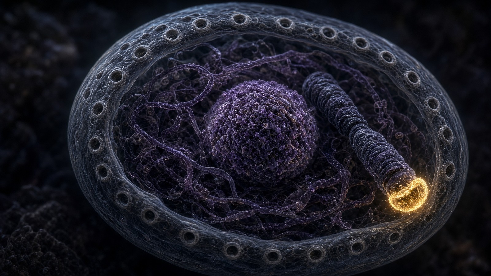
Human telomeres are TTAGGG repeats, typically five to fifteen kilobases at birth in a given cell type, bound by shelterin — TRF1, TRF2, POT1, TIN2, TPP1, Rap1 — and tucked into a t-loop so the end isn't an end. Lose TRF2 and you get end-fusions; the nucleus essay is that argument. Each S phase, because of the end-replication problem and because of processing, the repeats shorten. When they're short enough, shelterin can't hide the end, the DNA-damage response treats a chromosome end as a break, ATM/ATR fire, p53 and p21 and sometimes p16 engage, and the cell senesces or dies. Stem cells and the germline keep TERT on so the counter can be reset. Most somatic cells keep TERT transcriptionally off on purpose. The purpose is tumour suppression. A cell that can rebuild its telomeres forever is a cell that can become a clone that doesn't stop. Most cancers reactivate TERT or use ALT, a recombination workaround. The bargain is tissue renewal versus neoplastic risk. One-sided gift language skips the spindle. Draw both sides and the bargain is still interesting.
In short. Telomeres shorten each S phase. Too short, and ATM sees a break, p53 parks or kills the cell. TERT is off in most of your body on purpose. Cancers turn it back on.
That sentence is why Epithalon's literature is large relative to four residues. Khavinson's school reported that AEDG can move TERT, that TRAP assays in cultured fibroblasts can light up, that telomeres in those cultures can measure longer. Anisimov's rodent papers sit on lifespan and tumour incidence with epithalamin and the tetrapeptide. Those are gene-level claims: a promoter, a reverse transcriptase, a pineal melatonin amplitude in the dark. They aren't a cyclin-D curve. They aren't a CDK protocol. They aren't a human randomised trial of telomere length in adults, and we won't write one into existence. The nucleus essay held the claim next to shelterin and Casgevy and refused to inflate it. This essay holds the same claim next to Hayflick and p53 and refuses the same inflation. If TERT came on in a somatic cell that still had to decide about p53, the karyotype would be part of the methods. A TRAP band without a karyotype is an incomplete sentence. Incomplete isn't false. It's unfinished, and unfinished is allowed if you say so.
In short. Epithalon's papers claim TERT, not a cyclin. That's a promoter argument. No large Western trial of telomere length in adults. A TRAP assay without a karyotype is unfinished.
Mitosis, senescence, apoptosis: three fates, plus a reversible cousin
People use old, damaged, tired, and senescent as if they were one word. They're not. Mitosis is two living cells with partitioned genomes. Apoptosis is a living cell becoming a corpse on purpose, tidy, caspase-mediated, usually unnoticed by the innate immune system. Senescence is a living cell that has stopped dividing and often started secreting, metabolically active, resistant to apoptosis in many tissues, a problem for the neighbours. Quiescence is a living cell that has left the cycle and can come back when mitogen and space and the right transcription factors say so — a satellite cell, a hepatocyte after partial hepatectomy, a memory lymphocyte. Differentiation is a living cell that has locked an identity; iPS cells as a Nobel are the reminder that the lock is expensive to pick. Four or five nouns. A repair paper that uses none of them is an adjective with a chromatogram. You're allowed to find that slightly comic. You're also allowed to pick a noun, stain for it, and join the grown-up conversation. The nouns were always there.
In short. Mitosis: two cells. Apoptosis: a tidy death. Senescence: alive, not dividing, often shouting. Quiescence: parked and able to return. Name the noun. Repair without a noun is an adjective.
Senescence is a durable arrest with a secretome
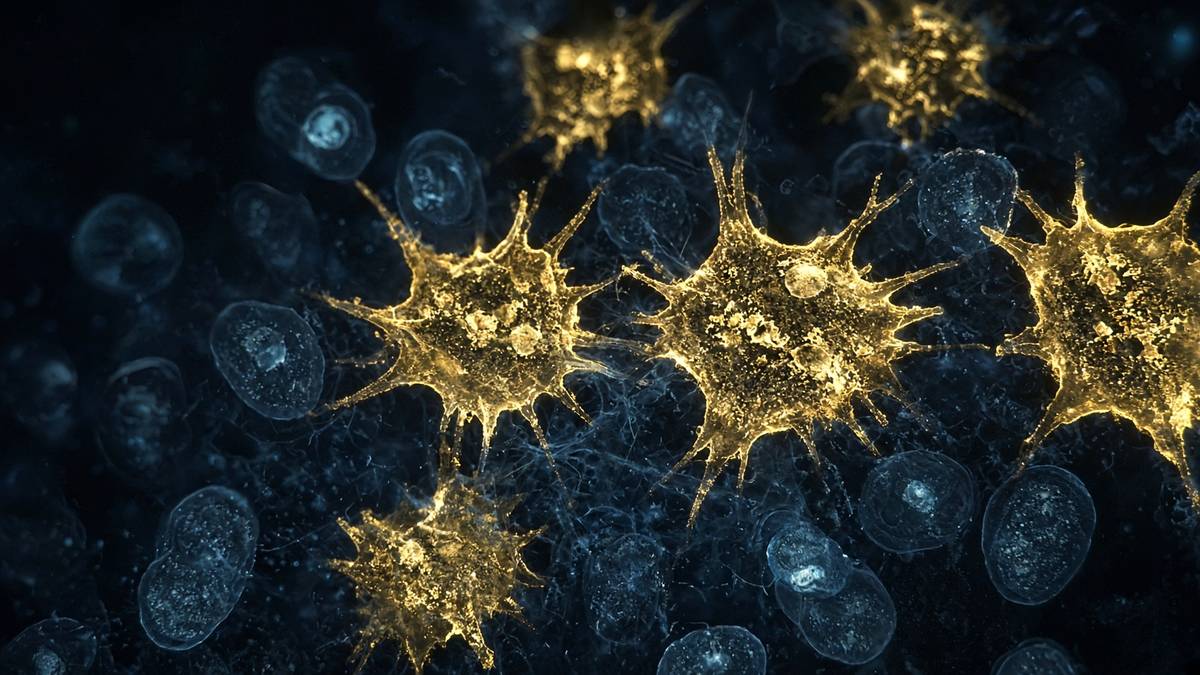
Replicative senescence is Hayflick's stop: short telomeres, a persistent DNA-damage response at chromosome ends, p53/p21 and often p16. Stress-induced, or premature, senescence is the same arrest from other insults: too much ROS, too much replication stress, a chemotherapeutic that left survivors, ionising radiation. Oncogene-induced senescence is the safety catch on Ras and Myc we already named — ARF, p53, p16, a cell that refuses to become a clone. Therapy-induced senescence is what some cytotoxics leave behind, a mixed blessing because a senescent tumour cell can sit in a SASP that the remaining tissue hears. The shared hardware is CDK inhibition that lasts, a chromatin state — SAHFs, senescence-associated heterochromatin foci, in some cells — SA-β-gal, and, in the cells Campisi taught us to worry about, a SASP. Different insults, one durable blockade. Once you can see the hardware, the insults stop looking like different diseases and start looking like different keys to the same lock. That's a unifying idea, and it's earned.
In short. Telomeres, ROS, oncogenes, some chemotherapies: different insults, same durable CDK blockade. p16, p21, SA-β-gal. SASP is what makes the arrest a tissue problem.
Judith Campisi's career, and Coppé, Patil, Rodier and colleagues in PLoS Biology 2008, made SASP a measured object: IL-6, IL-8, other cytokines and chemokines, matrix metalloproteinases, TGF-β neighbourhood signals, sometimes VEGF, a conditioned-medium experiment that changes what neighbours do, including epithelial cells that were considering transformation. In an acute wound a temporary senescent cohort can be useful — limit fibrosis, help resolution. In aged dermis, fat, endothelium, a persistent SASP is a fire alarm that will not reset: low-grade inflammation, matrix that is both degraded and poorly rebuilt, stem-cell niches that no longer feel like niches. Senolytics — dasatinib plus quercetin is the famous pair; navitoclax as a Bcl-2-family occupancy — try to kill the shouting cell. Senomorphics try to mute the shout. Neither class is a research peptide in the catalogue. A GPCR agonist isn't a senolytic because a forum used the word ageing. The senescence-SASP essay next door is that argument at full length. Here the job is to keep the fate distinct from mitosis and from death so a BPC-157 caption can't hide in the gap.
In short. SASP is a measured secretome that changes neighbours. Useful in a wound, corrosive when it persists. Senolytics are a different pharmacology, and they aren't in this catalogue.
Quiescence is not senescence, and G0 is not a dustbin
A quiescent cell has low CDK activity, often high p27, Rb on, E2F off, a metabolism that isn't a mitotic metabolism, and a door back. Serum, a growth factor, a lost neighbour in a liver that has been cut, an antigen: cyclin D can come back. Muscle satellite cells are the teaching case. Hepatocytes after partial hepatectomy are the dramatic case — a largely G0 organ that can re-enter S, which is why the liver is a regeneration story that doesn't need a peptide caption to be true. Memory T cells. Hair-follicle bulge stem cells. Quiescence is a fate with a return ticket. Senescence's ticket is one-way under ordinary physiology, which is why iPS reprogramming of senescent cells is a paper and not a weekend. Mixing G0 and senescence is how a resting tissue gets called aged, and how an aged tissue gets promised a mitogen as if mitogen were a senolytic. Different stains. Different kinases. Different honesty. I like the liver example because it already exists, in mammals, without anyone selling it.
In short. G0 is reversible: p27, Rb on, a door back for hepatocytes and satellite cells. Senescence isn't that door. Calling a resting cell aged mixes the fates.
Contact inhibition is the quiet G1 police of a monolayer: cadherins, Hippo, a Merlin–YAP neighbourhood, cyclin D down, p27 up, a dish that is confluent and stops. Lose it and you have a transformed focus, which is how tumour-virus assays used to be scored. Anchorage dependence is the same honesty in three dimensions: most epithelia need integrin occupancy to keep CDK activity on; lose the floor and they die a death called anoikis, which is apoptosis after a failed ECM handshake. A scratch assay — the wound on plastic that peptide papers love — is a contact-inhibition and motility experiment first. Filling the scratch can be proliferation, migration, or both. Without a mitotic index or an EdU pulse, you don't know which. BPC-157 papers that count endothelial migration are at least counting a behaviour. Papers that show a closed scratch and write regeneration have photographed a gap. The gap isn't a fate. Close it, then stain. That's a two-step method, and both steps are cheap compared with a new noun.
In short. Contact inhibition parks a monolayer in G1. A scratch assay mixes migration and proliferation. Close the gap and stain. A closed scratch isn't regeneration until you say which fate filled it.
A repair-peptide paper that cannot name a fate is still only a description
This is the disciplinary rule, and it's the reason this essay exists on a catalogue site rather than only in a textbook. Repair, regeneration, healing, anti-ageing, cellular health: all tissue words, all organism words, all at floor 7 and 8 of the pathophysiology stack. Under them, at floor 6, something happened to a fate. Tenocytes proliferated, or they didn't. Endothelial cells sprouted, or they didn't. Apoptosis in a crush model fell, or it didn't. Senescent cells left, or they didn't. Fibroblasts laid collagen without dividing, or they divided and then laid collagen. Each of those is a different cyclin, a different stain, a different honesty. If the methods can't say Ki-67, EdU, phospho-H3, cleaved caspase-3, TUNEL, p16, SA-β-gal, a SASP panel, a VEGFR2 phosphorylation, a collagen transcript, then the abstract's verb is an adjective. We'll sell you the sequence anyway, labelled for the bench. We won't help the adjective put on a white coat. That's the whole arrangement, and it's enough for a reagent.
In short. Repair is a tissue word. Under it a fate moved, or it didn't. Name the stain. If the paper can't, the verb is an adjective. The vial can still be a reagent.
GHK-Cu: a transcriptome is not a mitotic index
Loren Pickart isolated GHK from plasma fractions that made old liver tissue, in culture, synthesise proteins more like young tissue. Glycine-histidine-lysine, usually carrying Cu²⁺ in a square-planar complex. Plasma levels, on his numbers, about 200 ng/ml at twenty and about 80 ng/ml at sixty. Lysyl oxidase is a copper enzyme; SOD1 is a copper-zinc enzyme; free Cu²⁺ is a Fenton reagent. The ligand is a delivery story before it's anything else. The unusual claim is breadth: Pickart and Margolina's microarray work reports large sets of fibroblast transcripts moving — collagens, decorin, TIMPs up; some MMPs down; TGF-β and integrin neighbourhoods shifting. That's floor 2, a transcriptome. A transcriptome can be a step toward a fate. It isn't cyclin D. It isn't EdU. It isn't a Hayflick curve. Copper delivery to lysyl oxidase would already earn the complex a place on a matrix bench. Claiming that an array is a cell-cycle protocol is how a spreadsheet becomes a product story, and we'd rather keep the spreadsheet.
In short. GHK-Cu is a copper tripeptide with fibroblast arrays: matrix genes, not a cyclin curve. Copper to lysyl oxidase is already a reason to look. An array isn't a mitotic index.
What would a grown-up GHK-Cu cell-cycle paper look like? A defined fibroblast line, copper stoichiometry declared, a cell-cycle profile — FACS with DNA stain, a G1/S/G2/M fraction — EdU for S, a p21 and p16 blot if senescence is the claim, a SASP panel if the claim is that the secretome moved, and an RNA-seq with a pre-registered analysis rather than a 2000s array as the last word. If the claim is proliferation, show Ki-67 and a count. If the claim is that senescent fibroblasts look less senescent, show SA-β-gal and a telomere or damage-foci measurement, and don't call it reversal until the cell divides again. We stock Gly-His-Lys·Cu²⁺, 100 mg, HPLC, US-made, lyophilised, research use only. We don't stock a dermis, a hair follicle, or a G1 protocol. Cosmetic copper-peptide chemistry is a different regulatory object. Identity is still three letters, one metal, a chromatogram. That's a smaller claim than a cycle drug, and smaller claims are the ones you can actually test.
In short. A real GHK-Cu fate paper would show FACS, EdU, p16/p21, maybe SASP, with copper declared. We stock the complex for the bench, not a G1 protocol.
BPC-157: angiogenesis is a fate. ‘Healing’ is not.
BPC-157 is GEPPPGKPADDAGLV, a gastric pentadecapeptide that survives acid, which is why a 15-mer can be studied orally in a rat at all. Sikiric's Zagreb programme is large, sometimes messy, always interesting: NSAID lesions, tendon-to-bone, muscle crush, a recurring NO-system sentence. The cleaner mechanistic neighbourhood, as we've written before, is VEGFR2 internalisation, FAK–paxillin at focal adhesions, eNOS. That's an endothelial occupancy. Endothelial cells that occupy VEGFR2 do a fate: they migrate, they can proliferate, they form tubes. Angiogenesis is floor 6 becoming floor 7. A tendon readout in an animal can be a vessel readout in disguise, because a healing tendon needs vessels and a degenerating tendon often has the wrong ones. Independent labs have reproduced angiogenic and tenocyte-migration pieces more cleanly than the more extravagant CNS claims. That's how a large single-lab literature is supposed to age in public. Ageing in public is a compliment. It means the sequence is still worth arguing about.
In short. BPC-157's cleaner story is VEGFR2, FAK, eNOS — endothelium. Angiogenesis is a real fate. A tendon number in a rat may be a vessel number. Healing as a caption isn't a fate.
What the literature hasn't done, and what we won't pretend it has done, is a human trial we can file next to Jastreboff's retatrutide Phase 2. There isn't one on this desk. Animal crush, animal tendon, cell migration, a NO blot: those are the objects. A BPC-157 paper that reports a closed scratch without EdU hasn't distinguished migration from proliferation. A paper that reports less TUNEL in a crush has named apoptosis and is, at least, on the fate map. A paper that reports enhanced healing with a photograph and no cell-type stain is an adjective. We stock the 15-mer, 10 mg, HPLC, US-made, lyophilised, kit on peptide orders over £75, research use only. We don't stock a tendon protocol, a gut protocol, or a mitotic drug. The pathophysiology essay already said this in one paragraph. Here it is the whole point: put the 15-mer next to cyclin B and see whether the sentence survives. Sometimes the endothelial sentence does. The adjective doesn't. That's a useful filter, and you can run it on any abstract in five minutes.
In short. No human BPC-157 trial lives on this desk. Name TUNEL, EdU, VEGFR2, or the sentence is still open. We stock the 15-mer as a reagent, not a healing protocol.
Epithalon: a gene-level claim, not a cell-cycle protocol
Ala-Glu-Asp-Gly. 390 daltons. Khavinson's St Petersburg Institute of Bioregulation and Gerontology. The live claims, as the nucleus essay and the peptide essay both held, are TERT transcription and TRAP-measurable telomerase in cultured fibroblasts, telomere length in those cultures, nocturnal melatonin amplitude in aged animals, and rodent lifespan and tumour-incidence papers from Anisimov's group on epithalamin and the tetrapeptide. Mechanism remains the open joint: a complementary-peptide DNA argument most Western molecular biologists would still call unresolved, an unnamed receptor, a pineal-first model, a culture artefact. We won't pick a favourite from that list. What this essay adds is the floor-6 consequence. If TERT came on in a somatic fibroblast, the Hayflick parameter would move — more doublings before the telomere checkpoint. That's a fate claim, a big one, and it brings p53 and karyotype with it as mandatory methods, not as optional safety decoration. Stem cells keep TERT on with a genome-maintenance budget. A peptide that turns TERT on in a cell that was supposed to keep it off is doing oncology-adjacent work whether the abstract wanted that sentence or not.
In short. Epithalon's literature is TERT and melatonin, not cyclin D. If TERT came on, Hayflick would move, and karyotype and p53 would be mandatory methods. That still isn't a cycle protocol.
Contrast the three ligands on the same table, because the search bar will keep offering to blend them. GHK-Cu: copper complex, fibroblast transcriptome, matrix enzymes, floor 2 toward floor 7. BPC-157: gastric 15-mer, VEGFR2/FAK/eNOS, endothelial fate in animals, floor 3 toward floors 6 and 7. Epithalon: tetrapeptide, TERT/pineal literature, floor 1–2 with a possible Hayflick consequence if the TRAP is telling the truth, unresolved mechanism, no large Western RCT. CRISPR-Cas9, Casgevy, is the actual editor, licensed, a different legal class, not in the catalogue. Palbociclib is the actual G1 CDK4/6 occupancy, licensed, not in the catalogue. Nutlins occupy MDM2–p53, oncology, not in the catalogue. The catalogue is lyophilised sequences for a bench that can name an assay. Research use only is the class that keeps those sentences from collapsing into a stack you inject because a forum said repair. Three tools, three floors, and the licensed cycle drugs on a different till. That's a map, not a ranking.
In short. GHK-Cu is a transcriptome. BPC-157 is an endothelial animal fate. Epithalon is a TERT claim. CRISPR, palbociclib and nutlins are the real gene-and-cycle occupancies, and they aren't this catalogue.
Four residues claiming a reverse transcriptase is a promoter argument. A 15-mer claiming a closed tendon is an endothelial argument until stained otherwise. A copper tripeptide claiming a younger spreadsheet is a transcriptome argument. None of those is cyclin B. Stop writing as if they were.
Cancer is this machinery with the vetoes off
A tumour isn't a cell that divided. Your gut epithelium divides every few days and isn't a tumour. A tumour is a clone that divided when the checkpoints said no, or that never heard them. Rb lost, so the restriction point won't hold. p16 lost, so CDK4/6 won't sit down. p53 lost or MDM2 amplified, so damage won't write p21 and won't write PUMA. Myc up, so the nucleolus and the cyclins are loud. TERT on, so Hayflick won't save you. APC/C and the SAC compromised, so daughters are aneuploid and the next veto is weaker. This is Weinberg's hallmarks translated into the nouns of this essay, not a new theory. The peptide catalogue doesn't occupy those nodes. Filing a repair ligand under oncology support has left the building. Filing a TERT-on claim without a karyotype hasn't yet entered the part of science that tumour biology already paid for. The payment was decades of checkpoints. We can name that sentence. We don't have to sell its opposite.
In short. Tumours are clones that ignored restriction, p16, p53, the spindle checkpoint, Hayflick. Gut epithelium divides and isn't a tumour. A TERT-on claim owes a karyotype.
Mitotic catastrophe, chromothripsis, micronuclei that leak DNA into the cytosol and wake cGAS–STING: the cycle, when it fails in M, becomes an innate-immune stimulus, which is why the immunity desk is later in the pipeline and why a G2/M checkpoint isn't a private matter. Aneuploidy is a fate of the karyotype that becomes a fate of the tissue. p53 is one of the reasons a micronucleus should have been an apoptotic vote instead of a next mitosis. The error-rate diagram's first row is the archival bargain; cancer is what happens when the bargain is broken and the cell is still alive. We're a three-person lab with HPLC and a journal. We can name that sentence. We can't, and won't, sell a ligand as its opposite. Naming is the job. The cycle is older than the till. The till doesn't get to skip a shattered chromosome. If that sounds stern, it's only because the pictures are stern, and the pictures are public.
In short. A failed mitosis can shatter chromosomes, wake cGAS–STING, and feed the next tumour cycle. p53 should have made that a death. We name the sentence. We don't sell a ligand as its cure.
Stem, transit, differentiated: who is allowed to run the cycle
A tissue that renews is usually a hierarchy, not a democracy. A small stem cohort is quiescent or slowly cycling, TERT often on, p53 sensitive in ways that protect the archive. Transit-amplifying cells run the cycle hard, Hayflick-limited, and then differentiate. The differentiated end-cell — a keratinocyte squame, an enterocyte, a red cell without a nucleus — doesn't cycle. Cancers that look like they've forgotten this hierarchy have often captured a stem-like programme, which is why cancer stem cell is a real argument and a marketing magnet at once. GHK-Cu hair-follicle papers sit, if they sit anywhere honest, on the dermal papilla and the anagen programme, not on a promise to turn a differentiated keratinocyte into a stem cell. BPC-157 tendon papers sit on tenocytes and endothelium, cells that can cycle, not on a magic conversion of matrix into cells. Epithalon's fibroblast TRAP sits on a cultured somatic cell that was supposed to keep TERT off. Hierarchy first. Ligand second. Caption never. That's the order that keeps a tissue a tissue.
In short. Stem cells cycle sparingly with TERT on. Transit cells cycle hard. Differentiated cells mostly don't. Peptide papers have to say which cohort they touched. Most captions don't.
Partial hepatectomy is the existence proof that G0 isn't senescence and that a mammal already knows how to put a tissue back on the wheel. Remove two-thirds of a rodent liver; hepatocytes re-enter G1, pass restriction, run S, and the mass comes back on a clock measured in days. The signals are growth factors, IL-6, a Hippo-YAP unroofing, a metabolic shift, a p53 that must not veto a programme the organism needs. No research peptide is required for that sentence to be true. The sentence is here to keep regeneration expensive. If a 15-mer in a crush model is doing a fraction of a hepatectomy's work in a different tissue, show the DNA content, show the mitotic figures, show the cell type. If it's doing angiogenesis and that's why the crush looks better, show the vessels and stop saying regeneration as if it were hepatectomy. Honesty is cheaper than a new noun. The liver already ran the demonstration, in the 1930s, and it still looks like the real thing when you stain it.
In short. A liver can re-enter the cycle from G0 without a peptide. That's what regeneration looks like when it's real. A crush model owes you DNA content and a cell type.
The decision, the census, the adjective
Retrace once, without the caption. A dividing cell runs G1, S, G2, M. Cyclin D–CDK4/6 and cyclin E–CDK2 take restriction, Rb, E2F. Cyclin A takes S. Cyclin B–CDK1 takes mitosis, held off by Wee1 until Cdc25, held off again by Mad2 if a kinetochore is late, ended by APC/C. ATM and ATR veto S and M when the archive isn't fine. p53, eaten by MDM2 until it isn't, writes p21 or PUMA. Hayflick counted the doublings; the counter is TTAGGG; TERT is off in most of your cells on purpose. Senescence is a durable arrest plus, often, a SASP. Apoptosis is a pore and a caspase. Quiescence is a return ticket. Cancer is the vetoes off. GHK-Cu is a copper transcriptome. BPC-157 is an endothelial animal neighbourhood. Epithalon is a TERT-and-pineal literature, a gene-level claim, not a cell-cycle protocol. Palbociclib, nutlins, taxanes, Casgevy: different desks, different law. No human trial of these three ligands as fate drugs lives on this shelf, and we won't write one for the cadence.
In short. Phases, cyclins, checkpoints, p53, Hayflick, three fates. Three catalogue peptides occupy other nodes. Real cycle drugs and gene editors live on other desks. No invented human trial.
The vials are reagents. GHK-Cu 100 mg is Gly-His-Lys·Cu²⁺, HPLC, lyophilised, for a tube that can declare copper. BPC-157 10 mg is GEPPPGKPADDAGLV, HPLC, lyophilised, for a tube that can stain endothelium. Epithalon 50 mg is Ala-Glu-Asp-Gly, HPLC, lyophilised, for a tube that can run TRAP and, if it's being grown-up, a karyotype. Peptide orders over £75 include the sterile kit. Next-day UK postage is £5. Research use only, all three — not a protocol, not a dose, not a regenerative medicine, not an anti-ageing product, not a cell-cycle drug. A three-person lab with HPLC and a journal can hold a tetrapeptide and a metaphase plate in the same head without lying about either. The decision to divide is older than the catalogue. The catalogue doesn't get to skip it. That's the close. The machines in the first twelve headings are real whether or not a 15-mer ever sat on VEGFR2. Nucleus of the argument first. Papers second. Vial third, labelled, lyophilised, not a protocol.
In short. Three HPLC-characterised sequences, labelled for the bench, kit over £75, next-day UK £5. Not medicines. Not a cycle protocol. The decision to divide doesn't care what the till wanted.
A cell that cannot name whether it is about to copy a genome, stop forever, or die is not a healing cell. It is an adjective. We sell sequences. We do not sell the adjective.
Questions the essay actually answers
- What are G1, S, G2 and M?
- The four phases of a dividing somatic cell. G1 is growth and the restriction-point decision. S is DNA synthesis. G2 is the gap in which cyclin B accumulates and the cell asks whether replication finished cleanly. M is mitosis plus cytokinesis — prophase through telophase, then the cytoplasm splits. G0 is a reversible exit, not a fifth phase of the same clock.
- What do cyclins and CDKs actually do?
- Cyclin-dependent kinases are the engines. Cyclins are the matching licenses that appear and disappear in waves. Cyclin D–CDK4/6 in G1, cyclin E–CDK2 at G1/S, cyclin A–CDK2 in S, cyclin B–CDK1 at mitosis. Hartwell, Hunt and Nurse, Nobel 2001. Inhibitors (p16, p21, p27) and phosphatases (Cdc25) sit on the same engines. A peptide is not a cyclin.
- What is p53, and why does MDM2 matter?
- p53 is a tetrameric transcription factor, the product of TP53, that reads DNA damage, ribosomal stress and oncogene signalling. MDM2 is the E3 ligase that normally ubiquitinates it so the proteasome eats it; p53 transcribes MDM2, which is the negative feedback. When ATM/ATR/Chk2 mark p53, MDM2 cannot keep up, p53 stays, and it writes p21 (pause), DNA-repair genes, or PUMA/NOXA (apoptosis). Lane called it the guardian of the genome. It is not a peptide target in the catalogue.
- What is the Hayflick limit?
- Leonard Hayflick and Paul Moorhead, Experimental Cell Research, 1961: human diploid fibroblasts in culture divide a finite number of times — classically about 40–60 doublings — then stop. They had not yet named the counter. Olovnikov and Watson named the end-replication problem. Blackburn, Greider and Szostak found telomerase. The stop is replicative senescence, a DNA-damage checkpoint, not a mysterious vital essence running out.
- How is senescence different from apoptosis, quiescence and mitosis?
- Mitosis is two cells. Apoptosis is a tidy death: Bax/Bak, cytochrome c, caspases, a neighbour that eats the corpse. Quiescence (G0) is a reversible exit — a hepatocyte or a stem cell that can come back. Senescence is a durable arrest: p16 and/or p21, SA-β-gal, often a SASP that shouts at the tissue. Campisi’s sentence, not a synonym for old. Four fates. Name which one moved.
- Does Epithalon run the cell cycle?
- No. The Khavinson literature sits on TERT transcription and nocturnal melatonin — a gene-level claim about a tetrapeptide, Ala-Glu-Asp-Gly. That is a promoter argument, not a cyclin, not a CDK inhibitor, not a protocol for pushing G1. A TERT-on claim without a karyotype is incomplete. We stock the sequence as a characterised research ligand. We do not stock a cell-cycle drug.
- Why is a repair-peptide paper that cannot name a fate still only a description?
- Repair is a tissue word. Under it, some cells proliferated, some survived an apoptotic vote, some left senescence, some sprouted a vessel, some laid matrix. Ki-67, cleaved caspase-3, p16, SA-β-gal, a VEGFR2 blot, a collagen gene. If the paper cannot say which, it has not left marketing. BPC-157’s cleaner animal work at least names endothelium. GHK-Cu’s arrays name a transcriptome. Name the fate or sit down.
- Does GHK-Cu make cells divide?
- Pickart’s copper tripeptide has fibroblast microarray literature: collagen, TIMPs, some MMPs, a repair-looking spreadsheet. A transcriptome is not a cyclin-D induction curve, not a mitotic index, not a Hayflick experiment. Copper delivery to lysyl oxidase is already interesting. Claiming a cell-cycle protocol from an array is how a spreadsheet becomes a slogan. Research use only.
- What is the restriction point?
- Arthur Pardee, 1974: a place in G1 after which the cell no longer needs mitogen to finish the cycle. Molecularly it is Rb phosphorylation by cyclin D–CDK4/6 and cyclin E–CDK2, releasing E2F to transcribe S-phase genes. Before it, the cell can still return to G0. After it, it is committed, unless a checkpoint fires. Restriction is a decision, not a mood.
- Is this essay a protocol?
- No. It is the cell-cycle floor of the pathophysiology stack, written so a lyophilised ligand can be placed on the correct node. GHK-Cu, BPC-157 and Epithalon are HPLC-characterised research materials. Not medicines, not doses, not human trials we do not have. Research use only.
Hypothetical research reconstitution
How these vials are typically mixed
Hypothetical research reconstitution for the named catalogue vial. Not a protocol, not medical advice, not a use instruction. These amounts sit in published and commonly cited laboratory ranges. The vial is labelled for research use only — not for human or veterinary administration.
GHK-Cu
100mg
Mix with 5 ml bacteriostatic water → 20 mg/ml
- Hypothetical aliquot
- 1–2 mg
- 0.05–0.10 ml · 5–10 units on a U-100 syringe
- How often
- Once daily
- 4–8 weeks
Bench steps
- Let the vial sit until it is no longer cold to the touch.
- Wipe the stopper with 70% isopropyl alcohol. Let it dry.
- Draw 5 ml bacteriostatic water (0.9% benzyl alcohol).
- Run the water slowly down the inside glass — do not blast the cake.
- Roll between finger and thumb until the cake is gone. Do not shake.
- Label the date. Store the solution at 2–8 °C. Do not freeze. Use within 30 days unless the note below says otherwise.
The solution is blue if the copper is on the peptide. Grey or colourless is the wrong cake. 100mg wants 5 ml or the syringe marks get silly.
BPC-157
10mg
Mix with 2 ml bacteriostatic water → 5 mg/ml · 5,000 mcg/ml
- Hypothetical aliquot
- 250 mcg
- 0.05 ml · 5 units on a U-100 syringe
- How often
- Once or twice daily
- 2–4 weeks in the papers that actually run a course
Bench steps
- Let the vial sit until it is no longer cold to the touch.
- Wipe the stopper with 70% isopropyl alcohol. Let it dry.
- Draw 2 ml bacteriostatic water (0.9% benzyl alcohol).
- Run the water slowly down the inside glass — do not blast the cake.
- Roll between finger and thumb until the cake is gone. Do not shake.
- Label the date. Store the solution at 2–8 °C. Do not freeze. Use within 30 days unless the note below says otherwise.
Stable in bacteriostatic water in the fridge. 500 mcg is the upper end of what most bench notes call a daily aliquot; 250 mcg is the usual starting mark.
Epithalon
50mg
Mix with 5 ml bacteriostatic water → 10 mg/ml
- Hypothetical aliquot
- 5–10 mg
- 0.50–1.00 ml · 50–100 units on a U-100 syringe
- How often
- Once daily, evening, for 10–20 consecutive days
- 10–20 days, two cycles a year in the Khavinson-school notes
Bench steps
- Let the vial sit until it is no longer cold to the touch.
- Wipe the stopper with 70% isopropyl alcohol. Let it dry.
- Draw 5 ml bacteriostatic water (0.9% benzyl alcohol).
- Run the water slowly down the inside glass — do not blast the cake.
- Roll between finger and thumb until the cake is gone. Do not shake.
- Label the date. Store the solution at 2–8 °C. Do not freeze. Use within 30 days unless the note below says otherwise.
Tetrapeptide (AEDG). Short pulses, not a daily-forever molecule in that literature.
Bacteriostatic water and sterile syringes ship with peptide orders over £75. Kit details · 10 ml bacteriostatic water
The vials this essay sits on
Named sequences the essay maps — GHK-Cu, BPC-157, Epithalon. Hypothetical research neighbourhood, not a protocol, not a medicine. One press puts every in-stock vial in the bag.
 Research onlyOut of stock
Copper complex
GHK-Cu
100 mg GHK-Cu. Pickart’s copper tripeptide, lyophilised.
4.9(590)
110 browsing this now · 5 purchased in the last 24 hours
100mg
£35.00
 Research only
 Research only
Research use only. Not a combined-use instruction.
Read next
82 min · long read · The living cell
Pathophysiology from the genome to a person who notices
Disease is a stack: genome, transcriptome, proteome, metabolome, organelle, cell fate, tissue, organism. A peptide binds one node. The rest of the stack keeps running. CFTR, type 2 diabetes and a tendon as worked examples.
54 min · long read · The living cell
The nucleus, telomeres, and the four residues of Epithalon
Two metres of DNA folded into a nucleus a few micrometres across. TERT is off in most somatic cells on purpose. Epithalon is four residues, Ala-Glu-Asp-Gly, with a TERT and pineal literature. The machines are real. A large Western trial of telomere length in adults is not.
49 min · long read · Peptide research
Senescent cells refuse to die, and they talk too much
Hayflick watched fibroblasts stop dividing. Campisi named the SASP — the inflammatory letters those arrested cells keep sending. Clearing p16-high cells in mice (INK-ATTAC) delayed aging phenotypes. The pathology is accumulation, not the programme itself.
More in this desk
70 min · long read · The living cell
How peptides talk to cells: occupancy, amplification, arrestin
A peptide is a ligand. Most of the catalogue binds a GPCR on the cell surface: one occupancy, then enzymes make thousands of second messengers. That amplification is real, and it is not magic. Desensitisation is why more ligand is not more signal forever.
65 min · long read · The living cell
The living cell is a city, and you are 36 trillion of them
A 70 kg adult is on the order of 36 trillion cells, most of them red blood cells with no nucleus. A typical nucleated cell holds about ten billion proteins and two metres of DNA. The body recycles 40–60 kg of ATP a day. Those figures are published; this essay is what they mean.
64 min · long read · The living cell
From gene to protein: how a cell actually reads itself
DNA is transcribed, spliced, exported, translated and folded. A research peptide is the ligand at the end of that pipeline, already made. This essay is every machine in between — polymerases, spliceosome, ribosome — with the actual rates attached.
64 min · long read · The living cell
Mitochondria: the bacterium you kept, the genome it kept, the peptides it writes
You turn over 40–60 kg of ATP a day using a 16,569-base genome that still uses a bacterial genetic code. NAD+ is the hydride carrier Complex I spends. MOTS-c is a 16-mer translated from mitochondrial 12S rRNA — Lee, Kim, Cohen, 2015. That last sentence is real, and it is surprising.
Essays describe published research. They are not medical advice and they do not authorise human use of any catalogue item.