

The living cell · 64 min · 14,132 words
Mitochondria: the bacterium you kept, the genome it kept, the peptides it writes
You turn over 40–60 kg of ATP a day using a 16,569-base genome that still uses a bacterial genetic code. NAD+ is the hydride carrier Complex I spends. MOTS-c is a 16-mer translated from mitochondrial 12S rRNA — Lee, Kim, Cohen, 2015. That last sentence is real, and it is surprising.
What this essay actually tells you
- Human mtDNA is 16,569 bp and 37 genes, still using a bacterial genetic code (UGA means tryptophan). Thirteen respiratory subunits stayed; ~1,200 more proteins are nuclear and imported.
- Peter Rich, 2003: 40–60 kg of ATP recycled per day via a ~150 mV inner-membrane battery. NAD+ is the hydride coin Complex I spends. The 1000 mg vial is that cofactor as a reagent, not the flux, and not the clinic injection.
- MOTS-c (MRWQEMGYIFYPRKLR) is translated from mitochondrial 12S rRNA — Lee, Kim, Cohen, Cell Metab 2015. A peptide the organelle wrote itself. HPLC research sequence. Not a mitochondrial booster slogan.
What this actually means
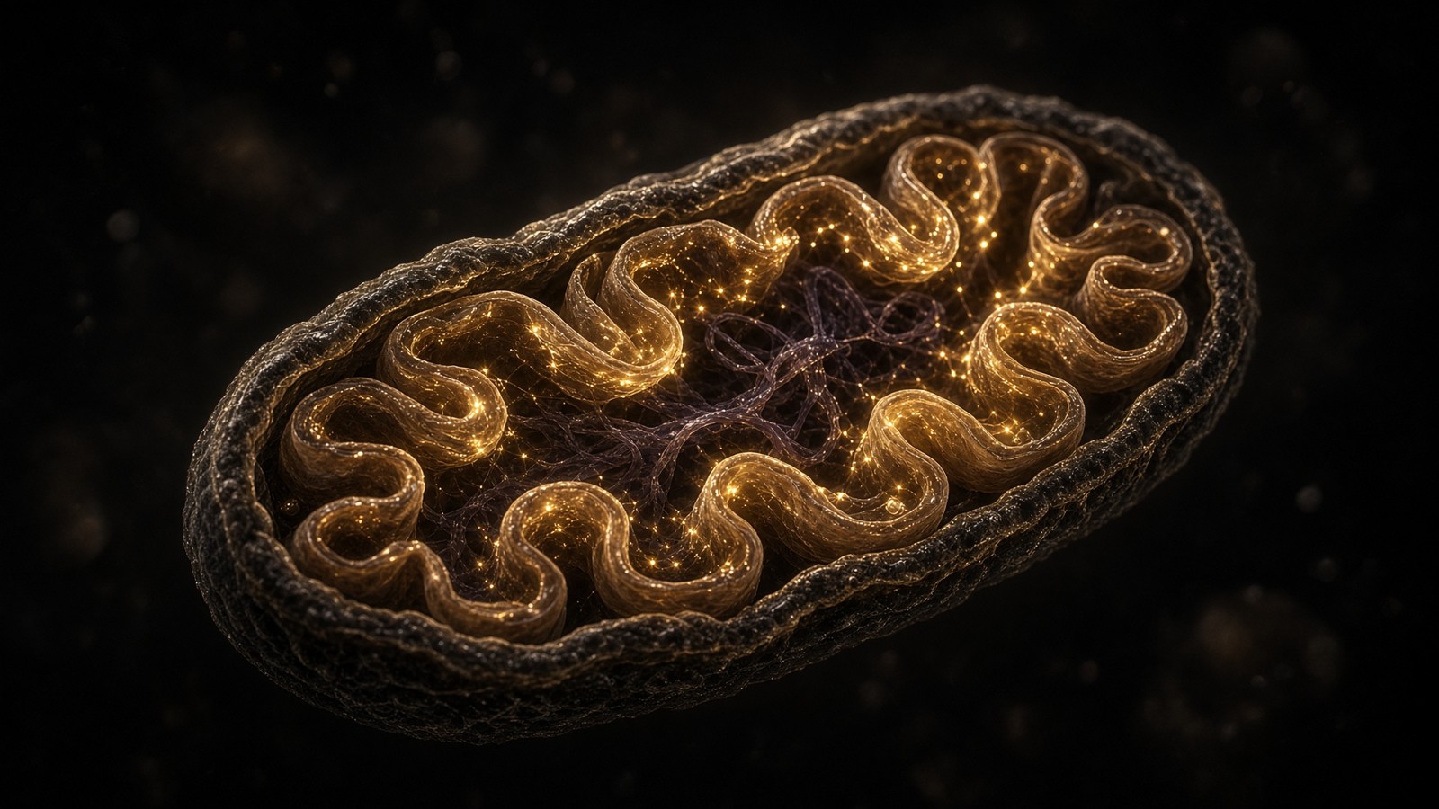
A mitochondrion is a former bacterium. Two membranes, its own circular DNA, 13 respiratory-chain proteins it still insists on encoding itself, tRNAs that use a slightly different genetic code (UGA means tryptophan, not stop). The nucleus encodes the other ~1,200 mitochondrial proteins and imports them. NADH from the TCA cycle feeds Complex I. Electrons run to oxygen. Protons are pumped. ATP synthase lets them back in and makes ATP. ~10²¹ times a second in a human. NAD+ is the oxidised coin. MOTS-c (MRWQEMGYIFYPRKLR) is translated from mitochondrial 12S rRNA in the reading frame of an RNA that was supposed to be a ribosome, not a message — Lee, Kim, Cohen, 2015. We stock both objects. They are not interchangeable. A 1000 mg cake of lyophilised β-NAD+ is a cofactor for the bench. An intramuscular NAD+ appointment at eLIVEate is a different product on a different till. Neither of them is the 40–60 kg of ATP you recycle while you read. Retatrutide occupies three GPCRs and changes how hungry an organism is; it does not 'boost mitochondria'. The organelle is a joint venture between two genomes and a voltage. The vials are reagents. Research use only.
Diagram
Matrix
- TCA cycle · β-oxidation · mtDNA nucleoids
- NADH produced here. Complex I spends it.
- MOTS-c (MRWQEMGYIFYPRKLR) from 12S rRNA.
Inner membrane
- I → II → III → IV → V (ATP synthase)
- ~150 mV proton-motive force
- ~40–60 kg of ATP turned over per human day
mtDNA is 16,569 bp, 37 genes, 13 proteins of the respiratory chain. Nuclear DNA encodes the other ~1,200 mitochondrial proteins. NAD+ is the hydride carrier between dehydrogenases and Complex I. MOTS-c is a 16-mer translated from 12S rRNA — a peptide the mitochondrion wrote itself.
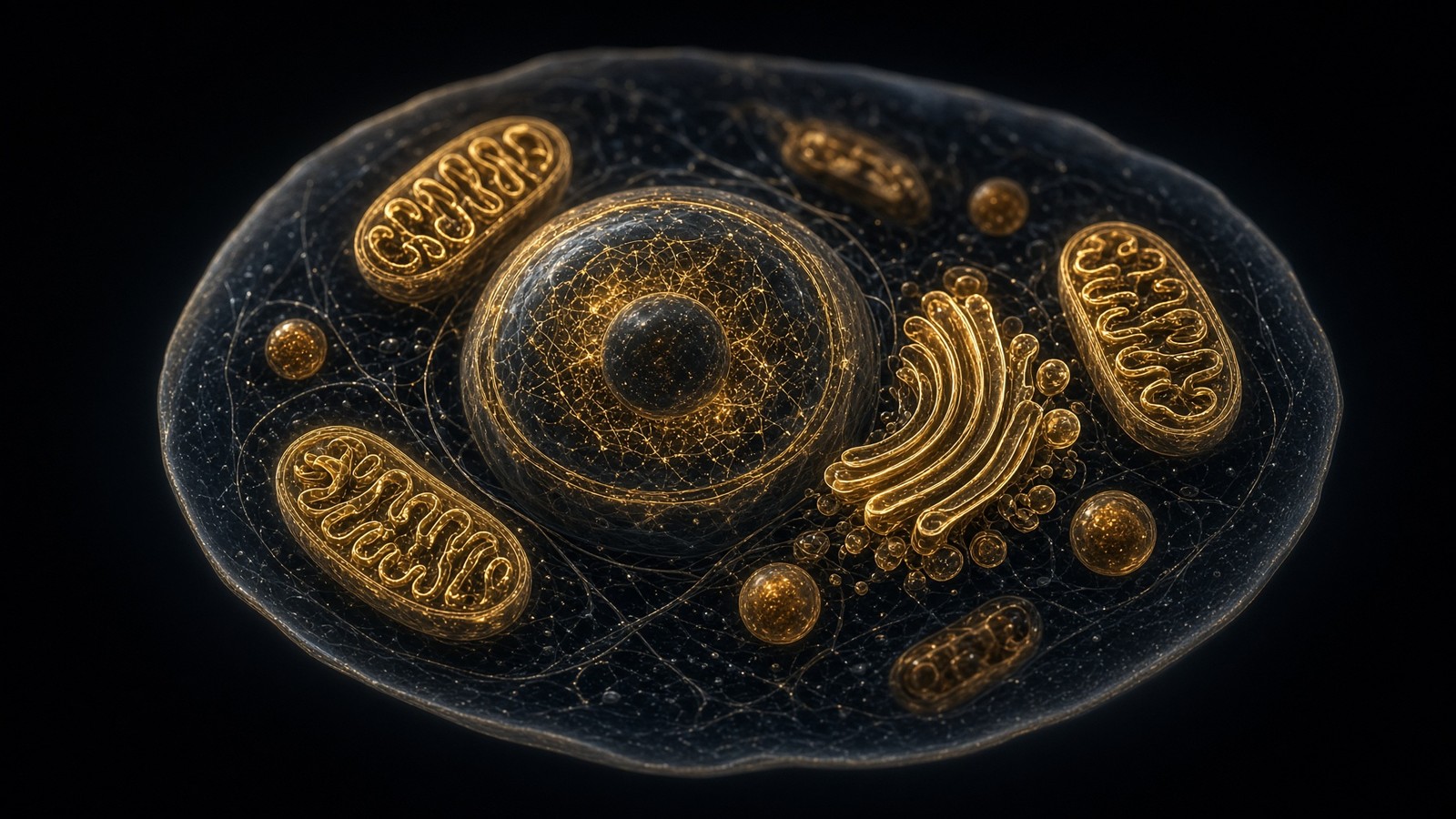
Lynn Margulis made endosymbiosis respectable; the genomes made it undeniable. Mitochondrial ribosomes are bacterial. The membrane lipids still argue. The division machinery (DRP1, FIS1, OPA1, MFN1/2) is how a former microbe now fissions and fuses inside you as a network, not a bean. A cardiomyocyte is a mitochondrial forest. A resting lymphocyte is a few dozen organelles. The census follows demand, PGC-1α, and whether you trained. Lynn Margulis spent years being told she was overstating a hunch. Then the rRNAs and the lipids and the circular chromosome lined up, and the hunch became furniture. I find that quietly thrilling: you are walking around with a captured microbe's power station in every cell that still bothers to respire, and the census of that station is a training variable, not a catalogue heading. PGC-1α is the coactivator that writes more organelles when you work. A vial doesn't.
In short. Mitochondria began as bacteria, and they still divide, fuse and keep bacterial ribosomes. How many you have follows demand and training, not a vial.
We're looking at the organelle as it actually is: a captured alphaproteobacterium that still keeps a genome, still uses a slightly different genetic code, still writes thirteen hydrophobic respiratory subunits on-site, and — this is the bit that shouldn't exist — still translates a 16-residue peptide from an rRNA that everyone thought they had finished annotating. Around that organelle sits a nuclear joint venture of roughly 1,200 imported proteins, a proton-motive force of about 150 millivolts across five nanometres, and a whole-body ATP turnover that Peter Rich put in the 40–60 kilogram range per day. NAD+ is the oxidised hydride coin that Complex I wants. MOTS-c is the 16-mer. Retatrutide is a different molecule on a different floor. The 1000 mg vial is a reagent. The clinic injection is a different till. None of those objects is 40 kg of ATP in a bottle, and the first job of a grown-up sentence is to stop pretending they are.
In short. A mitochondrion is a captured bacterium with two genomes and a voltage. NAD+ is the oxidised coin, MOTS-c is a 16-mer, and the 1000 mg vial isn't 40 kg of ATP.
- mtDNA
- 16,569 bp
- Respiratory subunits from mtDNA
- 13
- Nuclear mitochondrial proteins
- ~1,200
- Proton-motive force
- ~150 mV
- ATP turnover
- 40–60 kg/day
- MOTS-c
- 16 residues
37 genes. Compare 3.1 billion in the nucleus. Small, ruthless, essential.
The hydrophobic core of I, III, IV, V. Everything else is nuclear.
Imported as preproteins through TOM/TIM. The organelle is a joint venture.
Across 5 nm. The electric field is enormous. That is the battery.
Standing pool ~50 g. The mint never stops.
From 12S rRNA. A peptide that should not exist, published in 2015.
A bacterium, kept: endosymbiosis without the folklore
The idea is older than the respectability. Nineteenth-century microscopists noticed that mitochondria looked like bacteria and divided like bacteria. It took until 1967, and a paper in the Journal of Theoretical Biology under the name Lynn Sagan, for the argument to be made with enough stubbornness to survive a decade of polite refusal. Margulis (she published that one under her then-married name) said the eukaryotic cell is a merger, not a lonely inventor. An alphaproteobacterium — a relative of the lineage that now includes Rickettsia, though not Rickettsia itself, and the phylogeny has been rewritten more than once — was taken up by an archaeal host and never digested. Two billion years later you're still paying the electricity bill of that meal. You can disagree with parts of the energetic story and still have to live with the phylogeny. Two billion years later you're still paying the electricity bill of that meal, which I find quietly thrilling. The host was almost certainly an archaeon. The circular chromosome in the matrix is the receipt.
In short. Lynn Margulis argued the eukaryotic cell is a merger, not a lonely inventor. An alphaproteobacterium was taken up and never digested.
What made the argument undeniable wasn't the rhetoric. It was the genomes. Mitochondrial ribosomes are bacterial in architecture, 55S in mammals, with rRNAs transcribed from the organelle’s own 12S and 16S genes. The inner membrane still carries cardiolipin, a lipid that bacteria understand and that eukaryotic plasma membranes mostly do not. The division and fusion proteins are a later overlay — DRP1 recruited from the dynamin family, mitofusins and OPA1 doing the opposite job — but the organelle’s deepest habits remain prokaryotic. You don't get that combination from a nuclear invention. You get it from a cell that swallowed another cell and then spent geological time arguing about who was in charge. You don't get bacterial ribosomes, cardiolipin, and a circular chromosome from a nuclear invention. You get that combination from swallowing a cell. Division proteins came later — DRP1 from dynamin, mitofusins and OPA1 as the opposite job — an overlay on a prokaryotic habit that never quite left.
In short. The genomes settled the argument. Mitochondrial ribosomes are bacterial and the inner membrane still carries cardiolipin, habits you get from swallowing a cell.
The host was almost certainly an archaeon. Nick Lane and Bill Martin have spent a career pointing out that the energetic argument is the interesting one: a mitochondrion is how a eukaryotic cell afforded a large genome and a large cytoplasm, because the respiratory membrane was internalised and could be scaled by adding organelles rather than by stretching a plasma membrane around a bigger bag. You can disagree with parts of that energetic story and still have to live with the phylogeny. Mitochondria aren't ‘like bacteria’. They're the descendants of one, reduced, remodelled, and still running a circular chromosome in the matrix. The circular chromosome in the matrix is the receipt. Lane and Martin spent a career on the energetic half: once the respiratory membrane was internalised, you could scale ATP by adding organelles instead of stretching a plasma membrane around a bigger bag. That's how a eukaryotic cell afforded a large genome. I find that quietly thrilling.
In short. The host was almost certainly an archaeon. Mitochondria are descendants of a bacterium, reduced and remodelled, still running a circular chromosome in the matrix.
Two membranes, and cristae as an inverted bacterial inner membrane
A mitochondrion has two membranes because a bacterium has an inner membrane and the engulfment produced a second. The outer membrane is porous. VDAC, the voltage-dependent anion channel, lets metabolites through with a shrug; it isn't a vault. The intermembrane space is therefore in fairly free conversation with the cytosol for small solutes, which is why cytochrome c, once released through Bax/Bak pores, can reach the apoptosome without booking a transporter. The inner membrane is the opposite of porous. It's the reason the organelle exists. Almost no free proton leak except the regulated kind. Embedded in it, the respiratory complexes, ATP synthase, the adenine nucleotide translocator, the phosphate carrier, uncoupling proteins if the tissue has them. Folded into cristae — invaginations, not a bag of extra membrane stuffed in at random. Almost no free proton leak except the regulated kind. That's why cytochrome c, once it leaves through Bax/Bak pores, can reach the apoptosome without booking a transporter — the intermembrane space already talks to the cytosol for small solutes. The inner membrane is why the organelle exists, and cristae are how it packed the battery.
In short. A mitochondrion has two membranes because engulfment added a second. The outer is porous and the inner is the battery, with almost no free proton leak.
Cristae are the bacterial inner membrane, inverted and multiplied. Surface area is the point. A liver mitochondrion packs enough inner membrane to make the respiratory chain a crowded industrial estate rather than a cottage. MICOS complexes and OPA1 hold the crista junctions; lose those and the membrane becomes a slack balloon and ATP synthesis suffers. The old textbook bean, with a few cartoon folds, is a lie told to A-level students so they won't cry. A cardiomyocyte mitochondrion is cristae almost to the exclusion of matrix. A steroidogenic mitochondrion in the adrenal has a different folding habit again, because cytochrome P450scc sits on the inner membrane and cholesterol has to get in. Form follows the job. The job is a voltage across five nanometres. Form follows the job, and the job is about 150 millivolts across five nanometres. I find that field quietly thrilling. A slack balloon of inner membrane, MICOS gone, OPA1 gone, is a mitochondrion that still looks like a bean in a cartoon and no longer earns its keep as a mill.
In short. Cristae are the bacterial inner membrane, inverted and multiplied for surface area. Form follows the job, and the job is a voltage across five nanometres.
The gene heist, and the thirteen that refused to move
Most of the bacterial genome left. Endosymbiotic gene transfer isn't a metaphor; it is the reason the nuclear genome is littered with sequences of alphaproteobacterial origin and the reason the organelle now imports the vast majority of its proteome. MitoCarta and the Mootha laboratory’s census put the mammalian mitochondrial proteome at around 1,100–1,400 proteins depending on how strictly you count dual-localised ones. Call it 1,200 and you won't be laughed out of the room. Those proteins are made on cytosolic ribosomes as preproteins, most of them with an N-terminal amphipathic targeting peptide, and they queue at TOM, the translocase of the outer membrane. TIM23 takes the matrix-destined ones across the inner membrane, using the same Δψ that the chain is busy maintaining; TIM22 inserts the carrier family into the inner membrane. Chaperones catch them on the other side. The targeting peptide is cleaved. The organelle is a joint venture in which the nucleus holds most of the equity and the mitochondrion still insists on writing the hydrophobic core.
In short. Most of the bacterial genome left for the nucleus, and about 1,200 proteins are now imported through TOM and TIM. The mitochondrion still writes the hydrophobic core on-site.
Why keep any genes at all? The standard answer, which is probably true and is at least not silly, is hydrophobicity and timing. The thirteen proteins that remain on mtDNA — seven Complex I cores (ND1, ND2, ND3, ND4, ND4L, ND5, ND6), cytochrome b of Complex III, three Complex IV cores (COI, COII, COIII), and two ATP synthase subunits (ATP6, ATP8) — are transmembrane proteins of the inner membrane, awkward to import, and needed in stoichiometric register with assembly of the complexes. John Allen’s co-location for redox regulation hypothesis adds a second motive: the organelle keeps the genes whose expression ought to answer directly to the local redox state, rather than waiting for a memo from the nucleus. You don't have to pick a winner. Both pressures exist. The empirical fact is the list. Thirteen proteins, twenty-two tRNAs, two rRNAs. Everything else moved. The code, unsupervised by the nuclear apparatus for quite a long time, drifted.
In short. Thirteen hydrophobic respiratory subunits stayed on mtDNA, awkward to import and needed on-site. Twenty-two tRNAs and two rRNAs stayed with them, and the code drifted.
Sixteen thousand five hundred and sixty-nine letters
Human mtDNA is a closed circle of 16,569 base pairs. Anderson, Bankier, Barrell, de Bruijn, Coulson, Drouin, Eperon, Nierlich, Roe, Sanger, Schreier, Smith, Staden and Young, Nature, 9 April 1981. That's the Cambridge reference sequence, later revised in a few places and still the coordinate system everyone uses. Compare 3.1 billion base pairs in the haploid nucleus. The second genome is a rounding error by length and a catastrophe by omission: lose it and the cell can't respire. It's densely written. Almost no introns. Heavy strand and light strand, named for their buoyant density in an old centrifugation assay, not for a moral quality. A displacement loop, the D-loop, where a third strand of DNA sits as a replication and transcription control region and doesn't code for a protein. Two promoters on the heavy strand, one on the light. Polycistronic transcripts, punctuated by tRNAs that are cleaved out, leaving the mRNAs and rRNAs behind. It's an operon-ish habit the nucleus abandoned and the organelle never did.
In short. Human mtDNA is a closed circle of 16,569 base pairs, sequenced in 1981. Lose that densely written chromosome and the cell can't respire.
Thirty-seven genes isn't a lot, and every one of them is spoken for. Thirteen are proteins. Twenty-two are tRNAs, enough — just — to translate those thirteen with a wobble-heavy, stripped-down set that would make a cytosolic translator blush. Two are rRNAs: 12S (MT-RNR1) and 16S (MT-RNR2), the structural RNAs of the mitoribosome. There's no mtDNA-encoded 5S; mammals improvise. There are no mtDNA-encoded ribosomal proteins; those came from the nucleus, which is why a mitoribosome is a chimeric object, bacterial RNA wrapped in a protein shell that has been rewritten by eukaryotic history. The 13 protein genes aren't a random sample of metabolism. They're the cores of the chain. The thirteen proteins are the cores of the respiratory chain, not a random sample of metabolism. There is no mtDNA-encoded 5S; mammals improvise. There are no mtDNA-encoded ribosomal proteins; those came from the nucleus, which is why a mitoribosome is a chimeric object — bacterial RNA wrapped in a eukaryotic protein shell.
In short. Thirty-seven genes cover thirteen proteins, twenty-two tRNAs, and the 12S and 16S rRNAs. The thirteen proteins are the cores of the respiratory chain, not a random sample.
- Complex I cores: ND1, ND2, ND3, ND4, ND4L, ND5, ND6 — NADH dehydrogenase subunits, the hydrophobic heart of a ~45-subunit machine.
- Complex III: cytochrome b (MT-CYB) — the only mtDNA subunit of the bc1 complex, and the reason a Q-cycle can still be a mitochondrial story.
- Complex IV: COI, COII, COIII — cytochrome c oxidase, where O₂ becomes water and cyanide becomes a catastrophe.
- ATP synthase: ATP6 and ATP8 — Fo components. The rotary motor’s membrane-embedded half, written on-site.
- tRNAs: twenty-two, including the tRNA-Leu(UUR) whose m.3243A>G lesion is the usual MELAS mutation.
- rRNAs: 12S, from which MOTS-c is translated; 16S, from which humanin is translated. Structural RNAs with a second job nobody asked them for.
A genetic code that is almost, but not quite, the universal one
The so-called universal genetic code is a majority vote, not a law. Human mitochondrial translation uses a dialect. AUA, which the cytosol reads as isoleucine, is methionine in the mitochondrion — so there are two Met codons, AUG and AUA, and the initiator tRNA has to live with that. UGA, a stop codon in the nucleus, is tryptophan; the organelle has a tRNA that pairs with it and a release-factor habit that does not. AGA and AGG, arginine in the cytosol, are stops in human mitochondria. The codon table you memorised for A-level is the nuclear table. Feed it to a mitoribosome and you will write the wrong protein and fail to stop. This isn't a curiosity for trivia night. It's why a nuclear copy of an mtDNA gene is not, by itself, a working backup: even if you could import the protein, you would still have to rewrite the code or recode the gene. Gene therapy for mtDNA disease has to live with that, which is one reason it has been slower than the CRISPR headlines.
In short. Human mitochondria use a dialect of the genetic code: AUA is methionine, UGA is tryptophan, AGA and AGG are stops. The nuclear table writes the wrong protein here.
The dialect is a fossil of isolation. Once most genes had left, the remaining translation system was a small society with no reason to stay compatible with the cytosol. tRNAs were lost until twenty-two was the number that still worked, with superwobble covering what a full set would have covered. Release factors specialised. AUA drifted. None of this required a plan. It required time and a bottleneck. Every one of your mitochondria still runs that dialect, in every tissue, from a circle your mother gave you and her mother gave her, with no paternal mtDNA to argue. (Paternal leakage exists as a rare laboratory and clinical observation. It isn't how the system is supposed to work, and it isn't how almost any of us inherited the molecule.). Every one of your mitochondria still runs that dialect, in every tissue, from a circle your mother gave you and her mother gave her. Feed the nuclear codon table to a mitoribosome and you'll write the wrong protein and fail to stop. That's not trivia-night biology. It's why a nuclear copy of an mtDNA gene isn't, by itself, a working backup.
In short. Once most genes had left, the remaining translation system drifted out of step with the cytosol. Every mitochondrion still runs that dialect, inherited down the maternal line.
Nucleoids, not chromosomes: TFAM and polymerase γ
mtDNA isn't wrapped around histones. It's packed by TFAM — mitochondrial transcription factor A, a high-mobility-group protein that both coats the DNA and, at the right stoichiometry, enables transcription from the promoters. Nils-Göran Larsson’s work is the place to start: too little TFAM and the circles vanish; too much and they're so densely coated they can't be read. The complexes of DNA plus TFAM plus the rest of the transcription and replication machinery are nucleoids, one or a few circles each, visible as foci in the matrix, often parked near inner-membrane nucleoids’ favourite crista junctions. There are no nucleosomes, no histone code, no chromatin immunoprecipitation story of the nuclear kind. Packaging is TFAM abundance and a handful of other DNA-binding proteins. Regulation is copy number, which is a tissue variable, and transcription, which answers to cellular energy demand and to a set of nuclear-encoded factors (TFB2M, POLRMT, TEFM) that have to be imported like everything else.
In short. mtDNA is packed by TFAM into nucleoids, not wrapped around histones. Copy number and TFAM abundance decide whether the circles can be read.
Replication is polymerase γ, POLG, a nuclear-encoded enzyme with a proofreading exonuclease, assisted by TWINKLE helicase and the mitochondrial single-stranded binding protein. The classic model is strand-displacement: the heavy strand starts at OH in the D-loop, runs most of the way around, exposes OL, and the light strand then fires. There's a competing ribonucleotide-incorporating, more bidirectional picture from the Falkenberg and Holt laboratories. The field hasn't finished that argument and we'll not pretend it has. What isn't in dispute: POLG mutations are a major cause of primary mitochondrial disease (Alpers, PEO, ataxia-neuropathy), the polymerase is the only replicative DNA polymerase in the organelle, and nucleoside analogue drugs that POLG will mis-incorporate have been mitochondrial toxicities in the clinic. Copy number per cell isn't two. It's hundreds in a platelet, thousands in a neuron, tens of thousands in an oocyte. That abundance is why heteroplasmy can hide, and why a bottleneck can un-hide it.
In short. Replication is polymerase γ, the organelle's only replicative DNA polymerase. Copy number runs from hundreds to tens of thousands per cell, which is why heteroplasmy can hide.
Heteroplasmy is a threshold, not a coin-flip
A nuclear gene comes in two copies, and a dominant or recessive story follows. mtDNA comes in hundreds to thousands of copies per cell, and the story is a mixture. Heteroplasmy means more than one mtDNA genotype in the same cell, the same tissue, the same person. A pathogenic variant can sit at 10% and do nothing detectable, sit at 50% and do something in a high-demand tissue, sit at 90% and be a disease. The threshold is tissue-specific because the spare respiratory capacity is tissue-specific. Brain and heart notice first. Skin fibroblasts in culture can look almost ordinary at fractions that would drop a neuron. This is why a blood heteroplasmy measurement isn't a brain heteroplasmy measurement, and why counselling a family with a known mtDNA variant is a specialist job rather than a blog. Brain and heart notice first because spare respiratory capacity is smallest there. Skin fibroblasts in culture can look almost ordinary at fractions that would drop a neuron. That's why a blood heteroplasmy number isn't a brain number, and why counselling a family with a known mtDNA variant is a specialist job rather than a blog.
In short. Heteroplasmy is a mixture of mtDNA genotypes in one cell. A pathogenic variant can sit at 10% and do nothing, or at 90% and be disease, and the threshold is tissue-specific.
MELAS — mitochondrial encephalomyopathy, lactic acidosis, and stroke-like episodes — is the named lesson that usually lives at m.3243A>G in tRNA-Leu(UUR). Goto, Nonaka and Horai, Nature 1990, for the mutation; a large subsequent literature for the messy genotype–phenotype map. The same variant, at different loads, can look like diabetes and deafness, or like a stroke in a young adult that doesn't respect a vascular territory, or like almost nothing. Leber hereditary optic neuropathy is the other name that belongs in any honest paragraph: Wallace, Singh, Lott and colleagues, Science 1988, the m.11778G>A substitution in ND4, later joined by ND1 and ND6 primary mutations. Subacute, painless, bilateral central vision loss, often in a young man, maternal relatives carrying the same circle at high load without the eye phenotype. Incomplete penetrance isn't a mystery to be solved by a motivational poster. It's nuclear background, haplogroup, sex, and probably a bit of bad luck about which retinal ganglion cells crossed their own threshold.
In short. MELAS usually maps to m.3243A>G in tRNA-Leu, and Leber hereditary optic neuropathy often to m.11778G>A in ND4. Same circle, different loads, different faces.
Inheritance is maternal. Sperm mitochondria are usually destroyed after fertilisation, by ubiquitin-dependent mechanisms and by simple dilution: an oocyte holds on the order of 10⁵ mtDNA copies, a sperm a few hundred, and the sperm’s contribution isn't invited to stay. That's why a pedigree of mtDNA disease traces through the mothers and why a father with LHON doesn't transmit the risk. Between generations there is a bottleneck. The primordial germ cell doesn't take a representative sample of the mother’s tens of thousands of oocyte mtDNAs; it takes a small draw, and the draw is then expanded. Heteroplasmy can therefore shift, sometimes sharply, from mother to child. Prenatal prediction is imperfect. Mitochondrial replacement techniques — ‘three-person IVF’, spindle transfer or pronuclear transfer — exist as a regulatory and clinical fact in the United Kingdom precisely because this inheritance pattern doesn't give families a nuclear-style option. That's statute and HFEA, not a research-peptide conversation. It belongs in the paragraph because the second genome isn't an abstraction. It's a legal object.
In short. Inheritance is maternal, and sperm mitochondria are usually destroyed. A germline bottleneck can shift heteroplasmy between generations, which is why mitochondrial replacement exists in UK statute.
Doug Wallace has spent decades arguing that mtDNA variation, including common haplogroups, isn't just rare disease. Adaptation to climate, to diet, to latitude; a contribution to common metabolic and degenerative phenotypes; a clock. Some of that has held. Some of it has been trimmed by better population genetics. What hasn't been trimmed is the disease mechanism: a respiratory chain built from two genomes will fail when either genome fails, and the mitochondrial one fails in a heteroplasmic, thresholdy, maternally inherited way that nuclear Mendelian intuition keeps getting wrong. Schon, DiMauro, Turnbull, Chinnery, Suomalainen — the mitochondrial-medicine literature is a field, not a footnote. Primary mitochondrial disease is rare. Secondary mitochondrial dysfunction is everywhere the chain is asked to work in a bad neighbourhood: ischaemia-reperfusion, heart failure, neurodegeneration, the insulin-resistant hepatocyte. Different floors. Same organelle. Schon, DiMauro, Turnbull, Chinnery, Suomalainen — the mitochondrial-medicine literature is a field, not a footnote. Primary mitochondrial disease is rare. Secondary dysfunction is common wherever the chain is asked to work in a bad neighbourhood: ischaemia, a failing heart, a neuron running out of spare capacity. Same organelle. Different floors.
In short. A respiratory chain built from two genomes fails when either genome fails. Primary mitochondrial disease is rare, and secondary dysfunction is common wherever the chain works badly.
The second genome ages faster
Nuclear DNA replication, after polymerase proofreading and mismatch repair, runs at something like 10⁻⁹ to 10⁻¹⁰ errors per base per division. mtDNA is worse by a factor of ten to a hundred, depending on which assay and which region you trust. The reasons aren't mystical. The molecule sits in the matrix, next door to Complexes I and III, which leak superoxide when the chain is reduced and oxygen is around. It has no histones to soak up chemistry. Base-excision repair exists in mitochondria; nucleotide-excision repair of the nuclear kind mostly does not. POLG proofreads, and does it well, but the neighbourhood is still a respiratory chain. The D-loop is particularly untidy. Deletions accumulate in post-mitotic tissues with age — the ‘common deletion’ of 4,977 bp is a famous passenger in ageing muscle and brain — and point mutations accumulate too. Whether they cause ageing or merely decorate it is a long argument. That they accumulate is not.
In short. mtDNA mutates ten to a hundred times faster than nuclear DNA. It sits next to superoxide with no histones and a thinner repair budget, and damage accumulates with age.
Harman proposed mitochondrial free-radical ageing in the 1970s. The strong form of that theory (ROS cause ageing, antioxidants prevent it) didn't survive contact with the intervention trials, the long-lived mutants that make more ROS, and the signalling functions of the leak. The weak form is still standing: mtDNA is a nearby, poorly defended target, the damage is real, and some of the phenotype of old post-mitotic tissue is mosaic respiratory deficiency in individual cells that have drifted to high mutant load. COX-negative fibres in aged muscle are the histology. Clonal expansions of mutant mtDNA, not a uniform dusting, are the genetics. A neuron or a myocyte is a long-lived bag of circles, and some of the circles win. COX-negative fibres in aged muscle are the histology. Clonal expansions of mutant mtDNA, not a uniform dusting, are the genetics. A neuron or a myocyte is a long-lived bag of circles, and some of the circles win. The strong free-radical theory didn't survive the trials. This mosaic picture did.
In short. The strong free-radical theory of ageing didn't survive the trials. The weak form still stands: mtDNA is a nearby, poorly defended target, and old post-mitotic tissue is mosaic for it.
This is why the neighbouring error-rates diagram in the cell-at-scale essay puts the mitochondrion in a different column from the nucleus. The genome that has to last is nuclear, repaired, chromatinised, diploid. The genome that sits next to the furnace is not. MOTS-c and humanin, if you take the papers seriously, are the organelle writing peptides from the very RNAs that also build the mitoribosome — a second, overlapping use of a small chromosome that is already busy and already under fire. Whether that is an evolved signalling system or a translation accident that became useful is a question the 2015–2018 papers opened and didn't close. The error rate isn't a question. It's a measurement. Whether MOTS-c and humanin are evolved signals or useful accidents is still open, and we won't close it from a catalogue page. The error rate isn't a question. It's a measurement: the genome next to the furnace, no histones, a thinner repair budget, damage accumulating in post-mitotic tissue. That's already enough wonder for one paragraph.
In short. The genome next to the furnace ages faster. MOTS-c and humanin are peptides written from rRNAs that also build the mitoribosome, evolved signals or useful accidents, still open.
Diagram
- DNA replication + MMR10⁻⁹ to 10⁻¹⁰A genome of 6 Gbp (diploid) accumulates a handful of mutations per division.
- Transcription~10⁻⁵RNA is disposable. The cell can afford a wrong letter in a message that lasts hours.
- Translation~10⁻⁴One wrong amino acid per ten thousand. Proteins turn over. DNA does not.
- mtDNA10–100× nuclearNo histones, ROS next door, weaker repair. The second genome ages faster.
The genome is sacred, the message is cheap, the protein is cheaper. Ageing is partly what happens when the sacred copy still drifts — and when mitochondria, which never got the nuclear repair budget, drift faster.
Fuel becomes NADH, and NAD+ is the oxidised coin
Before there is a respiratory chain there is a hydride. Carbohydrate, fat and (less cleanly) amino acids are oxidised in the matrix until their electrons sit on nicotinamide. Pyruvate from glycolysis crosses the inner membrane via MPC1/2 and is decarboxylated by the pyruvate dehydrogenase complex — a mitochondrial machine the size of a ribosome, thiamine-dependent, phosphorylated off by PDKs when the cell has had enough. Acetyl-CoA enters the TCA cycle. Each turn of citrate–isocitrate–α-ketoglutarate–succinyl-CoA–succinate–fumarate–malate–oxaloacetate throws off three NADH and one QH2 (at succinate dehydrogenase, which is also Complex II) plus a GTP or ATP at succinyl-CoA synthetase. The NADH is the point. The carbon dioxide is a by-product you breathe out and don't think about. Pyruvate crosses via MPC1/2. PDH — a mitochondrial machine the size of a ribosome, thiamine-dependent — decarboxylates it, and PDKs phosphorylate the complex off when the cell has had enough. Acetyl-CoA enters the TCA cycle. The NADH is the point. The carbon dioxide is a by-product you breathe out and don't think about.
In short. Fuel is oxidised until its electrons sit on nicotinamide. The TCA cycle throws off NADH as the point, and carbon dioxide is a by-product you breathe out.
Fatty acids enter as acyl-CoA, are conjugated to carnitine by CPT1, cross as acyl-carnitine, and are handed back to CoA by CPT2. β-oxidation then shortens the chain two carbons at a time: FADH2 at acyl-CoA dehydrogenase (electrons into the ubiquinone pool via ETF), NADH at hydroxyacyl-CoA dehydrogenase, acetyl-CoA into TCA or, in liver, into ketogenesis when the cycle can't take the lot. Odd-chain and unsaturated fatty acids have extra steps; peroxisomes take the very-long-chain ones first. The accounting is the same. Reduced cofactors. The mitochondrion is an engine for making NADH and QH2 so that the inner membrane has something to oxidise. Odd-chain and unsaturated fatty acids have extra steps; peroxisomes take the very-long-chain ones first. The accounting is the same: reduced cofactors for the inner membrane. CPT1 is the gate the liver uses when it decides to make ketones instead. I find that bookkeeping quietly beautiful once you can see it.
In short. Fatty acids enter as acyl-carnitine and β-oxidation shortens them two carbons at a time. The mitochondrion is an engine for making NADH and QH2 so the inner membrane can oxidise them.
NAD+ is the oxidised form of the coin. A dehydrogenase takes a hydride from the substrate and parks it at nicotinamide C4; NAD+ becomes NADH. Complex I takes the hydride back, as two electrons into the chain and a proton into the N-side chemistry, and NAD+ is regenerated. If Complex I stops — rotenone, a severe ND mutation, anoxia upstream of a reduced pool — NADH rises, NAD+ falls, the TCA cycle stalls at the NAD+-dependent steps, and the cell looks for glycolysis and lactate dehydrogenase to regenerate NAD+ in the cytosol instead. That's lactic acidosis as a mitochondrial sentence, not as a training status. The NADH/NAD+ ratio is a gauge that dehydrogenases and sirtuins both read. It isn't a vitamin ranking. That's lactic acidosis as a mitochondrial sentence, not as a training status. The NADH/NAD+ ratio is a gauge that dehydrogenases and sirtuins both read. It isn't a vitamin ranking, and a lyophilised cake in a catalogue isn't a way to force the gauge from outside three membranes.
In short. NAD+ is the oxidised coin, and Complex I regenerates it from NADH. If the complex stops, NADH rises, the TCA cycle stalls, and the ratio is a gauge not a vitamin ranking.
The matrix NAD+ pool and the cytosolic NAD+ pool aren't freely mixed. The inner membrane doesn't let the dinucleotide wander. What shuttles are the reducing equivalents: malate–aspartate, glycerol phosphate, and a few specialist routes. This is why a cell can have a reduced cytosol and an oxidised matrix, or the reverse, and why a lyophilised NAD+ solid added to a culture dish isn't ‘topping up mitochondria’. Charged, ~663 Da, it doesn't stroll through a plasma membrane and then through two more. Cells that take it up at all do so through connexin hemichannels or after extracellular hydrolysis to nicotinamide or NMN and resynthesis inside. The 1000 mg cake in the catalogue is the cofactor as a reagent for an assay that you control. It isn't a delivery system for the matrix. If you skip that hasn't looked hard at the membrane.
In short. Matrix and cytosolic NAD+ pools don't mix, and a lyophilised solid doesn't stroll through three membranes. The 1000 mg cake is a bench reagent, not a delivery system.
Electron transport is a bucket chain with a voltage at the end
Diagram
- I. Pumps H⁺. ~45 subunits. The NADH coin is spent here.
- II. TCA entry. No proton pump. FADH₂ neighbourhood.
- Q. Lipid-soluble shuttle in the inner membrane.
- III. Q-cycle. Pumps H⁺. Superoxide leak site.
- c. Intermembrane space. The shuttle everyone has heard of.
- IV. O₂ → H₂O. The reason you breathe.
- V. F₁Fₒ rotary. Protons in, ATP out. ~10²¹ times a second in you.
Mitchell’s chemiosmotic theory (Nobel 1978): the inner membrane is a battery of ~150 mV. NAD+ is the hydride carrier that feeds Complex I. MOTS-c is a 16-mer the mitochondrion translated from 12S rRNA — a different object on the same campus.
Complex I takes two electrons from NADH and dumps them on ubiquinone, pumping four protons. Complex II (TCA’s succinate dehydrogenase) feeds FADH2-level electrons onto the same quinone pool without pumping. Complex III splits the path (Q-cycle) and pumps. Cytochrome c shuttles one electron at a time. Complex IV reduces O₂ to water and pumps. Oxygen is the terminal acceptor, which is why you breathe and why cyanide is a catastrophe. The proton gradient is both a voltage and a pH difference; together they're the proton-motive force that ATP synthase uses, rotary catalysis, Boyer and Walker’s Nobel, 3 ATP per full rotation in the textbook cartoon and a messier integer in a living inner membrane because slip and leak are real. Oxygen is the terminal acceptor, which is why you breathe and why cyanide is a catastrophe. The proton gradient is both a voltage and a pH difference; together they're the proton-motive force ATP synthase uses. Rotary catalysis, Boyer and Walker, a messier integer in a living inner membrane because slip and leak are real.
In short. Electrons run from NADH to oxygen while Complexes I, III and IV pump protons. Complex II feeds the quinone pool without pumping, and the voltage drives ATP synthase.
- NADH donates two electrons to Complex I (NADH:ubiquinone oxidoreductase). Four protons are pumped. Ubiquinone is reduced to ubiquinol.
- Complex II (succinate dehydrogenase) oxidises succinate, reduces the same quinone pool, and pumps nothing. It is a TCA enzyme wearing a respiratory hat.
- Ubiquinol diffuses in the membrane to Complex III (bc1). The Q-cycle splits two electrons: one to cytochrome c, one back into the quinone pool. Protons are released to the P-side.
- Cytochrome c carries one electron at a time across the intermembrane space to Complex IV.
- Complex IV (cytochrome c oxidase) reduces O₂ to H2O, pumps protons, and terminates the chain. Cyanide, CO and nitric oxide all know this enzyme.
- The resulting Δp — voltage plus ΔpH — drives F1Fo ATP synthase. Protons return to the matrix through Fo. ATP is made in F1.
Complex I: forty-five subunits, a hydride, and a long tunnel
Mammalian Complex I is an L-shaped assembly of about forty-five subunits, seven of them mitochondrial-encoded, the rest nuclear, plus a cloud of assembly factors that never make it into the mature enzyme and will ruin your week if they're missing. Youssef Hatefi dissected the chain into complexes in the 1960s; Sazanov’s crystal structures and the later cryo-EM from the Hirst and Sazanov groups are how we know where the electrons actually go. A flavin (FMN) takes the hydride from NADH. Iron–sulphur clusters walk the two electrons along the hydrophilic arm. At the far end, ubiquinone sits in a deep cavity and is reduced. The pumping, four protons per two electrons, is a conformational wave along the membrane arm, not a simple redox loop. Rotenone and piericidin bind in the quinone pocket. Metformin’s weak Complex I inhibition is a real pharmacology with a dose–response that doesn't license anyone to call a research peptide a metformin. Different molecule, different till, different literature.
In short. Mammalian Complex I is about forty-five subunits, seven of them mitochondrial-encoded. It takes a hydride from NADH, pumps four protons, and reduces ubiquinone.
Complex I is also a superoxide source. When the FMN is reduced and the downstream chain is blocked, or when reverse electron transport from a reduced quinone pool drives electrons backward — the succinate story in ischaemia-reperfusion, Chouchani and Murphy — oxygen picks up a single electron and you have O2•−. That isn't a morality. It's a flavin in water with a finite leak. ND mutations (LHON’s ND4, MELAS-adjacent coding lesions, the rare NARP-ish Complex I defects) drop the enzyme’s throughput and, depending on the site, can raise or lower the leak. Seven of the thirteen mtDNA proteins live here. If you were designing a genome to keep on-site, you would keep these. The bacterium did. A flavin takes the hydride from NADH. Iron–sulphur clusters walk the two electrons along the hydrophilic arm. At the far end, ubiquinone sits in a deep cavity and is reduced. Rotenone binds in that quinone pocket. Seven of the thirteen mtDNA proteins live here. The bacterium kept these, and you still do.
In short. Complex I leaks superoxide when the flavin is reduced and the chain is blocked. Seven of the thirteen mtDNA proteins live here, which is why they stayed on-site.
Complex II, the quinone pool, and Complex III’s Q-cycle
Complex II is succinate dehydrogenase, four nuclear-encoded subunits, FAD, iron–sulphur clusters, a haem that is more structural than catalytic, and no proton pumping. It's the only complex with no mtDNA subunit, which is why it is the histochemical control when you stain for COX-negative fibres: Complex II still works when the circle is deleted. Electrons from succinate reduce FAD, run the clusters, and reduce ubiquinone to ubiquinol. The same quinone pool also takes electrons from Complex I, from ETF:ubiquinone oxidoreductase (β-oxidation), from dihydroorotate dehydrogenase (pyrimidine synthesis), from glycerol-3-phosphate dehydrogenase. Ubiquinone is the membrane’s public bus. Its redox state is a more interesting number than most people who say ‘mitochondrial health’ have ever measured. The quinone pool also takes electrons from ETF:ubiquinone oxidoreductase in β-oxidation, from dihydroorotate dehydrogenase, from glycerol-3-phosphate dehydrogenase. Ubiquinone is the membrane's public bus. Its redox state is a more interesting number than most people who say mitochondrial health have ever measured.
In short. Complex II is succinate dehydrogenase: four nuclear subunits, no proton pumping, no mtDNA gene. It still works when the circle is deleted, and ubiquinone is the membrane's public bus.
Complex III, the bc1 complex, is where Mitchell’s Q-cycle lives. Ubiquinol oxidised at the Qo site releases its two protons to the intermembrane space and splits its two electrons: one to the Rieske iron–sulphur protein and on to cytochrome c1 and cytochrome c; one to cytochrome b and back to a quinone at the Qi site. Two turns of that cycle put four protons on the P-side for every two electrons that go forward to cytochrome c. Antimycin A blocks Qi. Myxothiazol and stigmatellin block Qo. The Qo site is the other famous superoxide source: a semiquinone that can reduce oxygen if the site is occupied wrongly. Cytochrome b is the mtDNA contribution. The rest is nuclear. Hatefi’s fractionation, Trumpower’s biochemistry, the Xia and Iwata structures — this is a solved machine with remaining arguments about the exact leak, not about whether the Q-cycle happens.
In short. Complex III runs Mitchell's Q-cycle and puts protons on the P-side. Cytochrome b is the mtDNA contribution, and the Qo site is the other famous superoxide source.
Cytochrome c, Complex IV, and why you breathe
Cytochrome c is a small haem protein in the intermembrane space, loosely held on the inner membrane by cardiolipin, shuttling one electron at a time from III to IV. It's also, when the outer membrane is permeabilised, the apoptosome’s trigger: APAF-1, dATP, caspase-9. The same protein is a courier and a death sentence. That economy is typical of the organelle. Complex IV, cytochrome c oxidase, takes four electrons from four cytochrome c molecules, four protons from the matrix for chemistry, and pumps four more, and reduces one O₂ to two H2O. COI, COII and COIII are the mtDNA cores; the copper A and copper B centres and the haem a / a3 pair are the chemistry. Cyanide binds ferric haem a3. Carbon monoxide binds ferrous. Nitric oxide binds and, at physiological concentrations, regulates. This is the step that makes oxygen the terminal acceptor. This is why a room without oxygen is a mitochondrial event before it is a psychological one.
In short. Cytochrome c shuttles one electron from III to IV, and triggers apoptosis if the outer membrane opens. Complex IV reduces oxygen to water, which is why you breathe.
Keilin saw the cytochromes as absorption bands in a spectroscope and named them. Warburg argued about Atmungsferment. Chance and Williams titrated the states of the chain in the 1950s and gave us the vocabulary (State 3, State 4) that bioenergeticists still use. Photobiomodulation at 630–850 nm, the Karu and Hamblin literature, treats Complex IV as a chromophore: nitric oxide photodissociated from CuB/haem a3, a transient lift in flux, a pulse of ROS used as a message. That's a clinic-adjacent conversation at eLIVEate’s Dermalux desk, and it is a different object from a lyophilised peptide. The enzyme doesn't care which essay you're reading. It absorbs photons, binds O₂, and pumps. Chance and Williams titrated the states of the chain in the 1950s and gave us State 3 and State 4, vocabulary bioenergeticists still use. Photobiomodulation treats Complex IV as a chromophore — a clinic-adjacent conversation, and a different object from a lyophilised peptide. The enzyme doesn't care which essay you're reading. It absorbs photons, binds O₂, and pumps.
In short. Keilin named the cytochromes. Photobiomodulation treats Complex IV as a chromophore, a clinic-adjacent conversation and a different object from a lyophilised peptide.
The proton-motive force: Mitchell, 1961, Nobel 1978
Peter Mitchell’s chemi-osmotic hypothesis, Nature 1961, wasn't welcomed. The field preferred a high-energy chemical intermediate, a squiggle, something covalent and civilised. Mitchell said the intermediate was a proton gradient across a topologically closed membrane. It took the better part of two decades, a great deal of unkindness, and a Nobel Prize in 1978 for the argument to become the furniture. Δp = Δψ − 2.303(RT/F)ΔpH, in the proper units, or in the corridor shorthand, Δp = Δψ + ΔpH. In a typical mitochondrion the voltage does most of the work: about 150 mV, negative in the matrix, which across 5 nm is an electric field of 30 million volts per metre. The pH difference is smaller, 0.5 units or so, matrix alkaline, but it is real and it is used. Ions, metabolites and the phosphate carrier all feel it. Calcium enters through the uniporter because of it. ATP is made because of it.
In short. Mitchell said the intermediate was a proton gradient, not a covalent squiggle. About 150 mV across 5 nm is an enormous field, and ATP is made because of it.
Collapse Δψ and the cell notices on a timescale of seconds. Uncouplers (DNP, FCCP) let protons back in without ATP synthase; the chain runs, heat is made, ATP is not, and at sufficient concentration the cell dies. The same physics, at a controlled leak, is how brown adipose tissue is a heater rather than a mint. Oligomycin blocks ATP synthase and Δψ rises until the chain can't pump against it. Ischaemia stops the chain at Complex IV for lack of oxygen; succinate backs up; reperfusion slams reverse electron transport through Complex I and a ROS burst that Murphy and colleagues have spent a career quantifying. Bioenergetics isn't a wellness category. It's a set of numbers you can put a microelectrode on. The same physics, at a controlled leak, is how brown adipose tissue is a heater rather than a mint. Ischaemia stops the chain at Complex IV for lack of oxygen; reperfusion slams reverse electron transport through Complex I and a ROS burst Murphy spent a career quantifying. Bioenergetics is a set of numbers you can put a microelectrode on, not a wellness category.
In short. Collapse the voltage and the cell notices in seconds. Uncouplers make heat instead of ATP, and bioenergetics is a set of numbers, not a wellness category.
ATP synthase: the rotary mint, and 40 kg a day
F1Fo ATP synthase is the reason the gradient exists as far as you're concerned. Fo is the membrane-embedded ring of c-subunits plus the a-subunit that lets protons through, one per c-subunit per full rotation. F1 is the catalytic knob in the matrix: three β-subunits, three catalytic sites, Boyer’s binding-change mechanism, a γ-subunit rotor that makes each site take its turn at loose, tight and open. Walker’s structure. Noji’s single-molecule rotation. Nobel, 1997, to Boyer and Walker (Skou shared it for the sodium–potassium pump, which is a different ATPase and a different story). In mammalian mitochondria the c-ring has eight subunits, so eight protons per 360 degrees, three ATP per turn, plus the extra proton cost of importing phosphate and exchanging ATP for ADP via ANT. Textbook ‘3 H+ per ATP’ is a rounding. Living inner membranes also slip. The integer is messier than the lecture.
In short. ATP synthase is a rotary motor: protons turn Fo and F1 makes ATP. Eight protons per turn in the mammalian c-ring yield three ATP, plus transport costs.
Peter Rich, Biochemical Society Transactions, 2003: resting ATP turnover in an adult is on the order of 40 kg per day. Athletes, fever, pregnancy and a cold room push it higher, toward 60 kg and past it. The standing pool of ATP in the whole body is about 50 g — a shot-glass, a coffee cup, not a warehouse. The only way those two numbers can both be true is if every molecule is recycled hundreds of times a day. Do the arithmetic once. 50 kg is 50,000 g. ATP’s molar mass is 507 g·mol⁻¹, so roughly 99 moles. Avogadro’s number is 6.022 × 10²³. That's ~6 × 10²⁵ molecules per day, or ~7 × 10²⁰ per second — call it 10²¹ if you like round numbers and a more active day. A billion trillion hydrolyses while you finish this paragraph. The neighbouring cell-at-scale essay does this sum as well. It doesn't get less obscene with repetition.
In short. Peter Rich put resting ATP turnover near 40 kg a day, against a 50 g standing pool. Every molecule is recycled hundreds of times, about 10²¹ hydrolyses a second.
Uncoupling proteins spend the same gradient as heat. UCP1 in brown adipose tissue, activated by fatty acids, inhibited by purine nucleotides, is the canonical heater: Cannon and Nedergaard’s organ, the PET-CT-visible fat of cold-adapted adults, beige adipocytes as a more recent complication. UCP2 and UCP3 aren't UCP1. They do something milder to the gradient and to ROS, and the literature has been through several identities. A proton leak that isn't a named UCP also exists; the inner membrane isn't a perfect insulator. The physiology is a fork: ATP or heat. A shivering muscle does both, badly. Brown fat does heat on purpose. Selling a peptide as thermogenesis without naming UCP1, a tissue and an assay is selling a mood. UCP2 and UCP3 aren't UCP1. They do something milder to the gradient and to ROS, and the literature has been through several identities. A proton leak that isn't a named UCP also exists; the inner membrane isn't a perfect insulator. Name UCP1, a tissue and an assay, or you're naming a mood.
In short. Uncoupling proteins spend the same gradient as heat, and UCP1 in brown fat is the canonical heater. A peptide sold as thermogenesis without naming UCP1, tissue and assay is a mood.
The 1000 mg of lyophilised β-NAD+ in the catalogue isn't 40 kg of ATP. It's 1.5 millimoles of the oxidised cofactor, about 9 × 10²⁰ molecules, coincidentally in the neighbourhood of one second of whole-body ATP turnover and not a coincidence to hang a marketing sentence on. MOTS-c 40 mg is about 20 micromoles of a 16-mer. Retatrutide is a GPCR agonist in microgram-to-milligram research quantities. None of these is the mint. The mint is Complex V, spinning, in tens of thousands of organelles per cardiomyocyte, in 36 trillion cells, all day. Reagents are how you study a node of that. They aren't a substitute for it. The mint is Complex V, spinning, in tens of thousands of organelles per cardiomyocyte, in something like 36 trillion cells, all day. Reagents are how you study a node of that. They aren't a substitute for it. I find Rich's 40 kg against a 50 g standing pool quietly obscene, and then we can move.
In short. The 1000 mg NAD+ vial isn't 40 kg of ATP, and MOTS-c is a mitochondrial 16-mer. None of these research-use-only reagents is the rotary mint.
ROS are not a morality
ROS aren't a morality, and I want to say that once so we can use the real names. Superoxide at Complexes I and III is chemistry that happens when the chain is reduced and oxygen is around. SOD2 in the matrix, SOD1 in the cytosol and intermembrane space, catalase, glutathione peroxidase. A trickle is signalling — HIF, phosphatases, a cysteine that was meant to notice. A flood is mtDNA damage, lipid peroxidation, permeability transition. Uncoupling proteins leak protons as heat in brown fat via UCP1, or as a milder safety valve in other tissues. If you say antioxidants are good without naming the species, the compartment and the dose, you haven't read the trial that killed that one-liner for primary prevention. Name the leak, name the mop, name the amount. Then we can talk, and the organelle stays a chemistry problem instead of a mood.
In short. Superoxide at Complexes I and III is chemistry, not a morality. A trickle is signalling and a flood is damage: name the species, the compartment and the dose.
The species matter. Superoxide is the parent leak. SOD2 (manganese SOD, matrix) and SOD1 (copper–zinc, cytosol and IMS) dismute it to hydrogen peroxide plus oxygen. H2O2 is the signalling species: it travels, it oxidises cysteines, it is removed by glutathione peroxidases (using GSH, itself regenerated by NADPH-dependent glutathione reductase), by peroxiredoxins, by catalase if the concentration is rude. Hydroxyl radical, from Fenton chemistry on H2O2 and a loose ferrous iron, is the vandal: not a signal, a lesion. Cardiolipin peroxidation on the inner membrane is how a ROS event becomes a structural one; cytochrome c detaches, cristae disorganise, the permeability transition pore has a worse day. Mike Murphy, Navdeep Chandel, Martin Brand — the modern literature is about sites, rates and signals, not about ‘toxins’ as a personality trait of oxygen. Cardiolipin peroxidation on the inner membrane is how a ROS event becomes a structural one: cytochrome c detaches, cristae disorganise, the permeability transition pore has a worse day. Murphy, Chandel, Brand — the modern literature is about sites, rates and signals, not about toxins as a personality trait of oxygen.
In short. Superoxide is the parent leak, and SOD makes hydrogen peroxide, the signalling species. Hydroxyl radical is the vandal, and the modern literature is about sites, rates and signals.
Leak as signal is why the strong antioxidant theory of ageing failed and why the field now talks about mitohormesis without always being able to measure it. A pulse of mitochondrial H2O2 can stabilise HIF, inhibit a phosphatase, change an AMPK neighbourhood, and leave the cell more respiratory than it started. A chronic flood can do the opposite. The difference is amount, place, time, and which protein’s cysteine was in the way. MOTS-c’s stress-translocation literature (Kim 2018) sits adjacent: metabolic stress, nuclear entry, transcriptional response. That isn't ‘an antioxidant peptide’. It's an organelle writing a memo when the neighbourhood is rude. Glutathione still does the actual thiol chemistry. NADP+/NADPH, not NAD+/NADH, pays for the regeneration. Different coin, related mint. MOTS-c's stress-translocation literature sits adjacent: metabolic stress, nuclear entry, a memo. That's not an antioxidant peptide. Glutathione still does the thiol chemistry. NADP+/NADPH, not NAD+/NADH, pays for the regeneration. Different coin, related mint. Amount, place, time, and which cysteine was in the way.
In short. A pulse of mitochondrial hydrogen peroxide can leave a cell more respiratory, and a chronic flood can do the opposite. MOTS-c stress-translocation is an organelle writing a memo, not an antioxidant peptide.
NAD+ is the coin, not the mint
The hydride at nicotinamide C4 is the oldest electron currency in life. Dehydrogenases reduce NAD+ to NADH. Complex I oxidises it. The NADH/NAD+ ratio gates the TCA cycle and glycolysis (GAPDH). Separately, sirtuins, PARPs and CD38 consume NAD+ as a substrate, not a cofactor — the nicotinamide is released, the ADP-ribose is spent, the pool drops. Salvage through NAMPT is the bottleneck in most mammalian cells. Ageing raises CD38 and chronic PARP tone; the pool falls; mitochondria notice. That's Imai, Guarente, Verdin, Brenner. The 1000 mg vial is β-NAD+, HPLC, lyophilised, the same molecule those enzymes bind. It isn't NMN, not NR, not an infusion, not a clinic appointment. Ageing raises CD38 and chronic PARP tone; the pool falls; mitochondria notice. That's Imai, Guarente, Verdin, Brenner. The 1000 mg vial is β-NAD+, HPLC, lyophilised, the same molecule those enzymes bind. It isn't NMN, not NR, not an infusion, not a clinic appointment. Salvage through NAMPT is the bottleneck.
In short. NAD+ is cycled by dehydrogenases and Complex I, and consumed by sirtuins, PARPs and CD38. The 1000 mg vial is β-NAD+, lyophilised, not NMN, not an infusion.
Sirtuins 3, 4 and 5 live in the mitochondrion. SIRT3 is the NAD+-dependent deacetylase of the matrix: SOD2, LCAD, Complex I subunits, IDH2, the list is long and still growing. When matrix NAD+ falls, SIRT3 does less, acetylation rises, and the organelle’s enzymes aren't the versions the textbooks drew. SIRT4 and SIRT5 do stranger chemistry — ADP-ribosylation, desuccinylation, demalonylation — on a related theme: the mitochondrion reads its own NAD+ pool as a permission slip. These enzymes compete with the dehydrogenases for the same dinucleotide, but they consume it rather than cycling it. A SIRT3 turnover event is a NAD+ destroyed, a nicotinamide released, a salvage to be done. The pool is a budget. It isn't a vitamin you either have or do not. When matrix NAD+ falls, SIRT3 does less, acetylation rises, and the organelle's enzymes aren't the versions the textbooks drew. A SIRT3 turnover event is a NAD+ destroyed, a nicotinamide released, a salvage to be done. The pool is a budget. It isn't a vitamin you either have or don't.
In short. SIRT3, 4 and 5 live in the mitochondrion and read the matrix NAD+ pool as a permission slip. They consume the dinucleotide rather than cycling it, so the pool is a budget.
The competition is worse than the mitochondrial family. PARP1 in the nucleus can, on a bad DNA-damage day, consume a large fraction of cellular NAD+ in an hour, making poly(ADP-ribose) as a scaffold for repair and leaving the cell in a bioenergetic hole that may proceed to death. CD38, an NADase whose expression rises with age and with inflammation, is the other sink; Camacho-Pereira and Chini’s work is the usual citation. Nuclear PARPs, CD38 on membranes, SIRT1 in the nucleus, SIRT3 in the matrix — all of them spend the same coin, and the coin is made by salvage (NAMPT, NMNAT1–3) or by de novo synthesis from tryptophan (kynurenine pathway, slower, tissue-restricted). NMN and NR are precursors that feed salvage. They aren't NAD+. They aren't this vial. The neighbouring NAD essay is the map. This paragraph is the mitochondrial corner of it.
In short. PARP1 and CD38 can drain cellular NAD+, while NMN and NR feed salvage. They aren't NAD+, they aren't this vial, and the neighbouring NAD essay is the map.
So the objects, named, because mixing them is how a journal becomes a caption. Patriot's 1000 mg is lyophilised β-NAD+, the oxidised dinucleotide, at least 98% by HPLC, a reagent for dehydrogenases, sirtuins and PARPs in a tube you control. eLIVEate's intramuscular NAD+ is a prepared solution put into a muscle belly on JP's diary in Buckinghamshire, a clinic appointment built on a real cofactor, with no large randomised literature that makes it a settled metabolic therapy, and with zero commission paid to us. The 40–60 kg of ATP is a whole-body flux, Rich 2003, not for sale. Three objects. Three tills, if you count physiology as a till. Same carbon skeleton on the first two. Same organelle as the third. Not the same product. Hold the floors apart and the cofactor stays a cofactor, the clinic stays a clinic, and the flux stays a measurement you can't bottle.
In short. Patriot's 1000 mg is lyophilised β-NAD+ for the bench, and eLIVEate's intramuscular NAD+ is a clinic appointment with no commission. The 40–60 kg of ATP is flux, not for sale.
MOTS-c: when an rRNA is also a peptide gene
Mitochondrial 12S rRNA is a structural RNA of the mitoribosome. In 2015 Changhan Lee, Kyung Hwa Kim, Pinchas Cohen and colleagues reported a 16-amino-acid open reading frame inside it: MOTS-c, Mitochondrial ORF of the 12S rRNA type-c, sequence MRWQEMGYIFYPRKLR. Expressed, bioactive, AMPK-linked, interacting with the folate–methionine cycle, capable of nuclear translocation under metabolic stress (2018 follow-up) to regulate transcription. That's a peptide encoded in the mitochondrial genome, in an RNA everyone thought they had finished annotating. The small genome still had a secret. We stock 40 mg of that named 16-mer, US-made, HPLC. Not an exercise mimetic as a product. A mitochondrial peptide as a reagent. Read that sequence again. Met-Arg-Trp-Gln-Glu-Met-Gly-Tyr-Ile-Phe-Tyr-Pro-Arg-Lys-Leu-Arg. Sixteen residues from a reading frame in MT-RNR1, which isn't supposed to be an mRNA. Translation appears to happen on a cytosolic ribosome after the rRNA, or a fragment of it, has left the organelle. The 2015 paper opened that routing argument and didn't close it.
In short. MOTS-c is a 16-residue peptide, MRWQEMGYIFYPRKLR, translated from mitochondrial 12S rRNA. We stock that named 16-mer, HPLC, research use only, not an exercise mimetic as a product.
Read that sequence again. Met-Arg-Trp-Gln-Glu-Met-Gly-Tyr-Ile-Phe-Tyr-Pro-Arg-Lys-Leu-Arg. Sixteen residues from a reading frame in MT-RNR1, the 12S rRNA gene, which isn't supposed to be an mRNA. Translation appears to happen on a cytosolic ribosome after the rRNA, or a fragment of it, has left the organelle — the 2015 paper and the later work are still arguing about the exact routing — and the peptide can then act in the cytosol or, under metabolic stress, go to the nucleus. Kim, Son, Benayoun and Lee, Cell Metabolism 2018: MOTS-c translocates to the nucleus in response to metabolic stress and regulates nuclear gene expression. An organelle peptide with a nuclear job. The direction of the memo is the reverse of the usual TOM/TIM import story. That's why the sentence shouldn't exist. The data didn't ask permission. The direction of the memo is the reverse of the usual TOM/TIM import story. That's why the sentence still surprises. An organelle peptide with a nuclear job didn't ask permission, and the data didn't either. Kim, Son, Benayoun and Lee, Cell Metabolism 2018, is the paper I'd put in your hand.
In short. The 16-mer is written in 12S rRNA, which isn't supposed to be a message. Under metabolic stress it can go to the nucleus, an organelle peptide with a nuclear job.
The reported biology, in mice and in cells, is AMPK activation, a folate–methionine-cycle interaction, improved insulin sensitivity, resistance to diet-induced obesity in the original Lee et al. experiments, and an exercise-linked literature that includes Reynolds et al., Nature Communications 2021: MOTS-c as an exercise-induced mitochondrial-encoded regulator of age-dependent physical decline and muscle homeostasis, with human plasma MOTS-c rising after exercise. That last paper is a human observation plus mouse genetics. It isn't a randomised trial of injected MOTS-c as a medicine in patients, and we won't invent one. Independent replication of the original metabolic claims is the live scientific question; the sequence is not. Citing a large Phase 3 MOTS-c weight-loss trial is citing a paper that doesn't exist. We don't write those. Independent replication of the original mouse metabolic claims is the live scientific question; the sequence isn't. Reynolds 2021 is a human observation plus mouse genetics. It isn't a randomised trial of injected MOTS-c as a medicine, and we won't invent one. Citing a large Phase 3 MOTS-c weight-loss trial is citing a paper that doesn't exist.
In short. Reported biology includes AMPK, the folate–methionine cycle, and a human exercise-linked rise in plasma MOTS-c. That isn't a Phase 3 trial of injected MOTS-c as a medicine.
What we actually hold is the primary structure, and I want to sit with that because it is almost too small a sentence for what it is. MRWQEMGYIFYPRKLR, sixteen residues, made by solid-phase synthesis, HPLC-characterised, lyophilised, labelled for research use only. Forty milligrams is a research quantity of a 16-mer, not a training plan and not an AMPK drug. The papers you assay it against are Lee 2015, Kim 2018, Reynolds 2021, and whatever honest work has followed them. The organelle wrote the sequence inside 12S rRNA, which wasn't supposed to be a message. A chemist wrote the vial on a resin, one residue per cycle, then a chromatogram as the receipt. Those two facts are related and they aren't identical. One is two billion years of a captured genome keeping a secret ORF. The other is a characterised reagent for a tube you control.
In short. We hold MRWQEMGYIFYPRKLR, HPLC, lyophilised, research use only. Forty milligrams is a research quantity of a mitochondrial 16-mer, not a training plan.
Humanin, SHLPs, and a genome that had not finished speaking
MOTS-c isn't the first mitochondrial peptide. Humanin, 24 residues, was reported in 2001 by Hashimoto, Niikura, Tajima, Nishimoto and colleagues as a rescue factor from a cDNA screen against familial Alzheimer’s gene insults — and it maps to 16S rRNA, MT-RNR2, another structural RNA with a secret ORF. SHLPs, small humanin-like peptides, were later picked out of the same 16S region by the Cohen laboratory (Cobb et al., Aging 2016): a set of short peptides with overlapping but not identical effects on apoptosis, insulin sensitivity and inflammatory markers in the assays they ran. The naming is inelegant. The observation is the same kind of observation as MOTS-c: the mitochondrial transcriptome isn't only rRNA, tRNA and thirteen mRNAs. It's also a handful of short ORFs that a ribosome can, under some conditions, translate. A family of short mitochondrial ORFs is a paragraph, not a product line. The naming is inelegant. The observation is the same kind as MOTS-c: the mitochondrial transcriptome isn't only rRNA, tRNA and thirteen mRNAs. It is also a handful of short ORFs that a ribosome can, under some conditions, translate.
In short. Humanin, 24 residues from 16S rRNA, was reported in 2001, and SHLPs followed from the same region. A family of short mitochondrial ORFs is a paragraph, not a product line.
Whether these are evolved signals or translational noise that a field has dressed as a family isn't a question the catalogue will settle. What we won't do is inflate them into a ‘mitochondrial peptidome therapy’. Humanin has a serious in-vitro and rodent literature and no authorised medicinal product. SHLPs are earlier still. MOTS-c is the one we stock, because it is a named 16-mer with a published sequence, a Cell Metabolism paper, a follow-up, and a clean synthesis. Catalogue honesty is a primary structure plus a chromatogram plus a paper. A family of related ORFs is a paragraph. It isn't a product line. Humanin has a serious in-vitro and rodent literature and no authorised medicinal product. SHLPs are earlier still. MOTS-c is the 16-mer we stock because the sequence is published and the synthesis is clean. Catalogue honesty is a primary structure plus a chromatogram plus a paper. A family of related ORFs stays a paragraph.
In short. We won't inflate these into mitochondrial peptidome therapy. MOTS-c is the 16-mer we stock: published sequence, clean synthesis, research use only.
Diagram
| Node | Catalogue | Conversation |
|---|---|---|
| GPCR | Ipamorelin, MT2, PT-141, retatrutide, CJC | Second messengers, secretion, appetite, pigment |
| RTK / IGF1R | IGF-1 LR3 | IRS–PI3K–Akt–mTOR and Shc–ERK |
| Cytokine receptor | Somatropin (HGH) | GHR–JAK2–STAT5b, hepatic IGF-1 |
| Cofactor | NAD+ | Sirtuins, PARPs, CD38, redox |
| Actin buffer | TB-500 / Tβ4 motif | G-actin sequestration, motility |
| Growth-factor-like | BPC-157 | VEGFR2 / FAK / eNOS neighbourhood |
| Copper ligand | GHK-Cu | Transcriptome shift in fibroblasts |
| MC fragment | KPV | NF-κB, PepT1, no pigment |
| Nuclear / pineal | Epithalon (AEDG) | TERT and melatonin literatures |
| mtORF peptide | MOTS-c | AMPK, folate–methionine cycle |
Each row is a different kind of molecular conversation. The catalogue peptides bind at these nodes; they are not interchangeable, and stacking them because a forum did mixes unrelated literatures.
Fusion, fission, mitophagy: the network, not the bean
The textbook bean is a snapshot of a network. Mitochondria fuse and divide continuously. Mitofusin 1 and mitofusin 2 tether and fuse outer membranes; OPA1, inner-membrane and intermembrane-space forms of it, fuses the inner membrane and holds cristae. DRP1, recruited from the cytosol to receptors (FIS1, MFF, MiD49/51), constricts and divides. The balance isn't decorative. Fusion lets the network share mtDNA, membrane potential and proteins; a damaged unit can be buffered by a healthy neighbour. Fission isolates a damaged fragment so that mitophagy can eat it without swallowing the whole reticulum. Mutations prove the jobs: MFN2 is Charcot–Marie–Tooth type 2A; OPA1 is dominant optic atrophy; DRP1 lesions are rare and usually ugly. The organelle you have at any given minute is a negotiation. Mutations prove the jobs: MFN2 is Charcot–Marie–Tooth type 2A; OPA1 is dominant optic atrophy; DRP1 lesions are rare and usually ugly. Fusion lets the network share mtDNA and membrane potential; fission isolates a damaged fragment so mitophagy can eat it without swallowing the whole reticulum. The organelle at any minute is a negotiation.
In short. Mitochondria fuse and divide continuously: fusion shares potential and proteins, fission isolates damage for mitophagy. The organelle at any minute is a negotiation, and mutations prove the jobs.
Mitophagy is how the negotiation ends for a unit that has lost the argument. PINK1 accumulates on the outer membrane when Δψ collapses, because it can no longer be imported and cleaved. Parkin, an E3 ubiquitin ligase, is then recruited and ubiquitinates outer-membrane proteins. The autophagosome machinery recognises the tags, the fragment is wrapped, the lysosome finishes it. Narendra, Youle, and a large subsequent literature; Parkin and PINK1 mutations are familial Parkinson’s, which is the clinical proof that eating damaged mitochondria isn't optional in a nigral neuron. Other mitophagy paths exist (BNIP3, NIX, FUNDC1, especially under hypoxia). The PINK1/Parkin path is the one the textbooks have caught up with. A ‘detox’ isn't mitophagy. A collapsed Δψ and a ubiquitin chain is. Parkin and PINK1 mutations are familial Parkinson's, which is the clinical proof that eating damaged mitochondria isn't optional in a nigral neuron. Other paths exist — BNIP3, NIX, FUNDC1, especially under hypoxia. The PINK1/Parkin path is the one the textbooks have caught up with. A detox isn't mitophagy. A collapsed Δψ and a ubiquitin chain is.
In short. Mitophagy eats a unit that has lost the argument, tagged by PINK1 and Parkin when voltage collapses. A detox isn't that; a collapsed voltage and a ubiquitin chain is.
Biogenesis is the opposite direction. PGC-1α, the transcriptional coactivator, is how endurance work becomes more organelles: AMPK and calcium-dependent kinases phosphorylate it or its partners, NRF1 and NRF2 (the nuclear respiratory factors, not the antioxidant one) transcribe nuclear mitochondrial genes, TFAM is made and imported, mtDNA copy number rises, the chain’s nuclear subunits arrive to meet the thirteen that were always going to be written on-site. The neighbouring metabolism essays cover the organism-level version. This paragraph is the organelle-level version. Exercise still wins because PGC-1α, calcium, AMPK and the actual fusion/fission cycle respond to work. A peptide doesn't do a VO₂ session. MOTS-c and NAD+ are how you study the node. Training is how the node was built. The neighbouring metabolism essays cover the organism-level version. This paragraph is the organelle-level version. Exercise still wins because PGC-1α, calcium, AMPK and the fusion/fission cycle respond to work. A peptide doesn't do a VO₂ session. MOTS-c and NAD+ are how you study the node. Training is how the node was built.
In short. Biogenesis is PGC-1α, nuclear mitochondrial genes, TFAM, and a rise in copy number. Exercise still wins because work builds the node, and a peptide doesn't do a VO₂ session.
The census follows the job: tissues are not equal
A cardiomyocyte is a mitochondrial forest. Volume fraction can exceed 30%. The organelles sit in rows between sarcomeres, a crystalline habit that a fibroblast wouldn't recognise, because the job is to make ATP as fast as the myosin can spend it and never drop Δψ during a tetanus that doesn't end for eighty years. mtDNA copy number is high. Heteroplasmy thresholds are unforgiving: a MELAS load that a leucocyte shrugs off is a cardiomyopathy. Fatty acids are the usual fuel; glucose and lactate are used when they're there. Creatine kinase buffers the millisecond gaps. This is why heart muscle is the histology slide in every mitochondrial-medicine talk, and why ‘boost mitochondria’ as a caption under a vial is an insult to a cell that already devoted a third of its volume to the organelle. Fatty acids are the usual fuel; glucose and lactate are used when they're there. Creatine kinase buffers the millisecond gaps. That's why heart muscle is the histology slide in every mitochondrial-medicine talk, and why a caption under a vial that says mitochondrial support is an insult to a cell that already devoted a third of its volume to the organelle.
In short. A cardiomyocyte is a mitochondrial forest, over 30% by volume, with unforgiving heteroplasmy thresholds. Calling a vial mitochondrial support is an insult to that cell.
A white adipocyte is the opposite bet: one fat droplet, a thin rim of cytoplasm, a handful of mitochondria whose job is to run enough metabolism to keep the cell alive and to handle the lipogenesis/lipolysis bookkeeping, not to oxidise the store. A brown adipocyte is a white adipocyte’s cousin who took a job as a heater: many small droplets, dense mitochondria, UCP1 in the inner membrane, PET-avid when cold or when a β-agonist is applied. A hepatocyte is a generalist — lots of mitochondria, lots of ER, the TCA cycle as a crossing of pathways rather than a furnace, β-oxidation, ketogenesis, urea cycle, cytochrome P450s in the ER that still need mitochondrial NADPH and haem. A cortical neuron is polarised: somal mitochondria, axonal trafficking on microtubules, synaptic mitochondria that are smaller and have a different proteome, because a bouton’s ATP demand is local and the axon is too long to wait for a shipment from the soma. Fail synaptic mitochondria and you fail the synapse before you fail the cell body. Neurodegeneration notices.
In short. A white adipocyte keeps a handful of mitochondria, and a brown one is a heater packed with UCP1. A neuron's synaptic mitochondria are local because the axon is too long to wait.
Skeletal muscle is a fibre-type story. Type I fibres are denser in mitochondria, more oxidative, more myoglobin; type II are the glycolytic sprinters with a smaller census. Training converts, within limits: PGC-1α, fibre-type gene programmes, a measurable rise in citrate synthase and in volume fraction. A resting lymphocyte is a few dozen organelles and a glycolytic habit; an activated T cell has a metabolic identity crisis that the immunology-metabolism field has spent fifteen years describing. Kidney proximal tubule is quietly one of the most mitochondrial tissues in the body, because reabsorbing sodium is expensive. If you say ‘mitochondria’ as if they were the same object in a brown adipocyte and a plasma cell hasn't looked at an electron micrograph. The organelle is a family of jobs sharing a genome. A resting lymphocyte is a few dozen organelles and a glycolytic habit; an activated T cell has a metabolic identity crisis the immunology-metabolism field has spent fifteen years describing. Kidney proximal tubule is quietly one of the most mitochondrial tissues in the body, because reabsorbing sodium is expensive. The organelle is a family of jobs sharing a genome.
In short. Type I fibres are denser in mitochondria than type II, and training raises the census within limits. Kidney proximal tubule is quietly one of the most mitochondrial tissues in the body.
Diagram
- 0.1 nmHydrogen atomA proton and an electron. Chemistry starts here.
- 0.3 nmWater molecule70% of a cell by mass. The solvent life is.
- 1 nmAmino acidTwenty kinds. Peptide bonds string them.
- 2–4 nmResearch peptideA named chain. BPC-157 is 1.4 kDa, 15 residues.
- 4–10 nmGlobular proteinHaemoglobin, a GPCR’s extracellular face.
- 25 nmRibosomeThe factory that reads mRNA into protein.
- 5 nmMembraneA lipid bilayer. Every compartment starts here.
- 0.5–1 µmMitochondrionA bacterium the cell swallowed and kept.
- 6–10 µmNucleusTwo metres of DNA folded into a sphere.
- 10–30 µmTypical cellA city. 10¹⁰ proteins. One genome.
- 1 mmTissue grainA thousand cells talking across ECM.
- 1.7 mYou~36 trillion human cells. Most of them are red blood cells.
Lengths are characteristic, not exact. A research peptide is closer in size to a water molecule than to the cell that assays it — which is why a 15-mer can occupy a receptor pocket a small-molecule drug also wants.
Retatrutide’s neighbourhood is fuel demand, not a mitochondrial pep-talk
Retatrutide (LY3437943) is a unimolecular triple agonist at GLP-1R, GIPR and GCGR. Jastreboff et al., New England Journal of Medicine 2023: 24.2% mean weight loss at 12 mg in the phase 2 obesity study, 48 weeks. That's a floor-8 organism-level number from a floor-3 occupancy of three class-B GPCRs. The vial in the catalogue is the published research structure, US-made, HPLC-MS, not Eli Lilly’s medicine, not a pen. Occupancy raises cAMP in incretin-responsive cells, slows gastric emptying, changes insulin and glucagon, reduces energy intake. Weight falls. Liver fat often falls with it. Insulin sensitivity often improves. Those are organism facts with tissue consequences. Occupancy raises cAMP in incretin-responsive cells, slows gastric emptying, changes insulin and glucagon, reduces energy intake. Weight falls. Liver fat often falls with it. Insulin sensitivity often improves. Those are organism facts with tissue consequences. The vial here is the published research structure, US-made, HPLC-MS, not Eli Lilly's medicine and not a pen.
In short. Retatrutide occupies GLP-1, GIP and glucagon receptors, and Phase 2 obesity data showed large weight loss. The vial here is the published research structure, not Eli Lilly's medicine.
Mitochondrial flux will change, because of course it will. A smaller person, eating less, with less ectopic fat in the hepatocyte, is a different substrate supply and a different NADH production rate. Brown fat and muscle may see a different work-rate. The quinone pool’s redox state won't be the one it was at 140 kg. None of that is retatrutide ‘boosting mitochondria’. None of it is retatrutide writing a 16-mer from 12S rRNA, occupying AMPK, or being a cofactor at Complex I. The peptide doesn't enter the matrix and give a talk. It sits on receptors at the plasma membrane of cells that have those receptors. The organelle notices later, the way a power station notices a city using less electricity. If you can't keep those floors apart you will write a sentence that isn't pathophysiology. The peptide doesn't enter the matrix and give a talk. It sits on receptors at the plasma membrane of cells that have those receptors. The organelle notices later, the way a power station notices a city using less electricity. If you can't keep those floors apart you'll write a sentence that isn't pathophysiology.
In short. Weight loss changes substrate supply, so mitochondrial flux will follow. That isn't retatrutide boosting mitochondria, writing a 16-mer, or sitting at Complex I.
This is the same stack as the pathophysiology essay, applied to one organelle. Retatrutide: proteome/receptor. NAD+: metabolome/cofactor. MOTS-c: a mitochondrial ORF with AMPK and nuclear-transcription papers. Training: PGC-1α, calcium, the actual fusion/fission cycle. Primary mitochondrial disease: mtDNA or POLG or a nuclear assembly factor, heteroplasmy, a named syndrome. They share a compartment in the way that a city shares a power grid. They aren't interchangeable interventions, and two of them aren't interventions at all — they're reagents and a published ligand. The honest sentence is occupancy, assay, species. The dishonest one is ‘mitochondrial support’ as a catalogue heading. They share a compartment in the way a city shares a power grid. They aren't interchangeable. Two of them aren't interventions at all — they're reagents and a published ligand. Occupancy, assay, species. The dishonest sentence is mitochondrial support as a catalogue heading. We'll keep the floors apart.
In short. Retatrutide is a receptor ligand, NAD+ a cofactor, and MOTS-c a mitochondrial 16-mer. They share a compartment, not an invoice, and the solids are research use only.
Diagram
01 Genome
Variant, CNV, methylation, telomere length
The script. Most of it never becomes a phenotype you can bill for.
02 Transcriptome
Which genes are on, splice isoforms, noncoding RNA
The script being read this hour. A cell type is a transcriptome.
03 Proteome
Abundance, PTMs, localisation, complexes
The machines. Phosphorylation can flip a pathway without new DNA.
04 Metabolome
NAD+/NADH, ATP/AMP, acetyl-CoA, ROS
The fuel gauges. They feed back onto the genome through sirtuins and chromatin.
05 Organelle
Mitochondria, ER stress, lysosome, nucleus
Compartments fail as units. A tired mitochondrion is a tired cell.
06 Cell fate
Proliferation, senescence, apoptosis, identity
Hayflick, SASP, p53. The cell decides whether to keep being a citizen.
07 Tissue
Inflammation, fibrosis, barrier, innervation
Where a person actually hurts. Collagen, endothelium, synapses.
08 Organism
Glucose curve, VO₂, sleep, fertility, lifespan
The readout. Everything above is allowed to be invisible until it isn’t.
Pathophysiology is this stack, not a single molecule. A research peptide occupies one node — a receptor, a cofactor, a cytoskeletal motif — and the rest of the stack is still running. That is why ‘what does it do?’ is a bad question and ‘where does it bind?’ is a good one.
When the organelle fails, the person eventually does
Primary mitochondrial disease is the hard end of this story: POLG, MELAS from mtDNA tRNA-Leu, Leigh syndrome, a heteroplasmy threshold, tissues that can't keep the inner-membrane voltage. Common pathophysiology borrows the same organelle without the spelling error — ischaemia-reperfusion, heart failure, neurodegeneration, the insulin-resistant hepatocyte stuffed with ectopic fat. The DiRECT literature is liver fat; mitochondria are downstream. Exercise still wins because PGC-1α, calcium, AMPK and the actual fusion/fission cycle respond to work. A peptide doesn't do a VO₂ session. MOTS-c and NAD+ are how you study the node. Training is how the node was built. Hold those floors apart and the organelle stays expensive in the right way. Named syndromes keep the word costly. A caption under a vial shouldn't spend it, and a clever 17-year-old can already see why once you name the lock, the tissue and the assay.
In short. Primary mitochondrial disease is POLG, MELAS, Leigh, a heteroplasmy threshold. Common pathophysiology borrows the same organelle, and training, not a peptide, is how the node was built.
Leigh syndrome is a paediatric, often fatal, bilateral basal-ganglia and brainstem necrosis that can come from mtDNA (ATP6 NARP/Leigh alleles at high load) or from a long list of nuclear genes for assembly factors and TCA enzymes. NARP — neuropathy, ataxia, retinitis pigmentosa — is often the same ATP6 allele at a lower heteroplasmy. MERRF is tRNA-Lys. Kearns–Sayre and Pearson are large deletions, usually sporadic, a bottleneck accident. The named list is longer than a paragraph and isn't our clinical desk. It's here to keep the word ‘mitochondria’ expensive. If a catalogue uses it as a mood, ask which complex, which genome, which tissue, which assay. If the answer is a smile, you're in a one-liner. NARP is often the same ATP6 allele at a lower heteroplasmy. MERRF is tRNA-Lys. Kearns–Sayre and Pearson are large deletions, usually sporadic, a bottleneck accident. The named list is longer than a paragraph and isn't our clinical desk. It's here to keep the word mitochondria expensive. Ask which complex, which genome, which tissue, which assay.
In short. Leigh, NARP, MERRF and Kearns–Sayre are named diseases that keep the word mitochondria expensive. Ask which complex, which genome, which assay.
Two genomes, one ATP budget
You contain an alphabet you didn't write. It runs a code your nucleus doesn't use. It mints, with the nuclear joint venture of 1,200 imported proteins, a body-weight of ATP a day, and it sits in a network that fuses, divides and is eaten when it fails. Thirteen of its proteins are still too hydrophobic or too local to have left. Two of its rRNAs have turned out to also be peptide genes. The error rate is worse than the nuclear one because the furnace is next door. Heteroplasmy is a threshold. Inheritance is maternal. The voltage is 150 mV across 5 nm. Peter Mitchell was right, and it took a Nobel to make the field say so. Rich’s 40–60 kg is the organism-level invoice. The 16-mer hidden in 12S rRNA is the least surprising surprise left in a 16-kilobase genome. We still weren't ready for it.
In short. You contain a second alphabet, a maternal circle, 150 mV, and a body-weight of ATP a day. MOTS-c is the 16-mer hidden in 12S rRNA that we weren't ready for.
The vials are reagents, and that's the legal class as well as the chemistry. β-NAD+ 1000 mg is the oxidised coin, HPLC, lyophilised, for a tube. MOTS-c 40 mg is MRWQEMGYIFYPRKLR, HPLC, lyophilised, for a tube. Retatrutide is a published triple agonist, HPLC-MS, for a tube. eLIVEate's intramuscular NAD+ is a clinic appointment on a different till, no commission. None of these is 40 kg of ATP. None of them is a training session. None of them is a protocol. Two genomes. One budget. Research use only on the solids. The organelle will still be a former bacterium in the morning, still running a dialect your nucleus doesn't use, still minting a body-weight of ATP while you finish the paragraph. I find that quietly thrilling, and then we can stop. The captured microbe doesn't need a caption from us to keep working.
In short. The vials are reagents: β-NAD+ 1000 mg is the oxidised coin, not 40 kg of ATP, and MOTS-c 40 mg is the mitochondrial 16-mer. Research use only on the solids.
You contain an alphabet you did not write, running a code your nucleus does not use, minting a body-weight of ATP a day. The 16-mer it hid in an rRNA is the least surprising surprise left in a 16-kilobase genome. We still were not ready for it.
Questions the essay actually answers
- Why do mitochondria have their own DNA?
- Endosymbiosis: a once-free-living alphaproteobacterium. Most genes moved to the nucleus. 13 hydrophobic respiratory subunits stayed, probably because they are hard to import. The code drifted. The organelle kept a genome.
- What is mtDNA, exactly?
- A 16,569-base-pair circular chromosome, sequenced by Anderson and colleagues in 1981. Thirty-seven genes: thirteen proteins of the respiratory chain, twenty-two tRNAs, and the 12S and 16S rRNAs. Packed as nucleoids with TFAM, copied by polymerase γ, inherited down the maternal line. Not a plasmid with a hobby. The second genome.
- What is MOTS-c, chemically?
- A 16-residue peptide, MRWQEMGYIFYPRKLR, translated from mitochondrial 12S rRNA. Reported to activate AMPK and interact with the folate–methionine cycle. Not a medicine and not an exercise mimetic as a product claim. Catalogue stock is that sequence, HPLC, research use only.
- How much ATP does a person make?
- On the order of 40–60 kg turned over per day, standing pool ~50 g, ~10²¹ molecules per second. Rich 2003. Recycled, not stored. A 1000 mg NAD+ vial is not that number in a bottle.
- What is the difference between NAD+ and NADH?
- The same dinucleotide in two redox states. NAD+ is oxidised; it accepts a hydride at nicotinamide C4 and becomes NADH. Dehydrogenases of the TCA cycle and β-oxidation mint NADH. Complex I oxidises it back. The ratio is a gauge. The 1000 mg listing is β-NAD+, the oxidised coin, lyophilised for the bench.
- Is NAD+ the same as an NAD+ drip?
- No. We’s 1000 mg is lyophilised β-NAD+ for the bench. eLIVEate’s appointment is intramuscular NAD+ on a clinic diary. Same carbon skeleton, different product, different till, different law. Patriot takes no commission on the booking.
- What is heteroplasmy?
- A cell can carry a mixture of mtDNA genotypes — wild-type circles and mutant ones — because there are hundreds to thousands of copies, not two. Phenotype appears when the mutant fraction crosses a tissue-specific threshold. MELAS and Leber hereditary optic neuropathy are the named lessons. Maternal inheritance, plus a bottleneck in the female germline that can shift the fraction between generations.
- Why does mitochondrial DNA mutate faster than nuclear DNA?
- It sits next to the electron-transport chain, which leaks superoxide. It has no histones, a thinner repair budget, and a polymerase (POLG) that, however careful, works in a worse neighbourhood. Textbook factor is ten- to a hundred-fold over the nuclear rate. The second genome ages faster. That is not a slogan. It is the arithmetic of ROS plus replication.
- Does retatrutide boost mitochondria?
- No. Retatrutide (LY3437943) occupies GLP-1, GIP and glucagon receptors. Organism-level fuel demand then changes, and mitochondrial flux in liver, muscle and brown fat will follow because that is what flux does when the animal eats less and oxidises more. Occupancy is GPCR. Different floor from a 16-mer written in 12S rRNA. Do not confuse two different molecules.
- What is the proton-motive force?
- The electrochemical gradient of protons across the inner membrane: a voltage (Δψ, about 150 mV, negative inside) plus a pH difference (ΔpH, matrix alkaline). Peter Mitchell, 1961; Nobel, 1978. ATP synthase is a rotary turbine that spends that gradient. Uncoupling proteins spend it as heat instead.
Hypothetical research reconstitution
How these vials are typically mixed
Hypothetical research reconstitution for the named catalogue vial. Not a protocol, not medical advice, not a use instruction. These amounts sit in published and commonly cited laboratory ranges. The vial is labelled for research use only — not for human or veterinary administration.
NAD+
1000mg
Mix with 10 ml bacteriostatic water → 100 mg/ml
- Hypothetical aliquot
- 50–100 mg
- 0.50–1.00 ml · 50–100 units on a U-100 syringe
- How often
- Two or three times per week in published infusion and assay notes
- 4–8 weeks, then a pause
Bench steps
- Let the vial sit until it is no longer cold to the touch.
- Wipe the stopper with 70% isopropyl alcohol. Let it dry.
- Draw 10 ml bacteriostatic water (0.9% benzyl alcohol).
- Run the water slowly down the inside glass — do not blast the cake.
- Roll between finger and thumb until the cake is gone. Do not shake.
- Label the date. Store the solution at 2–8 °C. Do not freeze. Use within 30 days unless the note below says otherwise.
A 1000mg cake wants 10 ml. Protect from light. Solution yellows as it oxidises — that is the cofactor dying, not a flavour. Use promptly.
MOTS-c
40mg
Mix with 2 ml bacteriostatic water → 20 mg/ml
- Hypothetical aliquot
- 5–10 mg
- 0.25–0.50 ml · 25–50 units on a U-100 syringe
- How often
- Two or three times per week
- 4–8 weeks
Bench steps
- Let the vial sit until it is no longer cold to the touch.
- Wipe the stopper with 70% isopropyl alcohol. Let it dry.
- Draw 2 ml bacteriostatic water (0.9% benzyl alcohol).
- Run the water slowly down the inside glass — do not blast the cake.
- Roll between finger and thumb until the cake is gone. Do not shake.
- Label the date. Store the solution at 2–8 °C. Do not freeze. Use within 30 days unless the note below says otherwise.
Mitochondrial 16-mer. Fridge. Do not freeze. The 5 mg mark is where most bench notes start.
Retatrutide
30mg
Mix with 3 ml bacteriostatic water → 10 mg/ml
- Hypothetical aliquot
- 1–2 mg to start; published trial arms ran higher by week
- 0.10–0.20 ml · 10–20 units on a U-100 syringe (at 1–2 mg)
- How often
- Once weekly
- The Jastreboff NEJM 2023 arms ran 48 weeks. That is a trial, not a shop protocol.
Bench steps
- Let the vial sit until it is no longer cold to the touch.
- Wipe the stopper with 70% isopropyl alcohol. Let it dry.
- Draw 3 ml bacteriostatic water (0.9% benzyl alcohol).
- Run the water slowly down the inside glass — do not blast the cake.
- Roll between finger and thumb until the cake is gone. Do not shake.
- Label the date. Store the solution at 2–8 °C. Do not freeze. Use within 30 days unless the note below says otherwise.
LY3437943 architecture. Weekly, not daily. Those milligram figures are what the papers used on the investigational medicine — they are not a use instruction for this reagent.
Bacteriostatic water and sterile syringes ship with peptide orders over £75. Kit details · 10 ml bacteriostatic water
The vials this essay sits on
Named sequences the essay maps — NAD+, MOTS-C, Retatrutide. Hypothetical research neighbourhood, not a protocol, not a medicine. One press puts every in-stock vial in the bag.
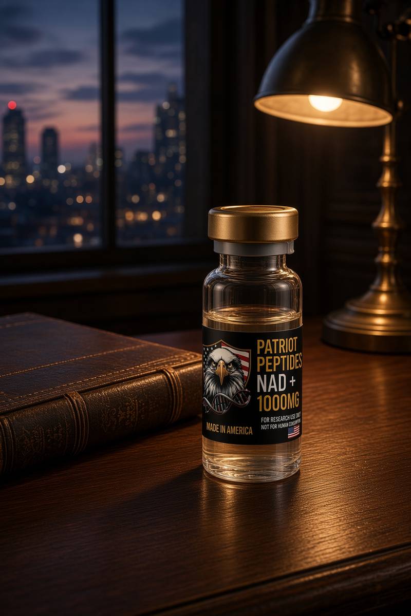 Research only
 Made in USA
 Made in USAOut of stock
Incretin
Retatrutide
US-made retatrutide 30mg — the published structure LY3437943, HPLC-MS verified.
4.6(609)
121 browsing this now · 5 purchased in the last 24 hours
30mg
£120.00
Research use only. Not a combined-use instruction.
Read next
65 min · long read · The living cell
The living cell is a city, and you are 36 trillion of them
A 70 kg adult is on the order of 36 trillion cells, most of them red blood cells with no nucleus. A typical nucleated cell holds about ten billion proteins and two metres of DNA. The body recycles 40–60 kg of ATP a day. Those figures are published; this essay is what they mean.
49 min · long read · Peptide research
NAD+: the currency of cellular time
NAD+ is nicotinamide adenine dinucleotide, the hydride coin Complex I wants oxidised and the stoichiometric substrate sirtuins, PARP1 and CD38 spend. Salvage through NAMPT is the kinetic bottleneck; the pool falls with age. Restoration in mice moves muscle, endothelium and stem-cell assays; human NR and NMN trials more often move the metabolome than a hard clinical endpoint. This essay is that topology, the named papers, and why a 1000 mg cake of lyophilised β-NAD+ is a laboratory reagent rather than an infusion.
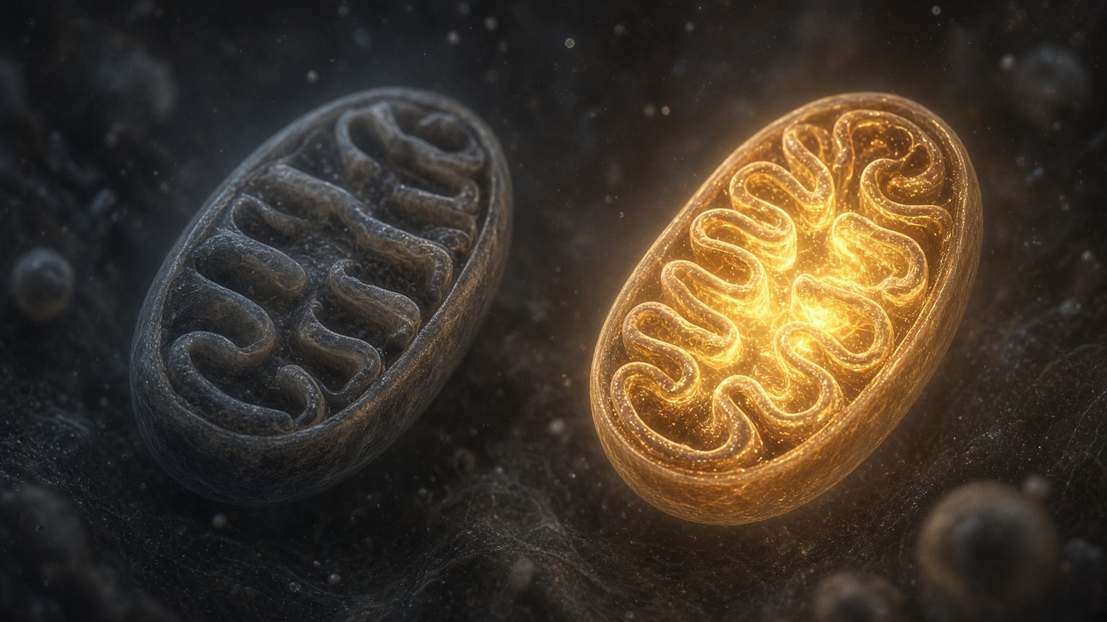
52 min · long read · Peptide research
Fatigue, cellular energy and the NAD+ / MOTS-c neighbourhood
NAD+ is the rechargeable chip every cell spends on fuel and DNA repair — and the pool shrinks with age. MOTS-c is a mitochondrial 16-mer sent out under metabolic stress. Two answers to 'I have no energy' that are not coffee.
50 min · long read · Peptide research
Sirtuins, caloric restriction and the NAD+ budget
Yeast SIR2, worm daf-16, mouse SIRT1 — a conserved story about spending NAD+ to deacylate the proteins that decide whether a cell is in famine or feast. The cofactor is the budget. We stock it at 1000mg.
82 min · long read · The living cell
Pathophysiology from the genome to a person who notices
Disease is a stack: genome, transcriptome, proteome, metabolome, organelle, cell fate, tissue, organism. A peptide binds one node. The rest of the stack keeps running. CFTR, type 2 diabetes and a tendon as worked examples.
More in this desk
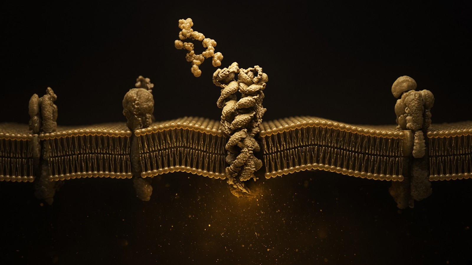
70 min · long read · The living cell
How peptides talk to cells: occupancy, amplification, arrestin
A peptide is a ligand. Most of the catalogue binds a GPCR on the cell surface: one occupancy, then enzymes make thousands of second messengers. That amplification is real, and it is not magic. Desensitisation is why more ligand is not more signal forever.
64 min · long read · The living cell
From gene to protein: how a cell actually reads itself
DNA is transcribed, spliced, exported, translated and folded. A research peptide is the ligand at the end of that pipeline, already made. This essay is every machine in between — polymerases, spliceosome, ribosome — with the actual rates attached.
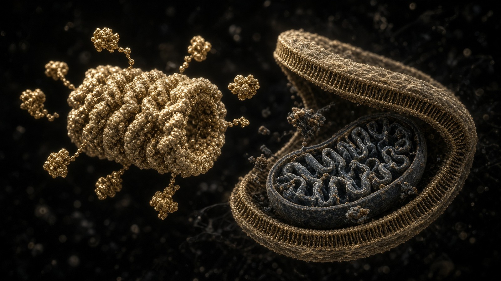
57 min · long read · The living cell
Proteostasis: the cell that eats its own mistakes
Ten billion proteins, a 76-residue tag, a 2.5-megadalton proteasome, and autophagy for whole organelles. Hershko, Ciechanover and Rose, 2004. Ohsumi, 2016. DSIP is a sleep-isolation nonapeptide, not an autophagy ligand.
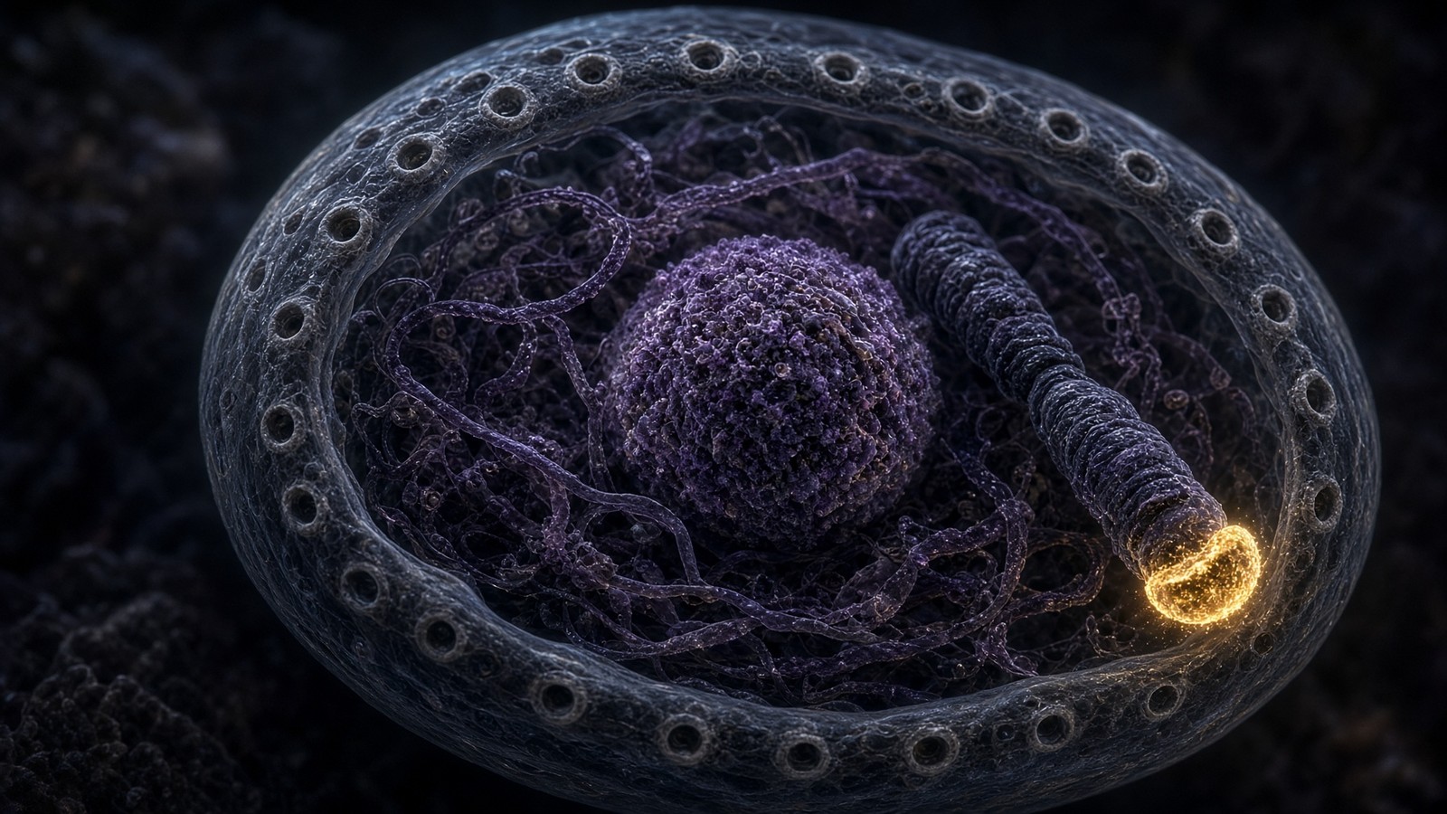
54 min · long read · The living cell
The nucleus, telomeres, and the four residues of Epithalon
Two metres of DNA folded into a nucleus a few micrometres across. TERT is off in most somatic cells on purpose. Epithalon is four residues, Ala-Glu-Asp-Gly, with a TERT and pineal literature. The machines are real. A large Western trial of telomere length in adults is not.
Essays describe published research. They are not medical advice and they do not authorise human use of any catalogue item.