

The living cell · 82 min · 18,092 words
Pathophysiology from the genome to a person who notices
Disease is a stack: genome, transcriptome, proteome, metabolome, organelle, cell fate, tissue, organism. A peptide binds one node. The rest of the stack keeps running. CFTR, type 2 diabetes and a tendon as worked examples.
What this essay actually tells you
- Pathophysiology is a stack: genome → transcriptome → proteome → metabolome → organelle → cell fate → tissue → organism. Most human disease is regulation, not a spelling error in DNA.
- A research peptide occupies one node — a GPCR, a cofactor pocket, an actin motif, a copper ligand, occasionally a claimed promoter. The rest of the stack keeps running. That is why ‘what does it do?’ is a bad question.
- Worked examples in the essay: CFTR ΔF508 (folding/trafficking), type 2 diabetes (ectopic fat, incretins, DiRECT), a tendon (BPC-157 vs TB-500 as different nodes). Named floors, named papers, no protocol.
What this actually means
A person notices a glucose curve, a tendon that will not settle, a 3 a.m. wake-up, a waist. Under that is a tissue. Under the tissue is a cell deciding whether to divide, senesce or die. Under the cell is an organelle leaking ROS or failing to make ATP. Under that is a metabolome (NAD+, acetyl-CoA, AMP). Under that is a proteome being phosphorylated. Under that is a transcriptome. Under that is a genome that may have a variant, a short telomere, or nothing wrong at all — because most pathophysiology is regulation, not spelling. Research peptides plug in at specific floors: a GPCR, a cofactor, a cytoskeletal motif, a copper-binding tripeptide. They do not replace the building.
Diagram
01 Genome
Variant, CNV, methylation, telomere length
The script. Most of it never becomes a phenotype you can bill for.
02 Transcriptome
Which genes are on, splice isoforms, noncoding RNA
The script being read this hour. A cell type is a transcriptome.
03 Proteome
Abundance, PTMs, localisation, complexes
The machines. Phosphorylation can flip a pathway without new DNA.
04 Metabolome
NAD+/NADH, ATP/AMP, acetyl-CoA, ROS
The fuel gauges. They feed back onto the genome through sirtuins and chromatin.
05 Organelle
Mitochondria, ER stress, lysosome, nucleus
Compartments fail as units. A tired mitochondrion is a tired cell.
06 Cell fate
Proliferation, senescence, apoptosis, identity
Hayflick, SASP, p53. The cell decides whether to keep being a citizen.
07 Tissue
Inflammation, fibrosis, barrier, innervation
Where a person actually hurts. Collagen, endothelium, synapses.
08 Organism
Glucose curve, VO₂, sleep, fertility, lifespan
The readout. Everything above is allowed to be invisible until it isn’t.
Pathophysiology is this stack, not a single molecule. A research peptide occupies one node — a receptor, a cofactor, a cytoskeletal motif — and the rest of the stack is still running. That is why ‘what does it do?’ is a bad question and ‘where does it bind?’ is a good one.
The word pathophysiology gets used as incense. We mean it as a stack. Something is wrong at one floor and the floors above start telling a story a mammal can feel. Cystic fibrosis is a CFTR spelling error that becomes a chloride-transport error that becomes a mucus error that becomes an infection error. Type 2 diabetes is often not a spelling error at all: it is ectopic fat in liver and pancreas, a first-phase insulin failure, a glucose curve. Same word — disease — wildly different floors. If you cannot name the floor you are on, you are not doing pathophysiology yet. You are still at the caption. The useful habit is to point: genome, transcriptome, proteome, metabolome, organelle, cell fate, tissue, organism. A peptide binds one node on one of those floors. The rest of the stack keeps running. That is not a disappointment. It is how biology is built, and it is why a 15-mer occupying a pocket is obviously not the building. Once you can see the floors, the vial stays the right size.
In short. Disease is a stack: cystic fibrosis starts as a CFTR spelling error, while type 2 diabetes is often ectopic fat and a failed first-phase insulin pulse.
Here is the long version of that sentence. Eight floors, a named disease or a named peptide occupancy on each, then three worked examples that refuse to skip steps, then a map of the catalogue as occupancies rather than moods. The point is not to make a research vial look like a therapy. The point is the opposite: once you can see the building, a 15-mer occupying a pocket is obviously not the building. That is the intellectually honest position, and it is also the legal one. Research use only, all the way down. Read the stack top-down when someone hands you a genome: what does this variant do to a message, a protein, a cell, a lung? Read it bottom-up when someone hands you a symptom: which tissue, which cell fate, which organelle, which metabolite, which phosphorylation, which transcript, and only then — maybe — which letter. Most of the time you will stop before the letter. That is not a failure of reductionism. That is the data. Common disease is polygenic and environmental and regulatory.
In short. Eight floors, named occupancies, three worked examples. The point is that a 15-mer occupying a pocket is not the building, and that is the legal position too.
- Genome
- 3.1 Gbp haploid
- Transcriptome
- ~10⁴ mRNAs / cell
- Proteome
- ~10¹⁰ proteins / cell
- Metabolome
- ATP, NAD+, SAM, ROS
- Organism
- 36 trillion cells
SNV, CNV, aneuploidy, CAG repeats, CpG methylation, telomere length. Most of it is not a gene.
A cell type is this list, plus isoforms, plus the RNAs that never make protein.
Abundance, phosphorylation, address, complex. Where almost every catalogue ligand actually sits.
The gauges that write back onto chromatin without changing a letter.
Glucose, sleep, waist, pain. The only floor a person is qualified to notice without a microscope.
Read the stack top-down when someone hands you a genome: what does this variant do to a message, a protein, a cell, a lung? Read it bottom-up when someone hands you a symptom: which tissue, which cell fate, which organelle, which metabolite, which phosphorylation, which transcript, and only then — maybe — which letter. Most of the time you will stop before the letter. That is not a failure of reductionism. That is the data. Common disease is polygenic and environmental and regulatory. Monogenic disease is the exception that taught us the wiring, and even then the interesting part is often what the misspelled protein does next — fold, traffic, get stuck, get chewed — not the codon itself. CFTR ΔF508 is the folding footnote we will walk in public: a three-base deletion, a protein one residue short, a channel that never arrives. Type 2 diabetes is the common case, and it usually stops several floors above the letter. Both are pathophysiology. They are not the same climb, and naming the floor is how you tell them apart.
In short. Read genome-first when you have a variant; symptom-first when you have a complaint. Most common disease stops before a letter, because it is polygenic and regulatory.
- Genome — letters, copies, repeats, methylation, telomeres. Occupancy that pretends to live here: Epithalon / TERT literature. Actual editors: CRISPR, base editors, Casgevy.
- Transcriptome — the genome being read this hour. Occupancy: GHK-Cu Pickart arrays in fibroblasts.
- Proteome — machines, tags, addresses. Occupancy: almost the entire catalogue. TB-500 at G-actin as the honest cytoskeletal example.
- Metabolome — ATP/AMP, NAD+/NADH, acetyl-CoA, SAM, ROS. Occupancy: the NAD+ vial, a cofactor, not a stimulant.
- Organelle — mitochondria, ER, lysosome, peroxisome. Occupancy: MOTS-c, a 16-mer the mitochondrion translated from 12S rRNA.
- Cell fate — divide, senesce, die, stay. Hayflick. Campisi SASP. A repair adjective that cannot name a fate is still only a description.
- Tissue — ECM, endothelium, barrier, nerve, infiltrate. Occupancy: BPC-157 in a VEGFR2/FAK/eNOS neighbourhood; KPV at NF-κB / PepT1.
- Organism — glucose curve, VO2, sleep, fertility, waist, pain. Occupancy: retatrutide as a Phase 2 medicine paper, not a research-vial instruction.
Floor 1 — Genome: spelling, copy number, and the silent majority
A single-nucleotide variant can be nothing (most of them), a splice-site disaster, or a Glu6Val in β-globin that sickles a red cell. Copy-number variants delete or duplicate genes. Aneuploidy is a chromosome too many or too few. Telomeres are the disposable DNA at the ends; when they shorten enough, DNA-damage checkpoints fire and the cell senesces or dies. Mitochondrial DNA is a second genome with a higher mutation rate, 37 genes, maternal inheritance, heteroplasmy. Most common disease is polygenic — thousands of variants of tiny effect, plus environment. Monogenic disease is the exception that taught us the stack, not the template for a peptide catalogue. A genome can fail as a letter, a copy-number, a chromosome, a telomere or a second mitochondrial script. CRISPR, base editors and prime editors write this floor. Casgevy is licensed. Nothing in the catalogue is that. If a peptide paper claims a transcriptional change, that is the transcriptome noticing a proteome event, or a claim about a promoter. Keep the floor labels honest and the argument gets possible.
In short. A genome can fail as a letter, a copy-number, a chromosome, a telomere or a second mitochondrial script. Most common disease is still polygenic, not a single typo.
Single-nucleotide variants: most of them are scenery
A diploid human genome is about six billion bases. Each newborn carries on the order of fifty to a hundred de novo single-nucleotide variants relative to the parents, and tens of thousands of standing differences from the reference. The overwhelming majority sit in non-coding sequence and do nothing anyone can measure. A minority change an amino acid and still do nothing, because proteins tolerate substitutions at surfaces that are not active sites, not interfaces, not splice signals. The ones that matter do one of a short list of rude things: they introduce a stop, they wreck a splice donor or acceptor, they change a residue the fold or the catalytic pocket cannot forgive, or they nudge a regulatory motif that a transcription factor used to find. Pathophysiology at this floor is the art of telling those four apart from the scenery. GnomAD exists so we stop calling every rare missense a villain. A newborn carries tens of thousands of differences from the reference. Most of them are scenery. The interesting ones are the rude four, and even those still have to climb the rest of the stack before a person notices.
In short. A newborn carries tens of thousands of differences from the reference. GnomAD exists so we stop calling every rare missense a villain.
Sickle cell is the teaching case because it is rude in public. Glu6Val in β-globin (HBB) creates a sticky patch. Deoxygenated haemoglobin polymerises. The red cell sickles. The phenotype is a polymer, then a shape, then a logjam in a capillary, then pain, infarct, infection. One letter, a cascade that never needed a committee. BRCA1 and BRCA2 are ruder in a quieter way: they are tumour suppressors in homologous recombination. Lose both copies in a cell and double-strand breaks are repaired sloppily. The phenotype is genomic instability, then a breast or ovarian epithelium that has rolled the dice too often. Still a spelling-adjacent story — truncated or unstable protein, failed repair complex — but the organism-level disease is probabilistic, age-dependent, and tissue-restricted. Same floor, different politics. One letter can be a polymer in a red cell or a dice-roll in an epithelium. Pathophysiology is naming which, and then watching the floors above tell the story a person can feel. That is already the stack, and we have not yet left the genome.
In short. Sickle cell is one letter that polymerises haemoglobin. BRCA loss is sloppy DNA repair, then a tissue that has rolled the dice too often.
Copy number and aneuploidy: too much, too little, a chromosome as a unit
A copy-number variant is a deletion or duplication from a few kilobases to a few megabases. Dosage is the disease. Too little of a haploinsufficient gene, too much of a gene that was supposed to be titrated, a fusion at a breakpoint that creates a chimera the cell never asked for. Aneuploidy is that idea at chromosome scale. Trisomy 21 is a gene-dosage storm, not a single villain locus, which is why the phenotype is a syndrome rather than a neat enzyme defect. Cancer genomes are aneuploidy as a treatment plan: whole-chromosome gains, arm-level losses, extrachromosomal circular DNA amplifying an oncogene until the copy number looks like a printing error. The peptide catalogue does not live here. Nothing we lyophilise inserts, deletes, or equalises a chromosome. If a caption implies otherwise, it has confused a ligand with a genome editor, and the floor labels are how you catch that. Dosage can be the disease: too little of a gene, too much of a chromosome, a cancer that prints oncogenes as circular DNA. Occupancy of a pocket does not equalise that.
In short. Dosage can be the disease: too little of a gene, too much of a chromosome, a cancer that prints oncogenes as circular DNA. Nothing we lyophilise equalises that.
Repeat expansion: Huntington as a length, not a typo
Huntington disease is a CAG trinucleotide repeat in HTT that encodes a polyglutamine tract. Below a threshold (classically 26 or fewer) the allele is fine. In a grey zone it may or may not. At 40 and above it is fully penetrant, with longer repeats bringing earlier onset — anticipation, especially through the paternal line, because spermatogenesis is sloppy with the slip. The protein is made. The polyQ stretch misfolds, aggregates, and the striatum notices first. This is still the genome floor, but the error is a length, not a letter, and the pathology is a proteostasis and circuit failure several floors up. Fragile X, myotonic dystrophy, some spinocerebellar ataxias: the same grammar of unstable repeats. A research peptide does not shorten a CAG tract. A caption that offers Huntington support as a sequence on a till has left the stack and entered a different industry. Huntington is a CAG length, not a typo: past forty repeats the polyglutamine misfolds and the striatum notices first. The interesting floors are 3 and 6 and 8. The letter was only the length.
In short. Huntington is a CAG length, not a typo: past forty repeats the polyglutamine misfolds and the striatum notices first. A research peptide does not shorten that tract.
Methylation: the mute button you can inherit for a while
Cytosine methylation at CpG, laid down by DNA methyltransferases and read by proteins such as MeCP2, is the longer-term mute on a promoter. Imprinting uses it so that some genes remember which parent they came from. CpG island hypermethylation can silence a tumour suppressor without deleting it — a regulatory lesion that looks, from the transcriptome down, like a loss-of-function mutation. Hypomethylation can wake a retrotransposon. Ageing methylomes drift in ways that clocks can read; whether the clock is a cause or a diary is still an argument, and we are not going to settle it in a caption. The relevant point for pathophysiology is simpler: a genome can be spelled correctly and still be unread, or read in the wrong cell, at the wrong time, at the wrong dose. That is already floor 1 leaking into floor 2 without a single SNV you would report on a diagnostic panel. A correctly spelled genome can still be unread. CpG methylation mutes promoters; imprinting and tumour-suppressor silencing are this floor leaking into RNA.
In short. A correctly spelled genome can still be unread. CpG methylation mutes promoters; imprinting and tumour-suppressor silencing are this floor leaking into RNA without a reportable SNV.
Diagram
- DNA replication + MMR10⁻⁹ to 10⁻¹⁰A genome of 6 Gbp (diploid) accumulates a handful of mutations per division.
- Transcription~10⁻⁵RNA is disposable. The cell can afford a wrong letter in a message that lasts hours.
- Translation~10⁻⁴One wrong amino acid per ten thousand. Proteins turn over. DNA does not.
- mtDNA10–100× nuclearNo histones, ROS next door, weaker repair. The second genome ages faster.
The genome is sacred, the message is cheap, the protein is cheaper. Ageing is partly what happens when the sacred copy still drifts — and when mitochondria, which never got the nuclear repair budget, drift faster.
Telomere length: the disposable end, the Hayflick limit, a Nobel
Linear chromosomes have an end-replication problem. DNA polymerase needs a primer; the lagging strand would shrink; a naked end looks like a double-strand break. The solution is telomeres — TTAGGG repeats in humans, bound by shelterin, tucked into a t-loop — and, in cells that are allowed to have it, telomerase: TERT plus the TERC RNA. Leonard Hayflick and Paul Moorhead, Experimental Cell Research, 1961: normal human fibroblasts do not divide forever. They have a limit, later understood as telomere erosion plus a DNA-damage checkpoint. Elizabeth Blackburn, Carol Greider and Jack Szostak took the 2009 Nobel for the telomere/telomerase solution to the end-replication problem. Stem cells keep TERT on. Most somatic cells keep it off on purpose, because a cell that divides without a limit is also a cell that can become a tumour. The bargain is tumour suppression versus tissue renewal. Short telomeres fire ATM/ATR, p53, p21; the cell senesces or dies. That is floor 1 talking directly to floor 6, and it is one of the most elegant bargains in biology.
In short. Linear chromosomes would shrink without telomeres and telomerase. Hayflick showed fibroblasts stop; Blackburn, Greider and Szostak explained why, and most somatic cells keep TERT off on purpose.
Most pathophysiology is regulation, not spelling — and CFTR ΔF508 is the folding footnote
This is the sentence the rest of the essay leans on. Genome-wide association studies of common disease find hundreds of loci of tiny effect, mostly non-coding, mostly regulatory, mostly in enhancers that a given cell type bothers to use. The letters are not wrong so much as the dimmer is in a slightly different position, in a specific tissue, in a specific decade, given a specific diet and sleep and infection history. Type 2 diabetes, coronary disease, height, BMI: polygenic scores, environment, a stack. The monogenic exceptions (CFTR, HBB, HTT, BRCA in the families that carry them) are precious because they made the wiring diagram drawable. They are not the template for how a middle-aged glucose curve goes wrong. GWAS of common disease finds hundreds of tiny, mostly regulatory loci. Monogenic exceptions taught us the wiring. Most of the clinic is the dimmer, the tissue, the decade, and last night's sleep. That is not a lesser pathophysiology. It is the common one, and it is why occupying one receptor rarely fixes a person.
In short. GWAS of common disease finds hundreds of tiny, mostly regulatory loci. Monogenic exceptions taught us the wiring; they are not the template for a middle-aged glucose curve.
Even the famous monogenic case is not a morality play about spelling. CFTR ΔF508 — a deletion of phenylalanine at position 508 — is a three-base deletion that produces a protein one residue short. The channel, if it reached the apical membrane and was gated, would retain substantial function. It does not reach the membrane. The fold is unstable. ER quality control retains it. The proteasome eats it. The pathophysiology is trafficking, which is floor 3 and floor 5, of a protein whose gene was named in 1989. Riordan, Rommens, Kerem, Tsui: Science, 1989. We will walk that lesion from letter to lung as a worked example. The preview is the point: a spelled protein can still fail, and a peptide does not fix CF. Correctors and potentiators that chaperone or open the channel are a different literature, small-molecule pharmacology aimed at fold and gate. Gene therapies and RNA approaches are other desks. The lung is six floors up from Riordan's gene paper. Everything interesting happens on the stairs.
In short. CFTR ΔF508 is almost spelled correctly, then fails to fold, is retained in the ER, and is degraded. The lung is six floors up from Riordan's 1989 gene paper.
Occupancy at this floor: Epithalon is a claim, CRISPR is an editor
CRISPR, base editors and prime editors write this floor. Casgevy (exa-cel) rewrites BCL11A's erythroid enhancer so foetal haemoglobin comes back. That is genome-floor medicine, licensed. Nothing in the catalogue is that. If a peptide paper claims a transcriptional change (GHK-Cu's microarray literature; Epithalon's TERT literature), that is the transcriptome noticing a proteome event, or a claim about a promoter. Keep the floor labels honest and the argument gets possible. A four-residue peptide that claims TERT is a hypothesis you can blot. A four-residue peptide that claims to be CRISPR is a category mix-up. The Nobel is not confused about this. We stock characterised ligands because the sequences are published and the assays are nameable. We do not stock a rewrite of the Hayflick limit. Holding those two sentences in the head at once is the whole literacy of this floor: occupancy of a pocket, or of a claimed promoter, is not an edit of the archive. Different acts. Different law. Different failure modes.
In short. CRISPR writes this floor; Casgevy is licensed. A peptide microarray or a TERT claim is the transcriptome noticing a ligand, not a gene editor.
Epithalon is Ala-Glu-Asp-Gly, 390 daltons, four residues from the Khavinson school in St Petersburg. The papers claim TERT induction, telomerase activity in cell assays, pineal melatonin effects, rodent lifespan. That is a genome-adjacent occupancy: a tetrapeptide alleged to talk to a promoter and a clock. It is not a nucleoprotein complex that reverse-transcribes TTAGGG onto a chromosome end. It is not Cas9. It is not a licensed telomere therapy. We stock the tetrapeptide as a characterised research ligand because the sequence is published and the assays are nameable. We do not stock a rewrite of the Hayflick limit. Those two sentences can live in the same head. Epithalon is four residues with a literature you can blot. CRISPR is an editor. The gap between them is the entire intellectual problem of writing a tetrapeptide next to the word telomere. The nucleus essay next door takes the papers at full length. This floor only needs the geometry: a promoter in a 3.1-billion-base search problem, and a polite refusal to confuse it with an edit.
In short. Epithalon is Ala-Glu-Asp-Gly, a tetrapeptide alleged to induce TERT. That is a published claim you can blot, not CRISPR and not a licensed telomere therapy.
A four-residue peptide that claims TERT is a hypothesis you can blot. A four-residue peptide that claims to be CRISPR is a category error. The Nobel is not confused about this. Vendors sometimes are.
Floor 2 — Transcriptome: the genome being read this hour
A cell type is a transcriptome. The DNA in a hepatocyte and a neuron is almost the same; the RNA is not. Bulk RNA-seq averages. Single-cell RNA-seq exploded the atlas: Tabula Sapiens, Human Cell Atlas, tens of millions of cells, hundreds of types, states that used to be called the same cell and are not. Splicing isoforms, RNA editing (ADAR), miRNA repression, lncRNA scaffolding, mRNA half-life (minutes to a day) — this floor is faster than the genome and slower than the metabolome. Inflammation is often a transcriptional programme (NF-κB, IRFs, STATs) rather than a new mutation. KPV's literature sits on NF-κB. That is this floor, reached from a tripeptide at a transporter and a receptor neighbourhood, not from a promoter. A cell type is which genes it is reading this hour, not its DNA. That is not fashion. It is resolution. Nineteenth-century anatomy named cells by shape and stain. We name them by the genes they transcribe, which is why a peptide paper without a gene list is often just a photograph of a well.
In short. A cell type is which genes it is reading this hour, not its DNA. Inflammation is often an NF-κB programme; KPV's literature sits there, reached from a tripeptide.
Diagram
A lyophilised research peptide skips every step after “protein”. It is the ligand already. That is the entire point of the catalogue, and the reason it is not a gene therapy.
Crick’s flow is still right. The numbers are the part textbooks skip: a mammalian polymerase is slow, splicing is a machine the size of a ribosome, and translation errors run about one in 10⁴ amino acids.
A cell type is a transcriptome, not a histology stain
Nineteenth-century anatomy named cells by shape and stain. The twenty-first century names them by the genes they are transcribing. That is not fashion. It is resolution. 'The fibroblast' is a family. 'The macrophage' is a cloud of states that textbooks still draw as M1 and M2 and that tissues refuse to honour. A senescent cell is a transcriptome with p16INK4a, a flattened morphology, and a SASP. A β-cell that has lost its identity under metabolic stress is a transcriptome that no longer bothers to say INS, MAFA, PDX1 at the right dose. Transdifferentiation, metaplasia, the slow identity leak of a chronically insulted epithelium: all of this is floor 2 before it is floor 6. If you want to know what a peptide did to a culture, the grown-up assay is often a gene list, not a photograph of the well with the word repair underneath. A cell type is a transcriptome, not a histology stain. The stain was a start. The gene list is how you stop averaging five cities into one name.
In short. Nineteenth-century anatomy named cells by stain. We name them by the genes they transcribe, which is why a peptide paper without a gene list is often just a photograph.
Alternative splicing: one gene, several proteins, several fates
About 95% of human protein-coding genes are alternatively spliced. Exon included or skipped, mutually exclusive exons, alternative 5' or 3' splice sites, intron retention: the spliceosome is not a tidy editor, it is the gene. Tropomyosin, DSCAM in the fly, the neurexins — identity can be an isoform. Disease can be an isoform too. A splice-site SNV that looks boring on a protein-coding annotation can drop an exon and wreck a channel. Antisense oligonucleotides that hide a splice site or force an exon to stay are genome-adjacent medicines that work on this floor; nusinersen in spinal muscular atrophy is the famous one. Nothing in the peptide catalogue is an ASO. If a paper reports that a peptide changed splicing, demand the RNA-seq, the event, and the cell type, then ask whether the peptide occupied a kinase that phosphorylates a splicing factor. That is a floor-3 story with a floor-2 readout, which is allowed, and is not magic. Most human genes are alternatively spliced, so a boring-looking splice-site variant can wreck a channel. The gene is the spliced product.
In short. Most human genes are alternatively spliced, so a boring-looking splice-site variant can wreck a channel. Nothing in the catalogue is an antisense oligonucleotide.
Diagram
Closed chromatin (H3K27me3, DNA methylation) hides the promoter. Pioneer factors and histone acetyltransferases open it.
PIC: TFIID, TFIIH, Mediator, Pol II. Ser5 phosphorylation of the CTD lets the polymerase leave the promoter.
Elongation ~20–40 nt/s. Capping, splicing, cleavage and polyadenylation happen on the still-growing RNA.
Human genes are islands in 3.1 billion base pairs of mostly noncoding sequence. Promoter, enhancers, chromatin state and the Mediator complex decide whether Pol II is allowed to fire. Epithalon’s literature sits on TERT and pineal clocks — two of the rare promoters anyone bothers to name in a peptide essay.
lncRNA and microRNA: modulators, not magic
MicroRNAs are ~22-nucleotide repressors that sit on the 3' UTR and take the edge off a message — cleavage or translational block, Argonaute, the RNA-induced silencing complex. They are rheostats, not on/off switches, which is why knocking one out so often does little until a stress arrives. Long non-coding RNAs are a census in search of a job description: some scaffold chromatin modifiers, some are transcriptional noise with a polyA, XIST is the genuine article and silences an X. The wellness industry has noticed both classes and started selling them as if a named ncRNA were a vitamin. It is not. A research peptide is also not an miRNA mimic. If a vendor's caption says a sequence regulates microRNAs without an assay, a cell type and a fold-change, they are using the floor as atmosphere. Modulators exist. Magic does not. MicroRNAs are short rheostats on a 3' UTR. A named ncRNA is not a vitamin, and a peptide is not an miRNA mimic until someone has shown the RISC, the UTR, and the fold-change.
In short. MicroRNAs are short rheostats on a 3' UTR, not on/off switches. A research peptide is not an miRNA mimic, and a named ncRNA is not a vitamin.
Occupancy: GHK-Cu, a 3-mer plus a metal, and Pickart's arrays
Glycyl-L-histidyl-L-lysine binds copper. Loren Pickart isolated the tripeptide from human plasma in the 1970s as a factor that appeared to modulate protein synthesis in liver tissue; the later literature is a long argument about copper delivery, integrin neighbourhoods, and — this is the floor-2 point — microarray studies in which GHK-Cu shifts large gene lists in fibroblasts toward matrix, anti-inflammatory and antioxidant programmes. That is transcriptome occupancy by a three-residue peptide plus a metal. It is not a transcription factor with a DNA-binding domain. It is not CRISPR. It is a small ligand whose downstream consequence, in those arrays, is that a fibroblast starts reading a different subset of its genome. Believe the arrays only as far as they were done: cell type, concentration in a dish, time point, the actual list. Do not promote a 3-mer to a gene therapist because a heatmap is colourful. GHK-Cu is a copper-binding tripeptide with a published fibroblast gene-expression fingerprint. The fingerprint is floor 2. The binding is chemistry. Matrix talk belongs to floor 7, where collagen lives.
In short. GHK-Cu is a copper-binding tripeptide whose fibroblast arrays shift matrix and antioxidant gene lists. That is chemistry with a transcriptome fingerprint, not a gene therapist.
Floor 3 — Proteome: machines, tags, addresses
A cell expresses ~10,000 protein species; the copy numbers run from tens to tens of millions. Phosphorylation (500+ human kinases) is the fast tag. Ubiquitin is the dispose-or-signal tag. Glycosylation is the extracellular identity tag. Localisation is destiny: a transcription factor that cannot enter the nucleus is not a transcription factor today. GPCRs, RTKs, channels, transporters, cytoskeletal polymers, ECM collagens — this is where almost every catalogue peptide actually lives. Occupancy. Affinity. Downstream phosphorylation. The assay is a blot, a cAMP kit, a scratch wound, a receptor internalisation image. It is not a mood. Ten billion protein machines in a hepatocyte, packed. A peptide ligand is a few nanometres of that city. Occupancy is a pocket, not a municipal renovation. Almost every catalogue peptide lives here: occupancy of a pocket, a phosphorylation, a blot. Ten thousand protein species, copy numbers from tens to tens of millions. Look at the exponents and you know what city you are in. Look at a peptide's target and you are usually looking at a rare machine.
In short. Almost every catalogue peptide lives here: occupancy of a pocket, a phosphorylation, a blot. Ten thousand protein species, copy numbers from tens to tens of millions.
Abundance: the cell is not a stoichiometric democracy
Copy number is destiny almost as often as sequence is. A transcription factor at a hundred molecules is a rumour. Tubulin at tens of millions of molecules is the furniture. Actin too. A GPCR may sit at a few thousand copies per cell and still run physiology because of amplification — one receptor, many G proteins, many cAMP, many PKA substrates — which is why occupancy of a scarce lock can move an organism. Proteomics (mass spectrometry, not a Western of your favourite band) is how you stop lying to yourself about what is actually present. Degradation is the other half of abundance: the proteasome, autophagy, a ubiquitin tag, a degron. CFTR ΔF508 is an abundance disease as much as a sequence disease: the protein is made and then destroyed. Increasing abundance of a misfolded channel without fixing the fold just feeds the proteasome. Correctors that help the fold are chaperone-adjacent pharmacology. Different desk from a research pentadecapeptide. Copy number is destiny almost as often as sequence. CFTR ΔF508 is an abundance disease: the protein is made, then the proteasome eats it.
In short. Copy number is destiny almost as often as sequence. CFTR ΔF508 is an abundance disease: the protein is made, then the proteasome eats it.
Post-translational modification: phosphorylation can flip a pathway with zero new DNA
More than 500 human kinases. A matching set of phosphatases. Serine, threonine, tyrosine. One phosphate is a two-charge change, a binding site for an SH2 or a 14-3-3, a conformational click, an activity that was off and is now on, or the reverse. Insulin does not rewrite the β-cell genome to lower glucose. Insulin occupies a receptor tyrosine kinase; the kinase autophosphorylates; IRS proteins; PI3K; Akt; GLUT4 vesicles in muscle and fat; glycogen synthase in liver. Zero new DNA. Minutes. That is floor 3 as physiology. Disease at this floor is a kinase that will not shut up (an oncogenic EGFR), a phosphatase that went missing, a receptor that desensitised, a substrate that cannot be phosphorylated because the residue was a variant. If your mental model of disease is 'a gene is broken', you will not see any of this, and you will not understand why a peptide ligand can be interesting without being a gene therapy. Ubiquitin, glycosylation and acetylation have their own politics, and they are how floor 4 writes on floor 3 without asking floor 1.
In short. Insulin lowers glucose without rewriting DNA: a receptor tyrosine kinase, a phosphate, GLUT4 vesicles in minutes. Floor 3 is why a ligand can matter without being gene therapy.
Localisation: the same protein in two postcodes is two proteins
A transcription factor in the cytosol is a paperweight. NF-κB held in the cytoplasm by IκB is off; degrade IκB and it walks through a nuclear pore and floor 2 notices. SREBP is a membrane protein in the ER until cholesterol is low; then it travels to the Golgi, is clipped, and the fragment becomes a transcription factor. Cytochrome c in the mitochondrial intermembrane space is a respiratory subunit; in the cytosol it is a death sentence. CFTR at the apical membrane is a chloride channel; CFTR in the ER is a quality-control substrate. MOTS-c, under metabolic stress, is reported to translocate to the nucleus (Kim et al., 2018) — an organelle peptide with a floor-2 ambition. Localisation is not a footnote. It is half of function. Any peptide paper that reports increased X without saying where X was is reporting a smear. The same protein in two postcodes is two proteins. NF-κB in the cytosol is off; cytochrome c in the cytosol is a death sentence. Address is destiny, and destiny is a Western with a fractionation, not a band on a whole-cell lysate.
In short. The same protein in two postcodes is two proteins. NF-κB in the cytosol is off; cytochrome c in the cytosol is a death sentence.
Complexes: nobody works alone, including the ligands
Ribosome, proteasome, spliceosome, pyruvate dehydrogenase, the respiratory chain, mTORC1, shelterin, the apoptosome: function is often a committee. Occupancy of one subunit can rewire the committee (rapamycin and FKBP12 on mTORC1 is the famous small-molecule version). A GPCR is a complex waiting to happen: receptor, G protein, then, if the ligand is that way inclined, GRK, β-arrestin, clathrin. Retatrutide occupies three receptors that each recruit their own committees. Ipamorelin occupies one (GHSR), chosen by Raun et al. in 1998 for GH release with little ACTH and prolactin — the older GHRPs dragged the HPA axis because their occupancy was less polite. Selectivity is a proteome fact. It is not branding. Function is often a committee: a GPCR, a G protein, then arrestin. Ipamorelin occupies GHSR with GH release and little ACTH; that is design, and the paper is occupancy plus selectivity, not a protocol for a research vial. Name the committee, and the ligand becomes a question you can actually ask of a dish.
In short. Function is often a committee: a GPCR, a G protein, then arrestin. Ipamorelin occupies GHSR with GH release and little ACTH; selectivity is design, not branding.
Occupancy: TB-500 / thymosin β4 as a G-actin buffer
Thymosin β4 is a 43-residue G-actin-sequestering protein. The actin-binding motif is LKKTETQ. TB-500 in the research trade is that idea as a fragment or as the parent sequence, depending on the supplier's honesty; our job is the characterised sequence on the page, not a nickname. The mechanism, when it is a mechanism, is cytoskeletal: buffering the pool of monomeric actin, changing what a cell can do with its shape, its motility, its leading edge. That is a proteome/cytoskeleton node. It is not a GPCR. It is not a growth factor. It is not repair juice. A motile cell in a wound is a tissue story only if the cells that crawl are the right ones — tenocytes, endothelial cells, fibroblasts, not a random leukocyte having a day. Floor 3 occupancy, floor 7 ambition. The animal literature has to climb those stairs in public. We will make it do that in the tendon example. Until then: actin buffer, not a mood. Name the motif, name the assay, and a motility claim becomes a number rather than a hope.
In short. Thymosin β4 buffers G-actin; the motif is LKKTETQ. That is a cytoskeletal occupancy, not a GPCR and not repair juice, until the right cells crawl in a tissue.
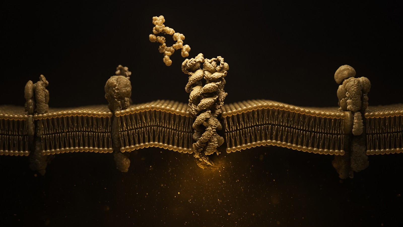
Floor 4 — Metabolome: the gauges that write back onto DNA
ATP/AMP (AMPK). NAD+/NADH (sirtuins, dehydrogenases). Acetyl-CoA (HATs, fatty-acid synthesis). SAM (methyltransferases). α-ketoglutarate (TET enzymes, histone demethylases). ROS (redox-sensitive cysteines, HIF in hypoxia). This floor is the reason a diet, a fast, a night of no sleep, or a failing mitochondrion can change transcription without a mutation. It is also why NAD+ is not anti-ageing as a caption and is a cofactor as a fact. MOTS-c's AMPK/folate–methionine literature is this floor talking to floor 1's one-carbon chemistry. Retatrutide's organism-level weight change is this floor plus endocrine GPCRs plus adipose tissue. Skip floors and the number becomes a superstition. ATP, NAD+, acetyl-CoA, SAM and ROS write on chromatin without changing a letter. A diet, a fast or a failing mitochondrion can rewrite transcription from this floor. That is pathophysiology as regulation, and it is why last night's sleep can wreck a glucose curve without a new SNV. The gauges moved. The genome did not.
In short. ATP, NAD+, acetyl-CoA, SAM and ROS write on chromatin without changing a letter. A diet, a fast or a failing mitochondrion can rewrite transcription from this floor.
ATP/AMP: AMPK as the budget committee
The cell does not store weeks of ATP. The standing pool in a human is on the order of 50 grams; daily turnover is 40–60 kilograms (Rich, 2003). Recycled, not warehoused. The ratio ATP/AMP is therefore a real-time budget, and AMPK (AMP-activated protein kinase) is the committee that reads it. High AMP: AMPK on; more catabolism, more fuel uptake, less of the expensive anabolic work, mitochondrial biogenesis via PGC-1α neighbourhoods, autophagy nudged. Low AMP, energy plentiful: the opposite. Metformin talks to this neighbourhood, which is why a diabetes medicine and an exercise physiology lecture share a kinase. MOTS-c's reported AMPK activation is a mitochondrial peptide arriving at this gauge. That is interesting if the assays hold. It is not an exercise mimetic as a product claim, and the catalogue will not say it is. The gauge is real. A glucose curve is, among other things, an ATP story in muscle, liver and β-cell. First-phase insulin release is a calcium-and-granule story that requires the β-cell to have enough ATP from glucose oxidation to close KATP channels and depolarise.
In short. The standing ATP pool is about 50 grams; daily turnover is tens of kilograms. AMPK reads the AMP ratio, and MOTS-c is reported to arrive at that gauge.
NAD+/NADH: the hydride coin, the sirtuins, the PARPs, CD38
NAD+ accepts a hydride and becomes NADH. That is the redox job: Complex I wants NADH; dehydrogenases want NAD+; the cell has to keep both pools in a ratio that matches the compartment. The signalling job is ruder. Sirtuins deacetylate using NAD+ as co-substrate, so a redox coin is a chromatin coin and a metabolic-enzyme coin. PARP1 spends NAD+ in a hurry when DNA is damaged, writing poly(ADP-ribose) chains; a large enough nick and the NAD+ pool collapses and the cell dies of bankruptcy rather than of the nick. CD38 is an NADase that rises with inflammation and age in some tissues, which is one of the proposed reasons the pool looks smaller in older animals. None of this makes a lyophilised NAD+ vial a stimulant, a nootropic, or a years-added product. It makes NAD+ a cofactor whose accounting is serious. The 1000 mg in the catalogue is β-NAD+, US-made, for the bench. eLIVEate's appointment is a different product on a different till. Same carbon skeleton. Different law. A metabolite can change transcription and organelle function and cell fate without a single genomic letter moving.
In short. NAD+ is a hydride coin and a signalling co-substrate for sirtuins and PARPs. The vial is β-NAD+ for the bench, not a stimulant and not years added.
Acetyl-CoA, SAM, ROS: the other pens the metabolome writes with
Acetyl-CoA is the acetyl donor for histone acetyltransferases and the substrate for fatty-acid synthesis and for ketones, depending on compartment and hunger. When the mitochondrion is reducing everything in sight, acetyl-CoA export to the cytosol changes, and chromatin notices. S-adenosylmethionine (SAM) is the methyl donor for DNA and histone methyltransferases; one-carbon metabolism (folate, methionine, B12) is therefore a chromatin input. MOTS-c's reported interaction with the folate–methionine cycle is this sentence. Reactive oxygen species are not a moral failing. They are a leak from the respiratory chain and from NADPH oxidases, a signal at low dose (HIF, redox-sensitive cysteines, insulin sensitivity in the short burst after exercise) and a damage programme at high dose (lipid peroxidation, mtDNA nicks, the inflammasome). A tired cell is often a cell whose ROS and NAD+ and AMP are telling a consistent story, which is an organelle story wearing a metabolite mask. Metabolites write on DNA without a mutation. That is the whole of this floor's gift, and its warning.
In short. Acetyl-CoA acetylates histones; SAM donates methyls; ROS are a leak that is a signal at low dose and damage at high. Metabolites write on DNA without a mutation.
Floor 5 — Organelle: when a compartment fails as a unit
Mitochondria: Δψm collapse, mtDNA damage, cytochrome c release (apoptosis), ROS leak, integrated stress response via DELE1–HRI–ATF4. ER: unfolded protein response (IRE1, PERK, ATF6), cholesterol sensing (SREBP), calcium store. Lysosome: pH 4.5–5, mTORC1's amino-acid radar, autophagy completion, storage diseases when one hydrolase is missing. Nucleus: laminaopathies, pore crowding, telomere damage foci. Peroxisome: very-long-chain fatty acids, plasmalogens. A tired cell is often a tired organelle. The peptide literature that actually names an organelle (NAD+ pool, MOTS-c, mitochondrial 12S, GHK-Cu and mitochondrial genes in some arrays) should be made to name the assay. A tired cell is often a tired compartment: mitochondrial membrane potential, ER unfolding, a lysosome that will not acidify. Name the organelle and the assay. When this compartment fails, floors 4, 6 and 8 notice. Cytochrome c in the cytosol is a death sentence. NAD+ depletion is a budget crisis. Compartments exist because chemistry is not one room.
In short. A tired cell is often a tired compartment: mitochondrial membrane potential, ER unfolding, a lysosome that will not acidify. Name the organelle and the assay.
Diagram
Matrix
- TCA cycle · β-oxidation · mtDNA nucleoids
- NADH produced here. Complex I spends it.
- MOTS-c (MRWQEMGYIFYPRKLR) from 12S rRNA.
Inner membrane
- I → II → III → IV → V (ATP synthase)
- ~150 mV proton-motive force
- ~40–60 kg of ATP turned over per human day
mtDNA is 16,569 bp, 37 genes, 13 proteins of the respiratory chain. Nuclear DNA encodes the other ~1,200 mitochondrial proteins. NAD+ is the hydride carrier between dehydrogenases and Complex I. MOTS-c is a 16-mer translated from 12S rRNA — a peptide the mitochondrion wrote itself.
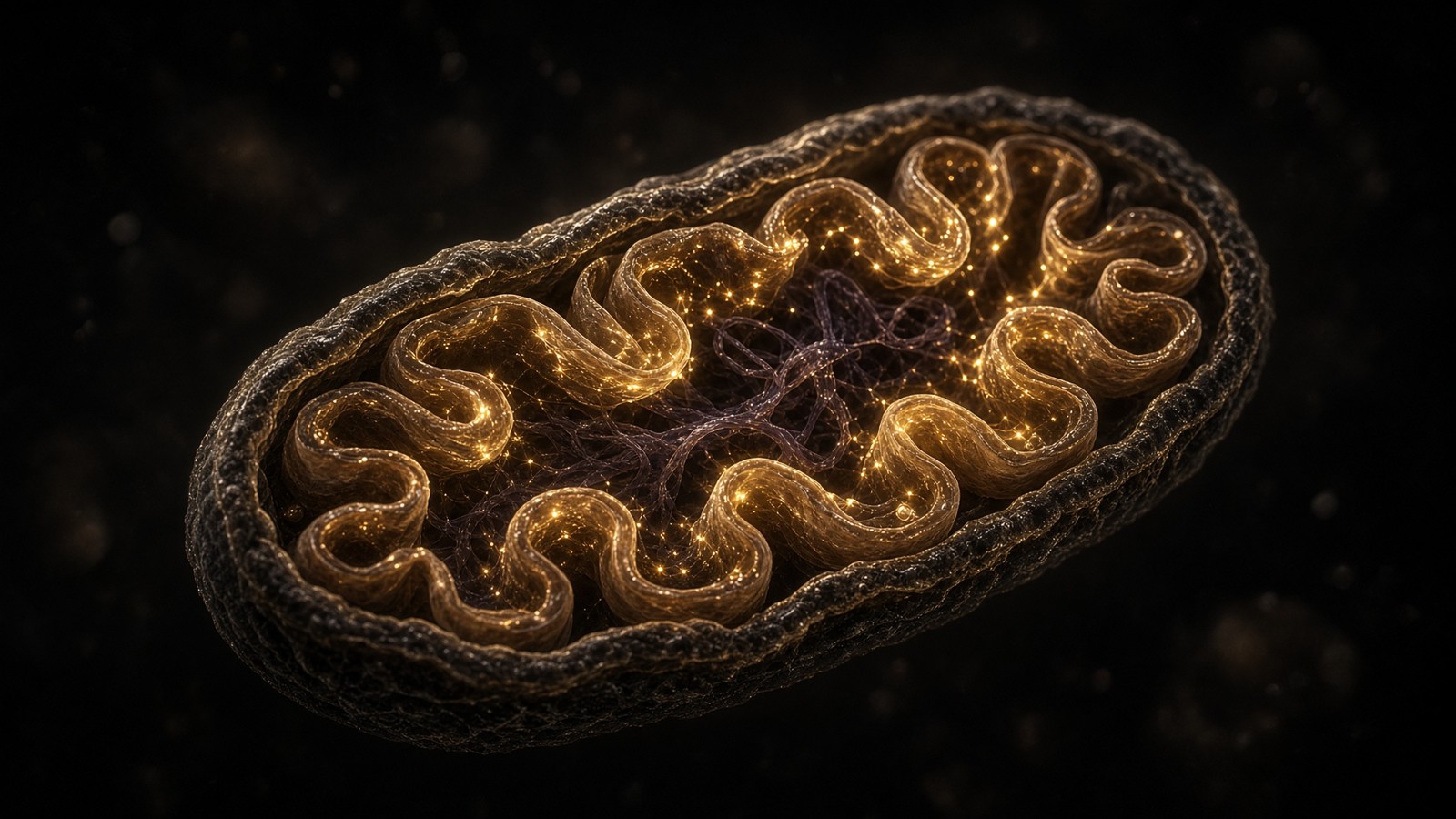
Mitochondria: Δψm, the second genome, the leak
A mitochondrion is a former bacterium with two membranes, a 16,569-base circular genome, 13 respiratory subunits it still encodes itself, and about 1,200 other proteins the nucleus makes and imports. The inner membrane holds a proton-motive force of roughly 150 millivolts. Electrons from NADH and succinate run through Complexes I–IV to oxygen. Protons are pumped. ATP synthase lets them back in. The currency is ATP. The waste, when the chain is reduced and oxygen is waiting, includes superoxide. mtDNA sits next to that leak, has no histones, and repairs less well than the nucleus, which is why the error-rate diagram was rude about it. Heteroplasmy — mixed mutant and wild-type mtDNA in one cell — is the inheritance plot: a threshold of mutant load, then a tissue that cares (brain, heart, muscle, β-cell) starts to fail. MELAS, MERRF, Leber hereditary optic neuropathy: named diseases of this floor. Common ageing of this floor is quieter and is part of why a 70-year-old mitochondrion is not a 20-year-old mitochondrion.
In short. Mitochondria keep 13 respiratory genes, a 150-millivolt inner membrane, and DNA sitting next to a superoxide leak. MELAS, MERRF and LHON are named failures of this floor.
The unfolded protein response: when the ER is full of regret
Secreted and membrane proteins are born into the endoplasmic reticulum. Folding is assisted (calnexin, calreticulin, PDI, BiP). Failure is recognised. The unfolded protein response has three sensors: IRE1 (which splices XBP1 mRNA — a floor-2 event triggered by a floor-5 problem), PERK (which phosphorylates eIF2α and slows translation), and ATF6 (which leaves for the Golgi and becomes a transcription factor). The aim is to catch up: more chaperones, less incoming peptide, ERAD of the hopeless through the proteasome. CFTR ΔF508 is a famous ERAD client. Too much UPR for too long is a cell-fate decision: ATF4, CHOP, apoptosis. Viral factories, antibody-secreting plasma cells, hepatocytes drowning in lipid and nascent protein — different insults, same organelle alarm. A peptide that reduces ER stress in a caption has not yet told you which sensor, which cell, which assay. Make it tell you. Secreted proteins fold in the ER; failure recruits IRE1, PERK and ATF6. CFTR ΔF508 is a famous ERAD client, which is competent quality control applied to a slightly incompetent fold. The cell is not broken. The protein is slightly wrong, and the ER is unforgiving.
In short. Secreted proteins fold in the ER; failure recruits IRE1, PERK and ATF6. CFTR ΔF508 is a famous ERAD client, which is competent quality control applied to a slightly incompetent fold.
Lysosomes and peroxisomes: pH, storage, very-long-chain fats
The lysosome is an acid vat (pH 4.5–5) with hydrolases that only work at that pH, which is why a leak is less catastrophic than it sounds and why a failure to acidify is a failure to digest. mTORC1 sits on the lysosomal surface and reads amino acids; autophagy completes here; storage diseases (Gaucher, Tay–Sachs, Pompe) are single hydrolases missing and a substrate that has nowhere to go. TFEB is the transcription factor that expands the system when it is allowed into the nucleus. Peroxisomes handle very-long-chain fatty acids and plasmalogen synthesis; their failures (Zellweger spectrum, X-linked adrenoleukodystrophy) are named diseases of this compartment. They are on the map because pathophysiology that cannot name a peroxisome will misread a set of metabolic presentations as mitochondrial out of habit. Compartments are not interchangeable. That is why they exist. The lysosome is an acid vat where autophagy finishes and storage diseases begin. Peroxisomes handle very-long-chain fats. Calling every metabolic failure mitochondrial is a habit, not a diagnosis, and the habit is how a rare disease gets a wrong room.
In short. The lysosome is an acid vat where autophagy finishes and storage diseases begin. Peroxisomes handle very-long-chain fats; calling every metabolic failure mitochondrial is a habit, not a diagnosis.
Occupancy: MOTS-c, a 16-mer the mitochondrion translated from 12S rRNA
Lee, Kim, Cohen and colleagues, Cell Metabolism, 2015: MOTS-c, sequence MRWQEMGYIFYPRKLR, translated from mitochondrial 12S rRNA in a reading frame of an RNA that was supposed to be a ribosome, not a message. Reported to act in the folate–methionine cycle and to activate AMPK; later work (Kim et al., 2018) reported nuclear translocation under metabolic stress. That sentence should not exist. It does. A mitochondrial open reading frame making a peptide that talks to nuclear programmes is the organelle writing back to floors 4, 2 and 1. We stock the 16-mer as a research ligand. We do not stock an exercise mimetic, a longevity drug, or a mitochondrial replacement. The paper is the occupancy. The vial is the sequence. The rest is a caption someone else can write on their own website. MOTS-c is a 16-mer translated from mitochondrial 12S rRNA, reported to talk to AMPK and one-carbon metabolism. A 16-residue peptide from an rRNA is one of the loveliest surprises in the last decade of mitochondrial biology. It is still sixteen residues, characterised, for the bench.
In short. MOTS-c is a 16-mer translated from mitochondrial 12S rRNA, reported to talk to AMPK and one-carbon metabolism. We stock the sequence, not an exercise mimetic.
Mitophagy: PINK1, Parkin, and the disposal of a failed bacterium
A mitochondrion that has lost its membrane potential can no longer import PINK1; PINK1 accumulates on the outer membrane and recruits the E3 ligase Parkin; ubiquitin coats the organelle; autophagosomes swallow it. PINK1 and PRKN mutations are monogenic Parkinson's disease, which is the cruel teaching case: a mitophagy failure in a nigral neuron is an organism-level movement disorder. More common, less clean: ageing networks that fuse and fission less well (MFN1/2, OPA1, DRP1), a PGC-1α programme that is quieter, a cell that keeps damaged organelles because autophagy is expensive and AMPK is not being listened to. MOTS-c is not Parkin. NAD+ is not PINK1. A research ligand in this neighbourhood has to name the assay — JC-1 or TMRM for Δψm, a mitophagy reporter, a Parkin recruitment image — or it is again an adjective. The organelle is allowed to die. The cell, sometimes, is healthier if it does. A depolarised mitochondrion recruits PINK1 and Parkin, then an autophagosome. Mutations there are monogenic Parkinson's. Name the assay, and a mitochondrial caption becomes a result.
In short. A depolarised mitochondrion recruits PINK1 and Parkin, then an autophagosome. Mutations there are monogenic Parkinson's; MOTS-c is not Parkin, and NAD+ is not PINK1.
Floor 6 — Cell fate: divide, senesce, die, or stay
Cyclins and CDKs run the cycle. Restriction point: G1, Rb, E2F. Checkpoints: ATM/ATR, p53, p21. Hayflick limit: telomeres, then senescence. Senescent cells stop dividing and start shouting (SASP: IL-6, IL-8, MMPs, TGF-β neighbourhood) — Campisi's sentence, and a measurable object rather than a mood. Apoptosis: Bax/Bak pores, cytochrome c, apoptosome, caspases. Necroptosis and pyroptosis are the louder deaths. Differentiation is a transcriptional identity that, once locked, is expensive to reverse — hence iPS cells as a Nobel, not a hobby. Peptide papers that claim repair without naming a fate (proliferation of tenocytes? angiogenesis? reduced apoptosis in a crush model?) are not yet pathophysiology. They are adjectives. A cell divides, senesces, dies or stays put. Those are not synonyms. Mitosis is a spindle and a cyclin-B wave. Senescence is a durable CDK blockade plus, often, a secretome. Apoptosis is a mitochondrial pore and a caspase cascade. Stay, if you are being precise, includes quiescence and terminal differentiation. Name which.
In short. A cell divides, senesces, dies or stays put. Peptide papers that claim repair without naming proliferation, angiogenesis or apoptosis have not yet left the adjective drawer.
Diagram
- Hayflick limit~40–60 doublingsHuman fibroblasts in 1961. They were not immortal. He counted.
- End-replication5–15 kb TTAGGGDNA polymerase needs a primer. The lagging strand shortens. Olovnikov named the problem.
- Shelterin6 proteinsTRF1, TRF2, POT1, TIN2, TPP1, Rap1. The end is hidden from the damage response.
- TERT offmost somatic cellsTumour suppression. Stem cells and germline keep it on. So do most cancers.
- SenescenceSASPp16, p21, the secretome Campisi named. A cell that refuses to die and talks too much.
Blackburn, Greider and Szostak, Nobel 2009. Epithalon’s literature sits on TERT and pineal melatonin — a tetrapeptide claiming two of the rare promoters anyone names in a peptide essay. The machines are real. A large Western RCT of telomere length in adults is not on the shelf next to the vial.
Cyclins, p53, p16INK4a, Bcl-2: the actual switches
The cell cycle is a series of kinases (CDKs) licensed by cyclins and blocked by CKIs. G1 restriction point: mitogen, cyclin D, CDK4/6, Rb phosphorylation, E2F, S phase. p16INK4a inhibits CDK4/6 and is a senescence marker when it stays on. p21, a p53 target, inhibits CDK2 and can pause the cycle at G1 or G2. p53 itself is the damage node: DNA nicks, telomere uncapping, ribosomal stress, oncogene signalling; MDM2 usually keeps it unstable; phosphorylation and acetylation let it stay and transcribe. The outputs are pause, repair, senescence, or apoptosis, depending on cell type, damage load, and which isoforms of which BH3-only proteins are around. Bcl-2 and Bcl-xL keep mitochondria from leaking cytochrome c; Bax and Bak make the pores; BH3-only proteins (Bim, Bid, Puma, Noxa) vote. This is not a metaphor. It is a Western blot, an annexin stain, a caspase cleavage, a cell that is either still a cell tomorrow or is not. The cycle is CDKs licensed by cyclins and blocked by p16 and p21. p53 pauses, repairs, senesces or kills.
In short. The cycle is CDKs licensed by cyclins and blocked by p16 and p21. p53 pauses, repairs, senesces or kills; Bax and Bak punch the mitochondrial pores.
Four deaths and a pause, and why the names matter
Apoptosis is tidy: caspases, phosphatidylserine on the outer leaflet, a neighbour that eats the corpse, little inflammation. Necroptosis is messy on purpose: RIPK3, MLKL, a ruptured plasma membrane, a danger signal. Pyroptosis is inflammatory death with gasdermin pores and IL-1β, the inflammasome's loud exit. Ferroptosis is iron-dependent lipid peroxidation, a membrane that fails as a chemistry problem. Autophagy is not a death, until it is: usually a recycling programme (AMPK on, mTORC1 off, ULK1, LC3, a lysosome), occasionally a last attempt that consumes the cell. Senescence is a pause that stuck: the cell is alive, metabolically active, not dividing, and — this is Campisi — often secretory. A paper that says a peptide prevented cell death without saying which death has not measured a fate. A paper that says anti-apoptotic in a tumour cell is a different moral object from anti-apoptotic in a neuron. The names matter because the biochemistry differs, the inflammation differs, and the tissue pays differently. Four deaths and a pause, and why the names matter: because a fate is an assay.
In short. Apoptosis is tidy; necroptosis and pyroptosis are loud; ferroptosis is iron-and-lipid chemistry. A paper that prevented cell death without saying which death has not measured a fate.
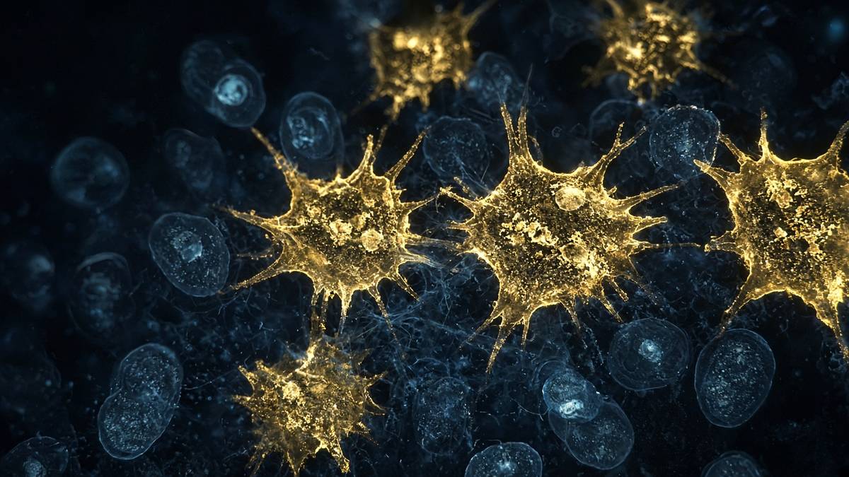
Hayflick, again, now as a fate rather than a chromosome end
The 1961 observation was a population of fibroblasts that stopped. The molecular account is telomere erosion, a DNA-damage response, p53 and p16, a durable cell-cycle arrest. That is replicative senescence. There are others: oncogene-induced senescence (a safety catch on Ras), stress-induced senescence (too much ROS, too much replication stress), therapy-induced senescence (what some chemotherapies leave behind). The Hayflick limit is therefore both a genome-floor fact (telomere length) and a fate-floor fact (the arrest). Blackburn's Nobel sits on the first. Campisi's career sits on the second, specifically on what the arrested cell does to its neighbours. Mixing them is allowed. Collapsing them into ageing as a single knob is how you get a supplement aisle. Replicative senescence is telomere erosion plus p53 and p16. Mixing that with oncogene-induced arrest and calling the pile ageing is how a fate becomes a vitamin. Keep the names. The assays differ. The tissue consequences differ. The peptide, if it has a literature here, has to say which arrest it thinks it moved.
In short. Replicative senescence is telomere erosion plus p53 and p16. Mixing that with oncogene-induced arrest and calling the pile ageing is how you get a supplement aisle.
Campisi and the SASP: a cell-fate floor with a tissue-floor megaphone
The senescence-associated secretory phenotype is a cocktail: IL-6, IL-8, other cytokines and chemokines, MMPs that remodel matrix, TGF-β neighbourhood signals, sometimes VEGF, sometimes activin. Coppé, Campisi and colleagues made this a measurable object rather than a mood — conditioned media, antibody arrays, the demonstration that senescent fibroblasts can change the behaviour of neighbours, including epithelial cells that were considering becoming a tumour. In a wound, a temporary SASP can be useful (clearance, plasticity). In a chronically inflamed, aged tissue, a persistent SASP is a fire alarm that will not silence: fibrosis, low-grade inflammation, stem-cell niches that no longer feel like niches. Senolytics try to kill the shouting cell. Senomorphics try to mute the shout. Neither is a research peptide in the catalogue, and we are not going to pretend a GPCR agonist is a senolytic because someone used the word ageing in a forum. SASP is IL-6, IL-8, MMPs and neighbours: a senescent cell shouting at a tissue. Context is the whole problem. The talk can be protective or corrosive.
In short. SASP is IL-6, IL-8, MMPs and neighbours: a senescent cell shouting at a tissue. Senolytics try to kill the shouter; a GPCR agonist is not one because a forum said ageing.
Occupancy that cannot name a fate is still only a description
This is the disciplinary rule for reading peptide papers that say repair, regeneration, anti-ageing, cellular health. Which fate moved? Did tenocytes proliferate (cyclin D, a Ki-67 stain, a count)? Did endothelial cells sprout (angiogenesis, a VEGFR2 story)? Did apoptosis fall in a crush model (TUNEL, cleaved caspase-3, a species, a time point)? Did senescent cells leave (p16, SA-β-gal, a SASP cytokine panel)? If the paper cannot answer, it has not yet left marketing. BPC-157's animal literature sometimes names angiogenesis and thus a fate of endothelial cells. TB-500's actin story names motility, which is not quite a fate but is at least a cell behaviour. GHK-Cu's arrays name a transcriptome, which is a step toward a fate. Epithalon's TERT claims name a genome-adjacent event that would, if real at scale, change a Hayflick parameter. Name it, assay it, species it. Then we can argue. Until then the word repair is an adjective stacked on a lyophilised cake. Name the fate that moved: Ki-67, TUNEL, p16, a SASP panel.
In short. Name the fate that moved: Ki-67, TUNEL, p16, a SASP panel. Until a paper can, repair is an adjective stacked on a lyophilised cake.
Floor 7 — Tissue: the first floor a person can point at
Extracellular matrix (collagens I/III/IV, elastin, proteoglycans, MMPs, TIMPs) — GHK-Cu's microarray literature lives here. Endothelium and angiogenesis — VEGF, nitric oxide, BPC-157's VEGFR2 neighbourhood. Barrier epithelia — tight junctions, mucus, IBS as a symptom cluster rather than a molecule. Immune infiltrate — neutrophils first, macrophages deciding M1/M2-ish programmes that are messier in humans than in the cartoon, T cells with actual antigen receptors. Fibrosis is collagen as a mistake that became a scar. Tendon is collagen as a job. The same proteins, different politics. TB-500's actin motif is a cell-motility story that becomes a tissue-repair story only if the cells that crawl are the right ones. Tissue is matrix, endothelium, barrier and infiltrate: the first floor a person can point at. A person notices a tendon, a rash, a cough. Under that is this chemistry in a pattern, not a bag of cells. Architecture is the result. Hydroxyproline is a number. The assay has to include the pattern.
In short. Tissue is matrix, endothelium, barrier and infiltrate: the first floor a person can point at. Fibrosis is collagen as a mistake; tendon is collagen as a job.
ECM: collagen I, III, IV, the MMPs and the TIMPs
Collagen I is bone, tendon, ligament, dermis — tensile, fibrillar, the rope. Collagen III is the finer companion in skin and vessels and a wound that is still deciding. Collagen IV is basement membrane, a sheet not a rope, the thing an epithelium sits on and a kidney glomerulus filters through. Elastin is recoil. Proteoglycans hold water and growth factors. MMPs cut the matrix; TIMPs hold the MMPs. A tissue is this chemistry in a pattern, not a bag of cells. Fibrosis is the pattern going wrong: too much I and III, cross-linked, contracted, a TGF-β programme that will not switch off, myofibroblasts that forgot to leave. GHK-Cu's fibroblast arrays often light up matrix genes, which is why this occupancy gets mentioned in the same breath as skin and as scarring, and why the breath should still include the words array, fibroblast and dish until a tissue-level human experiment has actually been done. We sell the tripeptide. We do not sell a dermis. Collagen I is the rope; collagen IV is the sheet. A gene list in a dish is not yet a human dermis.
In short. Collagen I is the rope; collagen IV is the sheet. GHK-Cu's fibroblast arrays light up matrix genes in a dish, which is not yet a human dermis.
Endothelium: the lining that is a tissue, not a wallpaper
Every blood vessel is an endothelial monolayer with a basement membrane and a decision to make about tone, leak, and new branches. Nitric oxide from eNOS relaxes smooth muscle. VEGF from a hypoxic neighbour occupies VEGFR2, and the endothelial cell sprouts. Angiogenesis is a cell-fate programme (migrate, proliferate, form a lumen) that becomes a tissue fact (a new capillary, or a leaky mess, or a tumour's private water supply). BPC-157's cleaner mechanistic neighbourhood in the animal literature is exactly here: VEGFR2 internalisation, FAK and paxillin at focal adhesions, eNOS. That is an endothelial and angiogenic occupancy. It is not a tendon-specific magic, even when the readout is a tendon, because a tendon that is healing needs vessels as well as collagen, and a tendon that is degenerating often has the wrong vessels in the wrong places. Occupancy is not outcome. Outcome is the rest of the stack, in a species, with controls. Every vessel is an endothelial decision about tone, leak and new branches. Name the receptor, and a wound vasculature becomes a question rather than a hope.
In short. Every vessel is an endothelial decision about tone, leak and new branches. BPC-157's cleaner animal neighbourhood is VEGFR2, FAK and eNOS, which is occupancy, not a tendon outcome.
Barriers: gut, skin, the quiet competence of tight junctions
An epithelium is a sheet that decides what may pass. Tight junctions (claudins, occludin, ZO proteins) are the seal. Mucus is the chemical moat. In the gut, a mucus layer, a microbiome, an immune system immediately underneath, and a surface area the size of a studio flat. Barrier failure is not a single molecule. It is a tissue state: leak, antigen flux, an immune conversation that was supposed to stay polite. IBS is a symptom cluster with a barrier and a pain-fibre and a brain in it, which is why naming one receptor as its cause is usually a conference talk, not a fact. Skin is the other barrier people point at: stratum corneum, lipids, a microbiome, keratinocytes that are a factory for a dead seal. KPV — Lys-Pro-Val, the C-terminal tripeptide of α-MSH — has an anti-inflammatory literature that sits on NF-κB and, in the gut, on PepT1 (SLC15A1), a proton-coupled di/tripeptide transporter in enterocytes. That is a barrier occupancy: a tripeptide that can be taken up and that talks to a transcriptional inflammatory programme. It is not a cure for a symptom cluster. It is a node.
In short. An epithelium decides what may pass: tight junctions, mucus, a studio-flat of gut surface. KPV sits on NF-κB and PepT1; that is a node, not a cure for a symptom cluster.
Innervation and immune infiltrate: the tissue is not sterile and not mute
A tendon has few nerves until it is angry, and then it has too many, which is why degenerative tendon pain is not a simple load-and-collagen story. A gut has an enteric nervous system that is a brain-scale object in its own right. Skin is an enormous sensory surface. Peptides that occupy melanocortin receptors (PT-141, MT2) or GHRH/GHSR axes are organism-level ligands partly because neural tissue expresses those GPCRs. The immune infiltrate is the other population a histology slide will not let you ignore: neutrophils first, then monocytes that become macrophages with a plasticity the M1/M2 cartoon only insults, T cells with antigen receptors, mast cells in barrier tissues. Chronic tissue pathology is often a failure to dismiss this infiltrate. KPV's NF-κB neighbourhood is an immune-transcription story. BPC-157 papers that count inflammatory cells in a lesion are at least counting something. Papers that say anti-inflammatory without a cell type are saying nothing yet. A degenerative tendon grows too many nerves; a gut has an enteric nervous system the size of a small brain. Name the cell.
In short. A degenerative tendon grows too many nerves; a gut has an enteric nervous system the size of a small brain. Anti-inflammatory without a cell type is saying nothing.
Fibrosis versus tendon: the same proteins, different politics
This is the distinction that keeps floor 7 honest. A healing tendon is collagen I, aligned, cross-linked, a tenocyte that did its job, a blood supply that came and then quietened, a nerve that did not sprout into the fascicles. A fibrotic liver is collagen I and III, disordered, a stellate cell that became a myofibroblast and stayed, TGF-β as a treatment plan, architecture that will not carry blood from portal tract to central vein. Same gene products. Opposite verdicts. A peptide that increases collagen has not yet told you which verdict it is serving. GHK-Cu's matrix-gene lists in fibroblasts could be either story depending on context. BPC-157's angiogenic neighbourhood could seed a useful wound vasculature or a disorganised one. TB-500's motility could deliver the right cell or the wrong one. Tissue is politics. The assay has to include architecture, not just a hydroxyproline number. A healing tendon is aligned collagen I; a fibrotic liver is disordered collagen that forgot to leave. Architecture is the result. Hydroxyproline is a number. Name which verdict you think you served.
In short. A healing tendon is aligned collagen I; a fibrotic liver is disordered collagen that forgot to leave. A peptide that increases collagen has not yet told you which verdict it serves.
Occupancy: BPC-157 in a VEGFR2/FAK/eNOS neighbourhood; KPV at NF-κB / PepT1
BPC-157 is a 15-residue fragment (GEPPPGKPADDAGLV) of a gastric protein, associated for decades with the Sikiric group's animal literature — crush, anastomosis, sphincter, tendon, a range of lesions in rodents that is broader than a cautious reader finds comfortable. The mechanistic threads that can be named without blushing are angiogenic and endothelial: VEGFR2, FAK, paxillin, nitric oxide. That is floor 3 occupancy with a floor 7 ambition. It is animal literature. It is not a randomised human tendon trial, and we will not write one into existence to help a caption. KPV is three residues, a melanocortin fragment that does not drag the pigment programme, PepT1 as a possible entry, NF-κB as a possible transcriptional target. Floor 7 (barrier, infiltrate) via floors 3 and 2. Different node from BPC-157. Treating them as a gut stack because both have gastrointestinal anecdotes is how you fail the oral exam this essay is writing. Different nodes, not a gut stack. Name the lock, name the cell, name the species, and both literatures get more interesting rather than less.
In short. BPC-157 is a gastric 15-mer with animal angiogenic literature at VEGFR2 and FAK; KPV is three residues at NF-κB and PepT1. Different nodes, not a gut stack.
Floor 8 — Organism: set-points, behaviour, time
Glucose, blood pressure, body temperature, sleep stage, appetite, VO₂, fertility, mood. These are controlled variables with sensors, set-points and effectors — Cannon's homeostasis, then allostasis when the set-point itself moves. Endocrine axes (GH, HPA, HPG, thyroid, incretins) are how floor 3 in one organ talks to floor 3 in another. A GLP-1/GIP/glucagon triple agonist (retatrutide, LY3437943) is a floor-3 ligand with a floor-8 readout so large it remade an industry. A gastric 15-mer with a rat tendon model is a floor-3/7 literature that has not climbed to 8 in a way a regulator would recognise. Both can be interesting. They are not the same climb. Glucose, sleep, waist and pain are controlled variables with set-points. Retatrutide is a floor-3 ligand with a floor-8 medicine paper; a rat tendon 15-mer is not the same climb. Occupancy is the mechanism. Weight is the readout in that paper. Those remain different sentences even when they appear in the same paragraph, and the paragraph is allowed to hold both.
In short. Glucose, sleep, waist and pain are controlled variables with set-points. Retatrutide is a floor-3 ligand with a floor-8 medicine paper; a rat tendon 15-mer is not the same climb.
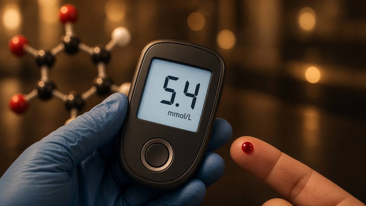
The glucose curve is not a molecule
An oral glucose tolerance test is a tissue conversation plotted as a line. Gut incretins (GLP-1, GIP) occupy β-cell GPCRs; cAMP; a first-phase insulin pulse that should already be in the portal vein before the glucose arrives. Liver takes the insulin hint and stops making glucose. Muscle and adipose take the later hint and open GLUT4. A curve that spikes and lingers is any of: a first phase that did not fire, a liver that is full of fat and deaf, an incretin conversation that is quiet, a muscle that is insulin-resistant, a stomach that emptied at the wrong speed, a sleep-debt from Van Cauter's world. Occupying GLP-1R, GIPR and GCGR with a unimolecular agonist is one way to rewrite several of those terms at once. Emptying the liver of fat, as Roy Taylor has spent a career showing, is another. They are not the same intervention. They can move the same number. Pathophysiology is naming which term you moved. An oral glucose test is gut, liver, muscle and last night's sleep plotted as a line. Skip those and the number is a superstition.
In short. An oral glucose test is gut, liver, muscle and last night's sleep plotted as a line. Occupying three incretin-family receptors rewrites several terms at once; emptying liver fat rewrites others.
VO2, sleep, fertility, waist, pain: the other traces a mammal notices
VO2 is oxygen consumption as a whole-body number: mitochondria, haemoglobin, cardiac output, muscle mass, training. A MOTS-c paper that reports treadmill time in a mouse has climbed from floor 5 to floor 8 in a rodent. A NAD+ caption that says energy has climbed nowhere yet. Sleep is a hypothalamic and brainstem state with a pineal footnote (melatonin, AANAT, the SCN). DSIP is a research peptide with a sleep literature that has never been as clean as its name; Epithalon's pineal claims live here as well. Fertility is an HPG axis, kisspeptin, GnRH, gonadotrophins, a gonad that needs a metabolic green light (leptin, energy availability) before it will play. Waist is adipose distribution, cortisol, sex steroids, incretins, a liver, a behaviour. Pain is nociceptors, spinal gain, inflammation, a tendon with too many nerves, a mood. Each of these is an organism-level readout with a stack under it. A peptide occupancy that cannot say which readout, in which species, with which control, is not yet floor 8. It is a hope. Name the trace.
In short. VO2, sleep, fertility, waist and pain are organism traces with stacks under them. A MOTS-c treadmill time in a mouse has climbed; a caption that says energy has not.
Diagram
Outside
Peptide ligand
Named sequence in the nM–µM pocket. Shape complementarity, not vibes. A 15-mer and a 4-mer do not fit the same hole.
Membrane
7-TM receptor
Helices rearrange. The cytoplasmic face becomes a GEF for a heterotrimeric G protein (Gs, Gi, Gq, G12/13).
Inside
Second messengers
cAMP, IP₃, Ca²⁺, β-arrestin. One occupied receptor can spawn thousands of messenger molecules. That is amplification.
~800 GPCRs in the human genome. Seven transmembrane helices, an extracellular ligand pocket, an intracellular G-protein handshake. Catalogue neighbours: ipamorelin at GHSR, PT-141/MT2 at melanocortin receptors, retatrutide at GLP-1R/GIPR/GCGR.
Occupancy: retatrutide as a Phase 2 medicine paper, not a vial instruction
Retatrutide (LY3437943) is a fatty-acylated 39-residue triple agonist at GLP-1R, GIPR and GCGR. Class B GPCRs, Gs, cAMP, the incretin and glucagon neighbourhoods, albumin binding via the fatty acid as the half-life trick. Jastreboff AM et al., New England Journal of Medicine, 2023; 389: 514–526: a Phase 2 obesity trial, 24.2% mean weight loss at the 12 mg dose at 48 weeks. That is a floor-8 number from a floor-3 occupancy of three receptors. The paper is public. The 12 mg and the 48 weeks are facts about a medicine trial, not a use instruction for a research vial, not a reconstitution guide, not a protocol. We sell the published research structure as a US-made, HPLC-MS characterised ligand, labelled for research use only. It is not Mounjaro, not Zepbound, not Eli Lilly's pen, not a licensed medicine. Occupancy is the mechanism. Weight is the readout in that paper. Those remain different sentences even when they appear in the same paragraph. The research vial is the structure. The medicine is a regulator's object.
In short. Retatrutide occupies GLP-1R, GIPR and GCGR. Jastreboff's 2023 Phase 2 paper reported 24.2% mean weight loss at 12 mg, 48 weeks: a medicine trial, not a vial instruction.
Van Cauter: sleep restriction as a metabolome insult with a glucose readout
Spiegel, Leproult and Van Cauter, Lancet, 1999: sleep debt in healthy young men, carbohydrate metabolism and endocrine function measured. The next day's glucose curve is worse; cortisol and sympathetic tone move; the organism has not acquired a new SNV overnight. That is floor 8 noticing floors 4 and 3 because floor 8's own set-point (sleep) was kicked. It is one of the cleanest demonstrations that you do not need a mutation to wreck a glucose trace, and that pathophysiology includes last night. A retatrutide paper and a Van Cauter paper can describe the same curve for incompatible reasons. If you cannot tell those reasons apart, you are not reading either paper. You are looking at a number and calling it a molecule. Restrict sleep in healthy young men and the glucose curve the next day is not the same curve. No new letters in the genome. Set-points moved. Floor 8 writing down onto floors 4 and 3. That is regulation, and it is why a peptide occupancy is not the only way a glucose number moves.
In short. Spiegel, Leproult and Van Cauter, Lancet 1999: sleep debt in healthy young men wrecks the next day's glucose curve. No new letters in the genome; set-points moved.
When someone asks what this peptide does in the body, the honest answer is a floor number, an assay, and a species. Anything else is a catalogue written by marketing. The rest of this essay is three worked examples that refuse to skip, then a map of the till as occupancies, then the reason the question was wrong. A ligand occupies a pocket in a cell type, at a concentration, for a time, in a species, and a cascade either happens or is compensated. What does retatrutide do is three GPCRs, cAMP, insulin, glucagon, gastric emptying, appetite, and, in a Phase 2 medicine paper, a weight. What does BPC-157 do is, at best, a VEGFR2 neighbourhood in an animal lesion. What does NAD+ do is a category mix-up if you wanted a receptor story; it is a cofactor accounting if you wanted the truth. The caption question invites a one-liner. The bench question names the pocket and the readout. We prefer the bench question. It is the only one a dish can answer, and the only one a chromatogram can keep honest.
In short. When someone asks what a peptide does in the body, the honest answer is a floor number, an assay and a species. Anything else is marketing with a sequence attached.
Worked example A — CFTR ΔF508, from a missing phenylalanine to a lung
Cystic fibrosis is the teaching case because every floor has a named object and the causal arrow actually holds. We will walk it without a peptide at the end, on purpose. A peptide does not fix CF. Correctors and potentiators (lumacaftor, ivacaftor, the triple combinations that changed clinical life) are small-molecule pharmacology aimed at fold and gate. Gene therapies and RNA approaches are other desks. The point of the walk is to show what from genome to a person who notices looks like when the arrow is actually complete. Every floor has a named object: a three-base deletion, a fold, a channel that never arrives, a mucus that will not flow, a lung a person can point at. That completeness is why CF is taught. It is also why it is a terrible template for common disease, and a worse template for a peptide catalogue. Nothing we lyophilise chaperones CFTR through the ER. The stack is for reading. It is not a permission slip. Walk it anyway. The stairs are the education.
In short. Cystic fibrosis is the teaching case because every floor has a named object. A peptide does not fix CF; correctors and potentiators chaperone fold and gate.
The letter: a three-base deletion that names a residue
Riordan et al., Science, 1989: the gene, the predicted protein, the common allele. ΔF508 is a deletion of three nucleotides, in-frame, so the protein is one phenylalanine short at position 508, in a nucleotide-binding domain. The gene is spelled almost correctly. Most of the coding sequence is intact. This is already a warning against the idea that genome-floor disease is always a stop codon or a frameshift. A missing residue in a domain that has to fold against an ATP-binding cassette architecture is enough. Heterozygotes are mostly well, which is why the allele could become common in some populations. Homozygotes, and compound heterozygotes with another serious CFTR allele, are not. ΔF508 is an in-frame deletion of one phenylalanine in CFTR, named in Science in 1989. A spelled protein can still fail. The interesting part is what happens next — fold, traffic, get stuck, get chewed — not the codon itself. That is already the stack, and we have not yet left the letter. The lung is six floors up. Everything interesting happens on the stairs.
In short. ΔF508 is an in-frame deletion of one phenylalanine in CFTR, named in Science in 1989. Heterozygotes are mostly well; homozygotes and serious compound heterozygotes are not.
The fold: ER retention, proteasome, a channel that never arrives
The nascent CFTR is born into the ER, glycosylated, inspected. ΔF508 makes a protein that is thermally unstable at 37 °C and is recognised as a failure. BiP, calnexin, the glycan code, ubiquitination, retrotranslocation, the proteasome. Floor 3 abundance collapses because floor 5 quality control is doing its job. Cool the cell in a dish and some of the mutant protein sneaks through — a classic experimental tell that the lesion is folding, not a total absence of function. If you artificially park ΔF508 at the membrane, the channel can work, albeit not perfectly. That observation is the entire rationale for correctors (help the fold, help the traffic) and potentiators (open the gate of whatever arrived). Pathophysiology here is a competent quality-control system applied to a slightly incompetent client. The cell is not broken. The protein is slightly wrong, and the ER is unforgiving. Cool the dish and some sneaks through, which is why correctors exist. A peptide does not do that job. Small-molecule pharmacology aimed at fold and gate does, in the clinic, in licensed combinations.
In short. The mutant CFTR is thermally unstable, retained in the ER, and eaten by the proteasome. Cool the dish and some sneaks through, which is why correctors exist.
The ion: no chloride, a mucus that will not flow
CFTR at the apical membrane of airway epithelium (and of pancreatic duct, sweat duct, gut, vas deferens) is a chloride and bicarbonate channel, and a regulator of other channels including ENaC, the sodium absorber. Without it, the airway surface liquid dehydrates, mucus becomes viscid, cilia cannot clear it. Floor 7: a barrier that has become a glue. The pancreas: ducts blocked, enzymes trapped, fat-soluble vitamins lost, an endocrine pancreas that may later fail as well. The vas deferens: often absent, fertility as a floor-8 readout of a floor-7 developmental failure. The sweat duct: salt on the skin, the diagnostic clue that predates the gene. Same protein, several tissues, several organism-level facts. That is already the stack, and we have not yet mentioned infection. Without apical CFTR, airway surface liquid dries, mucus thickens, and cilia cannot clear it. Same protein, several tissues: lung, pancreas, vas deferens, salt on the skin. One channel, a body that notices in four places. Pathophysiology is that one-to-many, not a single organ with a single complaint.
In short. Without apical CFTR, airway surface liquid dries, mucus thickens, and cilia cannot clear it. Same protein, several tissues: lung, pancreas, vas deferens, salt on the skin.
The lung: infection, inflammation, bronchiectasis, a person who notices
Stuck mucus is a culture medium. Staphylococcus, then Haemophilus, then Pseudomonas and the biofilm politics of a chronically colonised airway. Neutrophils arrive and do not leave; elastase from those neutrophils chews the airway wall; the wall dilates; bronchiectasis is now an architectural disease, not just a mucus disease. Floor 6 is a neutrophil that will not apoptose on schedule and an epithelium that is inflamed (NF-κB, a transcriptome). Floor 8 is cough, exacerbations, FEV1, oxygen, a life organised around physiotherapy and antibiotics and, in the lucky modern cohort, modulators that let some CFTR reach the membrane. The causal chain from three missing bases to bronchiectasis is unusually complete. That is why CF is taught. It is also why it is a terrible template for common disease, and a worse template for a peptide catalogue. Nothing we lyophilise chaperones CFTR through the ER. Claiming otherwise would be a different kind of missing residue: the one where intellectual honesty used to be. The chain from three missing bases to FEV1 is unusually complete, and a terrible peptide template.
In short. Stuck mucus becomes a culture medium; neutrophils stay; elastase chews the wall into bronchiectasis. The chain from three missing bases to FEV1 is unusually complete, and a terrible peptide template.
Worked example B — type 2 diabetes as regulation, not spelling
Now the common case. Type 2 diabetes can have a monogenic cousin (MODY: HNF1A, HNF4A, GCK — genuine spelling, genuine young adults, genuine single-gene clinics). Ordinary type 2 is not that. Ordinary type 2 is a polygenic risk score of small effect, a waist, a liver full of fat, a pancreas whose first-phase insulin pulse has gone quiet, an incretin conversation that is not amplifying what is left, and a glucose curve a person eventually notices. Roy Taylor's twin-cycle hypothesis and the DiRECT trial are the physiology. Jastreboff's retatrutide paper is a GPCR occupancy that moves the same organism-level numbers from a different floor. MOTS-c's AMPK literature is a third floor again. Same disease word. Three different stairs. Ordinary type 2 is polygenic risk, a fatty liver, a quiet first-phase insulin pulse, and a glucose curve. MODY is the monogenic cousin; most of the clinic is not that. Regulation, not a single spelling error. That is why occupying one receptor can move the number without rewriting the genome, and why emptying the liver of fat can move it from a different stair.
In short. Ordinary type 2 is polygenic risk, a fatty liver, a quiet first-phase insulin pulse, and a glucose curve. MODY is the monogenic cousin; most of the clinic is not that.
Ectopic fat, the first-phase pulse, and a liver that will not listen
Taylor's account, built from magnetic resonance spectroscopy and from people who actually lost weight: when calorie intake has been high for long enough, liver fat rises, hepatic insulin resistance rises, fasting glucose rises. The pancreas accumulates fat; β-cells lose the first-phase pulse — the dump of already-docked insulin granules that is supposed to meet a meal at the door. Second-phase insulin may still look noisy on a blood test; the first phase is the one that matters for the curve. This is metabolome and organelle and tissue (steatotic liver, steatotic pancreas) long before it is a new SNV. The genome is in the room as a polygenic willingness to store fat in the wrong postcode, and as the rare MODY exception. It is not dictating the daily glucose the way CFTR dictates chloride. Taylor's twin-cycle: liver fat, hepatic insulin resistance, a pancreas that loses its first-phase dump of docked granules. This is metabolome and tissue long before a new SNV. Empty the liver of fat and, in a defined cohort, the pulse can come back. That is reversible tissue state, which is a kind of wonder once you have seen the imaging.
In short. Taylor's twin-cycle: liver fat, hepatic insulin resistance, a pancreas that loses its first-phase dump of docked granules. This is metabolome and tissue long before a new SNV.
DiRECT: Lean, Taylor, a remission that is a tissue fact
Lean MEJ, Leslie WS, Barnes AC, et al., Lancet, 2018: the DiRECT cluster-randomised trial, Counterweight-Plus, a total-diet-replacement phase then stepped food reintroduction, in primary care. In a defined cohort of people with type 2 diabetes of limited duration, substantial weight loss put a substantial fraction into remission — off antidiabetic medication, HbA1c in the non-diabetic range — at one year, with the chance of remission tracking the weight lost. Taylor's imaging sub-studies showed liver fat collapsing, pancreas fat falling, first-phase insulin returning in those who remitted. That is pathophysiology as a reversible tissue state. It is not a peptide. It is not a genome editor. It is floor 8 (weight, glucose) writing down onto floors 7, 5 and 4, and floor 3 (insulin granules) coming back online because the environment of the β-cell changed. A research catalogue that talks about diabetes without being able to say this paragraph is talking about a market, not a mechanism. DiRECT put a defined cohort into remission with substantial weight loss: liver fat collapsed, first-phase insulin returned. Regulation. Reversible in a defined cohort.
In short. DiRECT put a defined cohort into remission with substantial weight loss: liver fat collapsed, first-phase insulin returned. That is reversible tissue state, not a research-vial claim.
Retatrutide occupies GLP-1R, GIPR and GCGR — a different stair, same building
The incretin axis is floor 3 in gut, β-cell, brain, adipose, liver. GLP-1R occupancy: more glucose-dependent insulin, less glucagon (in the intact α/β conversation), slower gastric emptying, less appetite via central receptors. GIPR occupancy: a more complicated adipose and β-cell sentence, and the reason a dual agonist is not just more GLP-1. GCGR occupancy: glucagon receptor, hepatic lipid oxidation, energy expenditure neighbourhoods, a lever that wants careful handling because glucagon also raises glucose. A unimolecular triple agonist occupies all three. Downstream of each: Gs, cAMP, a cell-type-specific dictionary. Organism-level: the Jastreboff curve, the weight, the metabolic laboratory sheet in a Phase 2 medicine paper. That paper is not a use instruction for the lyophilised research ligand we catalogue. It is the existence proof that this occupancy, in that formulation, in those humans, climbed to floor 8. The research vial is the structure. The medicine is a regulator's object. GLP-1R, GIPR and GCGR occupancy rewrites insulin, glucagon, emptying and appetite. Jastreboff is the existence proof in humans.
In short. GLP-1R, GIPR and GCGR occupancy rewrites insulin, glucagon, emptying and appetite. Jastreboff is the existence proof in humans; the research vial is the structure, not the pen.
MOTS-c and AMPK: a third stair, still not a protocol
Lee et al., 2015, again: a mitochondrial peptide, AMPK, folate–methionine, rodent insulin sensitivity and diet-induced obesity readouts. Floor 5 to floor 4 to, in a mouse, floor 8. Different receptors from retatrutide (there may not be a classical GPCR in the MOTS-c story at all). Different species from DiRECT. Different evidence grade from Jastreboff. Putting MOTS-c, retatrutide and a very-low-calorie diet in one sentence is allowed only if the sentence names the floors. All of these help glucose is not a sentence. It is a blending error. We stock MOTS-c and stock retatrutide as research ligands, US-made, HPLC-MS, and will not write a protocol that combines them, doses them, or aims them at a person. The pathophysiology is the map. The map is not a route. MOTS-c, retatrutide and a very-low-calorie diet can all move glucose from different floors. Saying they all help glucose, without naming the floor, is a blending error. Name the floor, the assay, the species, and the three stairs stay interesting rather than becoming a smoothie.
In short. MOTS-c, retatrutide and a very-low-calorie diet can all move glucose from different floors. Saying they all help glucose, without naming the floor, is a blending error.
Worked example C — a tendon that will not settle
A middle-aged tendon is a floor-7 object with a floor-8 complaint (pain, a failed return to load) and a floor-3/6 cellular argument underneath. Degenerative tendinopathy is not inflammation as a synonym for redness, and it is not a single missing molecule. It is a tenocyte that has changed its transcriptome, a collagen I architecture that has lost alignment, a proteoglycan swamp, vessels and nerves that have sprouted into a tissue that used to be almost avascular, and a failed attempt at repair that became a failed attempt at rest. Two catalogue ligands get pointed at this tissue constantly. They do not occupy the same node. The literature is largely animal. This is not a human protocol. It is a map of why the forum sentence BPC and TB for tendon is a category mix-up even when both papers exist. Degenerative tendinopathy is a tenocyte transcriptome, lost collagen alignment, and nerves that sprouted into a tissue that used to be almost avascular. BPC-157 and TB-500 do not occupy the same node. Name the node, and both literatures get more useful.
In short. Degenerative tendinopathy is a tenocyte transcriptome, lost collagen alignment, and nerves that sprouted into a tissue that used to be almost avascular. BPC-157 and TB-500 do not occupy the same node.
The tenocyte, the collagen, the vessel, the nerve
A healthy tendon is mostly extracellular matrix, mostly collagen I, tenocytes as the sparse residents that maintain it, a crimp pattern, a sliding fascicle, load as the language the tissue speaks. Degeneration: the tenocyte looks more like a chondrocyte on a bad day (rounder, more proteoglycan transcription), collagen becomes disorganised, type III appears where it does not belong, MMPs outrun TIMPs, the architecture that carried load starts to behave like a poor ligament. Angiogenesis into the tendon proper is not automatically salvation; neurovascular ingrowth is one of the pain stories. Load still helps many tendons, which is why physiotherapy is not a footnote. A ligand that cannot say whether it is talking to the tenocyte's transcriptome, the endothelial sprout, the nerve, or the macrophage is not yet a tendon story. It is a hope wearing a sequence. A healthy tendon is sparse tenocytes and aligned collagen I. Degeneration adds type III, proteoglycan swamp and neurovascular ingrowth. A ligand has to say which of those it is talking to, in which species, with which control.
In short. A healthy tendon is sparse tenocytes and aligned collagen I. Degeneration adds type III, proteoglycan swamp and neurovascular ingrowth; a ligand has to say which of those it is talking to.
BPC-157: angiogenesis and focal adhesions, in animals
The Sikiric corpus is large, rodent-heavy, and enthusiastic. The threads a cautious reader can hold are endothelial and cytoskeletal-adjacent: VEGFR2, FAK, paxillin, eNOS, a wound vasculature, a sphincter or an anastomosis that closed in a rat. Tendon and ligament models exist in that corpus. They are animal models. They are not a Phase 2 NEJM paper. Translating a rat Achilles crush into a human tendinopathy is a species jump plus a disease-definition jump (acute lesion versus chronic degeneration) plus a route-of-administration jump that we will not make in public, because that jump is a protocol. What we will say: if BPC-157 has a tendon-relevant occupancy, it is likely the angiogenic and endothelial neighbourhood, which may or may not be what a degenerative human tendon wants more of. That is already a more useful sentence than repair. The Sikiric corpus is rodent-heavy. If BPC-157 has a tendon-relevant occupancy, it is likely angiogenic, which a degenerative human tendon may or may not want more of. Occupancy is not outcome. Outcome is the rest of the stack.
In short. The Sikiric corpus is rodent-heavy. If BPC-157 has a tendon-relevant occupancy, it is likely angiogenic, which a degenerative human tendon may or may not want more of.
TB-500: a G-actin buffer, a different node, still not a protocol
Thymosin β4 / TB-500 occupies G-actin. Motility, a leading edge, a cell that can crawl. In a wound, cells that crawl include the ones you want (keratinocytes, endothelial cells, fibroblasts, tenocytes) and the ones you are less sure about. The fragment literature (LKKTETQ as the actin-binding motif) is a proteome story. Combining it with BPC-157 because both have been mentioned next to musculoskeletal models is like combining a wrench and a thermometer because both have been in a garage. Different nodes, different assays, different failure modes. A tenocyte that cannot deposit aligned collagen will not be rescued by more motility alone. An avascular mid-portion may want vessels; an already-ingrown painful tendon may not. Floor 7 is politics. Animal literature is a draft. Human tendinopathy is a different document. Research use only, both vials, no stack as a protocol, no diagram of a body. TB-500 occupies G-actin, a motility node, not BPC-157's angiogenic neighbourhood. Name the node. Then we can argue about whether the right cell crawled.
In short. TB-500 occupies G-actin, a motility node, not BPC-157's angiogenic neighbourhood. Combining them because both appeared next to musculoskeletal models is a wrench and a thermometer in the same garage.
A tendon is collagen as a job. A scar is collagen as a mistake. A peptide that 'increases collagen' has not told you which. Architecture is the result. Hydroxyproline is a number.
The peptide map as occupancies, not slogans
Here is the till, redrawn as a set of locks rather than a set of moods. Each row is a different conversation with a cell. Mixing them because a forum did is how you get a stack that is not a mechanism. The next essay — how peptides talk to cells — is the close-up of the GPCR rows. This is the atlas. Each ligand is a different conversation with a cell. Mixing them because a forum did is how a stack stops being a mechanism. Name the lock, or name the pocket, or name the metal, or name the motif. If you cannot, you do not have a mechanism yet. You have a brand colour. We will sell you a characterised sequence. We will not sell you the brand colour. GPCRs, receptor tyrosine kinases, a cofactor, an actin motif, a copper tripeptide, a melanocortin fragment, a tetrapeptide with a promoter-level literature, a mitochondrial ORF: different floors, different assays, different evidence grades. The map is how you read papers. It is not a permission to occupy a receptor in a person.
In short. The till redrawn as locks rather than moods: each ligand is a different conversation with a cell. Mixing them because a forum did is how a stack stops being a mechanism.
Diagram
| Node | Catalogue | Conversation |
|---|---|---|
| GPCR | Ipamorelin, MT2, PT-141, retatrutide, CJC | Second messengers, secretion, appetite, pigment |
| RTK / IGF1R | IGF-1 LR3 | IRS–PI3K–Akt–mTOR and Shc–ERK |
| Cytokine receptor | Somatropin (HGH) | GHR–JAK2–STAT5b, hepatic IGF-1 |
| Cofactor | NAD+ | Sirtuins, PARPs, CD38, redox |
| Actin buffer | TB-500 / Tβ4 motif | G-actin sequestration, motility |
| Growth-factor-like | BPC-157 | VEGFR2 / FAK / eNOS neighbourhood |
| Copper ligand | GHK-Cu | Transcriptome shift in fibroblasts |
| MC fragment | KPV | NF-κB, PepT1, no pigment |
| Nuclear / pineal | Epithalon (AEDG) | TERT and melatonin literatures |
| mtORF peptide | MOTS-c | AMPK, folate–methionine cycle |
Each row is a different kind of molecular conversation. The catalogue peptides bind at these nodes; they are not interchangeable, and stacking them because a forum did mixes unrelated literatures.
GPCRs: the majority of the interesting catalogue
Class A: GHSR (ipamorelin, the older GHRPs), melanocortin receptors (MT2, PT-141, and KPV as a fragment that does not write the same sentence). Class B: GHRHR (modified GRF 1-29 / CJC without DAC), GLP-1R, GIPR, GCGR (retatrutide as the unimolecular triple). Occupancy rearranges seven helices. A G protein spends GTP. cAMP or calcium or both. Amplification. Arrestin. Desensitisation. Lefkowitz and Kobilka, chemistry Nobel 2012, for the family. Raun 1998 for ipamorelin's selectivity. Jastreboff 2023 for the organism-level climb of the triple agonist. This is floor 3 as a profession. It is not signalling support. It is a Kd, an EC50, a cell type, a second messenger, a readout. Most of the interesting catalogue occupies GPCRs: GHSR, melanocortin receptors, GHRHR, GLP-1R, GIPR, GCGR. Occupancy rearranges seven helices and a G protein spends GTP. That is a Kd, not signalling support. Name the receptor class, and the ligand becomes a question a dish can answer. Skip the class, and a peptide becomes a mood.
In short. Most of the interesting catalogue occupies GPCRs: GHSR, melanocortin receptors, GHRHR, GLP-1R, GIPR, GCGR. Occupancy rearranges seven helices and a G protein spends GTP; that is a Kd, not signalling support.
Receptor tyrosine kinases and cytokine receptors: the longer ligands
IGF-1 LR3 occupies IGF1R, a receptor tyrosine kinase: dimer, autophosphorylation, IRS, PI3K–Akt–mTOR and Shc–ERK. Native IGF-1 is buffered by IGFBPs; Long R3 is an 83-residue analogue with collapsed binding-protein affinity, so more ligand reaches the kinase. That is the entire design. Somatropin occupies GHR, a class-I cytokine receptor: JAK2, STAT5b, hepatic IGF-1. 191 residues, recombinant, a different factory from a 15-mer on a resin. These are still occupancy. They are not GPCRs. Flattening them into growth peptides erases the lock, the second messenger, and the safety conversation (which is a different essay, and a different till, and still not a protocol). IGF-1 LR3 occupies IGF1R; somatropin occupies GHR via JAK2 and STAT5b. Still occupancy, not GPCRs. The lock, the second messenger, and the factory all differ. Name them, and a 191-mer and an 83-mer stop being synonyms. They were never synonyms. They share a catalogue and a word, growth, that is doing too much work. The receptors know the difference. The blot should too.
In short. IGF-1 LR3 occupies IGF1R; somatropin occupies GHR via JAK2 and STAT5b. Still occupancy, not GPCRs; flattening them into growth peptides erases the lock.
Cofactor, actin, copper, melanocortin fragment, nuclear/pineal, mtORF
- NAD+ — cofactor. Sirtuins, PARPs, dehydrogenases, CD38 as a consumer. Floor 4. Not a receptor ligand. Not a stimulant. Lyophilised β-NAD+ for the bench.
- TB-500 / thymosin β4 — G-actin buffer, LKKTETQ motif. Floor 3 cytoskeleton. Motility as a behaviour, tissue as an ambition.
- GHK-Cu — copper-binding tripeptide. Pickart. Fibroblast arrays (floor 2) and a matrix neighbourhood (floor 7). A 3-mer plus a metal, not a transcription factor.
- KPV — Lys-Pro-Val, α-MSH C-terminal fragment. NF-κB, PepT1. Barrier and infiltrate (floor 7) via a transcriptional programme (floor 2). Little pigment.
- Epithalon — AEDG. Khavinson literature on TERT and pineal melatonin. Genome-adjacent and clock-adjacent claims. Not CRISPR. Not a telomere therapy. A tetrapeptide you can blot or not.
- MOTS-c — MRWQEMGYIFYPRKLR, mtORF from 12S rRNA. Lee, Cell Metab 2015. AMPK, folate–methionine, nuclear translocation under stress. Floor 5 writing upward.
- BPC-157 — GEPPPGKPADDAGLV. Sikiric animal literature. VEGFR2 / FAK / eNOS neighbourhood. Floor 3 occupancy, floor 7 ambition, not a human protocol.
- DSIP — a sleep-named peptide whose literature has never been as tidy as the name. Organism-floor ambition, still a research ligand.
If you want a single rule for this map: name the lock, or name the pocket, or name the metal, or name the motif. If you cannot, you do not have a mechanism. You have a brand colour. We will sell you a characterised sequence. We will not sell you the brand colour. NAD+ is a cofactor. TB-500 is a G-actin buffer. GHK-Cu is a copper-binding tripeptide. KPV is a melanocortin fragment at NF-κB and PepT1. Epithalon is AEDG with a TERT literature. MOTS-c is a mitochondrial ORF. BPC-157 is a gastric 15-mer with animal angiogenic literature. DSIP is a sleep-named peptide whose literature has never been as tidy as the name. Each of those sentences names a conversation with a cell. Mixing them because a search bar did is how a stack stops being a mechanism. Name the lock. Then we can argue. Until then you are describing a feeling, and feelings are not Kd values. The vial is a ligand. The paper, if there is one, is a paper. The person is a stack that is still running.
In short. Name the lock, the pocket, the metal or the motif. If you cannot, you have a brand colour, and we will not sell you that.
Why 'what does it do?' is the wrong question
Because a ligand does not do a person. A ligand occupies a pocket in a cell type, at a concentration, for a time, in a species, and a cascade either happens or is compensated. What does retatrutide do is three GPCRs, cAMP, insulin, glucagon, gastric emptying, appetite, and, in a Phase 2 medicine paper, a weight. What does BPC-157 do is, at best, a VEGFR2 neighbourhood in an animal lesion, and at worst a list of forums. What does NAD+ do is a category mix-up if you wanted a receptor story; it is a cofactor accounting if you wanted the truth. What does Epithalon do is a TERT and pineal literature you have to read as literature, not as a rewrite of Blackburn's Nobel. The wrong question invites a one-line caption. The right question is longer, and is the only one a bench can answer. A ligand occupies a pocket in a cell type, at a concentration, in a species. The caption question invites a one-liner; the bench question names the pocket and the readout. We prefer the longer question. It is the only one that survives an experiment written in good faith.
In short. A ligand occupies a pocket in a cell type, at a concentration, in a species. The caption question invites a one-liner; the bench question names the pocket and the readout.
- Where does it bind? Receptor, motif, metal, cofactor pocket. Kd if anyone measured one.
- In which cell type? A GPCR in a β-cell is not the same GPCR in a neuron. A fibroblast is not a tenocyte.
- With which readout? cAMP, a phosphorylation, a gene list, a Δψm, a SASP cytokine, a histology, a glucose curve, a body weight. Name the floor of the readout.
- In which species, at which evidence grade? A rat crush is not a NEJM Phase 2. Both can be real. They are not interchangeable.
- What does the rest of the stack do after occupancy? Compensation, desensitisation, redundancy, 36 trillion cells. A clean blot is not a clinical endpoint.
That list is how you read we, and how you should read anyone else's. It is also why occupying one receptor does not fix a person: the stack has seven floors above the pocket, and a human is not a well. Spare receptors, arrestin, a liver that is still full of fat, a tendon whose architecture is already gone, a senescent neighbour shouting SASP into the wound, last night's sleep — all of these can swallow a beautiful occupancy and return a shrug. Jastreboff's paper is striking because, in that formulation, in those humans, the shrug did not happen at the weight readout. Most ligands never get that paper. Pretending they did is the original sin of a certain kind of catalogue. A binding assay is not a person. A person is not a well. Amplification can make a scarce occupancy look loud in a dish; tissue context can make a loud occupancy look silent in an animal; a Phase 2 can make a silent-looking occupancy look like an industry. Those are the floors. The vial is a ligand. The paper, if there is one, is a paper. The person is a stack that is still running.
In short. Seven floors sit above the pocket, and a human is not a well. Spare receptors, a fatty liver, last night's sleep and a SASP neighbour can swallow a beautiful occupancy.
The stack is still running after occupancy
You can occupy a GPCR and the genome does not notice. You can occupy a cofactor pocket and a sirtuin does. You can occupy actin and a cell changes shape. You can occupy nothing at all and a night of no sleep rewrites a glucose curve. Pathophysiology is the building. Research peptides are, at most, a key to one door on one floor. HPLC-MS tells you the key is the key it says it is. It does not tell you the building will rearrange itself for you. It will not. Compensation is older than your catalogue. Redundancy is why we are still here. Thirty-six trillion cells is why a well is a bad metaphor. You can occupy a GPCR and the genome does not notice, or occupy NAD+ and a sirtuin does. A research peptide is a key to one door, not a rearrangement of the building. The stack is still running after occupancy. That is the ethic, and it is also the law. Research use only is not a footnote. It is the only honest place a lyophilised occupancy can stand.
In short. You can occupy a GPCR and the genome does not notice, or occupy NAD+ and a sirtuin does. A research peptide is a key to one door, not a rearrangement of the building.
CRISPR writes floor 1. Casgevy is a licensed example. Nothing in the catalogue is that. Correctors and potentiators write CFTR's fold and gate; nothing in the catalogue is that either. A very-low-calorie diet, in DiRECT, wrote on liver fat and a first-phase pulse; we do not sell diets. A Phase 2 triple agonist wrote on weight in NEJM; we sell the published research structure as a research ligand, not the pen. Epithalon's papers claim TERT; we sell four residues, not a telomere. MOTS-c's paper claims AMPK and a mitochondrial ORF; we sell sixteen residues, not an exercise. GHK-Cu's arrays claim a fibroblast gene list; we sell a copper tripeptide, not a dermis. BPC-157 and TB-500 occupy different nodes in an animal literature; we sell the sequences, not a tendon. The pattern is the ethic. The ethic is the law. Research use only is not a footnote. It is the only honest place a lyophilised occupancy can stand. CRISPR, CFTR correctors and DiRECT write on floors we do not sell. We stock characterised sequences as research ligands.
In short. CRISPR, CFTR correctors and DiRECT write on floors we do not sell. We stock characterised sequences as research ligands; the pattern is the ethic, and the ethic is the law.
Name the floor. Name the assay. Name the species. Then we can argue. Until then you are describing a feeling.
If you have read this far you now have a way to place every vial on the till without asking it to be a treatment plan. Genome, transcriptome, proteome, metabolome, organelle, cell fate, tissue, organism. A disease is a failure with an address. A peptide is an occupancy with an address. When the addresses match, you have a hypothesis. When they do not, you have a forum. We prefer the hypothesis. We stock the ligand. We leave the building running. Name the floor. Name the assay. Name the species. Then we can argue. Until then you are describing a feeling, and feelings are not Kd values. A map is not a protocol. This stack is how we read papers. It is not a permission to occupy a receptor in a person. Every peptide listing here is labelled for research use only: HPLC-characterised ligands, not medicines, not CRISPR. The rest of the biology keeps running either way. That is the last sentence, and it is the kindest one: the building does not need you to rearrange it. It is already running. The ligand is a question. The stack is the answer-space.
In short. A disease is a failure with an address; a peptide is an occupancy with an address. When they match you have a hypothesis; when they do not, you have a forum.
Questions the essay actually answers
- What is pathophysiology, precisely?
- The mechanism of a disordered function: how a molecular change becomes a cell-state change becomes a tissue change becomes a sign a clinician can name. Not a synonym for 'disease', and not a protocol. The stack in this essay is the working definition: genome, transcriptome, proteome, metabolome, organelle, cell fate, tissue, organism.
- Do peptides work at the DNA level?
- Almost none of the catalogue is a transcription factor. They occupy receptors, cofactor pockets, actin, copper-binding sites. Epithalon's papers claim TERT and pineal effects — a gene-level literature, still not a gene editor. CRISPR is the gene editor. Different desk.
- What is the difference between genotype and phenotype?
- Genotype is the letters you inherited (and the copies, the methylation, the telomere length). Phenotype is what a cell, a tissue or a person actually does with them. Most common disease is a phenotype assembled from many small genotypic nudges plus environment. Cystic fibrosis is the rare case where one spelling error is enough. Type 2 diabetes usually is not.
- What is SASP?
- The senescence-associated secretory phenotype: a cell that has permanently left the cell cycle and started secreting inflammatory cytokines, chemokines, proteases and growth factors (IL-6, IL-8, MMPs, TGF-β neighbourhood). Campisi's sentence, and a measurable object rather than a mood. It is a cell-fate floor with a tissue-floor shout.
- How does retatrutide change a glucose curve?
- It occupies three class-B GPCRs — GLP-1R, GIPR and GCGR — on β-cells, brain, adipose and liver. Downstream: cAMP, insulin secretion, glucagon tone, gastric emptying, appetite. The glucose curve is an organism-floor readout of that proteome occupancy. Jastreboff NEJM 2023 is a Phase 2 medicine paper on the published ligand, not a use instruction for a research vial.
- Why doesn't occupying one receptor fix a person?
- Because the stack has seven floors above the receptor. Compensation, redundancy, tissue context and the fact that a human is 36 trillion cells, not one GPCR, are why a clean binding assay is not a clinical endpoint.
- Is cystic fibrosis a spelling error or a folding error?
- Both, in that order. ΔF508 deletes a phenylalanine in CFTR (Riordan et al., Science 1989 named the gene). The protein is spelled almost correctly, then fails to fold, is retained in the ER, and is degraded. Chloride never reaches the apical membrane. A peptide does not 'fix CF'. Correctors and potentiators that chaperone or open the channel are a different literature.
- Is type 2 diabetes written in the genome?
- Polygenic risk is real and small per locus. The physiology that actually breaks is often ectopic fat in liver and pancreas, a failed first-phase insulin pulse, and an incretin conversation at GPCRs. DiRECT (Lean, Taylor) showed that substantial weight loss can put the disease into remission in a defined cohort. Regulation, not a single spelling error.
- Where do BPC-157 and TB-500 sit on the stack?
- Different nodes. BPC-157's cleaner animal literature sits on VEGFR2, FAK/paxillin and eNOS — an angiogenic and endothelial neighbourhood at the proteome that becomes a tissue story. TB-500 / thymosin β4 is a G-actin buffer, a cytoskeletal occupancy. A tendon that will not settle has both angiogenesis and matrix-remodelling problems. They are not synonyms, and the literature is largely animal. Not a human protocol.
- What does research use only mean for a ligand that occupies a real receptor?
- It means the vial is a characterised chemical for the bench, not a medicine, not a prescription, and not CRISPR. Occupancy is a fact about a pocket. Licensed use is a fact about a regulator. We sell the first and label it as such. HPLC-MS, US-made, lyophilised. The stack is still running after you occupy a node.
Hypothetical research reconstitution
How these vials are typically mixed
Hypothetical research reconstitution for the named catalogue vial. Not a protocol, not medical advice, not a use instruction. These amounts sit in published and commonly cited laboratory ranges. The vial is labelled for research use only — not for human or veterinary administration.
BPC-157
10mg
Mix with 2 ml bacteriostatic water → 5 mg/ml · 5,000 mcg/ml
- Hypothetical aliquot
- 250 mcg
- 0.05 ml · 5 units on a U-100 syringe
- How often
- Once or twice daily
- 2–4 weeks in the papers that actually run a course
Bench steps
- Let the vial sit until it is no longer cold to the touch.
- Wipe the stopper with 70% isopropyl alcohol. Let it dry.
- Draw 2 ml bacteriostatic water (0.9% benzyl alcohol).
- Run the water slowly down the inside glass — do not blast the cake.
- Roll between finger and thumb until the cake is gone. Do not shake.
- Label the date. Store the solution at 2–8 °C. Do not freeze. Use within 30 days unless the note below says otherwise.
Stable in bacteriostatic water in the fridge. 500 mcg is the upper end of what most bench notes call a daily aliquot; 250 mcg is the usual starting mark.
TB-500
10mg
Mix with 2 ml bacteriostatic water → 5 mg/ml
- Hypothetical aliquot
- 2 mg
- 0.40 ml · 40 units on a U-100 syringe
- How often
- Twice weekly for four weeks, then once weekly
- 4–6 weeks loading, then a weekly hold if the assay continues
Bench steps
- Let the vial sit until it is no longer cold to the touch.
- Wipe the stopper with 70% isopropyl alcohol. Let it dry.
- Draw 2 ml bacteriostatic water (0.9% benzyl alcohol).
- Run the water slowly down the inside glass — do not blast the cake.
- Roll between finger and thumb until the cake is gone. Do not shake.
- Label the date. Store the solution at 2–8 °C. Do not freeze. Use within 30 days unless the note below says otherwise.
Thymosin β4 fragment. The literature uses milligrams, not micrograms — do not treat it like BPC-157. Same fridge rule.
NAD+
1000mg
Mix with 10 ml bacteriostatic water → 100 mg/ml
- Hypothetical aliquot
- 50–100 mg
- 0.50–1.00 ml · 50–100 units on a U-100 syringe
- How often
- Two or three times per week in published infusion and assay notes
- 4–8 weeks, then a pause
Bench steps
- Let the vial sit until it is no longer cold to the touch.
- Wipe the stopper with 70% isopropyl alcohol. Let it dry.
- Draw 10 ml bacteriostatic water (0.9% benzyl alcohol).
- Run the water slowly down the inside glass — do not blast the cake.
- Roll between finger and thumb until the cake is gone. Do not shake.
- Label the date. Store the solution at 2–8 °C. Do not freeze. Use within 30 days unless the note below says otherwise.
A 1000mg cake wants 10 ml. Protect from light. Solution yellows as it oxidises — that is the cofactor dying, not a flavour. Use promptly.
Retatrutide
30mg
Mix with 3 ml bacteriostatic water → 10 mg/ml
- Hypothetical aliquot
- 1–2 mg to start; published trial arms ran higher by week
- 0.10–0.20 ml · 10–20 units on a U-100 syringe (at 1–2 mg)
- How often
- Once weekly
- The Jastreboff NEJM 2023 arms ran 48 weeks. That is a trial, not a shop protocol.
Bench steps
- Let the vial sit until it is no longer cold to the touch.
- Wipe the stopper with 70% isopropyl alcohol. Let it dry.
- Draw 3 ml bacteriostatic water (0.9% benzyl alcohol).
- Run the water slowly down the inside glass — do not blast the cake.
- Roll between finger and thumb until the cake is gone. Do not shake.
- Label the date. Store the solution at 2–8 °C. Do not freeze. Use within 30 days unless the note below says otherwise.
LY3437943 architecture. Weekly, not daily. Those milligram figures are what the papers used on the investigational medicine — they are not a use instruction for this reagent.
GHK-Cu
100mg
Mix with 5 ml bacteriostatic water → 20 mg/ml
- Hypothetical aliquot
- 1–2 mg
- 0.05–0.10 ml · 5–10 units on a U-100 syringe
- How often
- Once daily
- 4–8 weeks
Bench steps
- Let the vial sit until it is no longer cold to the touch.
- Wipe the stopper with 70% isopropyl alcohol. Let it dry.
- Draw 5 ml bacteriostatic water (0.9% benzyl alcohol).
- Run the water slowly down the inside glass — do not blast the cake.
- Roll between finger and thumb until the cake is gone. Do not shake.
- Label the date. Store the solution at 2–8 °C. Do not freeze. Use within 30 days unless the note below says otherwise.
The solution is blue if the copper is on the peptide. Grey or colourless is the wrong cake. 100mg wants 5 ml or the syringe marks get silly.
Epithalon
50mg
Mix with 5 ml bacteriostatic water → 10 mg/ml
- Hypothetical aliquot
- 5–10 mg
- 0.50–1.00 ml · 50–100 units on a U-100 syringe
- How often
- Once daily, evening, for 10–20 consecutive days
- 10–20 days, two cycles a year in the Khavinson-school notes
Bench steps
- Let the vial sit until it is no longer cold to the touch.
- Wipe the stopper with 70% isopropyl alcohol. Let it dry.
- Draw 5 ml bacteriostatic water (0.9% benzyl alcohol).
- Run the water slowly down the inside glass — do not blast the cake.
- Roll between finger and thumb until the cake is gone. Do not shake.
- Label the date. Store the solution at 2–8 °C. Do not freeze. Use within 30 days unless the note below says otherwise.
Tetrapeptide (AEDG). Short pulses, not a daily-forever molecule in that literature.
MOTS-c
40mg
Mix with 2 ml bacteriostatic water → 20 mg/ml
- Hypothetical aliquot
- 5–10 mg
- 0.25–0.50 ml · 25–50 units on a U-100 syringe
- How often
- Two or three times per week
- 4–8 weeks
Bench steps
- Let the vial sit until it is no longer cold to the touch.
- Wipe the stopper with 70% isopropyl alcohol. Let it dry.
- Draw 2 ml bacteriostatic water (0.9% benzyl alcohol).
- Run the water slowly down the inside glass — do not blast the cake.
- Roll between finger and thumb until the cake is gone. Do not shake.
- Label the date. Store the solution at 2–8 °C. Do not freeze. Use within 30 days unless the note below says otherwise.
Mitochondrial 16-mer. Fridge. Do not freeze. The 5 mg mark is where most bench notes start.
Bacteriostatic water and sterile syringes ship with peptide orders over £75. Kit details · 10 ml bacteriostatic water
The vials this essay sits on
Named sequences the essay maps — BPC-157, TB-500, NAD+, Retatrutide, GHK-Cu, Epithalon, MOTS-C. Hypothetical research neighbourhood, not a protocol, not a medicine. One press puts every in-stock vial in the bag.
 Research only
 Research only
 Research only
 Made in USAOut of stock
Incretin
Retatrutide
US-made retatrutide 30mg — the published structure LY3437943, HPLC-MS verified.
4.6(609)
121 browsing this now · 5 purchased in the last 24 hours
30mg
£120.00
 Research onlyOut of stock
Copper complex
GHK-Cu
100 mg GHK-Cu. Pickart’s copper tripeptide, lyophilised.
4.9(590)
92 browsing this now · 5 purchased in the last 24 hours
100mg
£35.00
 Research only
 Made in USA
Research use only. Not a combined-use instruction.
Read next
65 min · long read · The living cell
The living cell is a city, and you are 36 trillion of them
A 70 kg adult is on the order of 36 trillion cells, most of them red blood cells with no nucleus. A typical nucleated cell holds about ten billion proteins and two metres of DNA. The body recycles 40–60 kg of ATP a day. Those figures are published; this essay is what they mean.
64 min · long read · The living cell
From gene to protein: how a cell actually reads itself
DNA is transcribed, spliced, exported, translated and folded. A research peptide is the ligand at the end of that pipeline, already made. This essay is every machine in between — polymerases, spliceosome, ribosome — with the actual rates attached.
70 min · long read · The living cell
How peptides talk to cells: occupancy, amplification, arrestin
A peptide is a ligand. Most of the catalogue binds a GPCR on the cell surface: one occupancy, then enzymes make thousands of second messengers. That amplification is real, and it is not magic. Desensitisation is why more ligand is not more signal forever.
49 min · long read · Peptide research
Senescent cells refuse to die, and they talk too much
Hayflick watched fibroblasts stop dividing. Campisi named the SASP — the inflammatory letters those arrested cells keep sending. Clearing p16-high cells in mice (INK-ATTAC) delayed aging phenotypes. The pathology is accumulation, not the programme itself.
46 min · long read · Peptide research
Retatrutide (LY3437943): the published triple-agonist structure
A fatty-acylated unimolecular agonist at GIPR, GLP-1R and GCGR. US-made, HPLC-MS verified, labelled for laboratory research — not a medicine.
More in this desk
64 min · long read · The living cell
Mitochondria: the bacterium you kept, the genome it kept, the peptides it writes
You turn over 40–60 kg of ATP a day using a 16,569-base genome that still uses a bacterial genetic code. NAD+ is the hydride carrier Complex I spends. MOTS-c is a 16-mer translated from mitochondrial 12S rRNA — Lee, Kim, Cohen, 2015. That last sentence is real, and it is surprising.
57 min · long read · The living cell
Proteostasis: the cell that eats its own mistakes
Ten billion proteins, a 76-residue tag, a 2.5-megadalton proteasome, and autophagy for whole organelles. Hershko, Ciechanover and Rose, 2004. Ohsumi, 2016. DSIP is a sleep-isolation nonapeptide, not an autophagy ligand.
54 min · long read · The living cell
The nucleus, telomeres, and the four residues of Epithalon
Two metres of DNA folded into a nucleus a few micrometres across. TERT is off in most somatic cells on purpose. Epithalon is four residues, Ala-Glu-Asp-Gly, with a TERT and pineal literature. The machines are real. A large Western trial of telomere length in adults is not.
51 min · long read · The living cell
Membranes: a five-nanometre wall
Every cell, every organelle, every synapse sits on a lipid bilayer about five nanometres thick. Singer and Nicolson, Hodgkin and Huxley, a billion lipids, and why a peptide ligand at a GPCR does not need to enter the cell.
Essays describe published research. They are not medical advice and they do not authorise human use of any catalogue item.