

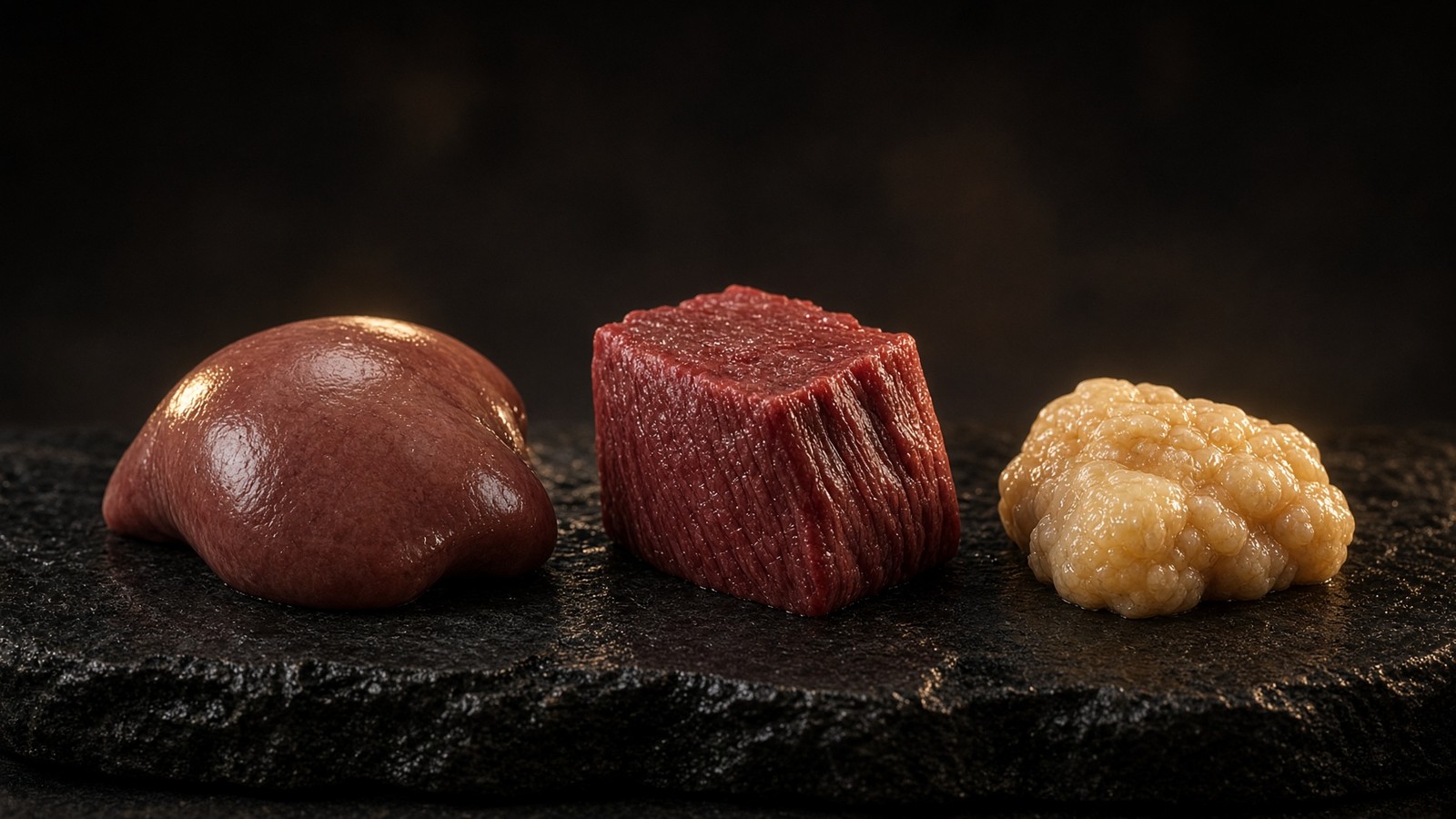
Metabolism · 51 min · 11,196 words
Human metabolism, without the slogan
Calories are bookkeeping. The actual system is a set of hormonally gated fluxes through glycogen, fat and amino acids — with the liver as air-traffic control.
What this essay actually tells you
- Energy in a human is ATP from glycolysis, β-oxidation and the electron-transport chain. The currency is reducing equivalents. Calories as a moral unit is a different, worse conversation.
- Insulin, glucagon, cortisol, thyroid hormone and catecholamines are the control layer that decides which fuel the mitochondrion sees. Five hormones. One organelle. That's the map.
- Liver, muscle and adipose are not interchangeable. A model that treats 'the body' as one tank will mis-predict every diet, which is most of the diets on the internet.
What this actually means
You do not burn food so much as you shunt it through a set of hormonally gated doors. Carbohydrate lands as glucose and glycogen, fat as fatty acids and ketones, protein as amino acids that either rebuild tissue or, if the liver is short of carbon, become glucose. Insulin, glucagon, adrenaline, cortisol, thyroid hormone and the incretins decide which door is open. The liver dispatches, muscle disposes and works, adipose buffers. Glycogen caps are small; everything past them overflows, often into liver and pancreas. Cahill showed the brain will take ketones and spare muscle. Exercise rewrites the doors for hours afterwards via AMPK and PGC-1α. So does a short night, a fever, and whatever you ate yesterday. Metabolism is not a furnace with one thermostat. It is fluxes, and the liver is doing air-traffic control whether anyone asked it to or not. NAD+, MOTS-c and retatrutide occupy a cofactor, a mitochondrial 16-mer and three gut-hormone receptors in this neighbourhood. They are research sequences, not a diet.
Calories are a unit of heat. A bomb calorimeter asks how much energy a food releases when you burn it in oxygen, and the answer is a number you can print on a packet. Conservation of energy still holds in a person: metabolisable energy that's not expended is stored, mostly as triglyceride. That is thermodynamics, and it's not optional. The useful conversation is the one the bomb calorimeter cannot have. A human is not a bomb. Glucose, fatty acids and amino acids enter named tissues through named transporters, meet named enzymes, and become acetyl-CoA, reducing equivalents, or new protein. Hormones decide which of those doors is open. The mitochondrion of a hepatocyte, a myofibre and an adipocyte then see different fuel, at different rates, for different jobs. Treating the whole animal as a furnace with one thermostat is how a diet written on a spreadsheet ends up mis-predicting the person who actually ate it. This page is the routing table: three fuels, three tissues, a control layer, a backup generator Cahill measured, and the kinases that rewrite the doors afterwards.
In short. Food energy still adds up. The interesting part is which tissues take which fuel, and which hormones open the doors.
Carbohydrate lands as glucose and as glycogen. Fat lands as fatty acids, as triglyceride, and, when the liver is overflowing acetyl-CoA, as ketones. Protein lands as amino acids that either rebuild tissue or, if the liver is short of carbon, become glucose. Those are three cargoes, not three personalities. The brain wants plasma glucose in a fairly tight band and will take ketones if it has to; Cahill's starvation work still is not taught hard enough. Muscle will oxidise whatever it has been trained and fed to oxidise: glycogen, fatty acids, ketones, even a bit of branched-chain amino acid if you are being unkind to it. Adipose tissue stores surplus and releases it when insulin drops. That is a buffer. The moral language people glue onto adipocytes is not a mechanism. The actual constraint is mitochondrial. You can't push more acetyl-CoA through the TCA cycle — the Krebs cycle, the mill that turns two-carbon units into hydrides — than the organelles will take, and you can't park more glycogen than a few hundred grams. Everything past that spills. Name the cargo. Name the tissue. Then the diet conversation has somewhere to sit.
In short. Carbohydrate, fat and protein are three different cargoes. Liver, muscle and fat cells handle them differently, and the leftover has to go somewhere.
We stock three sequences that live in this neighbourhood, and neighbourhood is not identity. NAD+ is nicotinamide adenine dinucleotide — a small molecule that shuttles electrons. It is the hydride coin Complex I wants oxidised and the stoichiometric substrate sirtuins and PARPs spend. MOTS-c is a sixteen-residue peptide a mitochondrion translated from its own 12S rRNA — Lee, Kim, Cohen, Cell Metabolism, 2015 — sitting on AMP-activated protein kinase, the cell's fuel-gauge. Retatrutide is the published LY3437943 structure, a unimolecular agonist at the receptors for glucose-dependent insulinotropic polypeptide, glucagon-like peptide-1 and glucagon. Those three objects occupy a cofactor, a fuel-gauge kinase, and three class-B G-protein-coupled receptors. They don't occupy a diet. This page is human fuel routing: glycolysis, β-oxidation, the electron-transport chain, the hormones that decide which fuel a mitochondrion sees, and why liver, muscle and adipose are not interchangeable. The catalogue items sit on the reading list because a bench that reads this physiology will meet their names. They're not a protocol stacked on the routing table, and they will not be written as one. Hold the floors apart and each object keeps the job it actually has.
In short. Three research sequences sit near this map. They occupy three different locks. This page is the physiology, not a stack.
Calories are bookkeeping. Reducing equivalents are the currency. ATP is the product. Hormones decide which fuel the mitochondrion sees.— Cahill measured the backup. Rich measured the ATP scale. The liver still dispatches.
The product is ATP. The currency is reducing equivalents.
Adenosine triphosphate is the phosphate currency. Almost every energy-requiring job in a cell — a sodium pump, a myosin stroke, a ligation, a cycle of a G protein — spends the γ-phosphate of ATP and leaves ADP plus inorganic phosphate. You don't store a warehouse of it. Peter Rich, writing in Biochemical Society Transactions in 2003, put whole-body turnover on the order of forty to sixty kilograms a day, against a standing pool of about fifty grams. Recycled, not stored. Something like 10²¹ hydrolyses a second in a living adult. That number still makes me sit up. That phosphate is minted in two places. Substrate-level phosphorylation in glycolysis and in the TCA cycle contributes a little. Oxidative phosphorylation on the inner mitochondrial membrane contributes almost all of the rest. The electron-transport chain does not burn calories as a metaphor. It oxidises reducing equivalents — NADH at Complex I, QH2 at Complex III — pumps protons, and lets ATP synthase spend the voltage. The currency of metabolism, before the phosphate, is those reducing equivalents. Forget that and you will keep talking about food as if it were firewood.
In short. Cells spend ATP on almost every job. They mint it from food electrons running down a chain to oxygen, and they recycle a body-weight of it daily.
Glycolysis is the cytosolic path from glucose to two molecules of pyruvate. Hexokinase — glucokinase in hepatocytes — phosphorylates glucose so it cannot leave. Phosphofructokinase-1 is the committed step, allosterically read by energy charge and by fructose-2,6-bisphosphate. Glyceraldehyde-3-phosphate dehydrogenase takes NAD+ and a phosphate and a three-carbon aldehyde and hands back NADH and a high-energy acyl phosphate; phosphoglycerate kinase and pyruvate kinase then mint ATP by substrate-level phosphorylation. Net yield from glucose to two pyruvates: two ATP and two NADH. The rest of the energy is still in the pyruvate. If the cytosol is reduced — high NADH, low NAD+ — GAPDH slows and the cell dumps pyruvate to lactate via lactate dehydrogenase in order to regenerate NAD+ in the cytosol. That's the Warburg observation as a nucleotide problem, and it is also what a sprinting myofibre does on purpose. Aerobic fate is different. Pyruvate crosses the inner membrane, and the pyruvate dehydrogenase complex decarboxylates it to acetyl-CoA, minting NADH and CO2. That's the gate from sugar into the matrix. Insulin and PDH phosphatases lean it open. Pyruvate dehydrogenase kinase, read by acetyl-CoA and NADH, leans it shut.
In short. Breaking glucose in the cytosol gives a little ATP and loaded electron carriers. Most of the remaining energy is still sitting in pyruvate.
The TCA cycle is how acetyl-CoA becomes reducing equivalents rather than how food becomes a story. Citrate synthase condenses acetyl-CoA with oxaloacetate. Aconitase and isocitrate dehydrogenase 3 produce NADH and CO2. The α-ketoglutarate dehydrogenase complex does the same on a five-carbon acid and is, chemically, a cousin of pyruvate dehydrogenase. Succinyl-CoA synthetase mints a GTP or ATP by substrate-level phosphorylation. Succinate dehydrogenase — Complex II — reduces ubiquinone to QH2. Malate dehydrogenase, near equilibrium, produces the last NADH; the mitochondrial NAD+/NADH ratio sets how hard that step pulls. Two carbons entered. Two CO2 left. The oxaloacetate was regenerated. What you actually banked is three NADH, one QH2, and one GTP per turn, plus whatever the anaplerotic traffic was doing to the pool of intermediates. A liver that is making glucose is pulling oxaloacetate out for phosphoenolpyruvate carboxykinase. A muscle that is working is running the cycle as a furnace. Same enzymes. Different withdrawals. The cycle is not a decorative wheel in a textbook. It is the mill that turns two-carbon units into the hydrides Complex I wants. Carbon dioxide is the exhaust. The hydrides are the point.
In short. The Krebs cycle takes two-carbon units and mints the loaded electron carriers the respiratory chain actually spends. Carbon dioxide is the exhaust.
β-oxidation is the spiral that turns a fatty acyl-CoA into acetyl-CoA, NADH and QH2. Carnitine palmitoyltransferase I on the outer membrane is the gate; malonyl-CoA, the product of acetyl-CoA carboxylase, inhibits it, which is how a cell that is writing fat does not simultaneously burn fat. The acyl chain is translocated as an acyl-carnitine, rebuilt as acyl-CoA in the matrix, then dehydrogenated, hydrated, dehydrogenated again, and cleaved. Each turn shortens the chain by two carbons and loads one FAD, which reduces Q, and one NAD+. Even-chain saturated fat is the clean case. Odd-chain fat leaves propionyl-CoA, which becomes succinyl-CoA and can be anaplerotic. Unsaturated fat needs extra enzymes. Peroxisomes handle very-long-chain species first. A liver that cannot reoxidise NADH and QH2 will stall the spiral and either make ketones or store the fat. Hormone-sensitive lipase and adipose triglyceride lipase decide whether the fatty acids arrive in the first place. Insulin suppresses those lipases. Catecholamines and glucagon raise them. The spiral itself is chemistry. The supply of substrate is a hormone decision. Confusing the two is how a β-oxidation lecture becomes a diet argument it was never built to settle.
In short. Fat is cut into two-carbon units inside mitochondria. Each turn of that spiral loads more electron carriers, but hormones decide whether the fat arrives.
The electron-transport chain is where those reducing equivalents are cashed. NADH:ubiquinone oxidoreductase, Complex I, oxidises NADH, walks the electrons down iron-sulphur clusters, reduces ubiquinone, and pumps four protons. Complex II feeds Q from succinate and does not pump. The Q-cycle at Complex III pumps more protons and reduces cytochrome c. Complex IV, cytochrome c oxidase, reduces oxygen to water and pumps again. Peter Mitchell's chemiosmotic theory is why this is a voltage rather than a series of substrate-level tricks: a proton-motive force of about 150 millivolts, negative inside, plus a pH difference, matrix alkaline. ATP synthase is the rotary turbine that lets protons home and phosphorylates ADP. Boyer and Walker got the Nobel for the catalytic mechanism; Mitchell got his in 1978 for insisting the gradient was the point. Oxygen is the terminal acceptor. Without it the chain stops, NADH backs up, the TCA cycle stalls at the NAD+-dependent steps, and glycolysis dumps to lactate. That's not a metaphor for tiredness. It is the reason ischaemic tissue acidifies, and the reason a Seahorse oxygen-consumption trace is a metabolic paper rather than a mood. Electrons walk to oxygen. The voltage pays for the phosphate.
In short. The inner membrane pumps protons as electrons walk to oxygen. ATP synthase spends that voltage on a phosphate. Oxygen is the last acceptor.
Diagram
- I. Pumps H⁺. ~45 subunits. The NADH coin is spent here.
- II. TCA entry. No proton pump. FADH₂ neighbourhood.
- Q. Lipid-soluble shuttle in the inner membrane.
- III. Q-cycle. Pumps H⁺. Superoxide leak site.
- c. Intermembrane space. The shuttle everyone has heard of.
- IV. O₂ → H₂O. The reason you breathe.
- V. F₁Fₒ rotary. Protons in, ATP out. ~10²¹ times a second in you.
Mitchell’s chemiosmotic theory (Nobel 1978): the inner membrane is a battery of ~150 mV. NAD+ is the hydride carrier that feeds Complex I. MOTS-c is a 16-mer the mitochondrion translated from 12S rRNA — a different object on the same campus.
The dinucleotides themselves don't cross the inner mitochondrial membrane. Cytosolic NADH from glycolysis has to hand its electrons in by shuttle. The malate-aspartate shuttle reconstitutes NADH in the matrix, so Complex I sees it. The glycerol-phosphate shuttle dumps electrons on Q and skips Complex I, a cheaper, less complete capture. A hepatocyte uses the first. A skeletal myofibre uses both, with the mix depending on fibre type. Mitochondrial NAD+ is therefore not in free equilibrium with the cytosol; SLC25A51 is the mammalian candidate importer, and matrix-targeted biosensors exist because homogenate kits cannot see the rooms. Compartmentation is why a whole-cell NAD+ number is a scout and not a mitochondrial paper, and why a catalogue sentence about 'boosting mitochondria' has no address. The reducing-equivalent currency has postcodes. Glycolysis spends and mints in the cytosol. β-oxidation and the TCA cycle mint in the matrix. The chain spends on the inner membrane. A story that treats those three addresses as one tank of energy has already failed the topology the enzymes actually live on. You can't average the rooms and call the average a mitochondrion. The rooms are the point.
In short. The electron-carrying coins cannot freely cross the inner membrane. Shuttles move the electrons between rooms of the cell. A blended average hides that.
A calorie, on a food label in this country, is a kilocalorie: the heat needed to raise a kilogram of water by one degree Celsius. Atwater factors — four, nine, four, roughly, for carbohydrate, fat and protein — are metabolisable-energy averages, not tissue-level jobs. They are good enough for a packet and bad at the thing people actually argue about, which is where the energy went and who decided. Thermic effect is real: protein costs more to process than fat. Palatability is real: Hall's NIH ward studies showed an ultra-processed pattern drove spontaneous overeating even when macronutrients were matched. Insulin is real: it routes. None of those facts repeals conservation of energy, and none of them makes a calorie a moral unit. People attach virtue to the number because the number is printable. The worse conversation is the one that treats a kilocalorie as a character test and then wonders why two isocaloric weeks, in two tissues, with two hormone backgrounds, don't produce the same person. Bookkeeping is required. Bookkeeping is not physiology. The rest of this page is the physiology the packet cannot print: which door opened, which tissue took the cargo, and which hormone wrote the instruction.
In short. A calorie is heat from a bomb calorimeter, useful for a label. It is not a moral score, and it does not say which tissue took the fuel.
Diagram
Matrix
- TCA cycle · β-oxidation · mtDNA nucleoids
- NADH produced here. Complex I spends it.
- MOTS-c (MRWQEMGYIFYPRKLR) from 12S rRNA.
Inner membrane
- I → II → III → IV → V (ATP synthase)
- ~150 mV proton-motive force
- ~40–60 kg of ATP turned over per human day
mtDNA is 16,569 bp, 37 genes, 13 proteins of the respiratory chain. Nuclear DNA encodes the other ~1,200 mitochondrial proteins. NAD+ is the hydride carrier between dehydrogenases and Complex I. MOTS-c is a 16-mer translated from 12S rRNA — a peptide the mitochondrion wrote itself.
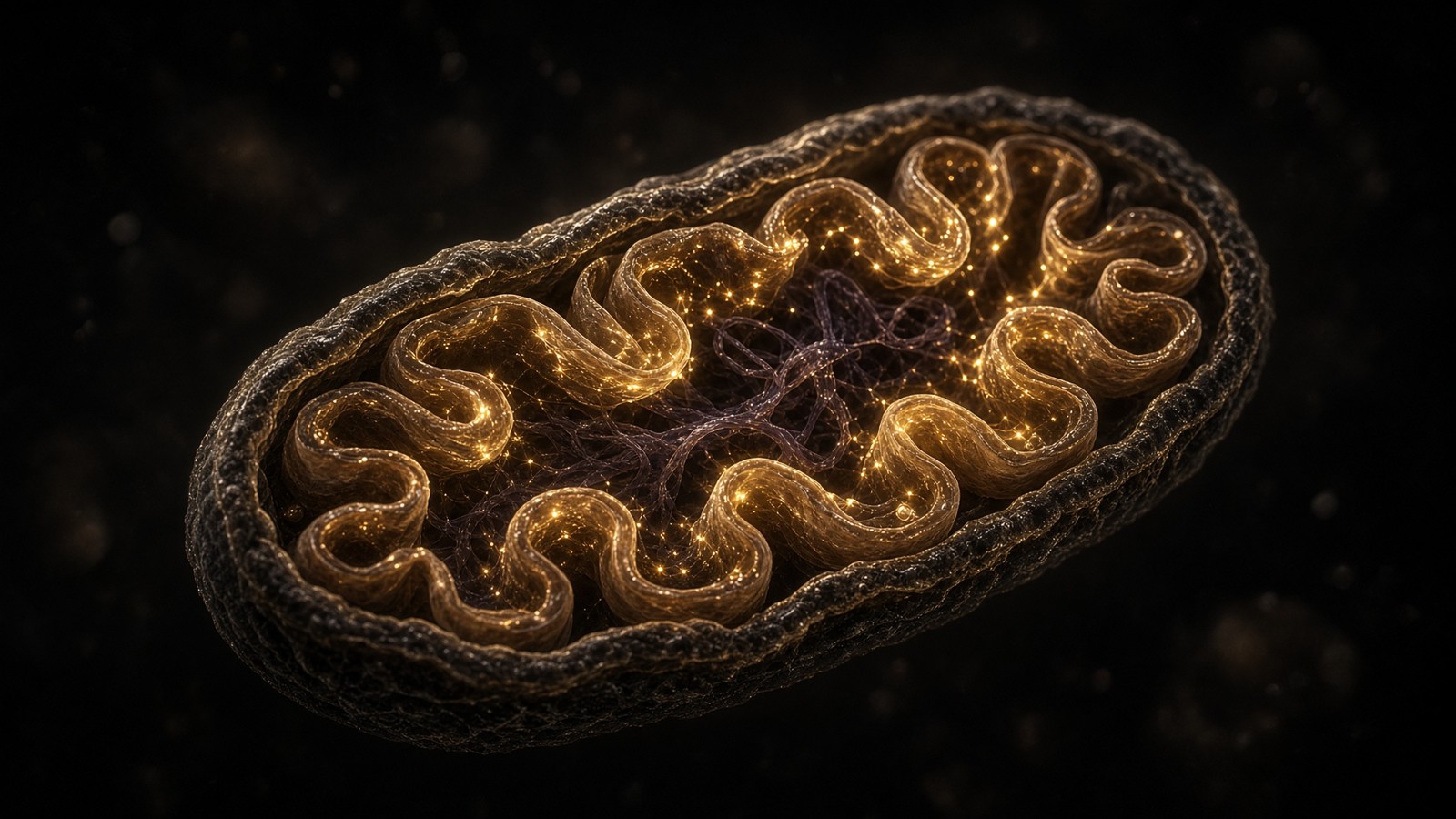
Three tissues, not one tank
The liver is air-traffic control. That analogy once, then the anatomy. A hepatocyte sits on the portal vein, sees what the gut absorbed before any other organ does, and decides what the rest of the blood will carry. It takes up glucose via GLUT2, a high-capacity, insulin-independent transporter, phosphorylates it with glucokinase, and either parks it as glycogen, runs it through glycolysis, or, when citrate and insulin say so, writes fatty acid via acetyl-CoA carboxylase and fatty-acid synthase. It also does the reverse: glycogenolysis and gluconeogenesis, the two halves of endogenous glucose production, which in a fasted adult can be the entire supply the brain is living on. It oxidises fatty acids and, when acetyl-CoA overflows, writes ketones. It takes amino acids, transaminates them, dumps the nitrogen as urea, and keeps the carbon as glucose or as TCA intermediates. No other organ does all of those jobs. Muscle cannot make glucose for export. Adipose cannot run the urea cycle. A model that treats 'the body' as one tank has already thrown away the dispatcher, and then it is surprised when a diet that looked good on a spreadsheet misbehaves in a person.
In short. The liver is the dispatcher. It sees a meal first, makes and takes glucose, writes fat and ketones, and decides what the blood will carry.
Endogenous glucose production is a hepatic sentence before it is a breakfast sentence. Overnight, liver glycogen is drawn down and gluconeogenesis from lactate, alanine and glycerol takes a growing share. Glucagon, cortisol and, in the small hours, a rise in growth hormone lean on that production. Insulin is supposed to mute it; in insulin resistance the mute button sticks, which is why morning glucose is often a hepatic number rather than a toast number. The Cori cycle — lactate from muscle and red cells, glucose back from the liver — is not a curiosity. It is how a working fibre pays its NAD+ debt without needing the rest of you to be in a sprint. The Cahill cycle, alanine out of muscle and glucose back, is the amino-acid version, and it is how a fast starts to cost contractile protein if it runs long enough. First-pass hepatic extraction of portal insulin is large, so the liver sees a higher insulin concentration than a muscle does. That is geometry, not a personality. If we write 'insulin resistance' as a whole-body phrase without saying which tissue is not listening, we have not yet chosen an experiment, or a diet to criticise honestly.
In short. Between meals the liver puts glucose into blood from glycogen and from amino acids. Morning glucose is often that hepatic number, not the breakfast.
Skeletal muscle is the main disposal sink after a meal and the organ that does mechanical work. GLUT4, insulin-translocated and contraction-translocated, is the door. Hexokinase, glycogen synthase, PFK-1, and a mitochondrial census that training can raise: those are the furniture. Post-prandial, most of the glucose that leaves the blood goes into muscle. A day of sitting is a GLUT4 door that never got the contraction signal, so the same insulin has more work to do at the same meal. Fibre type is not a lifestyle brand. Type I fibres are mitochondria-rich, fat-oxidising, recruited first. Type II fibres are glycolytic when they need to be, glycogen-hungry, the fibres a heavy set actually recruits. Training rewrites the census: more GLUT4, more hexokinase, more PGC-1α targets, more capillaries. That's why walking after a meal is a control-system intervention rather than a wellness tip, and why a programme that never empties glycogen never quite lifts insulin sensitivity in the fibres that were supposed to take the glucose. Muscle also oxidises fatty acids at modest intensity, and it will take ketones if asked. It will not export glucose. It is a sink and a motor. It's not a second liver.
In short. Muscle is the big glucose sink after a meal and the organ that does work. Training changes the doors. Sitting leaves them shut.
White adipose tissue is a warehouse, not a character flaw. One large lipid droplet per cell in the classical adipocyte, a thin cytoplasm, a modest mitochondrial census, and a receptor sheet that includes the insulin receptor, β-adrenergic receptors, and a growing list of GPCRs the adipokine literature keeps adding to. The storage job is triglyceride: insulin raises lipoprotein lipase and suppresses hormone-sensitive lipase and ATGL, so fatty acids enter and stay. The release job is the reverse: insulin falls, catecholamines rise, the lipases work, glycerol and non-esterified fatty acids leave. Leptin is the long-range message to the brain about how stocked the warehouse is. Adiponectin, resistin, and a hundred other peptides are the rest of the mail. Visceral and subcutaneous depots are different organs wearing the same colour; visceral fat drains to the portal vein and talks to the liver in a way a gluteal depot does not. An adipocyte is a unilocular cell with a lipid droplet. The moral language glued onto it — lazy, toxic, earned — is a conversation about a person, and it is a worse conversation than the lipase.
In short. Fat cells store surplus as triglyceride and release it when insulin falls. That is a warehouse job, not a verdict on character.
Glycogen caps are the constraint a one-tank model keeps forgetting. Liver glycogen in an adult is roughly eighty to a hundred and twenty grams, a few hours of brain fuel, stored with water, gone after a modest fast. Muscle glycogen is larger, perhaps three to five hundred grams in a trained adult, locked in the fibre that made it, useless to the brain because muscle lacks glucose-6-phosphatase. You can't park more. Supercompensation after depletion is real and is still a cap, not a warehouse. McArdle, Nilsson, Hultman, Bergström: the biopsy literature that put those numbers on paper is older than most of the diets arguing about them. Carbohydrate taken in above the cap does not vanish. It is oxidised at a higher respiratory quotient, or it is written into fat by de novo lipogenesis, which humans do, slowly, mostly in the liver, and which becomes obvious when the surplus is large and chronic. Fructose, in particular, is a hepatic substrate that bypasses PFK-1 and feeds DNL more readily than glucose. The cap is why 'eat less' and 'eat clean' both underspecify the problem. Less of what, into which store, past which cap.
In short. Liver glycogen is about a hundred grams. Muscle holds a few hundred more, locked in those fibres. Everything past those caps overflows.
Overflow is the pathophysiology, and it has addresses. De novo lipogenesis writes new palmitate from carbohydrate-derived acetyl-CoA; the fat is exported as VLDL or parked in the hepatocyte as triglyceride. Ectopic fat in liver and pancreas is the twin-cycle object Roy Taylor put under type 2 diabetes: a fatty liver overproduces glucose and VLDL; that fat lands in the pancreas; first-phase insulin goes quiet. Muscle can pick up lipid too, as intramyocellular triglyceride and as ceramides and diacylglycerols that interfere with insulin signalling. Adipose tissue that can still expand, subcutaneously, is in that sense protective: it parks surplus where the warehouse was built to park it. Adipose that cannot expand, or that is already inflamed and leaky, dumps fatty acids at the liver. A one-tank model looks at scale-weight and calls the job done. A three-tissue model asks where the surplus went. DiRECT emptied hepatic and pancreatic fat with a formula diet and put a large fraction of recent-onset type 2 into remission. That result is a routing result. It's not a sermon about willpower, and it's not a calorie-as-character argument. It is what happens when the overflow is withdrawn from the organs that were drowning in it.
In short. Overflow becomes new fat, including fat in liver and pancreas you would rather not park there. That ectopic fat is how type 2 often starts.
- Liver glycogen
- 80–120 g
- Muscle glycogen
- 300–500 g
- Brain glucose, fed
- ~120 g/day
- Whole-body ATP turnover
- 40–60 kg/day
- Standing ATP pool
- ~50 g
- Proton-motive force
- ~150 mV
- DiRECT remission
- 46% vs 4%
- Retatrutide Phase 2
- 24.2% at 12 mg
A few hours of brain fuel. Gone after a modest fast. Water stored with it.
Trained adult. Locked in the fibre. No glucose-6-phosphatase, so no export.
Cahill. Ketones take a large share once glycogen is gone.
Rich, Biochem Soc Trans, 2003. Recycled, not stored. Standing pool ~50 g.
A coffee-cup, not a warehouse. ~10²¹ hydrolyses a second in a living adult.
Mitchell. Negative inside. ATP synthase is the turbine.
Lean et al., Lancet 2018. Twelve months. Tracked weight loss, ~15 kg.
Jastreboff, NEJM 2023, 48 weeks. Investigational medicine. Not a protocol.
The control layer: five hormones, one organelle
The control layer is the set of hormones that decide which fuel the mitochondrion sees. Insulin, glucagon, cortisol, thyroid hormone and the catecholamines are the five named broadcasts this heading will keep. They're not a ranking. They are a simultaneous conversation. Insulin says store, grow, mute hepatic glucose, suppress lipolysis. Glucagon says the opposite, mostly at the liver: glycogenolysis, gluconeogenesis, β-oxidation. Catecholamines dump fuel for the next few minutes, via β-receptors, cAMP, and the same lipases insulin had been sitting on. Cortisol dumps fuel for the longer emergency and, left high for months, steals amino acids from muscle for gluconeogenesis. Thyroid hormone sets basal throughput: how many sodium pumps, how much uncoupling, how hard the transcriptional programmes for respiratory subunits run. In a living person they all run at once. A fasting morning is low insulin, higher glucagon, a cortisol peak, thyroid in the background, catecholamines if you stood up quickly. A mixed meal inverts the first two, leaves the others where they were, and adds the incretins. Arguing about which hormone is 'most important' is how a control system becomes a brand. The mitochondrion does not read brands. It reads substrate.
In short. Hormones decide which fuel a mitochondrion actually sees. Five named broadcasts run at once in a living person, not as a ranked list.
Insulin is a fifty-one-residue peptide from the β-cell, two chains linked by disulphides, the first peptide medicine, and still the storage-and-growth signal people love to blame for jobs it did not apply for. It occupies a receptor tyrosine kinase. IRS proteins, PI3K, Akt: the cascade that translocates GLUT4 in muscle and adipose, that suppresses FOXO1-driven gluconeogenic transcription in the liver, that activates phosphodiesterase 3B so cAMP falls and the lipases quiet, that feeds mTORC1 so translation ramps. In a hepatocyte it also promotes glucokinase and glycogen synthase and, chronically, the lipogenic transcription factor SREBP-1c. Banting and Best, 1921, is the origin story. The physiology is older than the argument that treats insulin as a prison. Lowering it — by carbohydrate restriction, by fasting, by weight loss itself — helps people whose liver is overproducing glucose. It's not required for fat loss if energy is down; Hall and others have shown isocaloric low-carbohydrate versus low-fat fat-loss is similar when protein is matched, with small advantages that don't match the size of the argument. Insulin routes. It does not repeal thermodynamics. It also does not deserve to be the villain of a supermarket.
In short. Insulin is the storage-and-growth signal: open the glucose doors, stop fat release, tell the liver to stand down. It routes fuel. It is not a prison.
First-phase insulin release is the under-celebrated bit of hardware. Within minutes of a glucose rise, stored granules dump and restrain the liver before the meal has even finished. In early type 2 this phase dies first. That's why two people can eat the same bread and paint very different curves: one still has the dump, the other is already borrowing from a slower, weaker second phase while the liver keeps making glucose in the background. Continuous glucose monitors made the missing first phase visible to people who are not in clinic. The control system was always there. We used to sample it four times a day and pretend that was a curve. Incretins, coming in the next heading, exist in part to make this dump larger when the glucose arrived by mouth. Sulphonylureas force a dump without the glucose-dependence, which is why they cause hypoglycaemia and why the incretin class does so less. Talk about 'insulin' without saying first phase versus second, portal versus peripheral, liver versus muscle, and you are talking about a cloud. The cloud is allowed in a waiting room. It's not allowed in a routing essay.
In short. The first insulin dump after a meal restrains the liver within minutes. That dump dies early in type 2 diabetes, which is why two curves can disagree.
Glucagon is the pancreatic α-cell's broadcast, twenty-nine residues cut from proglucagon, and it is aimed mostly at the liver. The receptor, GCGR, is a class-B GPCR, Gs-coupled, cAMP, protein kinase A. Occupancy raises glycogenolysis, gluconeogenesis, β-oxidation and ureagenesis. Hepatic glucose output rises. In a healthy fasting person that is how the brain keeps a number. In a person with type 2 it is often a broadcast that will not stand down, because the α-cell no longer hears insulin or somatostatin the way it should, and because the hepatocyte is insulin-resistant. Unger's radioimmunoassay work made glucagon a measurement rather than a rumour. The modern twist is that the same receptor is the third occupancy on retatrutide, put there as an energy-expenditure and lipid-oxidation arm, against the metabolic adaptation that usually attends large weight loss. That is a lawful sentence about a receptor. It is a longer walk to a lawful sentence about a person. Glucagon can raise energy expenditure in humans; Tan, Salem and others have measured it. Some of that is hepatic futile cycling. Some, in rodents, is brown fat. Species is a variable. The hormone remains the opposite of insulin at the organ that actually listens to it.
In short. Glucagon tells the liver to make glucose and burn fat. It is the opposite broadcast to insulin, aimed mostly at one organ.
Catecholamines are the minutes-scale dump. Sympathetic fibres release norepinephrine onto β-adrenergic receptors in adipose, liver, muscle and brown fat. Adrenal medulla adds circulating epinephrine for the larger emergency. Gs, cAMP, protein kinase A, the same lipases, a phosphorylation of glycogen phosphorylase via phosphorylase kinase, a rise in cardiac output so the fuel has somewhere to go. A sprint, a fright, a cold morning: the nerve got there first. The hormone in blood is backup. In brown adipose tissue the same occupancy frees fatty acids that open UCP1, and heat is the product rather than ATP; that's the neighbouring essay, and it is a different decision about the same voltage. In muscle the occupancy supports glycogenolysis for the contraction that is already happening. In liver it supports glucose output. β-blockers blunt the whole sheet, which is information you have to remember if the subject is on them. The control layer does not pause for a clinic letter. It just runs quieter. Catecholamines are not a personality called 'stress'. They are a receptor occupancy with a half-life of minutes and a transcriptional echo if they keep arriving for days.
In short. Adrenaline dumps fuel for the next few minutes. Nerves get there first; the hormone in blood is backup. Brown fat uses the same switch for heat.
Cortisol is the longer emergency. The hypothalamic–pituitary–adrenal axis writes it on a circadian clock — high in the morning, low at night — and then writes more of it if the emergency does not end. A glucocorticoid receptor, a nuclear receptor, a transcriptional programme: more gluconeogenic enzymes in the liver, more amino-acid mobilisation from muscle, more lipolysis in some depots and more visceral storage in others, a rise in appetite that the brain reads as a reason to eat the emergency better next time. Overnight, the morning cortisol peak is part of why endogenous glucose production is ready before breakfast. Chronically high, the same programme is how a person on long-term glucocorticoids loses muscle, gains central fat, and paints a glucose curve that looks like type 2. Spiegel, Leproult and Van Cauter wrecked next-day glucose tolerance in healthy young men by taking the night apart; cortisol was part of that wreckage. A short night is a control-layer event. Cushing taught the extreme. Ordinary sleep debt teaches the common case. The mitochondrion of a myofibre, the next day, sees more fatty acid and less of the glycogen it was supposed to refill.
In short. Cortisol dumps fuel for the longer emergency and, left high for months, steals it from muscle. A short night is already this hormone, not a mood.
Thyroid hormone sets basal throughput. The gland releases mostly T4; deiodinases in tissues convert it to T3, the ligand the nuclear thyroid-hormone receptor actually wants. Occupied, that receptor writes sodium–potassium ATPases, respiratory-chain subunits, uncoupling proteins in some tissues, and a general rise in protein turnover. Basal metabolic rate tracks T3. Hypothyroidism is a slow mitochondrion in the colloquial sense: fewer pumps, a quieter chain, a person who feels cold and whose weight creeps. Hyperthyroidism is the reverse, and the weight loss is not a protocol. Brown adipose tissue amplifies the local T3 signal with type 2 deiodinase, which is why a hypothyroid animal is bad at non-shivering thermogenesis. Muscle and liver listen too. TSH is the pituitary's readout, and a TSH in range is a clinic object before it is a forum object. 'Thyroid is metabolism' is a real sentence that still does not license a research peptide as a thyroid. The control layer has a set-point. Thyroid hormone is one of the few members of that layer whose absence you can measure with a single blood test and whose replacement is a licensed tablet. That is a different legal class from everything on the peptide shelf.
In short. Thyroid hormone sets how hard the whole system ticks. Too little and throughput falls; too much and it races. A blood test already names this one.
They all run at once. That's the sentence a control-layer heading has to end on, because we keep picking a favourite. An insulin-centric story of obesity is true about hepatic glucose and about lipolysis, and false as a claim that calories stopped counting. A glucagon-centric story of fasting is true about the liver, and incomplete about muscle. A cortisol story of modern life is true about sleep and about the waiting room, and is not a licence to treat every spare tyre as Cushing. A thyroid story is true about basal rate, and is the most over-requested blood test in people whose TSH is already boring. A catecholamine story is true about the next ten minutes, and is not a personality. Incretins, next, sit on top of the meal and don't replace any of the five. The mitochondrion sees the integral: how much glucose, how much fatty acyl-CoA, at what NAD+/NADH ratio, against what ADP. Hormones wrote that integral. Training and sleep wrote the receptor sheet the hormones act on. A living person is that stack, running. A favourite hormone is a slice of the stack, sold as the building.
In short. In a living person these hormones run together. Picking a favourite is how a control system gets sold as a single story.
Diagram
× 1
Ligand
One peptide in one pocket. nM–µM. Shape, not a mood.
× 10–10²
G proteins
The occupied GPCR is a GEF. Each Gα is a catalyst.
× 10³–10⁴
cAMP / IP₃ / Ca²⁺
Adenylyl cyclase and PLC do not make one molecule. They make a cloud.
× 10⁴–10⁶
PKA / PKC / CaMK
Kinases phosphorylate many substrates per messenger.
× tissue
Secretion, transcription, motility
The organism-level readout. Still not a protocol.
This is the only magic, and it is not magic. A nanomolar ligand can move a micromolar messenger because enzymes sit between them. Desensitisation (GRK, β-arrestin, endocytosis) is how the cell refuses to let ‘more ligand’ mean ‘more signal’ forever.
Incretins sit on top of the meal
The incretin effect is a paired curve before it is a drug. Give glucose by mouth and the insulin rise is larger than the rise you get from the same glucose load in a vein. McIntyre, Holdsworth and Turner put that contrast into the Lancet in 1964. Elrick, Stimmler, Hlad and Arai published the same shape the same year. The gut, they argued, must be releasing something that amplifies the β-cell's response to glucose. GIP, from K-cells in the proximal small intestine, was isolated first and misnamed gastric inhibitory polypeptide before the insulinotropic job stole the acronym. GLP-1, from L-cells denser toward the ileum and colon, is a post-translational product of proglucagon; Habener, Holst and Drucker made that hiding place a receptor you could occupy on purpose. Both peptides occupy class-B GPCRs, Gs-coupled, and both amplify glucose-stimulated insulin secretion. Both also slow the stomach, to different degrees. Native GLP-1 is destroyed in minutes by dipeptidyl peptidase-4. The analogue strategy occupies the receptor with a chain the enzyme cannot eat as fast. Sitagliptin occupies the enzyme. Both exist because the two-minute half-life is real, and because oral versus intravenous is still the measurement that gives this family a job.
In short. Gut hormones warn the pancreas that sugar arrived by mouth. That warning is why swallowed glucose raises more insulin than the same glucose in a vein.
Gastric emptying is half of the post-prandial curve people blame on 'carbs' as a monolith. A liquid sugar appears in the duodenum quickly, incretin tone and an insulin dump follow, and the liver and muscle have to catch a steep appearance rate. A mixed meal with protein, fibre and fat empties more slowly; appearance rate falls; the same starch paints a quieter curve. GLP-1R on the stomach and on the brainstem is why incretin agonists delay emptying and why nausea is on-family pharmacology rather than a contaminant. Appearance rate, incretin tone, and how much muscle you recruited yesterday will write more of a continuous-glucose trace than the macronutrient tribal affiliation. That's not an argument against carbohydrate restriction in a person whose liver is overproducing glucose. It is an argument against treating 'carbs' as a moral category when the control system is reading rate, route and receptor occupancy. Walk after the meal and you open GLUT4 by contraction. Eat the protein first and you slow the appearance. Sleep, because cortisol and insulin sensitivity both notice. Those levers are free and they move the variable. They are also, inconveniently, not a brand.
In short. Slowing the stomach is half of a meal's glucose curve. A liquid sugar hits differently from a steak for that reason, not because of virtue.
Retatrutide is LY3437943, a fatty-acylated unimolecular agonist at GIPR, GLP-1R and GCGR. Coskun and colleagues described the engineering in Cell Metabolism in 2018: one backbone, one lipid so albumin will carry the chain through a week, a set of relative potencies a medicinal chemist would call bias. Jastreboff and colleagues, New England Journal of Medicine 2023, Phase 2, obesity: 24.2 percent mean weight loss at 12 milligrams, 48 weeks. That paper is an investigational-medicine trial. It's not a diet. Semaglutide had already shown that GLP-1R occupancy, on its own, could move large clinical endpoints. Tirzepatide added GIPR. Retatrutide asks what a third occupancy, glucagon, does to energy expenditure on the same chain. We synthesise the published structure in the United States, HPLC-MS, labelled for research use. We're not Eli Lilly. Occupancy at those three receptors sits in the neighbourhood this page has been mapping: appearance rate, insulin dump, hepatic glucose, a possible expenditure lift. Neighbourhood is not a protocol. A research vial is a ligand for a tube. A Phase 2 curve is a paper. Human fuel routing is a set of tissues and hormones that were here before either object. Hold the three floors apart.
In short. A published three-receptor chain sits in this neighbourhood. Occupying those gut-hormone receptors is chemistry, and it is not a diet written as a vial.
Diagram
GLP-1R
β-cell, brainstem, stomach
Incretin, delayed emptying, satiety. Semaglutide’s occupancy.
GIPR
β-cell, adipocyte
Second incretin. Lipid handling. Tirzepatide added this.
GCGR
hepatocyte
Glycogenolysis and, biased, energy expenditure. The third occupancy.
LY3437943 is a fatty-acylated unimolecular agonist at GIPR, GLP-1R and GCGR (Coskun, Cell Metab 2018). Jastreboff, NEJM 2023: 24.2% mean weight loss at 48 weeks, 12 mg, Phase 2 — clinical literature, not a use instruction for a research vial.
Cahill, ketones, and the brain's second fuel
George Cahill's starvation studies, run in the 1960s on volunteers and on fasting obese patients at the Joslin, are still the document for what a human does when glycogen runs out. Brain glucose consumption in a fed adult is on the order of 120 grams a day. After a few days of starvation, hepatic glycogen is gone, gluconeogenesis from amino acids would have to cannibalise muscle to keep that number, and the brain has induced the transporters and enzymes that let it take β-hydroxybutyrate and acetoacetate instead. Cahill measured the switch: ketone uptake rising, glucose uptake falling, nitrogen loss slowing once the brain no longer demanded a full gluconeogenic ration. A well-formulated ketogenic diet is a milder, voluntary version of the same physics. The carnivore pattern, on this desk in another essay, is that idea plus an elimination trial. None of this is mystical. HMGCS2 commits hepatic acetyl-CoA overflow to ketogenesis; extrahepatic tissues oxidise the ketones back via BDH1 and SCOT. The backup generator is a liver writing a water-soluble, brain-permeant fuel from fat. A one-tank model that cannot name that generator will treat every low-carbohydrate week as a personality transplant. It is not.
In short. Cahill showed a starving brain will run on ketones and spare muscle. That backup generator is still under-taught, and it is ordinary liver chemistry.
The chemistry is an overflow valve. Hepatic β-oxidation throws off acetyl-CoA. When citrate synthase and the TCA cycle cannot take the lot — oxaloacetate has been withdrawn for gluconeogenesis, NADH is high, the cycle is full — mitochondrial HMG-CoA synthase 2 condenses acetoacetyl-CoA with another acetyl-CoA, and the HMG-CoA lyase cut produces acetoacetate. β-hydroxybutyrate dehydrogenase reduces a fraction to BHB, the species you measure in blood; a little spontaneously decarboxylates to acetone. Extrahepatic tissues, including brain, heart and kidney, take BHB back to acetyl-CoA and burn it. Newman and Verdin later showed that BHB is also an HDAC inhibitor; Youm showed it can quiet NLRP3. Fuel and signal, the neighbouring ketone essay. For this page the point is routing. Ketogenesis is what a liver does with fat when it cannot store it and cannot burn all of it through citrate synthase. Insulin suppresses HMGCS2 transcription and, by suppressing lipolysis, dries up the substrate. Glucagon and a low insulin lean the other way. People who 'stall' on a high-protein plate often turn out to be eating enough protein that glucose production and insulin never fully stand down. That is HMGCS2 competing with a busy TCA cycle.
In short. When the liver cannot park acetyl-CoA in the Krebs cycle, it writes ketones and exports them. Insulin dries that valve; fasting opens it.
Protein is not free glucose. The sentence is true and incomplete. Alanine and glutamine are gluconeogenic substrate; they feed hepatic glucose output via pyruvate and via the TCA cycle. A high-protein carnivore plate is therefore less ketogenic than a fat-heavy one, which is why rabbit-starvation is a protein problem as much as a fat-deficiency problem. Muscle, under cortisol or under a hard fast, exports alanine — the Cahill cycle already named — and the liver writes glucose that the brain, until ketones are up, still wants. In a fed person, dietary protein meets a leucine threshold, mTORC1 opens in the fibres that were loaded, and the amino acids become myofibrils rather than glucose. Tension without amino acids is a signal without bricks. Amino acids without tension mostly make expensive urea. Morton, Phillips, Schoenfeld: the hypertrophy literature that put 1.6 grams per kilogram near the ceiling for most trainees. The routing point is simpler. Amino acids have a choice of fates, and hormones plus mechanical tension decide the fate. A one-tank model that files protein under 'calories' will not predict a cut that kept muscle, and will not predict a fast that spent it.
In short. Protein is not free sugar, but some amino acids can become glucose. Whether they rebuild muscle or feed the liver depends on tension and on hormones.
Given time, the human brain will run mostly on ketones and spare the muscle it would otherwise have spent on gluconeogenesis. That is a measured switch, not a personality.— Cahill's starvation studies, in working English. Joslin, the 1960s, still the document.
AMPK and PGC-1α rewrite the doors
A bout of endurance work is a metabolic drug with a dose. It empties glycogen, which is still the most reliable way we have to lift subsequent insulin sensitivity in those same fibres. It raises AMP, which raises AMP-activated protein kinase. AMPK phosphorylates acetyl-CoA carboxylase, malonyl-CoA falls, CPT-I opens, fat oxidation rises. AMPK also leans on ULK1 and on TSC2, so autophagy ticks up and mTORC1 ticks down for the duration of the bout. Hours later, PGC-1α has been transcribed and coactivated, and nuclear genes for respiratory subunits and for TFAM get written. Mitochondrial biogenesis is a transcriptional programme, not a supplement. VO2 max is the organism-level readout of that programme plus the heart: stroke volume, blood volume, capillary density and mitochondrial capacity in series. Polarised versus pyramidal is a programming argument. The organelles are not. You either build them or you do not. Fat oxidation really does peak at modest intensity, often around the first lactate threshold, which is where the zone-2 conversation started before it became a brand. Skip one mode because a podcast picked a favourite and you end up with a big engine on a small chassis, or the reverse.
In short. Endurance work empties glycogen and switches on a fuel-gauge kinase, then a programme that builds more mitochondria. That is a dose, not a personality.
AMPK is the cell's fuel gauge. The heterotrimer — a catalytic α subunit, a β scaffold, a γ subunit that binds AMP and ADP — is activated when ATP falls relative to AMP, and by liver kinase B1 and by calcium-calmodulin kinase kinase 2. Hardie, Kahn, the reviews a physiology paper actually cites. Phosphorylation of ACC1 and ACC2 drops malonyl-CoA, which both slows de novo lipogenesis and lifts the brake on CPT-I. Phosphorylation of TBC1D1 assists GLUT4 translocation in muscle, insulin-independently, which is why a contraction can take glucose without an insulin dump. Phosphorylation of ULK1 starts autophagy. Phosphorylation of TSC2 quiets mTORC1. The kinase is ancient; yeast SNF1 is the orthologue. Metformin, whatever else it does, sits in this neighbourhood at the liver, which is why a diabetes tablet and a long run can rhyme on a blot without being the same object. MOTS-c, in the Lee papers, sits on AMPK too. That is a mitochondrial 16-mer occupying a fuel-gauge kinase, a different invoice from a contraction. AMPK is how a cell, not a person, notices that ATP is running behind. Whole-body 'activation' as a product claim is a category error the kinase did not ask for.
In short. AMPK notices when ATP is running behind. It leans the cell toward burning fuel and recycling parts rather than storing, until the charge recovers.
PGC-1α is the transcriptional coactivator that sits on the decision to make more mitochondria. AMPK, calcium-calmodulin kinases and p38 MAPK all converge on it during endurance work; SIRT1 deacetylation is the NAD+-gated half of the same sentence. Occupied, PGC-1α works with NRF1, NRF2 and ERRα, nuclear genes for respiratory subunits and for TFAM get written, the organelle census can rise. You get more cristae, more fatty-acid oxidation enzymes, better insulin-independent glucose uptake. The coactivator is also in brown fat, in heart, in liver; the partners change, the logic does not. Spiegelman's laboratory put PGC-1α on the map in the late 1990s and has spent the decades since watching people treat it as a supplement target. It is a protein the cell writes when the work has already happened. Cold, endurance, a genuine fast: those are the stimuli the papers measured. A catalogue that stocks NAD+ stocks the cofactor SIRT1 spends on PGC-1α, which is a real paper-trail and is not a biogenesis product. The training essay on this desk is the version that still has a session in it. You can't skip the work and keep the coactivator.
In short. PGC-1α is the coactivator that writes more mitochondria. Endurance work, cold and some fasts lean on it. The session still has to happen.
mTORC1 is the other switch, and concurrent training is the art of not letting one signal chronically cancel the other. Mechanical tension deforming the fibre, plus leucine via sestrin2 and the Rag–Ragulator complex at the lysosome, opens mTORC1. S6K1 and 4E-BP1 are phosphorylated, translation of myofibrillar proteins ramps. AMPK's veto during the endurance bout is real and temporary. People who row and squat solve this daily; people who post about interference solve it less often. Sleep is a control-layer reset: growth hormone pulses in slow-wave sleep, cortisol falls at night if the night is intact, insulin sensitivity the next morning tracks the hypnogram more honestly than it tracks the previous day's macros. A fever raises expenditure and rewrites hepatic glucose output. Yesterday's meal is still in the glycogen stores and in the intestinal incretin tone. Exercise rewrites the doors for hours afterwards. So does a short night. The control layer is not a static ranking of five hormones. It is a ranking that training, sleep, illness and the last plate keep editing. A routing essay that stopped at insulin and glucagon would have stopped before the kinases that decide how many mitochondria the next week will have.
In short. Heavy lifting uses a different switch to add contractile protein. Sleep, a fever and yesterday's meal rewrite the fuel doors for hours as well.
Why a one-tank model mis-predicts every diet
Conservation of energy is not a personality. If, over a month, ingested metabolisable energy exceeds expenditure, the difference is stored, mostly as triglyceride. That is true on keto, on carnivore, on a Mediterranean pattern, and on a ward diet a dietitian wrote. The live question is what actually moves the two sides of that equation in a human who has to live in a kitchen. A five-hundred-kilocalorie spreadsheet deficit that leaves you ravenous will be eaten back, usually from the cupboard you swore you had closed. A diet that quietly drops five hundred kilocalories because the food is filling will look like magic and still be arithmetic. The one-tank model stops at the arithmetic and then is confused by the person. The three-tissue model asks what happened to hepatic glucose output, to muscle glycogen, to adipose lipolysis, to leptin. Those are the levers that decide whether the arithmetic can be lived with. Named diets are different ways of leaning on one of those levers: more protein, less energy density, a forbidden list that cuts a cue. Pick the mix you can actually run. The physics does not care what you named it.
In short. Energy in minus energy out still holds. A one-tank model still mis-predicts diets because routing and appetite are the levers a person actually has to live with.
Kevin Hall's NIH ward studies are the inconvenient tables. Isocaloric low-carbohydrate versus low-fat, protein matched, in a metabolic chamber: fat loss is similar, with small advantages that don't match the size of the argument around them. An ultra-processed pattern, macronutrients matched to an unprocessed one, drove spontaneous overeating. The first result is why 'insulin is a prison' failed as a universal fat-loss theory. The second is why palatability is a control-layer problem as much as a moral one. Carbohydrate restriction can still be the right tool in a person whose liver is overproducing glucose, because you have stopped putting the variable into the system, and because insulin and hepatic glucose output fall, sometimes before heroic scale-weight change. Virta and the ketogenic-diet literature live there. Very-low-fat high-carbohydrate patterns can also drop liver fat if the carbohydrate is not liquid sugar and calories actually fall. Both roads fail if the energy surplus and the liver fat come back, which they will if the only plan was a heroic month. Hall did not repeal hormones. He measured them in a setting where the tank's two sides were actually known.
In short. When protein is matched, low-carb and low-fat lose similar fat in ward studies. The argument around them is larger than the measured difference.
Simpson and Raubenheimer's protein-leverage hypothesis: many animals eat until protein needs are met. Dilute protein in a high-fat, high-sugar food supply and they over-eat energy to get there. Raise protein and total energy often falls without a lecture. The thermic effect — roughly twenty to thirty percent of protein energy spent on processing — plus GLP-1, PYY and satiety add a hormonal layer. Energy density is the mechanical cousin. A stomach has a volume, and four hundred kilocalories of steak and potatoes occupy it differently from four hundred kilocalories of oil and refined starch. Ultra-processed palatability, salt, sugar, fat and a refined texture, is the third lever, the one that lets people eat past both protein and stretch signals. Those three — protein leverage, energy density, palatability — are why a named diet can 'work' without anyone having discovered a new law of physics. They are also why a spreadsheet that ignores them fails on a Friday. The routing table still applies. Protein that was used to rebuild a fibre did not become hepatic glucose. Starch that appeared slowly did not demand the same first-phase dump. Fat that stayed in a subcutaneous warehouse did not land in the pancreas.
In short. Protein leverage, how bulky the food is, and how engineered it is to be eaten quickly decide whether a person can live inside a deficit.
Type 2 diabetes, for a lot of people, is a liver and pancreas clogged with fat, a first-phase insulin response that has gone quiet, and muscle that ignores the insulin that does arrive. Roy Taylor's twin-cycle hypothesis put that in working English. DiRECT, 2018, cluster-randomised, UK primary care, tested a practical version: a formula very-low-calorie diet, then stepped food reintroduction, in people with type 2 of less than six years. At twelve months, 46 percent of the intervention arm were in remission against 4 percent in control. Remission tracked weight loss. Around fifteen kilograms the odds became very good. Two-year follow-up showed the expected fade in people who regained. Carbohydrate restriction is a parallel road: stop putting the variable in, let insulin and hepatic glucose output fall, empty ectopic fat over time. Both roads are three-tissue stories. A one-tank model looks at the fifteen kilograms and stops. A routing model asks whether hepatic fat fell, whether first-phase insulin came back. Remission is not the pancreas of a twenty-year-old. Long-standing type 2 and type 1 are different biology. Nobody competent claims a steak or a peptide vial is a substitute for medical care in a person on hypoglycaemic drugs.
In short. Type 2 remission trials emptied fat from liver and pancreas. That is a tissue story, not a furnace story, and it is not a licence to ignore clinic drugs.
Named reagents in the neighbourhood
NAD+ is nicotinamide adenine dinucleotide, the hydride coin this page has been spending without always naming. Carbon 4 of the nicotinamide ring accepts a hydride; the molecule becomes NADH; Complex I oxidises it back. That is still most of the pool's day job, the job GAPDH, malate dehydrogenase and the acyl-CoA dehydrogenases all have. Three enzyme families consume NAD+ as a substrate rather than recycle it as a coenzyme: sirtuins, PARP1, CD38. Salvage through NAMPT is the kinetic bottleneck. The pool falls with age in specific tissues, in part because CD38 rises. Mitochondria notice first, because Complex I and SIRT3 both sit on the pool, and because the matrix is not in free exchange with the cytosol. We stock lyophilised β-NAD+, a thousand milligrams, HPLC-characterised, because that's the cofactor the sirtuin, PARP and CD38 papers actually weigh into a tube. The NAD+ essay on this desk is the topology. This paragraph is the invoice: a routing essay that talks about reducing equivalents without naming the dinucleotide is leaving the currency off the page. A 1000 milligram cake is a reagent for an assay you control. It's not forty kilograms of ATP, and it's not a drip.
In short. NAD+ is the coin mitochondria use to move electrons, and some enzymes spend it rather than recycle it. The research solid is that coin, for a tube.
MOTS-c is Met-Arg-Trp-Gln-Glu-Met-Gly-Tyr-Ile-Phe-Tyr-Pro-Arg-Lys-Leu-Arg, written MRWQEMGYIFYPRKLR, sixteen residues translated from an open reading frame in mitochondrial 12S rRNA. Lee, Kim, Cohen, Cell Metabolism 2015: AMPK, the folate–methionine cycle, insulin-sensitivity assays in mice. Kim and Lee, 2018: nuclear translocation under metabolic stress. Reynolds, 2021: exercise-induced, age-dependent physiology in plasma. The organelle that houses the electron-transport chain is the organelle that wrote this peptide. That is a shared address, not a shared job. MOTS-c occupies none of GLP-1R, GIPR or GCGR. It's not NAD+. AMPK is the fuel-gauge kinase a contraction already occupies, which is why exercise-mimetic language in a mouse paper is a phenotype and not a product claim. We stock the sixteen-mer as HPLC-characterised research material because the papers are real. We will not file it under thermogenesis, under a diet, or under a smaller NAD+. A mitochondrial open reading frame is a surprising object, and it remains surprising if you keep its invoice separate. Confusing a 16-mer, a dinucleotide and a triple agonist is how a journal becomes a stack. Neighbourhood, on this page, is a courtesy on a reading list. Same organelle as the chain. Different job.
In short. MOTS-c is a short peptide a mitochondrion writes from its own RNA. It talks to the fuel-gauge kinase. Same organelle as the chain, different job.
Three related peptides, then, and three honest jobs. Retatrutide: class-B GPCR occupancy, intake and a glucagon-shaped energy-expenditure neighbourhood. MOTS-c: a mitochondrial open reading frame with AMPK papers. NAD+: the cofactor the chain and the sirtuins actually spend. You can read them on the same afternoon as Cahill, as Hall, as DiRECT, as Hardie on AMPK and Spiegelman on PGC-1α. You can't stack them into a protocol that replaces the routing table. A smaller person with a quieter liver is a different NADH supply to a different set of mitochondria; flux will follow because flux follows fuel. That is downstream weather. It's not occupancy at Complex I, and it's not a 16-mer becoming a triple agonist. The pathophysiology stack still holds: receptor or cofactor floor, then tissue, then organism. Skip a floor and you write a claim the assay cannot carry. We will sell you the named objects, HPLC-characterised, American-made where that's the listing. We will not design the blot, and we will not write a protocol that pretends a gram of cofactor is a sixteen-mer or a weekly incretin. The tissues will still be doing their jobs in the morning, whether or not anyone opened a vial.
In short. Three catalogue objects, three different jobs. A reading list can sit them together. A protocol cannot, and a diet cannot either.
Diagram
| Node | Catalogue | Conversation |
|---|---|---|
| GPCR | Ipamorelin, MT2, PT-141, retatrutide, CJC | Second messengers, secretion, appetite, pigment |
| RTK / IGF1R | IGF-1 LR3 | IRS–PI3K–Akt–mTOR and Shc–ERK |
| Cytokine receptor | Somatropin (HGH) | GHR–JAK2–STAT5b, hepatic IGF-1 |
| Cofactor | NAD+ | Sirtuins, PARPs, CD38, redox |
| Actin buffer | TB-500 / Tβ4 motif | G-actin sequestration, motility |
| Growth-factor-like | BPC-157 | VEGFR2 / FAK / eNOS neighbourhood |
| Copper ligand | GHK-Cu | Transcriptome shift in fibroblasts |
| MC fragment | KPV | NF-κB, PepT1, no pigment |
| Nuclear / pineal | Epithalon (AEDG) | TERT and melatonin literatures |
| mtORF peptide | MOTS-c | AMPK, folate–methionine cycle |
Each row is a different kind of molecular conversation. The catalogue peptides bind at these nodes; they are not interchangeable, and stacking them because a forum did mixes unrelated literatures.
Close: routing table, public papers, laboratory reagents
The picture, restated without the favourite-hormone stories. Energy in a human is ATP, minted by glycolysis, by β-oxidation feeding the TCA cycle, and by the electron-transport chain that cashes the reducing equivalents those paths produced. The currency, before the phosphate, is NADH and QH2. Calories are bookkeeping. Insulin, glucagon, cortisol, thyroid hormone and the catecholamines are the control layer that decides which fuel the mitochondrion sees; the incretins sit on top of the meal and warn the pancreas, slow the stomach, and, in the analogue literature, move intake. Liver, muscle and adipose are not interchangeable. Glycogen caps are real and small. Overflow is ectopic fat. Cahill's ketones are the backup the brain will take when the caps are empty. AMPK and PGC-1α are how work rewrites the organelle census. A one-tank model mis-predicts every diet because it has thrown away the dispatcher, the sink, the warehouse and the control layer. That's the whole page. The rest was names, numbers and the papers you would want before treating a packet, a CGM trace, or a catalogue listing as a physiology.
In short. Leave with the map: three fuels, three tissues, a control layer, a ketone backup, and kinases that rewrite the doors. Calories remain bookkeeping.
The public papers are the reading list, and they are short enough to actually read. Cahill, New England Journal of Medicine and the later Annual Review of Nutrition, fuel metabolism in starvation, the ketone switch measured. Rich, Biochemical Society Transactions, 2003, the ATP turnover number this journal keeps using because it is the right size. McIntyre, Lancet 1964, oral versus intravenous glucose, the incretin effect as a paired curve. Mitchell, 1961 and the 1978 Nobel, the gradient as the point of the chain. Hardie on AMPK; Spiegelman on PGC-1α. Hall's ward studies, the isocaloric tables and the ultra-processed ad-libitum intake. Lean, DiRECT, Lancet 2018, remission as emptied ectopic fat. Coskun, Cell Metabolism 2018, LY3437943 as chemistry; Jastreboff, NEJM 2023, as a Phase 2 curve, not a protocol. Lee, Cohen, Cell Metabolism 2015, MOTS-c. Imai and Guarente, Nature 2000, NAD+ as a sirtuin substrate. Taylor's twin-cycle reviews, so the overflow has an address. That is a fortnight of evenings, not a guru. The diet headlines will still be there when you come back, and they will look smaller, which is the only reliable effect of reading the papers they were built on.
In short. A short stack of named papers covers Cahill, the chain, incretins, ward diets, DiRECT and the exercise coactivator. Read those before any headline.
What you should leave with is a topology, not a shopping list. Reducing equivalents are the currency; ATP is the product; calories are the bookkeeping. The liver dispatches, muscle disposes and works, adipose buffers. Glycogen caps overflow into fat, including fat in organs that then misbehave. Five hormones, plus the incretins, write the integral the mitochondrion sees. Work writes AMPK and PGC-1α and, in the fibres you loaded, mTORC1. Ketones are a lawful backup, not a badge. Named research sequences occupy named nodes of this machinery — a cofactor, a mitochondrial 16-mer, three class-B receptors — and occupying a node is not owning the phenotype the node sits under. If your experiment needs the cofactor, weigh it and name the drain. If it needs the 16-mer, show AMPK. If it needs a triple agonist, occupy the receptors in a system you actually have the controls for. If it needs a medicine, this catalogue does not sell one. If it needs a diet, Cahill, Hall and DiRECT are still better company than a comments thread. The routing table does not care which of those sentences you came here for. It will still be running at breakfast.
In short. Leave with fluxes, not a shopping list. The liver is still dispatching, and a node is not the whole phenotype.
Treat the body as one tank and every diet will surprise you. A low-carbohydrate week that flattened a glucose curve did so at the liver and at appearance rate, not because thermodynamics took a holiday. A high-carbohydrate, high-fibre pattern that dropped liver fat did so because energy fell and because the hepatocyte was no longer being asked to write palmitate all afternoon. A high-protein cut that kept muscle did so because leucine and tension occupied mTORC1 while the deficit occupied adipose. A very-low-calorie formula that put type 2 into remission did so because hepatic and pancreatic fat fell. Each of those is a three-tissue story with a control layer. The tank story can only say that weight moved. Sometimes weight moving is the job. Often the job is the organ the surplus was drowning. Name the tissue. Name the hormone. Name the cap you were up against. Then talk. The neighbouring essays — blood sugar, ketones, liver fat, brown fat, mitochondria, training — are the rest of this map at a different zoom. This piece was the wide shot: hormonally gated flux, with the liver as dispatcher, and calories as a necessary, insufficient number on a packet.
In short. Treat the body as one tank and every diet will surprise you. Name the tissue, the hormone and the cap. Then the diet conversation has somewhere to sit.
The neighbouring reagents remain reagents. NAD+ is the hydride coin, a thousand milligrams of lyophilised β-NAD+ for the assays a routing paper actually runs: a sirtuin tube, a PARP assay, isolated mitochondria whose oxygen consumption you record. MOTS-c is MRWQEMGYIFYPRKLR, HPLC-characterised, a sixteen-mer for AMPK and one-carbon neighbourhoods a blot can name. Retatrutide is the published LY3437943 backbone, American-made, HPLC-MS, a ligand at three class-B receptors, not the investigational pen and not Eli Lilly. eLIVEate's intramuscular NAD+ appointment, where it exists, is a different product at a different company; Patriot takes no commission on that booking. Same carbon skeleton on a whiteboard, in the NAD+ case, and a different legal object in the room. Three floors of a pathophysiology stack. A reading list can travel all three. A reconstitution does not inherit a Phase 2 curve, a clamp, or a Cahill protocol. Adults can want a paper and an appointment. Bundling them is how a research reagent becomes a medical claim it's not allowed to be. This paragraph is the unmix. The physiology above does not depend on it. The labels do.
In short. The neighbouring vials are laboratory solids with named jobs. A clinic injection, where it exists, is a different product at a different company.
Research-use-only. Not for human consumption, not a medicine. The lyophilised solids on the related listings are laboratory reagents, HPLC-characterised, labelled for in-vitro work: a standard curve, a receptor assay, a kinase blot, a set of isolated mitochondria whose oxygen consumption you actually record. The physiology in the paragraphs above is public, cited, and older than any vial. Use it to design the experiment you have the controls for, with the tissue named, the hormone named, the compartment named, and the time point written down. Read Cahill, read Hall, read DiRECT, read Coskun, read Lee, then weigh the cake that matches the question, or eat the lunch that matches the cap. We will sell you the cofactor, the sixteen-mer and the published triple-agonist backbone. We will not tell you they are a routing table you can inject and draw as a new metabolism. Human metabolism is a set of hormonally gated fluxes. Calories are bookkeeping. The liver is still dispatching whether anyone asked it to or not. These fluxes you can measure, in a person, in a chamber, in a tube, with a chromatogram on the bench beside the last of those.
In short. The vials are research chemicals for experiments, not medicines and not food. The biology is public. Measure the flux, and keep the claim the size of the assay.
- Reducing equivalents are the currency. ATP is the product. Calories are bookkeeping.
- Liver dispatches, muscle disposes and works, adipose buffers. A one-tank model throws all three away.
- Glycogen caps: liver ~80–120 g, muscle ~300–500 g. Overflow is ectopic fat.
- Insulin, glucagon, catecholamines, cortisol, thyroid hormone: the control layer. Incretins sit on the meal.
- Cahill: the brain will take ketones and spare muscle. HMGCS2 is the overflow valve.
- AMPK and PGC-1α rewrite the organelle census. The session still has to happen.
- NAD+, MOTS-c and retatrutide occupy a cofactor, a 16-mer and three GPCRs. Neighbourhood, not a stack.
Questions the essay actually answers
- Is this medical advice?
- No. It is a physiology essay: hormonally gated flux through glycogen, fat and amino acids, with the liver as dispatcher. Nothing in it is a protocol, a diet prescription, or a reason to change a prescribed medicine.
- What actually regulates metabolism?
- Insulin, glucagon, adrenaline, cortisol, thyroid hormone and the incretins decide which fuel door is open. Exercise, sleep and yesterday's meal rewrite those doors. The mitochondrion reads the substrate that integral delivered.
- Do calories still count?
- Yes. Conservation of energy is not optional. A calorie is still a unit of heat, useful for a label and insufficient as a routing table. Hormones, palatability and tissue-level caps decide whether a spreadsheet deficit can be lived with.
- Why isn't the body one tank?
- Liver, muscle and adipose are not interchangeable. Muscle cannot export glucose. Adipose cannot run the urea cycle. The liver sees the portal vein first. A model that treats 'the body' as one tank mis-predicts every diet.
- What are the glycogen caps?
- Liver roughly 80–120 g, muscle perhaps 300–500 g in a trained adult. Muscle glycogen cannot feed the brain. Everything past those caps is oxidised or written into fat, including ectopic fat in liver and pancreas.
- What did Cahill actually show?
- In starvation the brain takes a large ketone share and nitrogen loss slows, because gluconeogenesis no longer has to feed a full 120 g/day glucose ration. Ketogenesis is hepatic acetyl-CoA overflow. A backup generator, measured, not a badge.
- What are incretins?
- GLP-1 from L-cells and GIP from K-cells amplify glucose-stimulated insulin secretion and slow the stomach. That is why oral glucose raises more insulin than the same glucose in a vein, and why incretin-receptor agonists move the post-prandial curve.
- What do AMPK and PGC-1α do?
- AMPK is the cell's fuel gauge: AMP up, burn and recycle rather than store. PGC-1α is the coactivator that writes more mitochondria after endurance work. Training is the stimulus. The proteins are the readout.
- Where do research peptides sit in this picture?
- Named sequences such as retatrutide, MOTS-c and NAD+ occupy specific nodes of this machinery — three class-B GPCRs, AMPK, a hydride-carrying cofactor. They are laboratory reagents, not a diet. Neighbourhood is not identity.
- Is a research peptide a substitute for DiRECT or for training?
- No. DiRECT was a food-and-weight trial. Mitochondrial biogenesis needs the session. We stock the published LY3437943 structure, a mitochondrial 16-mer and lyophilised β-NAD+. A vial did not run those experiments, and we do not claim it did.
Hypothetical research reconstitution
How these vials are typically mixed
Hypothetical research reconstitution for the named catalogue vial. Not a protocol, not medical advice, not a use instruction. These amounts sit in published and commonly cited laboratory ranges. The vial is labelled for research use only — not for human or veterinary administration.
NAD+
1000mg
Mix with 10 ml bacteriostatic water → 100 mg/ml
- Hypothetical aliquot
- 50–100 mg
- 0.50–1.00 ml · 50–100 units on a U-100 syringe
- How often
- Two or three times per week in published infusion and assay notes
- 4–8 weeks, then a pause
Bench steps
- Let the vial sit until it is no longer cold to the touch.
- Wipe the stopper with 70% isopropyl alcohol. Let it dry.
- Draw 10 ml bacteriostatic water (0.9% benzyl alcohol).
- Run the water slowly down the inside glass — do not blast the cake.
- Roll between finger and thumb until the cake is gone. Do not shake.
- Label the date. Store the solution at 2–8 °C. Do not freeze. Use within 30 days unless the note below says otherwise.
A 1000mg cake wants 10 ml. Protect from light. Solution yellows as it oxidises — that is the cofactor dying, not a flavour. Use promptly.
MOTS-c
40mg
Mix with 2 ml bacteriostatic water → 20 mg/ml
- Hypothetical aliquot
- 5–10 mg
- 0.25–0.50 ml · 25–50 units on a U-100 syringe
- How often
- Two or three times per week
- 4–8 weeks
Bench steps
- Let the vial sit until it is no longer cold to the touch.
- Wipe the stopper with 70% isopropyl alcohol. Let it dry.
- Draw 2 ml bacteriostatic water (0.9% benzyl alcohol).
- Run the water slowly down the inside glass — do not blast the cake.
- Roll between finger and thumb until the cake is gone. Do not shake.
- Label the date. Store the solution at 2–8 °C. Do not freeze. Use within 30 days unless the note below says otherwise.
Mitochondrial 16-mer. Fridge. Do not freeze. The 5 mg mark is where most bench notes start.
Retatrutide
30mg
Mix with 3 ml bacteriostatic water → 10 mg/ml
- Hypothetical aliquot
- 1–2 mg to start; published trial arms ran higher by week
- 0.10–0.20 ml · 10–20 units on a U-100 syringe (at 1–2 mg)
- How often
- Once weekly
- The Jastreboff NEJM 2023 arms ran 48 weeks. That is a trial, not a shop protocol.
Bench steps
- Let the vial sit until it is no longer cold to the touch.
- Wipe the stopper with 70% isopropyl alcohol. Let it dry.
- Draw 3 ml bacteriostatic water (0.9% benzyl alcohol).
- Run the water slowly down the inside glass — do not blast the cake.
- Roll between finger and thumb until the cake is gone. Do not shake.
- Label the date. Store the solution at 2–8 °C. Do not freeze. Use within 30 days unless the note below says otherwise.
LY3437943 architecture. Weekly, not daily. Those milligram figures are what the papers used on the investigational medicine — they are not a use instruction for this reagent.
Bacteriostatic water and sterile syringes ship with peptide orders over £75. Kit details · 10 ml bacteriostatic water
The vials this essay sits on
Named sequences the essay maps — NAD+, MOTS-C, Retatrutide. Hypothetical research neighbourhood, not a protocol, not a medicine. One press puts every in-stock vial in the bag.
 Research only
 Made in USA
 Made in USAOut of stock
Incretin
Retatrutide
US-made retatrutide 30mg — the published structure LY3437943, HPLC-MS verified.
4.6(609)
121 browsing this now · 5 purchased in the last 24 hours
30mg
£120.00
Research use only. Not a combined-use instruction.
Read next
39 min · long read · Metabolism
Brown fat, UCP1, and heat as a decision
Brown adipose tissue is a heater with a named protein, UCP1, that spends the mitochondrial proton gradient as warmth instead of ATP. Adults have some; PET-CT found it in 2009. Cannon and Nedergaard wrote the review. Occupancy at the glucagon receptor lives in that neighbourhood. It is not a brown-fat drug.
64 min · long read · The living cell
Mitochondria: the bacterium you kept, the genome it kept, the peptides it writes
You turn over 40–60 kg of ATP a day using a 16,569-base genome that still uses a bacterial genetic code. NAD+ is the hydride carrier Complex I spends. MOTS-c is a 16-mer translated from mitochondrial 12S rRNA — Lee, Kim, Cohen, 2015. That last sentence is real, and it is surprising.
49 min · long read · Metabolism
Regulating blood sugar: the control system
Glucose is a tightly bound variable. Here is the hardware — pancreas, liver, muscle, incretins — and the places it fails.

45 min · long read · Metabolism
Ketones are a fuel. They're also a signal.
β-hydroxybutyrate isn't just what you burn when glycogen runs low. It is an HDAC inhibitor, an NLRP3 dampener, and a message to the brain that the house is running on fat.

49 min · long read · Peptide research
NAD+: the currency of cellular time
NAD+ is nicotinamide adenine dinucleotide, the hydride coin Complex I wants oxidised and the stoichiometric substrate sirtuins, PARP1 and CD38 spend. Salvage through NAMPT is the kinetic bottleneck; the pool falls with age. Restoration in mice moves muscle, endothelium and stem-cell assays; human NR and NMN trials more often move the metabolome than a hard clinical endpoint. This essay is that topology, the named papers, and why a 1000 mg cake of lyophilised β-NAD+ is a laboratory reagent rather than an infusion.
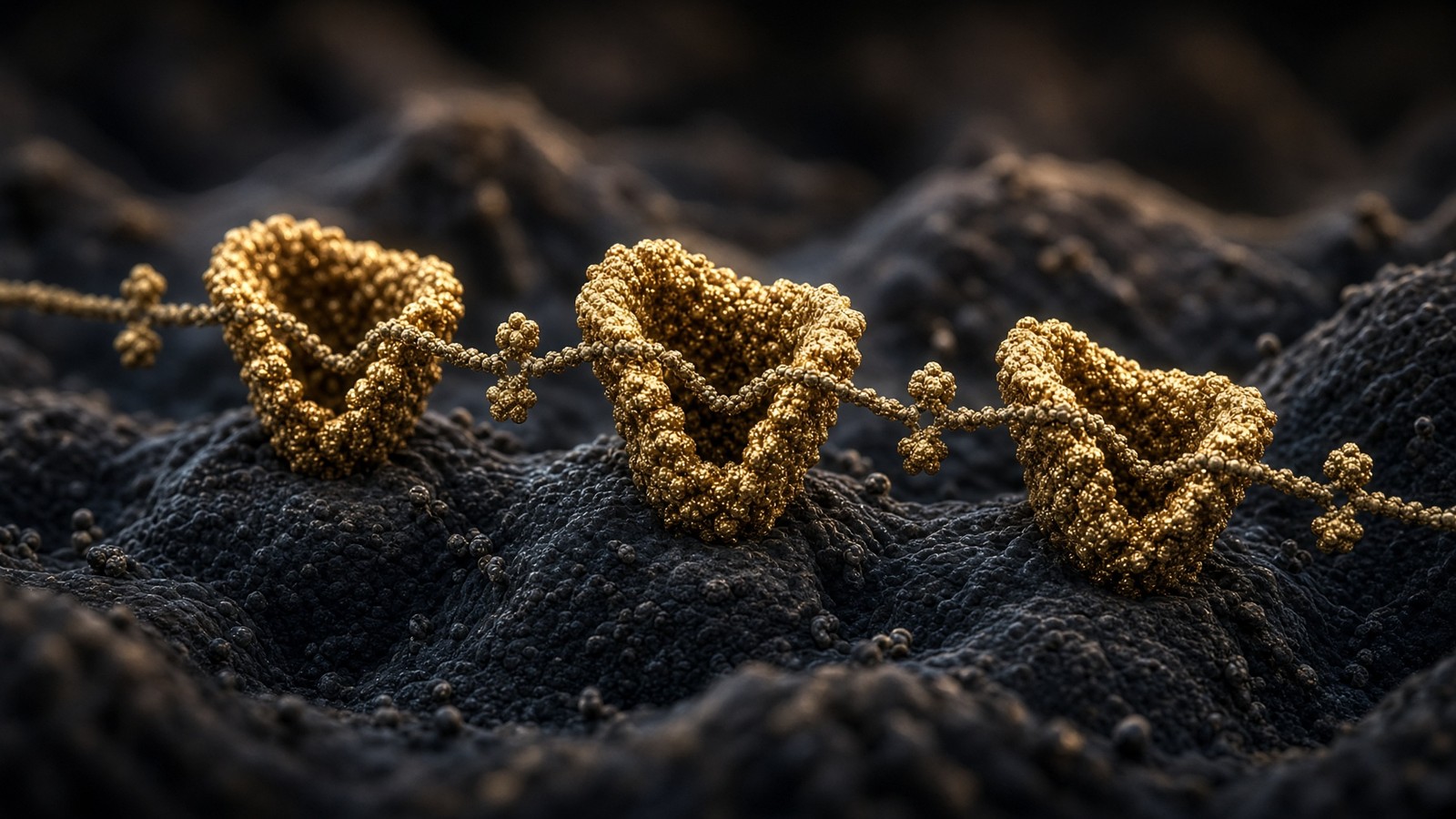
46 min · long read · Peptide research
Retatrutide (LY3437943): the published triple-agonist structure
A fatty-acylated unimolecular agonist at GIPR, GLP-1R and GCGR. US-made, HPLC-MS verified, labelled for laboratory research — not a medicine.
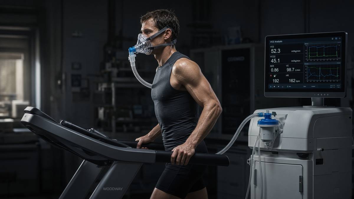
47 min · long read · Metabolism
Mitochondria, VO2 and why training still wins
Endurance work builds the engines. Heavy work builds the chassis. The molecular story is AMPK, PGC-1α, calcium and mTOR — not a shopping list.
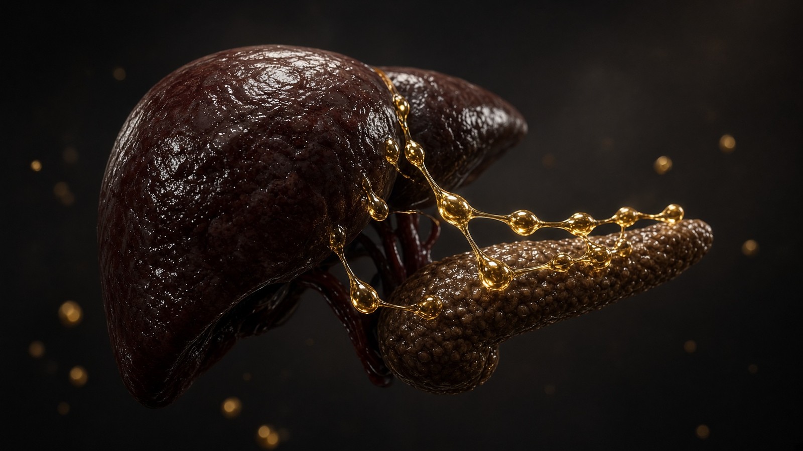
45 min · long read · Metabolism
How diets actually work
Calories are conservation of energy. Hormones are the routing table. Palatability is why the routing table keeps getting overwritten. Every named diet is a way of leaning on one of those three.
More in this desk
51 min · long read · Metabolism
Reversing type 2 diabetes is a published result, not a slogan
The DiRECT trial put type 2 diabetes into remission by emptying the liver and pancreas of surplus fat. Very-low-calorie and very-low-carbohydrate programmes can both get you there. The mechanism isn't mystical.

50 min · long read · Metabolism
Fasting, autophagy and the mTOR switch
Eat, and mTOR builds. Fast, and AMPK and ULK1 start recycling the cell. The molecular story is a switch, not a personality.
49 min · long read · Metabolism
Liver fat is the variable type 2 diabetes actually cares about
Ectopic fat in the liver overproduces glucose and VLDL. Empty it — by energy deficit, by carbohydrate restriction, or by both — and first-phase insulin can return. That is the twin-cycle in working clothes.

49 min · long read · Metabolism
Protein, training and muscle protein synthesis
Muscle isn't a mood. It is mechanical tension plus a leucine-gated translational programme. Here is the dose, the threshold, and why the gym still has to happen.
Essays describe published research. They are not medical advice and they do not authorise human use of any catalogue item.