


Metabolism · 45 min · 9,813 words
Ketones are a fuel. They're also a signal.
β-hydroxybutyrate isn't just what you burn when glycogen runs low. It is an HDAC inhibitor, an NLRP3 dampener, and a message to the brain that the house is running on fat.
What this essay actually tells you
- In starvation, Cahill showed the human brain can draw most of its energy from ketones (β-hydroxybutyrate and acetoacetate) and spare muscle. That's the survival argument for the pathway.
- BHB is also an HDAC inhibitor (Newman & Verdin) and an NLRP3 inflammasome damper (Youm et al., 2015). A millimolar metabolite is allowed to be a ligand. It already is.
- Physiological ketosis is roughly 0.5–3 mM BHB. Diabetic ketoacidosis is a different, dangerous state. They share a word. They do not share a clinic, a pH, or an ending.
What this actually means
When carbohydrate is scarce, the liver turns fat into ketones, mainly β-hydroxybutyrate and acetoacetate, so the brain has a fuel that isn't glucose. That's the textbook, and Cahill already measured it: given time, a human brain will run mostly on ketones and spare muscle. The last fifteen years added a second job. BHB can inhibit class I histone deacetylases, quiet the NLRP3 inflammasome, and occupy GPR109A, the niacin receptor, once you're into the higher millimolar range. A ketogenic diet or a serious fast is therefore both a fuel switch and a signalling state. Physiological ketosis sits roughly 0.5–3 mM and isn't diabetic ketoacidosis; they share a word, not a clinic, a pH, or an ending. Week-one 'keto flu' is mostly a brain still installing transporters plus a kidney dumping salt. That's why glucose often falls, why some people feel mentally clear after a lag, and why the useful question isn't 'did you enter ketosis' but which of those two jobs you actually wanted.
β-Hydroxybutyrate is a four-carbon hydroxy acid, formula C4H8O3, molecular weight 104.1, and it is the molecule we're going to keep on the board for the rest of the afternoon. Strictly it isn't a ketone: the carbonyl of acetoacetate has been reduced at carbon 3. Biochemists still file it with the ketone bodies because the liver mints it from the same acetyl-CoA overflow and extrahepatic tissues oxidise it back to the same two-carbon coin. Acetoacetate is the true ketone, CH3COCH2COO−. Acetone is what you smell when acetoacetate decarboxylates, spontaneously, in a quantity that's a sideshow of the flux rather than a fuel anyone bills. The biologically relevant enantiomer is D-β-hydroxybutyrate, also written (R)-3-hydroxybutyrate; the L-enantiomer is a different metabolite from a different path, and a racemic ketone salt is therefore two molecules wearing one name. Those are the facts we need on the board before anyone is allowed to talk about ketosis as a lifestyle. The pool that matters sits in plasma at hundreds of micromolar to low millimolar, depending on the last meal, liver glycogen, and how hard adipose is spilling fatty acids into the blood.
In short. The useful form is one-handed, not a mixture of mirror images. The main ketone in blood is a four-carbon acid made from fat. It isn't sugar.
The fuel job is older than the signalling job, and it is still most of the flux, which I find quietly thrilling. George Cahill's starvation studies — Owen, Morgan, Kemp, Sullivan, Herrera and Cahill, Journal of Clinical Investigation 1967, and the reviews Cahill kept having to write because the field preferred a catchphrase — showed that a human brain, given days rather than hours, will take most of its energy from β-hydroxybutyrate and acetoacetate and will spare the muscle that would otherwise have been converted to glucose. A fed brain is a glucose organ, something like a hundred grams a day. A starved brain is a ketone organ with a glucose residual. That switch is why a well-formulated ketogenic diet can flatten a glucose curve without a sermon, and why a three-day fast doesn't eat as much lean mass as a first-year textbook implied. The liver writes the ketone. Extrahepatic mitochondria cash it. The brain is the customer that made the pathway a survival argument rather than a curiosity of ruminants.
In short. The liver makes the fuel; the brain spends it. In a long fast the human brain can run mostly on ketones and so spare muscle. Cahill measured that.
The signalling job is newer, and it is why BHB shows up in papers that aren't about epilepsy or body weight — a fuel allowed to be a message. Shimazu, Hirschey, Newman, Verdin and colleagues, Science 2013: β-hydroxybutyrate is an endogenous inhibitor of class I histone deacetylases, HDAC1 and HDAC2 among the named enzymes, and the inhibition de-represses a set of oxidative-stress genes including FOXO3A. Youm, Kanneganti, Dixit and colleagues, Nature Medicine 2015: the same metabolite blocks the NLRP3 inflammasome, dropping IL-1β, by a mechanism that doesn't require the nicotinic-acid receptor. GPR109A, also called HCAR2, is a Gi-coupled GPCR for BHB once you're into the higher millimolar range; Taggart, Offermanns and the niacin literature put that lock on the map before anyone had a keto brand. A millimolar metabolite is allowed to be a ligand. It already is. We don't have to pick a lane for it. The cell didn't.
In short. A fuel is allowed to be a message. Ketones also talk. They can change which genes are readable, quiet a fire-alarm in immune cells, and occupy a receptor.
We're a peptide catalogue with a journal attached, not a ketogenic clinic with a millimolar badge. We stock retatrutide because it is the published LY3437943 structure, a unimolecular agonist at GIPR, GLP-1R and the glucagon receptor, American-made, HPLC-MS, the chain Coskun engineered and Jastreboff ran in Phase 2. We stock NAD+ because it is the hydride coin Complex I wants oxidised. We stock MOTS-c because it's a 16-mer a mitochondrion translated from its own 12S rRNA. Those three vials sit in the neighbourhood of fuel routing. Neighbourhood isn't identity. Occupancy at three class-B GPCRs is an honest sentence about body weight and hepatic fat. 'Retatrutide is a ketone drug' isn't. Let's walk the metabolite, the enzymes, the transporters, the two signalling floors, and the jobs we refuse to mix. This page isn't a meal plan. We don't tell you how many grams of carbohydrate to eat. The papers that already ran that experiment wrote the method down.
In short. This page is the metabolite, not a diet plan. Retatrutide, NAD+ and MOTS-c live near fuel biology. None of them is a ketone.
The liver writes a ketone when acetyl-CoA has nowhere to go
Hepatic β-oxidation throws off acetyl-CoA, the two-carbon coin the liver has to park or export. That's the ordinary job of a liver that has been told, by falling insulin and rising glucagon, to open adipose and burn the spill. Each turn of the spiral shortens a fatty acyl-CoA by two carbons, mints one FADH2 and one NADH, and drops an acetyl-CoA into the matrix. When the cell has enough oxaloacetate, citrate synthase condenses acetyl-CoA into the TCA cycle and the carbons become CO2. When the cell doesn't — because oxaloacetate has been siphoned into gluconeogenesis, which is exactly what a carbohydrate-poor liver is doing — acetyl-CoA accumulates. Thiolase condenses two of them to acetoacetyl-CoA. Mitochondrial HMG-CoA synthase 2, HMGCS2, adds a third acetyl-CoA and writes 3-hydroxy-3-methylglutaryl-CoA. That's the committed step. HMG-CoA lyase, HMGCL, then splits HMG-CoA to acetoacetate plus acetyl-CoA. The overflow has become a ketone. The mitochondrion that could not put the carbons into citrate has put them into a fuel the rest of the body can take.
In short. One named enzyme, HMGCS2, is the committed step. When the liver burns fat faster than the citric-acid cycle can take the fragments, it turns the leftovers into ketones.
HMGCS2 is the enzyme a ketogenesis paragraph actually has to name, because the cytosolic isoform HMGCS1 is cholesterol synthesis and a different invoice. The mitochondrial enzyme is transcriptionally hungry for the fasted state: Foxa2, PPARα, and a glucagon-cAMP programme write it; insulin and mTOR lean the other way. A liver that can't make HMGCS2 can't make ketones worth measuring, which is why inborn errors at this step, and at HMGCL, present as hypoketotic hypoglycaemia rather than as a keto-flu anecdote. The chemistry is a Claisen condensation followed by a lyase, not a mood. You can blot HMGCS2. You can assay HMG-CoA. You can, if you're a hepatocyte paper, knock the gene down and watch acetoacetate fall. Treating 'the liver makes ketones' as a mood is how a first-year pathway becomes a brand. Treating HMGCS2 as the committed step is how the experiment starts.
In short. HMGCS2 is that liver enzyme that decides leftover fat fragments become ketones. Fasting writes it. Insulin leans against it. Without it, ketones don't rise.
Acetoacetate remains the first ketone out of HMGCL. Mitochondrial BDH1, D-β-hydroxybutyrate dehydrogenase, then reduces most of it to BHB, using NADH, because a liver that is β-oxidising hard has NADH to spare and because BHB is the more reduced, more stable currency in blood. The BHB/acetoacetate ratio in plasma is therefore, among other things, a crude readout of hepatic mitochondrial NADH/NAD+. Acetone is the third body, and it's a leak: acetoacetate decarboxylates, enzymatically and not, to a three-carbon ketone that leaves on the breath. Nobody bills acetone as a brain fuel in a serious paper. The useful pair is BHB and acetoacetate, circulating in a ratio that is usually BHB-heavy once the pathway is up, transported on monocarboxylate carriers, and taken up by tissues that have the enzymes to write acetyl-CoA again. Name the pair. Name the dehydrogenase. A urine stick that only sees acetoacetate is looking at the less abundant sibling once adaptation has happened, which is one reason people 'leave ketosis' on a stick while a blood meter still reads millimolar BHB.
In short. That liver mostly ships β-hydroxybutyrate, the reduced partner of acetoacetate. Breath acetone is a leak, not the fuel. Blood BHB is the number that matters.
The liver almost doesn't consume what it makes. Succinyl-CoA:3-ketoacid CoA transferase — SCOT, gene OXCT1 — is the extrahepatic enzyme that activates acetoacetate back to acetoacetyl-CoA so thiolase can split it to two acetyl-CoA. Hepatocytes largely lack SCOT. That isn't a defect. It is the topology that makes ketogenesis an export pathway rather than a futile cycle in the same matrix. A liver that oxidised its own ketones would be burning a fuel it had just written from fat in order to... burn fat. Evolution declined. Extrahepatic mitochondria — brain, heart, kidney cortex, skeletal muscle — carry SCOT and BDH1 and take the delivery. The same topology is why a ketone measured in a hepatic vein is an export, and a ketone measured in an artery to the brain is a substrate. Compartmentation, again. A homogenate of 'the body' can't tell you who wrote the molecule and who spent it.
In short. Brain, heart, kidney and muscle cash the cheque. The liver almost can't burn the ketones it makes, because it lacks the enzyme SCOT. It exports them.
Insulin and glucagon set the writing, which remains why a carbohydrate cut and a genuine fast both raise BHB, and why an insulin-deficient patient can raise it into a different, dangerous range. Low insulin de-represses adipose lipolysis (ATGL, HSL, perilipin), floods the liver with non-esterified fatty acids, and takes the brake off HMGCS2 transcription. Glucagon, via cAMP and PKA, leans the hepatocyte toward β-oxidation and gluconeogenesis, which is the oxaloacetate siphon already named. A fed, high-insulin liver will re-esterify incoming fatty acids toward VLDL rather than toward ketones; that is one reading of a carbohydrate-rich surplus, and it is why liver fat and ketogenesis can move in opposite directions on the same fatty-acid delivery. The ketone is therefore a readout of hepatic acetyl-CoA overflow, not a badge you earned by buying a meter. Chasing a millimolar number while still overeating, or while liver fat remains, is how you miss the point the overflow was making.
In short. The blood number reports leftover fat fragments in the liver, not a gold star. Insulin down and glucagon up tell the liver to make ketones.
Diagram
Matrix
- TCA cycle · β-oxidation · mtDNA nucleoids
- NADH produced here. Complex I spends it.
- MOTS-c (MRWQEMGYIFYPRKLR) from 12S rRNA.
Inner membrane
- I → II → III → IV → V (ATP synthase)
- ~150 mV proton-motive force
- ~40–60 kg of ATP turned over per human day
mtDNA is 16,569 bp, 37 genes, 13 proteins of the respiratory chain. Nuclear DNA encodes the other ~1,200 mitochondrial proteins. NAD+ is the hydride carrier between dehydrogenases and Complex I. MOTS-c is a 16-mer translated from 12S rRNA — a peptide the mitochondrion wrote itself.
Extrahepatic tissues cash the cheque
Uptake is a monocarboxylate problem before it is an enzyme problem — the door has to exist before the matrix can cash the cheque. MCT1 (SLC16A1) and MCT2 (SLC16A7) move BHB and acetoacetate across plasma membranes, the same carriers that move lactate and pyruvate. Endothelium at the blood–brain barrier is MCT1-rich; neurons prefer MCT2. Proton-coupled, saturable, inducible. A brain that has been glucose-fed for decades doesn't have the full complement on day one of a carbohydrate cut, which is a large part of why week one can feel like a neurological insult even when the liver is already writing millimolar BHB. Enzyme induction follows: BDH1 and SCOT in extrahepatic mitochondria, and a set of TCA and respiratory subunits that have to handle the new acetyl-CoA. Days, not minutes. The metabolite can be in the blood this afternoon. The machinery that spends it, in the tissue you cared about, is a transcriptional programme. Confusing those two clocks is how 'I'm in ketosis' becomes a mood.
In short. Blood can be ready before the brain is. Ketones enter cells on the same doors as lactate. The brain takes days to put enough doors and enzymes in place.
Once inside a mitochondrion that has the kit, the chemistry is the reverse of the hepatic write, with a named transferase doing the activation. BDH1 oxidises BHB to acetoacetate and mints NADH. SCOT (OXCT1) transfers CoA from succinyl-CoA onto acetoacetate, writing acetoacetyl-CoA and succinate; that's why the enzyme is a CoA transferase and not a ligase, and why it couples to the TCA cycle at a succinyl node. Thiolase splits acetoacetyl-CoA to two acetyl-CoA. Acetyl-CoA enters citrate synthase if oxaloacetate is there, which in a neuron or a cardiomyocyte it usually is, because those cells aren't running gluconeogenesis. Two acetyl-CoA from one acetoacetate. NADH from the BDH1 step plus the NADH and QH2 from the isocitrate, α-ketoglutarate and malate dehydrogenases and from the β-oxidation the cell is also running. Complex I wants that NADH. The chain runs. Peter Mitchell's gradient is the product. ATP synthase is the turbine. A ketone is a fuel because it becomes acetyl-CoA in a matrix that can oxidise acetyl-CoA.
In short. The respiratory chain then spends it as usual. Inside the mitochondrion, ketones are turned back into the same two-carbon fuel sugar would have made.
Cahill's numbers are still the ones a brain paragraph has to carry, and they're still the right size. In the 1960s, at the Joslin and at Harvard, George Cahill, Oliver Owen and colleagues catheterised starving volunteers — obese patients undergoing prolonged therapeutic fasts, which is a sentence a modern ethics committee would rewrite and a measurement a field still uses — and showed that cerebral glucose uptake fell and cerebral ketone uptake rose until ketones accounted for most of the brain's oxygen consumption. Owen, Morgan, Kemp, Sullivan, Herrera, Cahill, J Clin Invest 1967: brain metabolism during fasting. Cahill's later Annual Review of Nutrition, 2006, is the document that put the whole starvation programme on one map: glycogen first, then gluconeogenesis from amino acids, then a ketone-adapted brain that lets nitrogen loss fall. A fed human brain is of the order of 100–120 g of glucose per day. An adapted starving brain might take 30–40 g of glucose and cover the rest with BHB and acetoacetate. That's the survival argument for the pathway. It is also why a well-formulated ketogenic diet can be an epilepsy therapy and a glucose-flattening tool without being a personality.
In short. Cahill's starvation studies showed an adapted human brain taking most of its energy from ketones. Glucose use fell. Muscle was spared. Those numbers still stand.
Muscle sparing is the sentence the pathway was for, at organism scale, and it is the sentence a protein-anxious fast still needs to hear. If the brain insisted on 100 g of glucose a day through a three-week fast, gluconeogenesis would have to come from somewhere, and a large fraction of somewhere is muscle amino acids — alanine, glutamine, the glucogenic spill of a proteolysed fibre. Ketones let the brain take a four-carbon fuel the liver wrote from fat, so the amino-acid bill shrinks. Nitrogen excretion falls after the first days, which Cahill measured, and which is why a starving human doesn't dissolve as fast as a simple glucose-budget predicted. Heart and kidney cortex are ketone-avid even in milder states; skeletal muscle takes them when they're offered and when glucose and fatty acids aren't outcompeting them. The hierarchy isn't a moral ranking. It is who has SCOT, who has MCT, and who is being asked to spare whom.
In short. Ketones let it burn fat instead, so lean mass lasts longer. If the brain had to keep eating sugar in a fast, it would eat muscle.
Heart, kidney and working muscle are the other extrahepatic customers, and they don't wait for a three-week fast. A cardiomyocyte will oxidise BHB readily; recent work has put ketone utilisation back into heart-failure papers as a fuel the failing organ will take when it can get it, which is a clinical literature we won't pretend to be. Kidney cortex is an oxidative tissue with a large ATP bill for sodium pumping and a respectable ketone capacity. Skeletal muscle is more opportunistic: glucose when insulin is high, fatty acids when they're high, ketones when they're high and the transporters are in. Exercise complicates the picture because a working fibre wants whatever is available and because MCT1 is also a lactate door. The point of the census isn't to memorise four one-liners. It is to stop writing 'the body burns ketones' as if tissues were interchangeable. The liver writes. Named extrahepatic mitochondria spend. The brain is why we care at starvation scale. The heart is why a millimolar BHB isn't only a brain story.
In short. Different organs, different appetite for the same fuel. Heart, kidney and muscle also burn ketones, and they don't need a three-week fast to start.
Diagram
- I. Pumps H⁺. ~45 subunits. The NADH coin is spent here.
- II. TCA entry. No proton pump. FADH₂ neighbourhood.
- Q. Lipid-soluble shuttle in the inner membrane.
- III. Q-cycle. Pumps H⁺. Superoxide leak site.
- c. Intermembrane space. The shuttle everyone has heard of.
- IV. O₂ → H₂O. The reason you breathe.
- V. F₁Fₒ rotary. Protons in, ATP out. ~10²¹ times a second in you.
Mitchell’s chemiosmotic theory (Nobel 1978): the inner membrane is a battery of ~150 mV. NAD+ is the hydride carrier that feeds Complex I. MOTS-c is a 16-mer the mitochondrion translated from 12S rRNA — a different object on the same campus.
Given time, the human brain will take most of its energy from β-hydroxybutyrate and acetoacetate. Muscle is what that switch spares.— Cahill's starvation programme, in working English
MCT1/2, induction, and the lag people call flu
Monocarboxylate transporters are the doors, and doors have kinetics, which is most of why week one can feel grim. MCT1 is widely expressed, including on endothelium and on many epithelia; MCT2 is the higher-affinity neuronal isoform; MCT4 is the lactate-export specialist of glycolytic cells and is less the ketone story. Halestrap's work put the SLC16 family on the map; Pierre and Pellerin put MCT2 on neurons and the lactate shuttle beside it. BHB is a substrate, not a VIP. It competes with lactate and pyruvate. A blood–brain barrier that has been running on glucose for years will increase MCT1 with sustained ketosis, which is a protein-synthesis job and a trafficking job, not a hashtag. Enzyme induction in neurons and glia — BDH1, SCOT, the TCA subunits — rides the same days-long clock. You can raise plasma BHB in an hour with a ketone ester. You can't raise brain MCT2 in an hour. That mismatch is physiology. It is also why exogenous ketones and a ketogenic week aren't the same experiment, a point the later heading will have to keep honest.
In short. A drink can raise blood ketones faster than the brain can learn to take them. Ketones share a door with lactate. The brain builds more of those doors over days, not minutes.
Keto flu remains the popular name for a first-week syndrome that is, in large part, two clocks plus a kidney. Clock one: the transporter and enzyme lag just named. A brain still waiting on MCT and SCOT, asked to run on a fuel it has not fully tooled for, will complain — headache, fog, a sense that the lights dimmed. Clock two: insulin natriuresis. Insulin tells the distal nephron to hold sodium; drop carbohydrate hard and insulin falls, the kidney lets sodium go, water follows, and magnesium and potassium hitch a ride. The electrolytes-and-the-kidney essay on this page is that nephron physics in working clothes. Ketone anions also obligate some urinary cation loss. The wrecked cohort in week one usually didn't salt the food and had not yet induced the transporters. The reborn cohort usually did both, or got lucky. Neither group underwent a spiritual test. The headache wasn't a detox. Calling it flu is how a forum medicalises a lag.
In short. That first-week misery is mostly a brain still installing ketone doors, plus a kidney dumping salt when insulin falls. It isn't a spiritual test.
Order of operations is most of the practical game, and it still isn't a meal plan. Replete sodium, potassium and magnesium on a hard carbohydrate cut unless a clinician already has those ions restricted — kidney disease, ACE inhibitors, potassium-sparing diuretics, a heart-failure clinic, those people have a different essay and a different telephone number. Then wait long enough that MCT and BDH1 and SCOT have had a chance to be written. Then decide whether you like the diet. Measuring BHB on day two and declaring failure, or declaring enlightenment, is a category error: the liver can write the metabolite before the brain can spend it. The neighbouring carnivore and how-diets-actually-work essays are the pattern-level arguments. This one is the lag. Treat week one as evidence of efficacy, or of toxicity, and you have skipped a transporter half-life. Let's keep the named papers in view as we go.
In short. Salt the food, wait for the brain's ketone machinery to catch up, then judge the diet. A day-two meter reading isn't a verdict.
Induction is tissue-specific, which is why a single blood number is a poor story about adaptation. Liver HMGCS2 can be up while neuronal MCT2 is still climbing. Skeletal muscle MCT1 responds to training as well as to diet, because lactate traffic is a training adaptation and BHB uses the same door. Heart takes ketones earlier than brain on most of the time courses people have actually drawn. Gut MCT isoforms see luminal as well as plasma substrate if you swallowed an ester. A paper that reports ketone adaptation without naming the tissue has reported a catchphrase. A paper that blots MCT1, MCT2, BDH1 and OXCT1 in the tissue it claims, and shows a time course, has reported a mechanism. The flu literature is almost all the former. The transporter literature is the latter. We'll stay with the latter, and we'll keep pointing at the kidney essay so the natriuresis half doesn't get forgotten every time someone buys a magnesium glycinate from a forum.
In short. Organs adapt on different clocks, so blood ketones can look ready while the brain is still catching up. Name the tissue before you call it adapted.
Physiological ketosis isn't ketoacidosis
Physiological ketosis sits roughly 0.5–3 mM BHB in plasma, a fuel state rather than an emergency. An overnight fast in a healthy person might tick 0.1–0.4 mM, a number that is real and not interesting as a lifestyle. Nutritional ketosis, the range a well-formulated ketogenic diet or a multi-day fast aims at, is the low millimolar band. Prolonged starvation, Cahill's volunteers, can sit around 5–7 mM and remain compensated: pH held, bicarbonate held, insulin low but not absent, a liver that is writing ketones and a periphery that is taking them. Those numbers are a fuel programme. They aren't an emergency department. The meter on a kitchen counter is allowed to read 1.5 mM without anyone calling an ambulance. A fingerstick that first crosses 0.5 mM is reporting hepatic overflow, not a personality, and not a reason to rewrite the rest of the week around a single strip. Treating every millimolar as a crisis is how a useful pathway inherited a word it has to spend half its essays unpicking.
In short. Everyday ketosis from fasting or a very low-carbohydrate diet is about 0.5 to 3 millimolar. That's a fuel state, not an emergency.
Diabetic ketoacidosis is a different, dangerous state — a word shared with everyday ketosis, and almost nothing else. Insulin absent or near-absent — typically type 1, or a ketosis-prone type 2 in a sick moment, or an SGLT2-associated euglycaemic variant a clinician already knows to fear — lipolysis unrestrained, hepatic ketogenesis unrestrained, extrahepatic uptake unable to keep up, plasma BHB often 15–25 mM, acetoacetate high, anion gap wide, bicarbonate down, pH down, the patient unwell. Volume depletion, potassium that looks normal in plasma while the body is empty of it, a precipitant (infection, missed insulin, a new drug). They share a word with physiological ketosis. They don't share a clinic, a pH, or an ending. A paper that writes ketosis for both has not yet started. A social post that treats a 1.2 mM morning reading as DKA has not looked at a blood gas. The ketone isn't the danger. The insulin deficiency, the acid, and the unwell person are the danger. The metabolite is the readout that got into the name.
In short. It shares a word with ordinary ketosis, not a ward, a pH, or an ending. Diabetic ketoacidosis is much higher ketones plus no insulin plus acid blood plus a sick patient.
pH is the discriminator a meter can't see, which is why a meter isn't a diagnosis. Physiological ketosis is buffered. The kidneys hold bicarbonate, ventilation isn't Kussmaul, and the person is, metabolically, compensated. DKA is an acidosis with an anion gap, and the gap is the ketone salts plus whatever else the unwell metabolism has thrown off. Point-of-care BHB is useful in a clinic precisely because urine acetoacetate sticks lag and because the decision is 'is this person acidotic and insulin-deficient', not 'is there a ketone in the room'. Home meters that report 0.8 mM after a gym session and a skipped breakfast are reporting physiology. They aren't reporting DKA. The reverse error exists too: SGLT2-associated euglycaemic DKA can present with glucose that doesn't look frightening and ketones that do. That is clinician territory. This page will name it so nobody leaves thinking glucose is the whole story, and will then stop, because a journal paragraph isn't an emergency protocol.
In short. Ordinary ketosis keeps blood pH. Acidosis plus ketones plus a sick, insulin-deficient patient is the emergency. A kitchen meter can't see pH.
SGLT2 inhibitors, type 1 diabetes, and pregnancy are the three neighbourhoods a metabolism essay names so that it doesn't pretend to be a clinic. SGLT2 drugs dump glucose in urine, lower insulin, and can, in the wrong setting, permit a ketoacidosis at glucose concentrations that look reassuring — euglycaemic DKA, now a labelled risk. Type 1 is insulin-deficient by definition; ketones there are a sick-day measurement, not a diet badge. Pregnancy has a lower threshold for ketosis and a separate argument about whether maternal ketones are a fetal problem; that argument is obstetric, contested, and not ours to close. Anyone in those three sets who wants a ketogenic experiment has a clinician, or needs one, and this page isn't that appointment. The rest of us are allowed to know that 1 mM BHB after a fast isn't DKA, and that DKA isn't 'being in ketosis harder'. Shared word. Different physics. Different ending.
In short. SGLT2 drugs, type 1 diabetes and pregnancy make ketones a clinical problem, not a lifestyle number. Those people need a clinician. A diet meter isn't that visit.
Compensation is the unglamorous half of the starvation range, and it is why Cahill's 5–7 mM volunteers weren't acidotic. Insulin was low, not absent. Lipolysis was up, not ungoverned. The periphery was taking ketones as fast as a brain and a heart can take them, so the pool sat in a high-but-stable band rather than running away. Bicarbonate held. The people were hungry and cold and studying, not acidotic and vomiting. That's a fuel programme at its upper physiological end. Push the same chemistry without insulin, or with a second illness, and the pool doesn't saturate; it climbs, the acid load outruns the kidney, and you have a different diagnosis. The meter can't tell those two stories apart if you only give it a BHB number. Clinical context can. We'll keep saying that until the word ketosis stops being asked to do both jobs. They share a carbon skeleton. They don't share a ward.
In short. Starvation can raise ketones higher than a diet does and still keep pH, because some insulin is still and tissues keep burning the fuel. Without insulin the same chemistry runs away.
- Overnight fast
- ~0.1–0.4 mM BHB
- Physiological ketosis
- 0.5–3 mM BHB
- Prolonged starvation
- ~5–7 mM BHB
- Diabetic ketoacidosis
- often 15–25 mM
- Brain on ketones
- most of its energy
- HMGCS2
- committed step
- SCOT (OXCT1)
- extrahepatic only
- GPR109A / HCAR2
- millimolar ligand
Real, small, not a lifestyle.
Nutritional ketosis. Buffered. Not DKA.
Cahill. Compensated. Insulin low, not absent.
Insulin-deficient, acidotic, unwell. A clinic.
Owen, Cahill, J Clin Invest 1967. Given time.
Hepatic mitochondrial HMG-CoA synthase 2. Not the cholesterol isoform.
Liver lacks it, so the ketone is an export.
Niacin receptor. Gi. Higher millimolar BHB.
A millimolar metabolite is allowed to be a ligand
Fuels aren't supposed to be ligands in the cartoon a first-year course hands out, and yet this one is. Hormones are ligands: nanomolar, receptors, amplification. Metabolites are substrates: millimolar, enzymes, flux. The last twenty years of metabolite signalling — succinate at GPR91, lactate at GPR81, kynurenine at AhR, BHB at HDACs and at GPR109A and at NLRP3 — is the cartoon coming apart. A molecule present at millimolar can still bind a protein and change that protein's behaviour, if the binding site has a millimolar Kd. That isn't a promotion to a hormone. It is ordinary physical chemistry. Newman and Verdin have been saying this about BHB in reviews a field actually has to cite: the metabolite is a fuel and a signal, and the two jobs run on the same carbon skeleton without asking anyone's permission. We don't have to pick a lane. The HDAC paper, the inflammasome paper, and the GPCR paper are three ligand jobs. The fuel job remains most of the flux. Both halves of that sentence are required.
In short. One fuel present at millimolar can still bind proteins and change them. Ketones do that. They didn't stop being fuel. They added a second job.
Shimazu, Hirschey, Newman, Verdin and colleagues, Science 2013, 339: 211–214, remains the HDAC document. β-Hydroxybutyrate inhibits class I histone deacetylases — HDAC1, HDAC2, HDAC3 among the named enzymes — at concentrations that overlap physiological ketosis. Histone acetylation rises. FOXO3A and metallothionein 2, among other oxidative-stress genes, are de-repressed. The chemistry isn't sirtuin chemistry. Classical HDACs (classes I, II, IV) hydrolyse acetyl-lysine with a zinc ion and a water; sirtuins, class III, spend NAD+ and were the subject of a neighbouring essay. BHB is a class I HDAC inhibitor, an endogenous one, which is why a millimolar metabolite gets to change transcription without being a transcription factor. Newman and Verdin's later reviews (including Annu Rev Nutr 2017) put the signalling map in one place: HDAC, NLRP3, GPR109A, and the fuel. Read the Science paper before a brochure that says ketones 'turn on longevity genes'. The genes in the paper were stress-response genes. Longevity is a different claim, in a different animal, with a different editor.
In short. That isn't the same family of enzymes that spend NAD+. BHB can block the enzymes that take acetyl badges off histones. Genes for handling stress become easier to write.
Transcription is the floor that HDAC inhibition actually sits on, and it's a slower floor than a fuel. Acetyl marks on histones H3 and H4 change how tightly nucleosomes hold DNA and how readily a promoter sees Pol II. FOXO3A is a forkhead transcription factor the stress literature already knew; de-repressing it's a gene-expression programme, hours, not a millisecond GPCR. BDNF — brain-derived neurotrophic factor — shows up in ketogenic and BHB papers via this neighbourhood and via others (Marosi, Mattson, a set of neuronal-culture and rodent assays). It's a real literature and a messy one, because BDNF is also exercise, also glutamate, also a dozen other inputs, and because a neuronal culture at 4 mM BHB isn't a person on a diet. The honest sentence is that BHB can change chromatin and can, in named assays, lean a neuron toward a BDNF-linked plasticity programme. The dishonest sentence is that a ketogenic week is a nootropic. We'll keep the first. The transcription diagram that follows is the floor, not a product.
In short. When those enzymes are blocked, a cell can read a different set of genes — stress-response genes and, in some neuron papers, a growth-factor gene. Hours, not seconds.
Class I HDAC inhibition is also why BHB and the sirtuin story get confused in the same paragraph, and they should not. Sirtuins spend NAD+ to take acyl groups off lysines; BHB doesn't spend NAD+ to inhibit HDAC1. Different enzymes, different chemistry, different budgets. A ketogenic diet can raise NAD+/NADH in some tissues because β-oxidation and a slower glycolytic NADH production change the redox ratio, and that can feed sirtuins as a side-effect of the fuel switch. That's a redox sentence, not an HDAC sentence. Verdin's laboratory has published on both floors, which is a reason the confusion happens and not a reason to merge the floors. If your blot is acetyl-histone, you're in the HDAC story. If your blot is acetyl-PGC-1α or a NAMPT number, you're in the sirtuin story. If you have neither blot, you have a BHB meter and a story. The neighbouring NAD+ and sirtuin essays are the other budget. This one is the millimolar inhibitor.
In short. Ketones blocking HDAC enzymes isn't the same as sirtuins spending NAD+. A diet can touch both, for different reasons. Don't merge the two stories.
Diagram
Closed chromatin (H3K27me3, DNA methylation) hides the promoter. Pioneer factors and histone acetyltransferases open it.
PIC: TFIID, TFIIH, Mediator, Pol II. Ser5 phosphorylation of the CTD lets the polymerase leave the promoter.
Elongation ~20–40 nt/s. Capping, splicing, cleavage and polyadenylation happen on the still-growing RNA.
Human genes are islands in 3.1 billion base pairs of mostly noncoding sequence. Promoter, enhancers, chromatin state and the Mediator complex decide whether Pol II is allowed to fire. Epithalon’s literature sits on TERT and pineal clocks — two of the rare promoters anyone bothers to name in a peptide essay.
β-Hydroxybutyrate is an endogenous HDAC inhibitor — a fuel that changes transcription.— Newman and Verdin's line of work, in one sentence
NLRP3 and GPR109A: two other ligand jobs
Youm, Nguyen, Grant, Goldberg, Bodogai, Kim, Dutta, Snyder, Goubert, Kang, Waschek, Barber, Somerville, Rossi, Blasco, Fridman, Bronson, Frenck, Hensley-McBain, Nikolich-Zugich, Biragyn, Robey, Van Gool, Kanneganti and Dixit, Nature Medicine 2015, 21: 263–269, remains the inflammasome document, and the author list is the joke a field tells about collaborative mouse work. The finding isn't a joke. β-Hydroxybutyrate inhibits the NLRP3 inflammasome, reducing caspase-1 activation and the maturation of IL-1β and IL-18, in macrophages and in mouse models of NLRP3-driven inflammation (Muckle–Wells, urate, a familial-cold neighbourhood). The inhibition didn't require GPR109A. It didn't require the HDAC job in the assays they ran to check. It tracked reduced potassium efflux from the cell, a known upstream of NLRP3 assembly. A millimolar metabolite, an innate-immune machine, a cytokine that a lot of sterile inflammation bills. That's a ligand job. It isn't the fuel job wearing a lab coat.
In short. It didn't need the niacin receptor to do that. A 2015 paper showed BHB quieting the NLRP3 inflammasome, an immune alarm that releases IL-1β.
NLRP3 remains a pattern-recognition complex, not a personality. In a macrophage it assembles, with ASC and caspase-1, when the cell sees a set of danger signals — ATP at P2X7, crystals, a potassium leak, mitochondrial ROS in some assays — and it then cuts pro-IL-1β and pro-IL-18 into the secreted forms. Those cytokines are how sterile inflammation talks. Gout, some autoinflammatory syndromes, parts of the metabolic-inflammation argument: NLRP3 sits on those lists. BHB, in Youm's hands, made assembly harder. Subsequent papers have both used and argued with the potassium-efflux mechanism, which is how a finding ought to age. What has survived is that physiological BHB can damp this particular inflammasome in named cells, and that the effect is separable from GPR109A. A ketogenic diet paper that reports lower IL-1β without blotting NLRP3, or without a BHB number, has reported a diet. A paper that does both has reported a metabolite. Demand the second if the claim is the metabolite.
In short. If you claim that job, show the alarm and the ketone, not only a diet. NLRP3 is the cell's crystal-and-danger alarm. BHB can make it harder to assemble.
GPR109A is the other receptor, and it's a GPCR, which means we're on a different floor from chromatin and from NLRP3. Also called HCAR2, hydroxycarboxylic acid receptor 2, it was cloned as the nicotinic-acid receptor: the lock niacin occupies to drop lipolysis in adipose and to flush the skin via Langerhans cells and prostaglandins. Taggart, Kero, Gan, Offermanns and colleagues put BHB on it as an endogenous ligand, with an EC50 in the high-hundreds-of-micromolar to low-millimolar band — exactly the band physiological ketosis occupies, and below typical DKA. The coupling is Gi. Occupancy lowers cAMP in the cells that carry the receptor. In adipocytes that can mean a brake on lipolysis, a negative feedback on the very spill that is feeding ketogenesis, which is a tidy piece of control theory if it holds at the concentrations you actually have. In immune cells the Gi signal is a different invoice. Niacin flush is this receptor. BHB doesn't usually flush, which is a reminder that occupancy and efficacy aren't the same sentence, and that millimolar BHB isn't millimolar niacin.
In short. GPR109A is that niacin receptor. BHB can occupy it at the high end of ordinary ketosis and lower cAMP. That's a true receptor, not a metaphor.
Second messengers are how a GPCR job remains allowed to be fast, and how it isn't allowed to be confused with HDAC inhibition. Occupancy at GPR109A, Gi, less adenylyl cyclase, less cAMP, less protein-kinase-A tone in that nanodomain. Minutes. Reversible. A phosphodiesterase away from baseline. HDAC inhibition is a millimolar occupancy of a zinc enzyme in the nucleus, a change in histone marks, a transcriptional programme, hours. NLRP3 is an oligomer in the cytosol, potassium, caspase-1, a cytokine, also faster than transcription and slower than a pure cAMP blip depending on the assay. Three ligand jobs, three timescales, three compartments. A diagram of clouds and kinases sits below so that the GPCR floor is visible, and so that nobody borrows amplification language for the HDAC job, which is stoichiometric inhibition, or for the fuel job, which is flux. cAMP is amplified from an occupancy. BHB at HDAC isn't. BHB at Complex I, after SCOT, is a hydride. Keep the floors.
In short. That receptor job is fast and uses cyclic AMP. The gene job is slow and uses histone marks. The immune-alarm job is another room again. Same molecule, three floors.
Separability is the experimental demand, and it is why a single BHB number can't tell you which ligand job you paid for. GPR109A-knockout mice still show the NLRP3 finding in Youm's paper. HDAC assays don't require the GPCR. Fuel oxidation requires BDH1 and SCOT and doesn't require either signalling protein. If you want the receptor, blot HCAR2, use a Gi inhibitor or a knockout, and show cAMP. If you want the inflammasome, show IL-1β, caspase-1, and an NLRP3-deficient control. If you want chromatin, show acetyl-histone and a class I HDAC assay, and don't call it sirtuin. If you want fuel, show oxygen consumption, 13C-BHB into TCA intermediates, and a SCOT-dependent step. Four experiments. One metabolite. A literature that reports a ketogenic diet and a phenotype, and nothing about which job, is the literature that doesn't replicate when someone else changes the diet's protein or its salt. Design the assay as if a careful reader will have to believe it. That reader is who we're writing for.
In short. You can knock out the receptor and still see the immune-alarm effect. Fuel burning needs different enzymes again. Say which job you actually measured.
Diagram
× 1
Ligand
One peptide in one pocket. nM–µM. Shape, not a mood.
× 10–10²
G proteins
The occupied GPCR is a GEF. Each Gα is a catalyst.
× 10³–10⁴
cAMP / IP₃ / Ca²⁺
Adenylyl cyclase and PLC do not make one molecule. They make a cloud.
× 10⁴–10⁶
PKA / PKC / CaMK
Kinases phosphorylate many substrates per messenger.
× tissue
Secretion, transcription, motility
The organism-level readout. Still not a protocol.
This is the only magic, and it is not magic. A nanomolar ligand can move a micromolar messenger because enzymes sit between them. Desensitisation (GRK, β-arrestin, endocytosis) is how the cell refuses to let ‘more ligand’ mean ‘more signal’ forever.
Diet, drinks, and a receptor occupancy are three different jobs
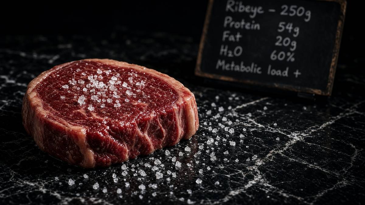
Carnivore and strict keto both raise BHB, and they're still not the same diet. They aren't the same diet. Carnivore is an elimination plus a protein load: meat, usually, and the carbohydrate that was in the other aisles is gone. Keto is a macronutrient ratio: carbohydrate low enough, fat high enough, protein moderate enough that hepatic acetyl-CoA overflows. Both can put you in ketosis. Both can fail if energy surplus and liver fat remain, because a stuffed hepatocyte has other things to do with fatty acids than write HMG-CoA. Protein is the unglamorous variable. Too little and you lose the muscle Cahill was trying to spare. Too much, in some people, and gluconeogenesis plus insulin blunt the overflow, so BHB stays shy of the millimolar band you were chasing. The ketone is a readout of hepatic acetyl-CoA overflow, not a badge. Chasing a millimolar number while still overeating steak is how you miss the point. The neighbouring carnivore and how-diets-actually-work essays are the pattern-level arguments. This one is the metabolite they sometimes raise.
In short. The blood number reports liver overflow, not which tribe you joined, and not whether you're overeating. Carnivore and keto can both raise ketones. They're different diets.
Exogenous ketone drinks raise circulating BHB without you having to restrict carbohydrate — fuel without the dietary rewrite. Esters (usually a BHB-butanediol or BHB-glycerol ester) and salts (usually sodium, potassium, calcium, magnesium racemates, which is already a stereochemistry problem) are the two shop objects. They do what they say: plasma BHB rises, often into the 1–3 mM band, on a timescale of minutes to an hour. They don't automatically recreate the insulin, glucagon, glycogen and adipose-lipolysis state of a ketogenic diet or a fast. You bought the metabolite. The hormonal context of a fast or a keto week is a different purchase. HDAC occupancy, NLRP3 occupancy, GPR109A occupancy might, in principle, follow the millimolar number; the fuel-switch programme in the liver won't, because the liver is still seeing carbohydrate. A paper that gives an ester and reports a signalling endpoint has done a metabolite experiment. A paper that gives an ester and claims it has done a diet has not.
In short. You didn't buy the low-insulin, empty-glycogen state of a fast. Ketone drinks can raise blood BHB without a diet. You bought the molecule.
Racemic salts are the stereochemistry the shop doesn't always print. D-BHB is the hepatic product and the HDAC and fuel molecule the papers named. L-BHB is handled differently, is a weaker or non-substrate for BDH1 in the physiological direction, and isn't a free extra millimole of the same job. A sodium-BHB powder that's a 50:50 racemate delivers half the relevant enantiomer and a sodium load the kidney will notice. Esters can be written as the D-enantiomer if the chemist bothered; some did, some didn't. If your assay is HDAC inhibition or 13C incorporation into TCA, the enantiomer is a methods line. If your assay is a lifestyle blog, the enantiomer is invisible. This catalogue doesn't sell a ketone ester. The sentence belongs here so that a millimolar reading after a drink isn't compared, naively, with Cahill's D-BHB. Identity is a carbon skeleton plus a stereocentre. Racemate is two identities.
In short. Read the label before you compare a drink to a fast. Many ketone salts are a 50:50 mix of the useful form and its mirror image. The liver makes only the useful form.
Retatrutide is occupancy at three class-B GPCRs, and it is the catalogue object this page actually carries into the fuel conversation. LY3437943, Coskun, Cell Metabolism 2018: a fatty-acylated unimolecular agonist at GIPR, GLP-1R and GCGR. Jastreboff, New England Journal of Medicine 2023: Phase 2, obesity, 24.2 percent mean weight loss at 12 mg, 48 weeks, a glucose and liver-fat neighbourhood that the twin-cycle essay cares about. We synthesise the published structure in the United States, HPLC-MS, labelled for research. We aren't Eli Lilly. We don't have an MHRA marketing authorisation to pretend otherwise. Incretin and glucagon-receptor occupancy move body weight and hepatic fat by a different door from HMGCS2. Ketones are what you get from the diet side when the liver overflows. The receptor is what the peptide occupies. We keep both on the same reading list because fuel routing is the subject, and pretending they're rivals is a category error. Use whichever actually moves the variable you're measuring. Don't staple them into a protocol.
In short. Retatrutide occupies three gut-hormone receptors and can move weight and liver fat. That's a different door from making ketones. Same subject, fuel routing. Not the same tool.
MOTS-c and NAD+ sit on the same organelle campus as ketone oxidation and don't share a mechanism with BHB. MOTS-c is MRWQEMGYIFYPRKLR, sixteen residues from mitochondrial 12S rRNA — Lee, Kim, Cohen, Cell Metabolism 2015 — AMPK-adjacent, a folate–methionine neighbourhood. NAD+ is the hydride coin BDH1 and Complex I actually spend; the 1000 mg cake is lyophilised β-NAD+ for a tube. A ketone becomes NADH at BDH1 and then at the TCA dehydrogenases; that NADH wants NAD+ back at Complex I. Shared redox, different molecule. Confusing a millimolar BHB, a dinucleotide, a 16-mer and a triple agonist is how a journal becomes a smoothie. We'll sell you the named objects. We won't design the blot, and we won't write a protocol that pretends a weekly incretin, a mitochondrial peptide, a cofactor and a diet-derived four-carbon acid are one juice. Neighbourhood, in this journal, is a courtesy on a reading list. It isn't a combination claim.
In short. One mitochondrial peptide, NAD+, a triple gut-hormone agonist and a ketone share an energy campus. They're four objects. Don't run them as one juice.
How to measure a ketone without lying to yourself
Decide what you remain measuring before you open a meter. Plasma or capillary D-BHB is the number the physiology above actually named. Enzymatic point-of-care meters (β-hydroxybutyrate dehydrogenase, a dye, a strip) are good enough for the 0.1–6 mM band in a human who isn't in DKA, and they aren't a mass spectrometer. Urine acetoacetate sticks (nitroprusside) see the ketone the kidney spilled, lag the blood, and go faint once BHB predominates, which is why adapted people 'leave ketosis' on a stick while a blood meter still reads 1.5 mM. Breath acetone is a leak, useful as a trend, hopeless as a concentration of BHB. Laboratory plasma BHB, enzymatic or GC-MS or LC-MS with a chiral column if the ester was racemic, is how a paper earns the word metabolome. NADPH isn't this assay. Acetoacetate is a different, less stable analyte and wants faster handling. Write which body you measured. Write the matrix. Write the time since the last meal. A number without those is a rumour, not a method.
In short. Breath acetone is a smell, not a concentration. Blood BHB is the measurement that matches the physiology. Urine sticks lag and can fade as you adapt.
If I were drawing the assay next, I'd start with the unglamorous half: which hand of the molecule you measured, and when you stopped the chemistry. D-BHB is the physiological molecule. L-BHB contaminates racemic salts and some labelled tracers. A chiral method, or an enzymatic assay that is D-specific, is how you stop adding 50 percent of the wrong hand. Acetoacetate decarboxylates on a warm bench; snap-freeze, acid or the method your lab actually validated, internal standard at quench not at injection. A time course after an ester is minutes. A time course after a carbohydrate cut is days, because HMGCS2 and MCT induction are days. If you add BHB to a medium and harvest at twenty-four hours, you have done a signalling-plus-fuel experiment in a dish whose glucose you must also name, because most media are 25 mM glucose and that isn't a ketogenic liver. Write the time. Write the glucose. Write the standard. The millimolar in the well is only as informative as the minute you chose to stop the chemistry.
In short. A ketone added to sugary medium isn't a fasting liver. The useful ketone is one-handed. Freeze samples fast. Write the time point and the sugar in the dish.
Machines, named, because we measured ketones isn't yet a measurement. A handheld enzymatic meter is a strip and a dehydrogenase. A clinical chemistry analyser is the same enzyme, calibrated, on venous plasma. GC-MS or LC-MS/MS, with 13C-BHB if you're doing flux, is how you separate BHB, acetoacetate, acetone, and the enantiomer if you asked. A Seahorse XF or an Oroboros O2k is oxygen consumption, the Complex I invoice, once you have given the tissue BHB instead of pyruvate and shown the SCOT dependence. Westerns for HMGCS2, BDH1, OXCT1, MCT1, MCT2, HDAC1/2, NLRP3, HCAR2. ELISA or multiplex for IL-1β if the inflammasome is the claim. Acetyl-histone blots, or mass-spec proteomics of histone marks, if HDAC is the claim. cAMP if GPR109A is the claim. None of that is glamorous. All of it is how you stop a metabolite essay becoming a mood. The diet is allowed as the intervention. It isn't allowed as the only figure in a ligand title.
In short. A strip for blood BHB, a mass spectrometer if you need the family, oxygen consumption if you claim fuel, blots if you claim a ligand job. Name the machine.
A BHB number remains not a ligand occupancy, not a fuel flux, and not a diet. Occupancy at HDAC wants a concentration in the nucleus, or at least a histone mark and an in-vitro IC50 that overlaps the plasma you measured. Occupancy at GPR109A wants cAMP in a cell that actually expresses HCAR2. Fuel flux wants 13C into citrate, or a measured arteriovenous difference, or an oxygen-consumption increment that dies when you block SCOT. A diet wants weighed food and a time. You can have millimolar BHB and no HDAC phenotype if the tissue didn't see it. You can have an HDAC phenotype in a dish at 5 mM and no such concentration in the neuron you cared about. You can have a ketogenic diet and a lower IL-1β because weight fell, or salt changed, or the microbiome changed, and not because NLRP3 saw BHB. The restoration literature in the NAD+ essay has this exact problem at a different node. Same demand. Name the job. Then measure the job. The meter is a scout.
In short. One blood-ketone number isn't proof you occupied an enzyme, a receptor, or a fuel pathway. Measure the job you claimed. The meter is only a scout.
- Name the body: D-BHB, acetoacetate, or acetone. Urine isn't plasma. Breath isn't blood.
- Name the matrix and the time: capillary, venous, tissue; hours after a meal, days into a cut, minutes after an ester.
- Name the enantiomer if the intervention was a salt or a labelled tracer. D is the physiological hand.
- Name the job: fuel (13C, OCR, SCOT), HDAC (acetyl-histone, class I assay), NLRP3 (IL-1β, caspase-1), GPR109A (cAMP, knockout).
- Name the tissue. Liver writes. Brain, heart, muscle spend on different clocks. A homogenate is a scout.
- Write the quench. Acetoacetate won't wait. Glucose in the dish is a methods line, not a footnote.
Close: two jobs, named enzymes, laboratory neighbours
The topology is small enough to hold, and it is the only reason a 1967 catheter paper, a 2013 HDAC paper and a 2015 inflammasome paper can sit in one piece without being a collage. Adipose spills fatty acids when insulin falls. The liver β-oxidises them, HMGCS2 commits the acetyl-CoA overflow to ketogenesis, BDH1 reduces most of the acetoacetate to BHB, and the ketone leaves because hepatocytes lack SCOT. Extrahepatic mitochondria take BHB on MCT1/2, oxidise it through BDH1 and SCOT, and feed acetyl-CoA to a chain that still wants NADH at Complex I. Given days, a human brain will take most of its energy this way and spare muscle: Cahill. Independently, millimolar BHB inhibits class I HDACs, damps NLRP3, and occupies GPR109A. Physiological ketosis is 0.5–3 mM, buffered. DKA is a different state. Keto flu is transporter lag plus natriuresis. Exogenous esters raise the metabolite without the hormonal context. Retatrutide occupies a different door onto the same fuel-routing subject. That's the map. The popular story got loud because the millimolar number is easy to print. The work got interesting when the number acquired ligand jobs.
In short. Liver makes ketones from fat overflow. Brain and other organs burn them. The same molecule also talks to genes, immune alarms and a receptor. Ordinary ketosis isn't acidosis.
Here, the public papers are the reading list, and they're short enough to actually read. Owen, Cahill, Journal of Clinical Investigation 1967, brain metabolism during fasting; Cahill, Annual Review of Nutrition 2006, the starvation map. Shimazu, Newman, Verdin, Science 2013, BHB as a class I HDAC inhibitor. Newman and Verdin, Annual Review of Nutrition 2017, the signalling review. Youm, Dixit, Nature Medicine 2015, BHB and NLRP3. Taggart, Offermanns, the GPR109A/niacin receptor papers, so the GPCR floor stays named. Halestrap, Pierre, Pellerin, the MCT literature, so the flu is a transporter lag and not a myth. The inborn-error clinics on HMGCS2 and OXCT1, so the enzymes stay real. Jastreboff, NEJM 2023, and Coskun, Cell Metabolism 2018, so the triple-agonist door stays a different door. That's a fortnight of evenings, not a guru. The ketosis headlines will still be there when you come back, and they will look smaller.
In short. One short stack of named papers covers the starving brain, the gene job, the immune-alarm job, the receptor, the doors, and the separate peptide door. Read those before any headline.
The neighbouring pages complete the campus — fasting, electrolytes, diets — and this one stays with the metabolite. Fasting, autophagy and the mTOR switch: the other programme a carbohydrate cut leans on, AMPK, ULK1, a recycle shift this ketone essay isn't. Electrolytes and the kidney: insulin natriuresis, the other half of week one. Liver fat, the real enemy: the depot type 2 actually cares about, and a reason ketogenesis and de novo lipogenesis can be opposite readings of the same hepatocyte. Carnivore diet mechanisms, and how diets actually work: pattern-level arguments that sometimes raise BHB and sometimes don't. Human metabolism, without the catchphrase. NAD+ and sirtuins: the other millimolar budget, the one that spends a dinucleotide rather than inhibiting a zinc HDAC. Brown fat: UCP1, a different decision about a mitochondrial gradient. Retatrutide: the published LY3437943 structure, three class-B GPCRs, a weight and liver-fat curve. Stay here if you wanted the four-carbon metabolite explained. Go there if you wanted the diet, the kidney, the depot, or the receptor.
In short. This page is the ketone as fuel and as signal. Next door: fasting and autophagy, salt and the kidney, liver fat, diets, NAD+, brown fat, and the triple agonist.
Leave with the map, not a shopping list. BHB and acetoacetate are fuels the liver writes from acetyl-CoA overflow and extrahepatic mitochondria spend through BDH1 and SCOT. Cahill showed the human brain will take most of its energy that way and spare muscle. MCT1/2 and enzyme induction take days, which is most of keto flu; the rest is natriuresis. Physiological ketosis is roughly 0.5–3 mM and buffered. DKA is insulin deficiency plus acid plus an unwell person. Independently, BHB is a class I HDAC inhibitor, an NLRP3 damper, and a millimolar ligand at GPR109A. A ketogenic pattern raises the metabolite in a hormonal context. An ester raises the metabolite without that context. Retatrutide occupies GIPR, GLP-1R and GCGR, a different door onto fuel routing. MOTS-c and NAD+ are mitochondrial reagents on the same campus. If your experiment needs the metabolite, measure the enantiomer, the tissue, and the job. If it needs a diet, weigh the food. If it needs a medicine, this catalogue doesn't sell one.
In short. Leave with the map: liver overflow, brain fuel, doors that take days, ordinary ketosis versus acidosis, three ligand jobs, and a peptide that's a different door. Measure the job you named.
Research-use-only. Not for human consumption / not a medicine. The lyophilised retatrutide on the related listing is the published LY3437943 structure, synthesised in the United States, HPLC-MS characterised, labelled for in-vitro work: a binding isotherm, a cAMP assay, a transfected well whose receptor you can actually name. We aren't Eli Lilly. The 1000 mg NAD+ cake and the MOTS-c 16-mer are laboratory reagents on the same energy campus, different jobs. The physiology in the paragraphs above is public, cited, and older than any of those vials. Use it to design the experiment you have the controls for, with the enzyme named, the transporter named, the ligand job named, and the time point written down. Read Cahill, read Newman and Verdin, read Youm, then weigh the cake that matches the question if the question was a receptor or a cofactor. We'll sell you the characterised objects. We won't tell you they're ketones, and we won't tell you a millimolar BHB is a protocol. Fuel routing is a set of rates. This rate you can measure, in a tube or in a plasma, with a chromatogram or a meter on the bench beside it.
In short. Those related vials are research chemicals for experiments, not ketones and not a diet. The biology is public. Name the job, then measure it.
Questions the essay actually answers
- Do exogenous ketone drinks do the same thing as a ketogenic diet?
- They raise circulating D-BHB, or a racemic mix, without you having to restrict carbohydrate. They don't automatically recreate the insulin, glucagon and glycogen state of a ketogenic diet or a fast. HDAC, NLRP3 and GPR109A occupancy might follow the millimolar number; hepatic ketogenesis won't. You bought the metabolite. The hormonal context is a different purchase.
- Is ketosis dangerous?
- Physiological ketosis sits roughly 0.5–3 mM BHB and is buffered. Diabetic ketoacidosis is a different beast: usually much higher, with a sick, insulin-deficient patient, a wide anion gap and a falling pH. They share a word, not a clinic, a pH, or an ending. SGLT2 inhibitors, type 1 diabetes and pregnancy are clinician territory, not blog territory.
- What did Cahill actually show?
- That a starving human brain, given days, takes most of its energy from β-hydroxybutyrate and acetoacetate and so spares muscle that would otherwise have been converted to glucose. Owen et al., J Clin Invest 1967; Cahill, Annu Rev Nutr 2006. That's the survival argument for the pathway, not a lifestyle badge.
- How is BHB a signal as well as a fuel?
- It inhibits class I HDACs (Shimazu, Newman, Verdin, Science 2013), damps the NLRP3 inflammasome (Youm et al., Nat Med 2015), and occupies GPR109A/HCAR2 in the higher millimolar range. A millimolar metabolite is allowed to be a ligand. The fuel job through BDH1 and SCOT remains most of the flux.
- What are HMGCS2, BDH1 and SCOT?
- HMGCS2 is mitochondrial HMG-CoA synthase 2, the committed step of hepatic ketogenesis. BDH1 is the dehydrogenase that interconverts acetoacetate and D-BHB. SCOT (OXCT1) activates acetoacetate in extrahepatic tissues; the liver largely lacks it, which is why the ketone is an export.
- Why does ‘keto flu’ happen?
- Mostly two clocks. MCT1/2 and extrahepatic BDH1/SCOT take days to induce, so the brain can see millimolar BHB before it can spend it. Independently, falling insulin makes the kidney dump sodium; magnesium and potassium follow. Salt the food. Then decide whether you like the diet. The electrolytes page is the nephron half.
- Is retatrutide a ketone drug?
- No. Retatrutide is published LY3437943: occupancy at GIPR, GLP-1R and GCGR. It moves weight and hepatic fat from the receptor side. Ketones are what a liver writes from acetyl-CoA overflow. Same subject — fuel routing — different door. We stock the structure as a research ligand. We aren't Eli Lilly.
- Carnivore or keto — which raises BHB?
- Both can. Carnivore is an elimination plus a protein load; keto is a macronutrient ratio. Protein high enough to blunt overflow will keep BHB shy of the millimolar band in some people. Energy surplus and liver fat can defeat both. The ketone is a readout, not a tribe.
- How should ketones be measured honestly?
- Say D-BHB, acetoacetate or acetone, and say plasma, urine or breath. Blood enzymatic BHB matches the physiology; urine sticks lag and fade as BHB predominates; breath acetone is a leak. If the claim is a ligand job, show the job (acetyl-histone, IL-1β, cAMP), not only the meter. Quench fast. Name the enantiomer if you swallowed a salt.
- Is this a supplement or a medicine?
- Neither. This page is physiology. The related listing is characterised LY3437943 for laboratory work, labelled for in-vitro use, not a ketone ester and not a diet.
Hypothetical research reconstitution
How these vials are typically mixed
Hypothetical research reconstitution for the named catalogue vial. Not a protocol, not medical advice, not a use instruction. These amounts sit in published and commonly cited laboratory ranges. The vial is labelled for research use only — not for human or veterinary administration.
Retatrutide
30mg
Mix with 3 ml bacteriostatic water → 10 mg/ml
- Hypothetical aliquot
- 1–2 mg to start; published trial arms ran higher by week
- 0.10–0.20 ml · 10–20 units on a U-100 syringe (at 1–2 mg)
- How often
- Once weekly
- The Jastreboff NEJM 2023 arms ran 48 weeks. That is a trial, not a shop protocol.
Bench steps
- Let the vial sit until it is no longer cold to the touch.
- Wipe the stopper with 70% isopropyl alcohol. Let it dry.
- Draw 3 ml bacteriostatic water (0.9% benzyl alcohol).
- Run the water slowly down the inside glass — do not blast the cake.
- Roll between finger and thumb until the cake is gone. Do not shake.
- Label the date. Store the solution at 2–8 °C. Do not freeze. Use within 30 days unless the note below says otherwise.
LY3437943 architecture. Weekly, not daily. Those milligram figures are what the papers used on the investigational medicine — they are not a use instruction for this reagent.
MOTS-c
40mg
Mix with 2 ml bacteriostatic water → 20 mg/ml
- Hypothetical aliquot
- 5–10 mg
- 0.25–0.50 ml · 25–50 units on a U-100 syringe
- How often
- Two or three times per week
- 4–8 weeks
Bench steps
- Let the vial sit until it is no longer cold to the touch.
- Wipe the stopper with 70% isopropyl alcohol. Let it dry.
- Draw 2 ml bacteriostatic water (0.9% benzyl alcohol).
- Run the water slowly down the inside glass — do not blast the cake.
- Roll between finger and thumb until the cake is gone. Do not shake.
- Label the date. Store the solution at 2–8 °C. Do not freeze. Use within 30 days unless the note below says otherwise.
Mitochondrial 16-mer. Fridge. Do not freeze. The 5 mg mark is where most bench notes start.
NAD+
1000mg
Mix with 10 ml bacteriostatic water → 100 mg/ml
- Hypothetical aliquot
- 50–100 mg
- 0.50–1.00 ml · 50–100 units on a U-100 syringe
- How often
- Two or three times per week in published infusion and assay notes
- 4–8 weeks, then a pause
Bench steps
- Let the vial sit until it is no longer cold to the touch.
- Wipe the stopper with 70% isopropyl alcohol. Let it dry.
- Draw 10 ml bacteriostatic water (0.9% benzyl alcohol).
- Run the water slowly down the inside glass — do not blast the cake.
- Roll between finger and thumb until the cake is gone. Do not shake.
- Label the date. Store the solution at 2–8 °C. Do not freeze. Use within 30 days unless the note below says otherwise.
A 1000mg cake wants 10 ml. Protect from light. Solution yellows as it oxidises — that is the cofactor dying, not a flavour. Use promptly.
Bacteriostatic water and sterile syringes ship with peptide orders over £75. Kit details · 10 ml bacteriostatic water
The vials this essay sits on
Named sequences the essay maps — Retatrutide, MOTS-C, NAD+. Hypothetical research neighbourhood, not a protocol, not a medicine. One press puts every in-stock vial in the bag.
 Made in USAOut of stock
Incretin
Retatrutide
US-made retatrutide 30mg — the published structure LY3437943, HPLC-MS verified.
4.6(609)
67 browsing this now · 5 purchased in the last 24 hours
30mg
£120.00
 Made in USA
 Research only
Research use only. Not a combined-use instruction.
Read next
48 min · long read · Metabolism
How a carnivore diet actually works
Eliminate plants, eat animals. The interesting part is not the internet. It is ketosis, gluconeogenesis, electrolytes and what happens to a microbiome when fibre hits zero.
45 min · long read · Metabolism
How diets actually work
Calories are conservation of energy. Hormones are the routing table. Palatability is why the routing table keeps getting overwritten. Every named diet is a way of leaning on one of those three.
51 min · long read · Metabolism
Human metabolism, without the slogan
Calories are bookkeeping. The actual system is a set of hormonally gated fluxes through glycogen, fat and amino acids — with the liver as air-traffic control.

50 min · long read · Metabolism
Fasting, autophagy and the mTOR switch
Eat, and mTOR builds. Fast, and AMPK and ULK1 start recycling the cell. The molecular story is a switch, not a personality.
More in this desk
51 min · long read · Metabolism
Reversing type 2 diabetes is a published result, not a slogan
The DiRECT trial put type 2 diabetes into remission by emptying the liver and pancreas of surplus fat. Very-low-calorie and very-low-carbohydrate programmes can both get you there. The mechanism isn't mystical.
49 min · long read · Metabolism
Liver fat is the variable type 2 diabetes actually cares about
Ectopic fat in the liver overproduces glucose and VLDL. Empty it — by energy deficit, by carbohydrate restriction, or by both — and first-phase insulin can return. That is the twin-cycle in working clothes.

49 min · long read · Metabolism
Protein, training and muscle protein synthesis
Muscle isn't a mood. It is mechanical tension plus a leucine-gated translational programme. Here is the dose, the threshold, and why the gym still has to happen.
49 min · long read · Metabolism
Regulating blood sugar: the control system
Glucose is a tightly bound variable. Here is the hardware — pancreas, liver, muscle, incretins — and the places it fails.
Essays describe published research. They are not medical advice and they do not authorise human use of any catalogue item.