

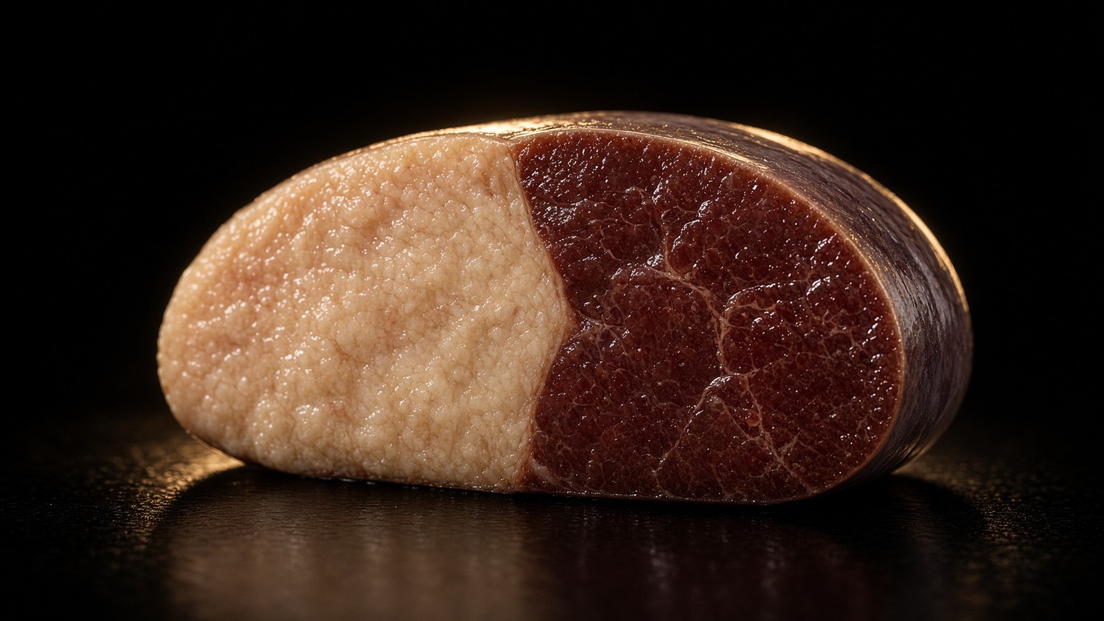
Metabolism · 49 min · 10,865 words
Liver fat is the variable type 2 diabetes actually cares about
Ectopic fat in the liver overproduces glucose and VLDL. Empty it — by energy deficit, by carbohydrate restriction, or by both — and first-phase insulin can return. That is the twin-cycle in working clothes.
What this essay actually tells you
- Taylor's Twin Cycle hypothesis: excess liver fat drives hepatic insulin resistance and excess pancreatic fat impairs insulin secretion. Two stores. One disease, if he's right, and DiRECT suggests he is.
- The Counterpoint and DiRECT trials showed that substantial weight loss can put type 2 diabetes into remission by emptying those stores. Liver fat down, glucose follows.
- Liver fat is the variable. Calories, ketones, incretin agonists and surgery are different tools that can move it. Pick the tool. Watch the variable. Don't marry the tribe.
What this actually means
Type 2 diabetes is often described as a pancreas problem. Roy Taylor's twin-cycle work says look at the liver first. Surplus energy becomes liver fat; a fatty liver keeps making glucose in the morning and ships VLDL out; that fat lands in the pancreas and first-phase insulin goes quiet. Pull the liver fat down and a large fraction of people in the first years of the disease get their glucose back. DiRECT showed it with a formula diet. Low-carbohydrate clinics show it with a different tactic. Incretin triple agonists (retatrutide's 24% weight-loss curve in NEJM) empty the same depot from the receptor side. The variable is hepatic fat. The tools are whatever actually empties it, and arguing about which tool is 'pure' is how the depot stays full.
If you only remember one organ from the diabetes pages, remember the liver. Muscle is the disposal sink after a meal. The pancreas is the controller. The liver is the factory that won't clock off when it's stuffed with fat. Morning glucose is often a liver number, not a bread number. That's why some people can skip breakfast carbohydrate and watch the dawn phenomenon shrink, and why a five-kilogram loss that happens to be hepatic is worth more than a five-kilogram loss that isn't. We still weigh people on a scale. The interesting mass is inside the ribcage, sitting in hepatocytes as triglyceride, measured in percent by magnetic resonance, not in kilograms on a bathroom floor. Roy Taylor spent a career saying this out loud from Newcastle, and the trials that followed him are why the sentence is no longer a hunch. Type 2 diabetes, for a large fraction of people in the first years of the disease, is ectopic fat in two organs. Empty the first store and the second often follows. Glucose follows both.
In short. Morning blood sugar is often the liver still making glucose, not the toast. Fat inside liver cells is the switch that keeps that factory on.
Type 2 diabetes is still described, in too many waiting rooms, as a pancreas problem that only ever gets worse. The β-cell fails, insulin falls behind, drugs stack, and the poster on the wall treats remission as a rumour. Taylor's twin-cycle work says look at the liver first. Surplus energy becomes intrahepatic triglyceride. A fatty liver keeps making glucose at dawn and ships very-low-density lipoprotein out into blood. That fat lands in viscera and in the pancreas. First-phase insulin goes quiet. Two stores. One disease, if he is right, and DiRECT suggests he is. Pull the liver fat down and a large fraction of people in the first years of the disease get their glucose back. That isn't a comments-thread catchphrase. It's a cluster-randomised trial in UK primary care, a magnetic-resonance time course from an eight-week liquid diet, and a biochemistry of gluconeogenesis and de novo lipogenesis that already had names before anyone put a formula shake in a GP surgery. The pancreas isn't innocent. It's second, and we should treat it that way.
In short. Type 2 is often blamed on the pancreas. A lot of it starts as fat in the liver that keeps making sugar and then spills fat into the pancreas.
This page is the variable, the two stores, the named trials, and the tools that actually move intrahepatic triglyceride. Calories, ketones, incretin agonists and surgery are different instruments. They aren't a purity contest. The catalogue neighbourhood includes retatrutide, the published LY3437943 structure, a unimolecular agonist at GIPR, GLP-1R and GCGR, because glucagon-receptor tone on a hepatocyte is a lipid-oxidation cue and because the Phase 2 weight curve empties the same depot from the receptor side. MOTS-c is a 16-mer a mitochondrion translated from its own 12S rRNA. NAD+ is the hydride coin Complex I wants oxidised. Sharing a shelf isn't sharing a mechanism. A formula diet, a carbohydrate cut, a Roux-en-Y, and a class-B occupancy can all lower liver fat. Pretending only one of them is 'real' is how the depot stays full while the argument gets louder. The job of this page is to name the depot, show how you see it, and keep the tools in their lanes. It isn't a protocol, and it isn't a clinic appointment written as physiology.
In short. Fat in the liver is the thing to watch. Diet, ketones, gut-hormone drugs and surgery are different tools that can lower it. Argue about the tool later.
Intrahepatic triglyceride isn't a personality and it isn't 'fat' in the sense a waist tape measures. A hepatocyte that has parked more than about 5.5 percent of its volume as lipid, the Szczepaniak magnetic-resonance spectroscopy threshold a field still uses, is steatotic. That lipid is mostly triglyceride. Some of it's diacylglycerol, which is the species Gerald Shulman's group keep finding next to hepatic insulin resistance. The cell is still a hepatocyte: glycogen, urea, cytochrome P450, bile acids, a first-pass customs post for everything the portal vein brought. Steatosis doesn't suspend those jobs. It biases them. Gluconeogenesis becomes harder for insulin to mute. De novo lipogenesis, already written by SREBP-1c and ChREBP, has more substrate and more transcription. VLDL assembly via apoB-100 and microsomal triglyceride transfer protein ramps, because the cell is trying to export what it can't oxidise. Export is how the second store fills. We'll drop the factory analogy here. The rest is enzymes, a membrane voltage, and two imaging methods that can see the depot without asking the person how they feel about bread.
In short. Fat inside liver cells is a measured percentage, not a feeling. Once it's there, the liver keeps making sugar and ships fat out in particles that land elsewhere.
Two stores. One disease, if he is right
Roy Taylor put the twin-cycle hypothesis on the page in Diabetologia in 2008, then again as a Diabetes Care review in 2013. Chronic positive energy balance, in a person who has exceeded what he later called their personal fat threshold, parks triglyceride in the liver. Hepatic insulin resistance follows: insulin no longer fully suppresses glucose production, so fasting glucose and the dawn rise climb even if the last meal was modest. The same liver overproduces VLDL. Those particles deliver triglyceride to viscera, to muscle, and to the pancreas. Pancreatic fat isn't an innocent bystander on an MRI. It's lipotoxicity at the β-cell: first-phase insulin secretion fails, the controller that should have shut the liver up after a carbohydrate load arrives late and small, and the cycle feeds itself. If you reverse the energy balance hard enough, liver fat falls within days. Pancreatic fat follows on a slower clock. First-phase can return. That's the hypothesis as a pair of stores, not as a morality of willpower, and it's why an eight-week liquid diet and a year of primary-care formula both belong in the same essay as a receptor occupancy.
In short. Taylor's idea: extra energy fills the liver with fat, the liver overmakes sugar and ships fat to the pancreas, and insulin release then fails. Empty the stores and the disease can quiet.
The personal fat threshold is the sentence that stops BMI from being a diagnosis. Taylor and Holman, Clinical Science 2015: people develop type 2 when they exceed their own capacity to store fat safely in subcutaneous adipose tissue, not when they cross a universal waist or a universal body-mass index. A South Asian adult at 23 kg/m² can have a fatty liver and a quiet first-phase. A Northern European at 35 can still have a liver that's metabolically quiet. Twin-cycle biology is ectopic fat, not a lecture about obesity as a character. Genetics, sex, age, and how long the surplus has been running all move the threshold. PNPLA3 I148M is the common steatosis allele a hepatologist already names; TM6SF2 and GCKR sit nearby. Polygenic risk for type 2 is real and small per locus. The physiology that actually breaks, in the people DiRECT enrolled, is still a liver full of fat and a pancreas that then fails its dump. Holding both — a threshold that's personal, and a depot you can empty — is how you stop telling a thin diabetic person that they have the wrong disease.
In short. You don't need to be very overweight to get type 2. Each person has a point where extra fat spills into liver and pancreas. That point is personal, not a BMI chart.
Hepatic insulin resistance is the first cycle as a measurement, not as an adjective. In a healthy liver, insulin, arriving in the portal vein at concentrations the rest of the body never sees, phosphorylates FOXO1 out of the nucleus and shuts off the gluconeogenic programme: phosphoenolpyruvate carboxykinase, glucose-6-phosphatase, the enzymes that make glucose from lactate, alanine and glycerol. Insulin also tells glycogen synthase to store what just arrived. A steatotic hepatocyte hears that insulin badly. Fasting glucose production stays on. Clamp studies, from DeFronzo through Petersen and Shulman, have been saying this for decades: the liver is insulin-resistant early, often before muscle is, and modest weight loss can reverse that hepatic resistance while the muscle clamp is still ugly. Counterpoint put a week on the clock. Fasting plasma glucose and hepatic glucose production fell into the non-diabetic range by day seven, in parallel with a collapse of liver fat, while peripheral insulin sensitivity had barely moved. That time course is the argument. The factory clocks off when the lipid leaves. It doesn't wait for you to join a gym.
In short. A fatty liver ignores insulin and keeps making glucose overnight. Empty the fat and that glucose production can fall within a week, before muscle has caught up.
VLDL is how the first store fills the second. A hepatocyte that can't oxidise or export-as-bile the triglyceride it has made will package it: apoB-100 as the structural protein, microsomal triglyceride transfer protein as the assembler, a particle that leaves through the Space of Disse into the sinusoid and then into the circulation. Adipose lipoprotein lipase and hepatic lipase remodel those particles. What the periphery doesn't take, and what the liver takes back as remnants, still leaves a trail of fatty acids in tissues that were never designed as warehouses. Visceral fat is one landing site. The pancreas is the one Taylor cares about. Donnelly, Parks and colleagues, Journal of Clinical Investigation 2005, already measured where intrahepatic triglyceride comes from in people with fatty liver: a large fraction from circulating non-esterified fatty acids, a non-trivial fraction from de novo lipogenesis, a smaller fraction from the meal itself. Surplus carbohydrate, particularly fructose, feeds DNL. Surplus energy of any macronutrient, once adipose is past its personal threshold, feeds the NEFA spill. Either way the hepatocyte writes VLDL. Either way the pancreas can receive the cargo. Export isn't a cure; it's how the second store fills. I want you to hold that.
In short. The fatty liver ships fat out in blood particles. Some of that fat lands in the pancreas. Clearing the liver therefore matters for the pancreas, not only for the liver.
First-phase insulin is a ten-minute event, and it's the controller the second store breaks. Glucose enters the β-cell through GLUT1 and GLUT2, glucokinase phosphorylates it, ATP rises, KATP channels close, the membrane depolarises, calcium comes in, ready-to-release granules fuse. That dump should already be in the portal vein before the meal's glucose has finished arriving. Incretins — GIP from K-cells, GLP-1 from L-cells — amplify it when the glucose came by mouth, which is the 1964 measurement, not a 2020s product story. Lipotoxicity at the β-cell, the Unger and Poitout literature, is what happens when fatty acids and their derivatives, ceramides among them, sit on that machine for months and years: ER stress, oxidative stress, a first-phase that flattens, a second-phase that may still limp. Function can return before mass does. That distinction is why an eight-week diet can restore a dump in recent-onset type 2 without growing a new islet, and why twenty years of type 2, with insulin already on the list, remits less often. The second store is pancreatic fat. The readout is first-phase. The clock we're on is shorter than a lifetime and longer than a weekend.
In short. The pancreas should dump insulin in the first minutes after sugar arrives. Fat in and around those cells can silence that dump. In early type 2, emptying the fat can bring it back.
Type 2 diabetes is a potentially reversible metabolic state precipitated by chronic excess energy intake, with the twin-cycle of liver and pancreas fat as the mechanism.— Taylor R. Type 2 diabetes: etiology and reversibility. Diabetes Care. 2013; 36: 1047–1055. The twin-cycle in the sentence he actually wrote.
What a fatty hepatocyte actually does
De novo lipogenesis is the liver writing fat from carbohydrate, and it isn't a rumour. Sterol regulatory element-binding protein 1c, SREBP-1c, is the insulin-written transcription factor that turns on acetyl-CoA carboxylase, fatty-acid synthase, ATP-citrate lyase, the kit that takes citrate out of the mitochondrion and makes palmitate. Carbohydrate-responsive element-binding protein, ChREBP, is the glucose- and fructose-written partner: xylulose-5-phosphate, protein phosphatase 2A, nuclear entry, the same lipogenic genes plus pyruvate kinase. A high-insulin, high-sugar portal vein therefore writes the programme twice. Fructose is a special case because it's phosphorylated by ketohexokinase, bypasses the phosphofructokinase gate that throttles glycolysis, and floods the hepatocyte with triose phosphates that ChREBP can see. Alcohol is another special case: ethanol to acetaldehyde to acetate, NADH rising, β-oxidation stalling, fat parking. Energy surplus of any kind still matters more than a single villain, which is why a calorically restricted formula empties the depot even when the remaining calories include carbohydrate. DNL is a named flux we can measure. It isn't a licence to pretend protein and fat can't fill a liver if the adipose threshold is already past.
In short. The liver can turn sugar into fat. Insulin and sugar both switch that programme on. Fructose and alcohol are extra pushes. Too many calories of any kind still fill the organ.
Gluconeogenesis is the morning number. Phosphoenolpyruvate carboxykinase converts oxaloacetate to phosphoenolpyruvate. Glucose-6-phosphatase takes the phosphate off glucose-6-phosphate so free glucose can leave through GLUT2 into the hepatic vein. Both genes are FOXO1 and CREB targets. Glucagon and cortisol write them on. Insulin should write them off. A steatotic liver, insulin-resistant at the receptor-kinase step, leaves them on at 4 a.m. while the person is still asleep. That's the dawn phenomenon as hepatic glucose output, not as a personality of breakfast. Van Cauter's sleep work sits next door: a short night makes the same output worse, because cortisol timing and GH pulses are part of the overnight invoice. This page's job is the lipid. A hepatocyte full of triglyceride is a hepatocyte whose gluconeogenic enzymes insulin can't fully mute. Metformin, for all the AMPK fuss around it, still earns a large fraction of its glucose effect by quieting hepatic glucose production. Emptying the fat does the same job from the substrate side. Two interventions. One output. If you're wearing a CGM at dawn, you're reading the liver, and it will keep reading the liver until the lipid leaves or a drug mutes the enzymes.
In short. Overnight the liver should stop making much sugar. A fatty liver often doesn't, so glucose is already high before breakfast. That's a liver reading, not a willpower reading.
Diacylglycerol and protein kinase C-ε are the molecular sentence Shulman's laboratory keep putting under hepatic insulin resistance, and they belong here so 'fatty liver' doesn't stay a histology caption. Excess fatty-acyl-CoA that isn't oxidised or exported is esterified. Triglyceride is the warehouse. Diacylglycerol is the signalling species: it activates PKCε, which phosphorylates the insulin receptor on an inhibitory threonine, which blunts insulin-receptor kinase activity, which means IRS-2 is less phosphorylated, which means the FOXO1 programme stays nuclear. Samuel, Petersen, Shulman, the Nature and JCI papers of the 2000s and 2010s: knock down PKCε in a rodent and hepatic insulin sensitivity returns even if some lipid remains; drop DAG with a modest weight loss in a person and the clamp improves. Triglyceride on an MRI-PDFF is a proxy for this neighbourhood, not identical to it. A liver can be steatotic and relatively insulin-sensitive if the DAG-PKCε arm is quiet, which is one reason imaging and clamps sometimes disagree. For clinical type 2, the proxy is good enough. For a proper enzyme assay, DAG species, PKCε translocation, and an insulin-receptor blot are how you stop a percent-fat number from pretending to be a kinase.
In short. A particular fat species inside the liver cell can jam the insulin receptor. That's a named enzyme story, not simply 'the liver looks fatty on a scan'.
Mitochondria of the hepatocyte are the other door out of the lipid. β-oxidation in the matrix, acyl-CoA dehydrogenase, NADH and QH2 on every turn, ketogenesis via HMGCS2 when acetyl-CoA overflows what citrate synthase will take. A liver that can't reoxidise NADH — because the respiratory chain is congested, because alcohol has already reduced the pool, because the cell is stuffed — stalls fatty-acid oxidation and stores the fat instead. Complex I wants oxidised NAD+. That's the neighbouring cofactor essay, and it's why a millimolar nucleotide sits on the same reading list as a twin-cycle trial without being the trial. SIRT3 deacylates matrix enzymes if NAD+ is there to spend. MOTS-c, MRWQEMGYIFYPRKLR, is a 16-mer from 12S rRNA that talks to AMPK, which can phosphorylate acetyl-CoA carboxylase and lift the malonyl-CoA brake on carnitine palmitoyltransferase-1. Same organelle campus. Different objects. A homogenate NAD+ kit isn't a PDFF. An AMPK peptide isn't a formula diet. The hepatocyte still has to oxidise or export the triglyceride. The chain, the cofactor, and the carnitine shuttle are how oxidation happens. Energy deficit is how you stop writing new lipid faster than those routes can clear it.
In short. Liver cells burn fat in mitochondria or they store it. If the engines are congested, storage wins. Diet and some hormones change how much new fat is written, not the laws of the organelle.
Fructose and alcohol are named insults because they hit this biochemistry at different rungs, and because the internet picked one each and made it a brand. Fructose, from sucrose or from sweetened drinks, is almost entirely hepatic at first pass. Ketohexokinase has a high capacity and a low restraint. ATP can dip. Uric acid can rise. Triose phosphates feed DNL and feed ChREBP. Soft-drink trials and isocaloric fructose-versus-glucose work both exist; the honest reading is that fructose is a potent DNL substrate and not the only way to fill a liver. Ethanol is a redox insult first: cytosolic alcohol dehydrogenase, then aldehyde dehydrogenase, NADH up, the NAD+/NADH ratio falling, β-oxidation losing the oxidised cofactor it needs, fat parking as a redox consequence. The metabolic fatty liver is now called MASLD. The alcohol version is still alcohol-related liver disease. Plenty of people have both. Neither gets a monopoly, which is a sentence the stub already earned and which the enzymology supports. Genetics, energy surplus, refined carbohydrate, ethanol, and time. The job is still to empty the depot. Naming the insult tells you which lever is nearest. It doesn't make the other levers imaginary.
In short. Sugar drinks and alcohol can both park fat in the liver, by different routes. So can eating too much in general. None of them owns the whole story.
Diagram
Matrix
- TCA cycle · β-oxidation · mtDNA nucleoids
- NADH produced here. Complex I spends it.
- MOTS-c (MRWQEMGYIFYPRKLR) from 12S rRNA.
Inner membrane
- I → II → III → IV → V (ATP synthase)
- ~150 mV proton-motive force
- ~40–60 kg of ATP turned over per human day
mtDNA is 16,569 bp, 37 genes, 13 proteins of the respiratory chain. Nuclear DNA encodes the other ~1,200 mitochondrial proteins. NAD+ is the hydride carrier between dehydrogenases and Complex I. MOTS-c is a 16-mer translated from 12S rRNA — a peptide the mitochondrion wrote itself.
- Normal liver fat (MRS)
- <5.5%
- Counterpoint
- 8 weeks, ~600 kcal/day
- DiRECT, 12 months
- 46% vs 4%
- Weight-loss threshold
- ~15 kg
- Jastreboff 2023, 12 mg
- 24.2% at 48 weeks
- Sanyal 2024, MRI-PDFF
- large relative drop
- Personal fat threshold
- not a BMI
- ALT
- noisy proxy
Szczepaniak. Above that, the hepatocyte is steatotic. MRI-PDFF is the trial endpoint.
Lim, Taylor, Diabetologia 2011. Liver fat and glucose production fell by day 7.
Remission, HbA1c <48 mmol/mol off drugs. Lean, Lancet 2018. Cluster-randomised, UK primary care.
In DiRECT, 86% of those who lost 15 kg or more were in remission. The depot itself.
Retatrutide Phase 2 mean weight change. Same depot from the receptor side.
Retatrutide in MASLD, 24 weeks. Intrahepatic fat moved with the chain. Read the figure, not a caption.
Taylor & Holman, Clin Sci 2015. Ectopic fat, not a universal waist.
A normal ALT on a fatty liver is a known trap. Image the depot, or admit you did not.
Counterpoint: eight weeks that named the mechanism
Lim, Hollingsworth, Taylor and colleagues, Diabetologia 2011, is the paper a twin-cycle essay has to sit with before it's allowed to talk about remission as a primary-care statistic. Eleven people with type 2 diabetes, short duration, came off their glucose-lowering drugs and ate a liquid formula of about 600 kilocalories a day for eight weeks, with a small amount of non-starchy vegetable for sanity. Controls without diabetes sat on the scanner for the comparison. Liver triacylglycerol, pancreatic triacylglycerol, hepatic glucose production, and first-phase insulin secretion were the measurements, not a well-being score. Magnetic resonance spectroscopy is how they saw the two stores. A hyperinsulinaemic clamp and a glucose load are how they saw the controller. The study is small. It's also the cleanest time course we have of Taylor's hypothesis run forwards as a treatment. Cite DiRECT without Counterpoint and you've got the outcome without the mechanism. Cite a comments thread before either and you've got neither.
In short. A small 2011 study put people with type 2 on a very-low-calorie liquid diet for eight weeks and scanned liver and pancreas. The fat left. Insulin release returned.
Day seven is the hepatic sentence. Liver fat had already collapsed toward the normal range. Fasting plasma glucose had fallen into the non-diabetic band. Basal hepatic glucose production had normalised. Peripheral insulin sensitivity, the muscle clamp, had barely moved. That dissociation is the whole of the first cycle as an experiment: the factory clocks off when its lipid leaves, and it leaves fast, because intrahepatic triglyceride is a small pool with a high turnover once the writing stops. Glycogen-bound water leaves with the first days as well, which is why the scale is theatrical in week one and why a waist tape and a glucose curve are the grown-up readouts. Counterpoint didn't need a gym, an incretin, or a personality theory to get this far. It needed an energy deficit large enough that the hepatocyte oxidised and exported more lipid than it made. The 600-kilocalorie formula is a tool that enforces that deficit. It isn't a sacrament. Any pattern that actually produces a deficit of that magnitude will move the same pool. The paper measured the pool. That's why we still cite it.
In short. Within a week, liver fat and overnight sugar production had fallen hard. Muscle hadn't yet changed. The liver is the fast store.
Week eight is the pancreatic sentence. Pancreatic fat had fallen. First-phase insulin secretion, the ten-minute dump, had returned toward the control curve. Maximal insulin secretion improved. The people weren't twenty again, and the paper doesn't claim they were. They had a controller that could hear glucose. That's a different object from a new islet. β-cell mass, if it was reduced, wasn't rebuilt in eight weeks; β-cell function, stunned by lipid, was. Al-Mrabeh's later morphology work on the pancreas in this programme — irregular borders, a volume conversation, a fat fraction — sits on the same scanner and the same hypothesis. The slow store is still a store. It's why an eight-week diet is a mechanism study and a year-long primary-care trial is an outcome study, and why people who regain the energy surplus regain the glucose. Function that returned because fat left will fail again if fat returns. That's the least surprising sentence in the file, and it's the one we keep having to repeat.
In short. By eight weeks, fat in the pancreas had fallen and the first insulin dump had come back. That's stunned cells recovering, not a new pancreas growing.
The formula is a tool, and treating it as a religion is how a mechanism study becomes a brand. Six hundred kilocalories, mostly as a liquid, for eight weeks, is how Counterpoint guaranteed an energy deficit in eleven people who would otherwise have negotiated with a plate. It isn't proof that liquid is magic, or that carbohydrate must be zero, or that a person must buy a particular shake. It's proof that a large, enforced deficit empties intrahepatic triglyceride on a days-to-weeks clock. DiRECT later used a slightly less severe formula, 825 to 853 kilocalories, because primary care has to keep people in the room. Ketogenic clinics empty the same depot by another door: insulin down, DNL down, often a deficit as well, glucose falling sometimes before the scale is impressive. Bariatric surgery empties it by restricting the pouch and by a gut-hormone storm. The tool is whatever actually creates the empty. Counterpoint chose liquid because the group needed a clean time course, not because the liver is a customer of a brand. Read the methods. Then pick a tool you can actually execute without becoming a different person.
In short. The liquid diet was a way to force a big calorie cut so the team could watch the fat leave. It isn't the only way, and the shake isn't the medicine.
Counterbalance, Steven, Hollingsworth, Al-Mrabeh, Taylor, Diabetes Care 2016, is the paper that asked who comes back and who doesn't. Very-low-calorie diet again, then six months of weight stability. Responders, defined by fasting glucose, had shorter diabetes duration, a first-phase that could still be recruited, a pancreas that still had something to recover. Non-responders were, on average, further into the disease. The twin-cycle isn't a promise to a person who has been insulin-dependent for fifteen years. It's a mechanism that works while there's still a first-phase to un-stun. Duration is the covariate DiRECT later built into its inclusion criteria by taking people with type 2 of less than six years. That cut isn't a law of nature. It's an enrolment decision that made remission common enough to measure. A trial that enrols everybody with a diabetes label and then calls the hypothesis falsified hasn't read Counterbalance. A clinic that promises remission to a long-standing, insulin-treated, C-peptide-quiet patient hasn't read it either. The second store can empty. The β-cell has to still be there for you to notice.
In short. People earlier in type 2 were more likely to respond. After many years, emptying the fat may not be enough, because too many insulin-making cells are gone.
DiRECT: primary care, a formula, a number
Lean, Leslie, Barnes, Taylor and the DiRECT investigators, Lancet 2018, took Counterpoint's mechanism and asked whether ordinary UK general practice could run it. Cluster-randomised: intervention practices versus control practices, so a GP surgery was the unit, not a super-selected metabolic-ward volunteer. People with type 2 of less than six years, not on insulin, BMI in a defined band, came off antidiabetic drugs on day one of a total-diet-replacement phase — Counterweight-Plus, 825 to 853 kilocalories a day, three to five months — then stepped food reintroduction, then a weight-maintenance programme. Two hundred and ninety-eight people in the primary analysis. Remission was HbA1c below 48 millimoles per mole, 6.5 percent in the old units, off all glucose-lowering drugs. That's a hard endpoint. It isn't a fasting glucose on a good Tuesday. Control practices delivered usual care. The trial is open-label, because you can't blind a formula diet, and the paper is honest about that. What you can't wave away is the difference that followed.
In short. DiRECT tested the same idea in ordinary GP surgeries: a low-calorie formula, then food back, in people with recent type 2. Remission was a real blood-test definition, off drugs.
At twelve months, 46 percent of the intervention arm were in remission against 4 percent in control. Remission tracked weight loss in a curve a child could draw. Of those who lost 15 kilograms or more, 86 percent were in remission. Below that, the odds fell in steps. Lean's group had the grace to put that relationship in the paper rather than in a press release, which is why 'around 15 kg the odds became very good' is a working-English sentence this journal already used and will keep using. A lot of that weight wasn't the interesting mass — glycogen, water, some lean tissue, subcutaneous fat that was never the enemy — but the interesting mass moved with it. Hepatic and pancreatic fat, in the MRI substudies, fell as the scale fell. Glucose followed the liver. First-phase followed the pancreas. The control arm's 4 percent is the reminder that spontaneous remission exists and is rare, and that a trial without a control would have been a brochure. Forty-six versus four is why we can stop believing waiting-room posters that say type 2 only ever progresses.
In short. Almost half the formula group were in remission at a year, against four percent in usual care. Losing about fifteen kilograms made remission very likely.
Two-year follow-up, Lean and colleagues, Lancet Diabetes & Endocrinology 2019, showed the expected fade. Thirty-six percent of the intervention arm were still in remission, against 3 percent in control. People who regained weight regained glucose. People who kept the weight off kept the remission. That's the least surprising sentence in the paper and the one the internet likes least, because a heroic month is a better story than a maintenance decade. The twin-cycle didn't fail. The energy surplus came back, the stores refilled, the factory clocked on, the first-phase quieted. DiRECT-3 and the five-year extension work keep saying the same thing at longer range: the mechanism is reversible while the stores stay empty, and it isn't a cure in the sense a resected tumour is a cure. Relapse is the natural history of a filled liver, not a moral verdict on the person who filled it. If we can't talk about maintenance, we haven't finished reading the trial we cite.
In short. By two years some people had regained weight and with it the diabetes. Keep the weight off, keep the remission. That's maintenance, not a miracle wearing off.
Remission isn't the pancreas of a twenty-year-old, and nobody competent claims it's. β-cell mass may still be reduced. The personal fat threshold hasn't moved; the person is living below it. Long-standing type 2, insulin dependence, and type 1 are different biology, and lumping them is how a useful trial becomes a cruel promise. Type 1 is an autoimmune loss of β-cells. A fatty liver can coexist with it; emptying that liver won't resurrect an islet the immune system destroyed. Ketosis-prone and LADA sit in the middle and belong with a C-peptide and an antibody, not with a blog. Hypoglycaemia is the practical danger when someone on sulphonylureas or insulin suddenly eats 850 kilocalories: the drugs overshoot, the person drops, and a mechanism paper becomes an ambulance. DiRECT stopped those drugs on day one for that reason. A peptide vial, a steak, or a fast isn't a substitute for that medical attention. The trial was food and weight, in a defined cohort, with clinicians in the room. Hold the 46 percent. Hold the cohort. Hold the clinic room you actually have.
In short. Remission means non-diabetic blood tests off the drugs, not a brand-new pancreas. It works best early, and people on insulin or sulphonylureas need a doctor watching if food drops hard.
Remission of type 2 diabetes is possible in primary care. The strongest predictor was weight loss; around 15 kg the odds became very good. A lot of that weight was hepatic and pancreatic fat.— Lean MEJ et al. Primary care-led weight management for remission of type 2 diabetes (DiRECT). Lancet. 2018; 391: 541–551. Working English of the relationship the paper actually drew.
The second store, and why first-phase goes quiet
A β-cell is a glucose sensor that happens to be an endocrine cell, and the sensing is the part lipotoxicity breaks first. Glucokinase, with its high Km, is the true sensor: phosphorylation of glucose scales with millimolar concentration in the physiological band, unlike hexokinase in muscle. The ATP/ADP ratio follows. KATP, the Kir6.2/SUR1 channel that sulphonylureas close from the other direction, is how that ratio becomes voltage. Voltage-gated calcium channels do the rest. First-phase is the already-docked granule pool fusing in minutes. Second-phase is recruitment from further back, slower, still glucose-dependent. Incretin GPCRs on the same cell raise cAMP and amplify both phases when the glucose came by mouth. A fatty acid sitting on this machine for months doesn't have to kill the cell to wreck the dump. It has to blunt glucokinase, or KATP, or granule priming, or the incretin amplification, enough that the portal vein doesn't see insulin when it needs it. The liver, still full, keeps making glucose. The cycle is now both stores, talking to each other — that's the picture we need.
In short. Insulin-making cells sense sugar and dump a first wave in minutes. Fat can jam that sensing without immediately killing the cell. The liver then never gets the message to stop.
Lipotoxicity is a set of named species, not a mood about frying pans. Long-chain acyl-CoAs and ceramides, the Unger lipotoxicity papers and the Poitout glucolipotoxicity papers, sit on β-cell mitochondria and on ER membranes. Unfolded-protein response, CHOP, a calcium store that no longer behaves, superoxide from a chain that's being fed fatty-acid reducing power it didn't ask for: those are the intracellular jobs. Islets in a steatotic pancreas also have an inflammatory conversation — macrophages, cytokines, a local IL-1β literature that's real and also over-claimed — which is why some people want to file type 2 as an autoinflammatory disease and skip the fat. The fat is still the load. Inflammation is one of the ways a loaded islet complains. Taylor's contribution wasn't to discover ceramide. It was to show, in people, that a falling pancreatic fat fraction on MRI tracks a returning first-phase, and that you don't need to name every sphingolipid to run the clinical experiment. Ceramide remains the methods-section species. Pancreatic triacylglycerol remains the trial species. Both can be true.
In short. Fat derivatives can stress the insulin-making cell from the inside. Scans of pancreatic fat are the human readout. The chemistry underneath has names; the trial still watches the scan.
Duration of diabetes is a proxy for how much controller is left, and it's a blunt proxy, which is why C-peptide is the measurement a clinic actually wants. Short-duration type 2, the DiRECT cohort, still has a first-phase that can be recruited once the lipid leaves. Long-duration type 2 may have lost mass as well as function: amyloid in the islet, a smaller β-cell census, a pancreas that has been quietly shrinking on serial imaging. Steven and Taylor's 2015 work on duration, and Counterbalance's responder split, are the documents. Insulin therapy in type 2 isn't a moral failure; it's sometimes a census. Remission rates fall as years accumulate because the second store is no longer the only problem. The first store still matters — a fatty liver will keep overproducing glucose in a person who has been diabetic for twenty years, and emptying it will still flatten a fasting number — but the dump may not return. Honesty about that slope is how you stop a 2018 trial from being weaponised against a person who did everything asked and whose C-peptide was already quiet.
In short. The longer type 2 has been running, the less likely a full comeback, because cells may have been lost, not just stunned. A quiet C-peptide is the warning, not a personal failure.
Al-Mrabeh, Hollingsworth and the Newcastle imaging papers made the pancreas an organ you could watch rather than a rumour inside a retroperitoneum. Pancreatic volume, the irregularity of the border, the fat fraction by MRI: recent-onset type 2 shows a smaller, fattier gland; weight loss in the Counterpoint and DiRECT MRI subsets returns some of that morphology toward the control shape. The pancreas isn't a slab of steatosis the way a liver can be. Fat sits in septa, in adipocytes that infiltrated, in acinar neighbourhoods, and, in the argument that matters here, in and around islets. Resolution is the limit. A clinical MRI doesn't give you islet lipid the way a biopsy and a mass spectrometer would. It gives you an organ-level fat fraction that tracks the functional recovery well enough to be a research endpoint. Write 'we emptied the β-cell of fat' on the back of a whole-pancreas PDFF and you've overclaimed the voxel. Ignore the imaging because the voxel is impure and you've thrown away the only human time course we've. Hold the organ-level scan. Hold the first-phase curve. Together they're the second store as a measurement.
In short. Scans can watch the pancreas get less fatty as weight drops, and insulin release often improves with that. The scan is of the whole organ, not a single islet.
How you actually see the depot
Magnetic resonance spectroscopy and MRI-proton-density fat fraction are how a trial sees intrahepatic triglyceride. Szczepaniak, Browning and colleagues put the 5.56 percent MRS threshold under 'normal' in the mid-2000s; MRI-PDFF is the imaging endpoint modern NASH and MASLD trials actually file, because it maps the whole liver rather than a single voxel and because it can be repeated. A 30 percent relative reduction in PDFF is the sort of number a drug paper treats as a liver signal; Sanyal's retatrutide MASLD study moved far more than that at the higher doses, which is a Phase 2a figure to read in the paper, not a caption for a vial. PDFF doesn't tell you inflammation. It doesn't tell you fibrosis. It tells you lipid. For twin-cycle work that's the right first measurement, because Taylor's variable is triglyceride, not a Kleiner score. A research group that claims to have emptied the depot without imaging, or at least without a CAP, is claiming a feeling. Feelings aren't percent fat.
In short. The proper way to see liver fat is an MRI method that reports a percentage. That number is fat, not scarring and not inflammation. Trials use it because you can repeat it.
Controlled attenuation parameter, CAP, on a FibroScan, is the clinic's cheaper cousin. Transient elastography was built for stiffness, which is a fibrosis conversation; CAP is the ultrasound attenuation that tracks steatosis. It isn't PDFF. It's good enough, in experienced hands, to say 'this liver is steatotic' and 'this liver is less steatotic than last time', which is often the question a person and a clinician actually have. Operator skill, body habitus, and a meal are confounds. A CAP of 300 dB/m isn't a percent. Conversion tables exist and are approximate. For a twin-cycle essay, CAP is a scouting tool and a follow-up tool, not a trial's primary endpoint. Ultrasound without CAP can see a bright liver and can miss a moderate one. It's still more information than a 'normal' ALT. If the choice is CAP versus guessing, take the CAP. If the choice is CAP versus PDFF in a paper you're about to cite as mechanism, take the PDFF.
In short. A clinic can estimate liver fat with a specialised ultrasound. It's rougher than MRI but far better than guessing, and it can show change over time.
ALT is a noisy proxy and we've all been burned by a 'normal' ALT on a fatty liver. Alanine aminotransferase leaks from hepatocytes when membranes are unhappy; it's an injury hint, not a lipid assay. Plenty of steatotic livers sit inside the laboratory reference range, especially in people who don't drink, and plenty of raised ALTs are alcohol, drugs, thyroid, muscle, or a viral insult that has nothing to do with the twin-cycle. AST, GGT, a raised ferritin, a low HDL and high triglycerides as a cluster: those can raise the pre-test probability. None of them is intrahepatic triglyceride. The British reference ranges are also not a hepatocyte's opinion of its own lipid. Imaging the depot, or at least CAP, is how you stop a blood test from lying politely. A falling ALT on a diet that also dropped waist and fasting insulin is supportive. A normal ALT at baseline was never reassurance. The stub already said we've been burned. The enzymology is why.
In short. The usual liver blood test can be normal while the organ is still full of fat. It can also be high for other reasons. Don't let it be the only evidence.
The poor man's proxies are waist, fasting insulin, HOMA-IR, a shrinking dawn phenomenon, and a CGM that stops climbing at 4 a.m. They beat guessing. They don't beat imaging. A falling waist means visceral and subcutaneous mass are moving; hepatic fat often moves with them but not always in lockstep. Fasting insulin and HOMA-IR fall when hepatic insulin resistance falls, and also when β-cells are failing, which is why a low insulin with a high glucose is a census warning rather than a victory. The dawn phenomenon shrinking on a carbohydrate cut is often the liver clocking off, and it's one of the more honest home experiments a CGM allows, provided the person isn't on a drug that will overshoot. Sleep still sits underneath: Spiegel, Leproult and Van Cauter already showed that a short night wrecks next-day glucose without a single extra gram of liver fat. Read the proxies as a bundle. Then, if the question is the depot, image the depot. Taylor imaged the depot. DiRECT imaged a subset. A blog that never does, and still talks about emptying the liver, is only talking. We can do better.
In short. Waist, fasting insulin and a quieter dawn glucose are clues you can gather at home. They are clues. A scan is how you know the liver fat actually moved.
Four tools, one variable
Energy deficit is door one, and it's the door Counterpoint and DiRECT walked through. A hepatocyte writes less lipid when insulin is lower and when there's less substrate, and it oxidises more of what it already has when glucagon, catecholamines and a rising AMP/ATP ratio say so. Magnitude matters. A 500-kilocalorie daily deficit will move the depot; a 1500-kilocalorie deficit will move it faster; the formula diets sit at the fast end because they remove negotiation. Hall, in the isocaloric work a diet-war keeps trying to delete, showed that fat loss from the whole body is mostly energy, not a macronutrient camp, when protein is matched. The liver is a slightly different object: carbohydrate restriction can drop intrahepatic triglyceride even before whole-body fat loss is impressive, because DNL is carbohydrate-written. Both sentences can be true. DiRECT didn't need to resolve that argument. It needed a deficit large enough, for long enough, in enough people, that 46 percent crossed a glucose line. The variable was liver and pancreatic fat. The tool was a formula that enforced the empty. Calories are a gap between what you eat and what you burn that a hepatocyte notices as substrate and as hormone.
In short. Eating less than you burn empties liver fat. A very-low-calorie formula is just a strict way to make that gap. Bigger gaps empty the liver faster.
Carbohydrate restriction is door two, and it can move glucose before the scale has done anything heroic. Drop the starch and the sugar, insulin falls, SREBP-1c has less to say, ChREBP loses its triose flood, DNL collapses, hepatic glucose output falls because glycogen runs down and because the gluconeogenic programme is no longer being fed a portal glucose load. Ketones, mainly β-hydroxybutyrate, appear when hepatic acetyl-CoA overflows the TCA cycle: a readout of that overflow, and also a signal, as the neighbouring ketone essay already argued. Virta Health and Hallberg's ketogenic-clinic series showed large reductions in HbA1c and in diabetes-drug use, with weight loss in the mix and a carbohydrate cut as the tactic. Some people prefer that to an 800-kilocalorie shake. The physiology rhymes with DiRECT: lower insulin, less hepatic glucose, less lipotoxicity over time. It isn't proof that calories don't count. People who overeat steak and still have a fatty liver are running an energy surplus with a ketone hobby. Chasing a millimolar BHB while the PDFF stays high is how you miss the variable. Ketosis is a tool. The depot is the assignment.
In short. Cutting starch and sugar can flatten glucose even before huge weight loss, because the liver stops being told to make fat from carbohydrate. Ketones are a sign of that shift, not a trophy.
Incretin agonists are door three, and they empty the depot from the intake side first. GLP-1R occupancy, on brainstem and stomach, shrinks meal size and slows appearance rate; the person eats less, the energy gap opens, liver fat follows whole-body fat. Semaglutide's NASH literature — Newsome, Lancet 2021, resolution of steatohepatitis without worsening fibrosis in a fraction of a Phase 2 cohort — is that door as a liver histology endpoint, not only as a scale. Tirzepatide added GIPR and moved weight further in SURMOUNT-1; liver-fat substudies in the dual-agonist programme keep showing PDFF falling with the weight. The mechanism is still largely 'the animal ate less', with a gastric-emptying contribution to post-prandial glucose that's pharmacology, not magic. That's enough to be a major tool. It isn't a reason to file a weekly analogue as a hepatocyte drug. The hepatocyte notices the missing substrate. The receptor that was occupied was mostly not on that hepatocyte. Honesty about which floor we're on — GPCR in brain and gut, then energy intake, then IHTG — is how a peptide journal stays next to a metabolism journal without fusing them.
In short. Gut-hormone medicines shrink meals. Liver fat then falls because less energy is coming in. That isn't a drug whose main job sits inside the liver cell.
Glucagon-receptor tone is the theoretically interesting extra, and it's why a triple agonist isn't just a fatter GLP-1. GCGR on the hepatocyte is Gs-coupled, cAMP, a push on glycogenolysis and gluconeogenesis that everyone already knows, and a push on lipid oxidation and on energy expenditure — futile cycling of glycogen and of urea among the proposed accounts — that medicinal chemistry spent a decade trying to keep without wrecking glycaemia. Coskun, Cell Metabolism 2018, wrote LY3437943 as that bias: enough GCGR for the lipid and expenditure arm, not so much that hepatic glucose output hands back the incretin gains. Sanyal and colleagues, Nature Medicine 2024, put retatrutide into a Phase 2a MASLD trial with MRI-PDFF as the liver endpoint and saw large relative reductions at 24 weeks, dose-related, in a small randomised cohort. This isn't DiRECT and not a licensed pen. Jastreboff, NEJM 2023, remains the weight paper: 24.2 percent mean at 12 milligrams and 48 weeks, n=338. Weight loss of that size empties livers. A GCGR arm may empty them a little more directly. The variable is still intrahepatic triglyceride. The occupancy is still three class-B receptors on one fatty-acylated chain, and we can name each receptor on it.
In short. The third receptor sits on the liver and can encourage fat burning. With eating less, trial liver fat has fallen a long way. A study result, not a home protocol.
Surgery is door four, and it's still the most effective single intervention we have for type 2 in people whose BMI and whose surgical risk sit in the band a bariatric service will accept. Pories, 1995, already asked who would have thought an operation would be the most effective therapy for adult-onset diabetes. Sjöström's SOS cohort, Mingrone's randomised surgical-versus-medical papers in the New England Journal, Schauer's STAMPEDE: remission rates that make DiRECT look modest, with a gut-hormone storm — GLP-1, PYY, a bile-acid and FXR conversation after Roux-en-Y — on top of the restriction. Liver fat collapses after bariatric surgery on a clock that rhymes with Counterpoint, because the energy deficit is brutal and the incretin change is real. Dumping, micronutrients, a missing piece of stomach, a lifelong follow-up: those are the jobs. I won't pretend a formula and a gastric bypass are the same afternoon. They move the same variable. A person who can empty the depot without an anastomosis should. A person who can't, and whose diabetes is eating their kidneys, already has a surgical literature, and it's older than Taylor's reviews.
In short. Weight-loss surgery can put type 2 into remission more often than diets, by cutting intake and by changing gut hormones. It's major surgery. It still works by emptying the same fat stores.
Pick the tool. Watch the variable. Don't marry the tribe. Calories, ketones, incretin agonists and surgery are different keys to the same depot, and the internet's favourite mistake is to pick a key and then deny the lock exists for anyone using a different one. A DiRECT formula that emptied a liver isn't cancelled by a ketogenic clinic that emptied another. A retatrutide PDFF curve isn't cancelled by a Roux-en-Y. Hall's isocaloric work isn't cancelled by a person whose glucose normalised on carbohydrate restriction at a modest weight change. The measurement is intrahepatic triglyceride, or the best proxy you can afford, plus a glucose curve, plus a first-phase or a C-peptide if the question is the second store. Tools that can't move those numbers are hobbies. Tools that move them are tools. We stock the published LY3437943 structure because the receptor physics of emptying a liver belong next to the diet physics of emptying a liver. We don't dose it as a medicine. We don't pretend a vial ran DiRECT. Same depot. Four keys. The argument about purity is how the fat stays.
In short. Use whatever actually lowers liver fat and glucose. Formula diets, low-carb plates, gut-hormone drugs and surgery can all do it. Loyalty to one camp isn't a mechanism.
Diagram
GLP-1R
β-cell, brainstem, stomach
Incretin, delayed emptying, satiety. Semaglutide’s occupancy.
GIPR
β-cell, adipocyte
Second incretin. Lipid handling. Tirzepatide added this.
GCGR
hepatocyte
Glycogenolysis and, biased, energy expenditure. The third occupancy.
LY3437943 is a fatty-acylated unimolecular agonist at GIPR, GLP-1R and GCGR (Coskun, Cell Metab 2018). Jastreboff, NEJM 2023: 24.2% mean weight loss at 48 weeks, 12 mg, Phase 2 — clinical literature, not a use instruction for a research vial.
Diagram
× 1
Ligand
One peptide in one pocket. nM–µM. Shape, not a mood.
× 10–10²
G proteins
The occupied GPCR is a GEF. Each Gα is a catalyst.
× 10³–10⁴
cAMP / IP₃ / Ca²⁺
Adenylyl cyclase and PLC do not make one molecule. They make a cloud.
× 10⁴–10⁶
PKA / PKC / CaMK
Kinases phosphorylate many substrates per messenger.
× tissue
Secretion, transcription, motility
The organism-level readout. Still not a protocol.
This is the only magic, and it is not magic. A nanomolar ligand can move a micromolar messenger because enzymes sit between them. Desensitisation (GRK, β-arrestin, endocytosis) is how the cell refuses to let ‘more ligand’ mean ‘more signal’ forever.
MASLD is a rename. The job did not change.
Non-alcoholic fatty liver disease was always a name that defined a disease by what it wasn't. In 2023 a Delphi process, Rinella and colleagues in Hepatology, retired NAFLD for metabolic dysfunction-associated steatotic liver disease, MASLD, and retired NASH for MASH. Steatosis plus at least one cardiometabolic risk factor, alcohol not excluded at low intake, a spectrum that still ends in inflammation, ballooning, fibrosis, cirrhosis, and hepatocellular carcinoma. The rename is taxonomy. The hepatocyte didn't notice. Intrahepatic triglyceride is still the early, reversible pool. Inflammation and fibrosis are later jobs, slower to write, slower to erase, and not the twin-cycle's first claim. DiRECT and Counterpoint emptied steatosis and glucose. They didn't run a fibrosis regression trial. Semaglutide's NASH Phase 2, Newsome 2021, and the later Phase 3 histology programmes, are the documents for the inflammatory end. A twin-cycle essay that pretends PDFF is a Kleiner score has jumped a floor. A hepatology essay that pretends glucose isn't the reason to empty the early pool has jumped the other way. Hold the rename. Hold the pool. Our job is still to empty the depot before the later jobs land.
In short. The field renamed fatty liver to put metabolism in the title. The fat in the cells didn't change. Empty it early, before inflammation and scarring become the story.
Alcohol, sugar, surplus and genetics all park fat in hepatocytes, and the internet's monopoly claims are all wrong. Ethanol reduces NAD+ and stalls β-oxidation. Fructose feeds ChREBP and DNL. Energy surplus of mixed macronutrients spills NEFA from adipose that has crossed its personal threshold. PNPLA3 I148M, sitting on lipid-droplet remodelling, makes some livers worse at exporting or hydrolysing what they stored, which is why two people with the same waist can have different PDFFs. TM6SF2 does a VLDL-assembly job; a loss-of-function allele leaves fat in the liver and is, ironically, a little kinder to the plasma apoB. GCKR moves glucokinase. None of these alleles is a sentence to a lifetime of type 2, and none of them is a reason to ignore the surplus. Gene plus load. The load is still the lever a formula, a carbohydrate cut, a weekly analogue, or a bypass can pull. A polygenic risk score that can't see the PDFF is a risk score. A PDFF that can't see the allele is still a depot. Measure both if you've them. Treat the depot either way.
In short. Genes, alcohol, sugar and extra calories can all fill the liver. Genes change how easily it fills. The practical lever is still emptying it.
Fibrosis is the later invoice, and it's why a twin-cycle victory on glucose isn't a hepatology discharge. Stellate cells, sitting in the Space of Disse, activate when the hepatocyte neighbourhood is inflamed and when TGF-β says so; they write collagen. Transient elastography and MRE see stiffness. A NAFLD fibrosis score and a FIB-4 are blood-test guesses. Cirrhosis is the end, portal hypertension the complication, carcinoma the long tail. Weight loss of DiRECT magnitude improves steatosis reliably, inflammation often, fibrosis less often and more slowly. Bariatric surgery has a fibrosis literature that's real and mixed. Incretin histology trials are the current bet for MASH, and they're running. This page won't steal their endpoints. It will say: the early pool is lipid, the early clinical prize is glucose, and the later prize is a liver that doesn't scar. Emptying the depot is necessary for both and sufficient only for the first. A person with known fibrosis already has a hepatologist. Taylor's trials weren't that clinic. Honesty about the floor you're on is how a metabolism essay stays useful next to a liver-clinic one.
In short. If the liver has started to scar, emptying fat still helps but may not undo the scar quickly. Sugar control can return sooner than a stiff liver softens.
Diagram
| Node | Catalogue | Conversation |
|---|---|---|
| GPCR | Ipamorelin, MT2, PT-141, retatrutide, CJC | Second messengers, secretion, appetite, pigment |
| RTK / IGF1R | IGF-1 LR3 | IRS–PI3K–Akt–mTOR and Shc–ERK |
| Cytokine receptor | Somatropin (HGH) | GHR–JAK2–STAT5b, hepatic IGF-1 |
| Cofactor | NAD+ | Sirtuins, PARPs, CD38, redox |
| Actin buffer | TB-500 / Tβ4 motif | G-actin sequestration, motility |
| Growth-factor-like | BPC-157 | VEGFR2 / FAK / eNOS neighbourhood |
| Copper ligand | GHK-Cu | Transcriptome shift in fibroblasts |
| MC fragment | KPV | NF-κB, PepT1, no pigment |
| Nuclear / pineal | Epithalon (AEDG) | TERT and melatonin literatures |
| mtORF peptide | MOTS-c | AMPK, folate–methionine cycle |
Each row is a different kind of molecular conversation. The catalogue peptides bind at these nodes; they are not interchangeable, and stacking them because a forum did mixes unrelated literatures.
How to measure the node honestly
Decide what you're measuring before you talk about emptying anything. Intrahepatic triglyceride is a percent on MRS or MRI-PDFF. CAP is an attenuation in dB/m. ALT is an enzyme in units per litre. Fasting glucose is a millimolar concentration at a clock time. HbA1c is a three-month glycation integral. First-phase insulin is a curve in the first ten minutes of a glucose load or a clamp. C-peptide is residual β-cell function. HOMA-IR is a fasting product that conflates liver and pancreas. Those are different objects. A paper that reports 'the liver improved' without saying which of them moved hasn't yet started. A social post that treats a falling scale-weight as a PDFF hasn't looked at a scanner. Taylor's group measured liver TAG, pancreas TAG, hepatic glucose production and first-phase in the same people. That's the template. You may not have the magnet. You still have to name the proxy and the thing it's standing in for. Percent fat isn't a kinase. A kinase isn't an HbA1c. An HbA1c isn't a fibrosis stage. Write which.
In short. Say whether you mean liver fat percent, a blood enzyme, fasting sugar, or insulin release. They can move apart. A lighter person isn't automatically a less-fatty liver.
Time course is a control, not a courtesy. Liver fat and hepatic glucose production can move in days, as Counterpoint's day-seven data showed. Pancreatic fat and first-phase move in weeks. Whole-body weight of DiRECT magnitude moves in months. Fibrosis, if it moves, moves in a year or more. A four-week supplement trial that claims twin-cycle remission has the wrong clock. A three-day fast that drops glucose has glycogen and salt and a hepatic glucose-output change, and may not yet have a new PDFF. Alcohol abstinence of a fortnight can drop liver fat in a drinker without touching the personal fat threshold. Acute illness raises glucose and can raise ALT. Glycogen depletion on a low-carbohydrate week changes water and can change some ultrasound readouts without the triglyceride story you think you're telling. Write the day. Write what was still on the plate. Write whether anyone was still drinking. The depot has a half-life. Pretending it's a light switch is how a weekend becomes a protocol.
In short. Liver fat can fall in days, pancreas recovery takes weeks, and scarring, if it changes, takes much longer. Match the claim to the clock.
Confounds a careful paper has to name, because fatty liver is a sociable finding. Alcohol, even at intakes people call social. Methotrexate, tamoxifen, amiodarone, glucocorticoids, a list of drugs a hepatologist already recites. Pregnancy. Hepatitis C, still. Total parenteral nutrition. Starvation, paradoxically, can steatose a liver. Refeeding can too. A late meal on a dawn CGM. A short night, Van Cauter, already cited. Ethnicity, because the personal fat threshold isn't the same in every population the BMI chart was drawn on. Sex, because oestrogen isn't a bystander at this depot. Age, because the threshold falls. If you're about to attribute a PDFF change to a peptide, a ketone ester, or a new plate, you need to know which of those moved as well. FK866 isn't the tool here; a drinks diary is. The twin-cycle is robust enough to survive honest confound lists. It isn't robust enough to survive a paper that never took them.
In short. Drink, some medicines, ethnicity, sex, age and a bad night's sleep all move these numbers. Name them before you credit the diet or the drug.
Machines, named, because 'we looked at the liver' isn't yet a measurement. A 3T scanner running an MRI-PDFF protocol, or a 1H-MRS voxel in the right lobe, is the research standard. A FibroScan with CAP is the clinic scout. A DEXA is body composition, not IHTG. A Seahorse XF or an Oroboros O2k is oxygen consumption in a dish or in isolated mitochondria, the Complex I invoice, useful if your question is the organelle and not the person. A clamp — hyperinsulinaemic-euglycaemic, with tracers if you want hepatic glucose production rather than a whole-body glucose infusion rate — is still the gold standard for insulin sensitivity, the measurement Yoshino used in the NMN paper and the measurement Counterpoint used for the liver. An OGTT with insulin timed at 0, 2, 5, 10 minutes is a first-phase you can run without a clamp. LC-MS for ceramides and DAGs is the species Shulman would want. None of that's glamorous. All of it's how you stop a liver-fat essay becoming a mood. The formula, the carbohydrate cut, the analogue and the bypass are interventions. The machines tell you whether the depot moved.
In short. Name the machine: an MRI for fat percent, a specialised ultrasound in clinic, a proper insulin test if you claim the pancreas woke up. 'The liver looks better' isn't a method.
- Name the pool: MRI-PDFF or MRS percent, or CAP as a scout. ALT is not a pool.
- Name the output: fasting glucose, hepatic glucose production, dawn CGM. HbA1c is a three-month integral.
- Name the second store: pancreatic fat fraction if you have it, first-phase insulin or C-peptide if you do not.
- Name the tool: deficit, carbohydrate cut, incretin occupancy, anastomosis. One sentence each.
- Write the clock: days for liver fat, weeks for first-phase, months for DiRECT-scale weight, longer for fibrosis.
- Write the confounds: alcohol, drugs, ethnicity, sleep, what was still on the plate.
Close: empty the depot, keep the variable
The public papers are the reading list, and they're short enough to actually read. Taylor, Diabetologia 2008 and Diabetes Care 2013, the twin-cycle as a hypothesis you could run backwards. Taylor and Holman, Clinical Science 2015, the personal fat threshold. Lim, Diabetologia 2011, Counterpoint, eleven people, eight weeks, both stores on a magnet. Steven, Diabetes Care 2016, Counterbalance, who responds. Lean, Lancet 2018, DiRECT, 46 percent versus 4 percent. Lean, Lancet Diabetes & Endocrinology 2019, two years, the fade with regain. Szczepaniak, the 5.56 percent. Donnelly, JCI 2005, where the triglyceride comes from. Samuel and Shulman, DAG and PKCε. Petersen, the clamp that moves with modest weight loss. Newsome, Lancet 2021, semaglutide and NASH histology. Jastreboff, NEJM 2023, 24.2 percent. Sanyal, Nature Medicine 2024, retatrutide and MRI-PDFF. Rinella, Hepatology 2023, the rename. Pories, SOS, Mingrone, the surgical door. Hall, the isocaloric reminder. That's a fortnight of evenings, not a guru. The tribe arguments will still be there when you come back, and they'll look smaller, which is the correct size for a percent of a liver.
In short. A short stack of named papers covers the idea, the eight-week scans, the GP trial, the fat chemistry, the gut-hormone studies and the surgery. Read those before any camp.
What you should leave with is a map, not a shopping list. Excess liver fat drives hepatic insulin resistance. Excess pancreatic fat impairs insulin secretion. Two stores. One disease if Taylor is right, and DiRECT suggests he is. Counterpoint showed that a large energy deficit empties the first store in days and the second in weeks, with first-phase returning. DiRECT showed that primary care can run a version of that empty and put 46 percent of a recent-onset cohort into remission at a year, tracking 15 kilograms. Carbohydrate restriction, incretin agonists including a glucagon-receptor arm, and bariatric surgery are other keys. The variable is intrahepatic triglyceride. MRI-PDFF sees it. CAP scouts it. ALT lies. Morning glucose is often a liver number. Remission isn't a twenty-year-old pancreas, and long-duration disease remits less. MOTS-c and NAD+ live on the mitochondrial campus of the same hepatocyte and don't occupy GIPR, GLP-1R or GCGR. Retatrutide does. A formula diet doesn't occupy them either. If your experiment needs the ligand, weigh it and name the receptor. If it needs a diet, write the deficit. If it needs a medicine, this catalogue doesn't sell one.
In short. Leave with the map: fat in liver, fat in pancreas, empty them and early type 2 can quiet. Several tools work. Watch liver fat, not the loudest tribe.
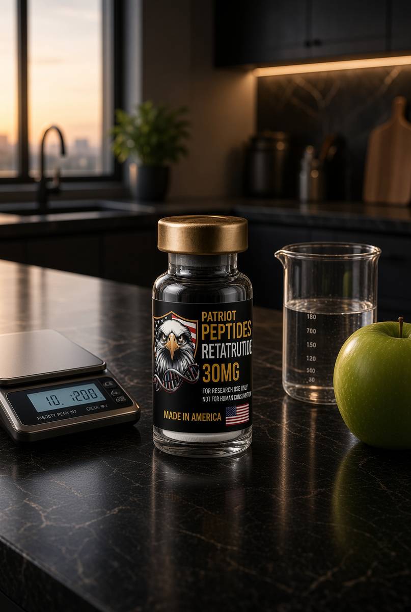
Research-use-only. Not for human consumption / not a medicine. The LY3437943 on this listing is a laboratory ligand, US-synthesised, HPLC-MS characterised, labelled for in-vitro work: a receptor assay, a hepatocyte dish, a concentration you write down. The physiology in the paragraphs above is public, cited, and older than the vial. Counterpoint was a liquid diet. DiRECT was food and weight in a GP surgery. Sanyal and Jastreboff were clinical trials of an investigational analogue we don't sell as a pen. Use the biochemistry to design the experiment you have the controls for, with the depot named, the clock named, and the tool named. Read Taylor, read Lean, read Shulman, then measure the percent. We'll sell you the published chain. We won't tell you it ran DiRECT, and we won't tell you a formula shake is a class-B occupancy. Intrahepatic triglyceride is a pool you can empty. The keys are several. This key you can weigh, in a tube, with a chromatogram on the bench beside it.
In short. The vial is a research chemical for experiments, not a medicine and not a diet. The liver-fat biology is public. Measure the percent, name the tool, and keep the claim the size of the data.
Questions the essay actually answers
- What is the twin-cycle hypothesis?
- Roy Taylor's account of type 2: excess liver fat drives hepatic insulin resistance and glucose overproduction; VLDL then delivers fat to the pancreas and first-phase insulin fails. Two stores. One disease if he is right, and DiRECT suggests he is.
- What did Counterpoint actually show?
- Lim, Taylor, Diabetologia 2011: eleven people with type 2, eight weeks at about 600 kcal/day. Liver fat and hepatic glucose production fell by day seven. Pancreatic fat and first-phase insulin followed by week eight. Mechanism, on a magnet.
- What did DiRECT actually show?
- Lean et al., Lancet 2018: cluster-randomised UK primary care, total-diet replacement, type 2 of less than six years. At twelve months, 46% of the intervention arm were in remission (HbA1c <48 mmol/mol off drugs) against 4% in control. Of those who lost 15 kg or more, 86% remitted. Two-year follow-up showed the expected fade with regain.
- Can I measure liver fat at home?
- Not really. ALT can hint and also lie. Clinics use MRI-PDFF, or at least a decent ultrasound/CAP. Waist, fasting insulin and a shrinking dawn phenomenon are the poor man's proxies, and they beat guessing.
- Is fatty liver only from alcohol or only from sugar?
- Neither gets a monopoly. Energy surplus, refined carbohydrate, alcohol and genetics all park fat in hepatocytes. The metabolic version is now called MASLD. The job is still to empty the depot.
- Do ketones empty liver fat?
- A ketogenic pattern can drop insulin and de novo lipogenesis, and liver fat often falls, sometimes before heroic scale-weight change. Ketones themselves are a readout of hepatic acetyl-CoA overflow. Chasing a millimolar BHB while energy surplus remains is how you miss the variable.
- Is retatrutide a liver-fat drug?
- It's a unimolecular agonist at GIPR, GLP-1R and GCGR. Weight falls; MRI-PDFF in a MASLD Phase 2a cohort fell with it (Sanyal, Nat Med 2024). GCGR on the hepatocyte is a lipid-oxidation cue. The listing is the published LY3437943 structure for the bench, not a licensed indication.
- Does bariatric surgery work by the same variable?
- Yes. Restriction plus a gut-hormone storm empties hepatic and pancreatic fat on a clock that rhymes with Counterpoint, with higher remission rates in surgical trials. Different key. Same lock. Lifelong follow-up is the invoice.
- Why does long-standing type 2 remit less often?
- The second store is fat; the controller is β-cell function and, later, mass. Counterbalance and DiRECT's six-year cut are the documents. C-peptide is the measurement. A quiet dump after twenty years is a census, not a moral.
- Is this a medicine or a diet plan?
- Neither. Counterpoint and DiRECT were food-and-weight trials. The catalogue item in the neighbourhood is a characterised laboratory ligand. This page is the variable those tools move.
Hypothetical research reconstitution
How this vial is typically mixed
Hypothetical research reconstitution for the named catalogue vial. Not a protocol, not medical advice, not a use instruction. These amounts sit in published and commonly cited laboratory ranges. The vial is labelled for research use only — not for human or veterinary administration.
Retatrutide
30mg
Mix with 3 ml bacteriostatic water → 10 mg/ml
- Hypothetical aliquot
- 1–2 mg to start; published trial arms ran higher by week
- 0.10–0.20 ml · 10–20 units on a U-100 syringe (at 1–2 mg)
- How often
- Once weekly
- The Jastreboff NEJM 2023 arms ran 48 weeks. That is a trial, not a shop protocol.
Bench steps
- Let the vial sit until it is no longer cold to the touch.
- Wipe the stopper with 70% isopropyl alcohol. Let it dry.
- Draw 3 ml bacteriostatic water (0.9% benzyl alcohol).
- Run the water slowly down the inside glass — do not blast the cake.
- Roll between finger and thumb until the cake is gone. Do not shake.
- Label the date. Store the solution at 2–8 °C. Do not freeze. Use within 30 days unless the note below says otherwise.
LY3437943 architecture. Weekly, not daily. Those milligram figures are what the papers used on the investigational medicine — they are not a use instruction for this reagent.
Bacteriostatic water and sterile syringes ship with peptide orders over £75. Kit details · 10 ml bacteriostatic water
The American-made molecule
Identical to Eli Lilly’s LY3437943. Synthesised in the United States. HPLC-characterised.
 Made in USAOut of stock
Incretin
Retatrutide
US-made retatrutide 30mg — the published structure LY3437943, HPLC-MS verified.
4.6(609)
50 browsing this now · 5 purchased in the last 24 hours
30mg
£120.00
Read next
51 min · long read · Metabolism
Reversing type 2 diabetes is a published result, not a slogan
The DiRECT trial put type 2 diabetes into remission by emptying the liver and pancreas of surplus fat. Very-low-calorie and very-low-carbohydrate programmes can both get you there. The mechanism isn't mystical.
49 min · long read · Metabolism
Regulating blood sugar: the control system
Glucose is a tightly bound variable. Here is the hardware — pancreas, liver, muscle, incretins — and the places it fails.
46 min · long read · Peptide research
Retatrutide (LY3437943): the published triple-agonist structure
A fatty-acylated unimolecular agonist at GIPR, GLP-1R and GCGR. US-made, HPLC-MS verified, labelled for laboratory research — not a medicine.
45 min · long read · Metabolism
How diets actually work
Calories are conservation of energy. Hormones are the routing table. Palatability is why the routing table keeps getting overwritten. Every named diet is a way of leaning on one of those three.
More in this desk
51 min · long read · Metabolism
Human metabolism, without the slogan
Calories are bookkeeping. The actual system is a set of hormonally gated fluxes through glycogen, fat and amino acids — with the liver as air-traffic control.

50 min · long read · Metabolism
Fasting, autophagy and the mTOR switch
Eat, and mTOR builds. Fast, and AMPK and ULK1 start recycling the cell. The molecular story is a switch, not a personality.
49 min · long read · Metabolism
Protein, training and muscle protein synthesis
Muscle isn't a mood. It is mechanical tension plus a leucine-gated translational programme. Here is the dose, the threshold, and why the gym still has to happen.
48 min · long read · Metabolism
When insulin falls, the kidney dumps sodium. That is the ‘keto flu’.
Insulin tells the kidney to hold salt. Carbohydrate restriction drops insulin, you diurese, and magnesium and potassium follow. The headache was not a test of character.
Essays describe published research. They are not medical advice and they do not authorise human use of any catalogue item.