

Metabolism · 49 min · 10,855 words
Regulating blood sugar: the control system
Glucose is a tightly bound variable. Here is the hardware — pancreas, liver, muscle, incretins — and the places it fails.
What this essay actually tells you
- Glucose is held in a narrow band by insulin (store, suppress glucose output) and glucagon (raise it). Incretins amplify insulin when the glucose is oral. That's the incretin effect, sitting on the same axis.
- Skeletal muscle is the largest insulin-stimulated glucose sink. That's why walking after a meal is a control-system intervention, not a wellness tip, and why we keep saying it.
- HbA1c is a 2–3 month integral of glycaemia. A CGM is the derivative. Both are useful. They answer different questions, and arguing about which one is 'real' is a waste of a clinic slot.
What this actually means
Healthy blood sugar is a closed loop. You eat, incretins warn the pancreas, insulin opens muscle and shuts the liver's glucose factory, glucagon waits for the next fast. Break the loop at the β-cell (not enough insulin), at the muscle (insulin resistance), or at the liver (glucose production that won't stand down) and the variable, plasma glucose, drifts. Continuous glucose monitors made that variable visible to people who aren't in clinic. The control system was always there. We just used to sample it four times a day and pretend that was a curve.
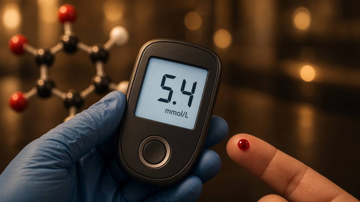
Let's start with the number on the board, because everything else this afternoon is trying to hold it still. A healthy adult holds it, fasting, in a band of roughly 3.9 to 5.6 millimoles per litre, and the brain is the customer that won't negotiate: neurons take glucose constitutively, GLUT1 and GLUT3 at the blood–brain barrier, and they will take ketones if they must, as Cahill's starvation work still shows, but they won't sit quietly through a two-millimolar trough. Claude Bernard put glycogen in the liver in the 1850s and gave physiology the milieu intérieur. Banting, Best, Macleod and Collip put insulin into a dying child in 1922 and made the actuator visible. Yalow and Berson's radioimmunoassay, 1960, made that actuator measurable in picomoles. The hardware is older than the diet arguments: pancreas, liver, skeletal muscle, adipose, gut endocrine cells, kidney in a supporting role. Break the loop at any of those nodes and the variable drifts. Let's walk that loop — who writes the hormones, who reads them, where the glucose actually goes, and which instruments report the result.
In short. Insulin and glucagon are the two main controls. Blood sugar is held in a tight band because the brain needs it that way.
The two pancreatic hormones do opposite jobs on the same number, which is a lovely piece of design once you see it drawn. Insulin is the storage-and-growth signal: GLUT4 translocated to the sarcolemma and adipocyte membrane, hormone-sensitive lipase suppressed, glycogen synthase de-inhibited, amino acids pushed into muscle, hepatocytes told to stop making glucose. Glucagon is the opposite broadcast, aimed mostly at the liver: glycogenolysis, gluconeogenesis, a push toward β-oxidation. Incretins sit on top of the meal. GIP from K-cells and GLP-1 from L-cells amplify insulin when glucose arrives by mouth rather than by vein, which is the incretin effect as a measurement before it was a drug class. McIntyre, Holdsworth and Turner put that oral-versus-intravenous contrast into the Lancet in 1964; Elrick's group published the same shape the same year. Swallow the glucose and the gut has already warned the islet. Put the same millimoles in a vein and you skip the warning. That difference is why a liquid sugar and a steak write different curves for reasons that have nothing to do with virtue.
In short. Gut hormones boost insulin when you swallow glucose, not when it is put in a vein. Insulin stores sugar and stops the liver making more. Glucagon raises it.
Skeletal muscle is the largest insulin-stimulated glucose sink, and if I had a piece of chalk I would circle it twice. DeFronzo's clamp work, and the literature that followed it, puts something like eighty to ninety percent of insulin-stimulated glucose disposal into those fibres in a healthy adult. That's a measured share, not a motivational poster. GLUT4 is the transporter that does the last millimetre of the job, and it lives in vesicles until insulin, or a contraction, tells it to fuse. Walking after a meal is therefore a control-system intervention: you raised the sink's conductance while appearance rate was still high. A day of sitting is a GLUT4 door that never got the contraction signal, so the same insulin has more work to do at the same plate. Training and glycogen depletion raise that conductance for hours afterwards. None of that requires a peptide. All of it is the loop doing what the loop was built to do, and it is the sentence we keep having to write, because a walk after lunch still gets filed under wellness.
In short. A walk after eating is a real control, not a lifestyle tip. Most sugar leaving the blood after a meal goes into muscle.
Two instruments report on the same variable and answer different questions, which is worth getting straight before we argue about breakfast. HbA1c is a two-to-three-month integral of glycaemia, written onto the N-terminal valine of haemoglobin as red cells live out roughly a hundred and twenty days. A continuous glucose monitor is the derivative: interstitial glucose, minutes, a trace you can overlay on a meal. Arguing about which one is real wastes a clinic slot. The average asks what the last season did to proteins. The trace asks what the next hour will do to the brain and the pancreas. Neighbouring pages take type 2 remission as a published food-and-weight result, human metabolism as hormonally gated flux, and the incretin century as chemistry. Retatrutide — the published LY3437943 structure, a unimolecular agonist at GIPR, GLP-1R and GCGR — sits in that literature because those three receptors already run this control system. It's a literature neighbour, not a use instruction. What follows is the physiology you would want before treating a screenshot of a curve as a mechanism, or a vial as a loop.
In short. This page is the hardware of that loop. A published three-receptor chain sits next door in the papers. It isn't a plan for you.
A variable held in a narrow band
The numbers are worth saying in SI, because the American milligrams-per-decilitre scale hides how small the band really is. 3.9 to 5.6 millimoles per litre fasting is 70 to 100 milligrams per decilitre; the conversion is multiply millimoles by eighteen. A two-hour oral glucose-tolerance value under 7.8 millimoles is the non-diabetic convention; 11.1 and above at two hours, or 7.0 fasting, or an HbA1c of 48 millimoles per mole (6.5 percent on the old percentage scale), is the diagnostic neighbourhood the WHO and the ADA have argued over for decades. Hypoglycaemia that the brain notices often sits under 3.0. Whipple's triad — low measured glucose, symptoms, recovery when you raise it — is still how you stop a dizzy spell being called a hypo without a number. The point of the census isn't to memorise cut-offs. It is to see that the entire diagnostic apparatus of a common disease is a conversation about a few millimoles in a few litres of plasma. Tight band. Large consequences when it slips.
In short. Healthy fasting blood sugar sits in a tight millimolar window. Too low and the brain fails; too high and vessels and proteins pay.
Insulin is a 51-residue heterodimer with three disulphides — the storage-and-growth signal, not a prison and not a personality. Sanger sequenced it in the 1950s; the receptor is a receptor tyrosine kinase, two α chains outside and two β chains spanning the membrane, which is a different grammar from the seven-helix receptors the incretins occupy. Occupancy autophosphorylates the β chain, IRS proteins dock, PI3K makes PIP3, Akt is recruited, and the metabolic work follows. In muscle and adipose that work includes TBC1D4/AS160 and the Rab-dependent arrival of GLUT4 at the surface. In the adipocyte it includes suppressing hormone-sensitive lipase, so triglyceride stays triglyceride. In the hepatocyte it includes telling glycogen synthase to run and glucose-6-phosphatase to stand down, so the factory stops exporting. Amino acids are pushed into muscle. The list is long because a growth-and-storage hormone with that many client tissues will find jobs. Blame insulin for the jobs it actually has. Don't pin a surplus you ate, and then stored, on the hormone that stored it.
In short. Insulin is that store-and-stop signal: open muscle, quiet the liver's sugar factory, and hold fat in fat cells.
Glucagon is a 29-residue product of proglucagon, written by pancreatic α-cells, and I'd draw its receptor as a class-B GPCR, Gs-coupled, raising cyclic AMP in hepatocytes. Protein kinase A then phosphorylates phosphorylase kinase, glycogen phosphorylase runs, and glucose-6-phosphate appears from glycogen on a timescale of minutes. Gluconeogenesis is the slower programme: pyruvate carboxylase, PEPCK, fructose-1,6-bisphosphatase, glucose-6-phosphatase, the enzymes a first-year class can still recite, fed by lactate, alanine and glycerol. Glucagon leans that programme on. It also leans the hepatocyte toward β-oxidation and, when acetyl-CoA overflows, toward ketogenesis. Unger's radioimmunoassay work made glucagon a quantitative object rather than a rumour of an opposite to insulin. The receptor lives mostly on hepatocytes, which is why glucagon is a liver hormone in working English even though the peptide is pancreatic. Adipose and heart carry some GCGR; the glucose story is still hepatic. Spend a night without food, or sit with a type 1 who still has α-cells when the β-cells are gone, and glucagon stops looking like a minor curiosity.
In short. That is how a fast doesn't become a faint. Glucagon talks mainly to the liver and tells it to break glycogen and make new glucose.
The two hormones are made a few microns apart, which is the sort of anatomy that still makes me grin when we draw the islet. Islets of Langerhans, 1–2 percent of pancreatic mass, β-cells in the core in rodents and more intermingled in humans, α-cells around and among them, δ-cells writing somatostatin as a local brake. Insulin arriving in the islet capillary can suppress glucagon directly; that paracrine hush is why a first-phase dump is a glucagon event as well as an insulin event. Lose the dump and the α-cell keeps talking while the meal is still arriving. The ratio, not either concentration in isolation, is what a hepatocyte actually reads. Alan Cherrington's Vanderbilt dog work spent decades on that ratio: clamp one hormone, vary the other, watch hepatic glucose output move. It is still the cleanest demonstration that the liver is a servo, not a furnace. Somatostatin, GLP-1, circulating glucose itself and autonomic nerves all write on the same α-cell. The textbook pair — insulin down, glucagon up, and vice versa — is the first harmonic. The rest of the overtones are why a person and a diagram aren't the same experiment.
In short. That two hormones are made in neighbouring islet cells, and insulin can hush glucagon on the spot. The ratio is the message.
First-phase insulin release is the under-celebrated bit of hardware, and it is the bit I would draw first if we had a trace on the desk. Within three to ten minutes of a sharp glucose rise, a ready-releasable pool of granules fuses, and the dump is large enough to restrain hepatic glucose output before the meal has even finished. A slower second phase then draws on a reserve pool and on new synthesis. Intravenous glucose-tolerance tests still show this biphasic shape in people who have it. In early type 2 the first phase dies first. That's why two people can eat the same bread and paint very different curves: one still has the dump, the other is already borrowing from a weaker second phase while the liver keeps making glucose in the background. Porte, Kahn, the Seattle work, and every later clamp-and-curve paper that bothered to look at the first ten minutes, are the papers I would put in your hand. You can see the missing first phase on a real trace. You can also see people blame the toast. The toast is appearance rate. The missing dump is the actuator that was supposed to meet it.
In short. In the first minutes of a sugar rise, stored insulin dumps and shuts the liver before you've even finished eating.
An overnight fast is a hepatic story wearing a glucose number, which is a surprise the first time you see it. After twelve hours without food, almost all the glucose appearing in plasma was made by the liver — glycogenolysis early, gluconeogenesis taking a larger share as the glycogen drops — at something like two milligrams per kilogram per minute in a lean adult, a flux Rothman and Shulman put NMR numbers on, and that Landau's pioneering work had already framed with labelled precursors. Muscle isn't taking much, because insulin is low and GLUT4 is inside. The brain is still taking its hundred-odd grams a day. Morning glucose is therefore often a hepatic number, not a breakfast number. The dawn phenomenon — a rise in the early hours driven by growth hormone and cortisol against a liver that is already on — is this paragraph with a clock on it. Eat breakfast into an unrestrained factory and the curve starts from a raised floor. That isn't a moral failure of porridge. It's a mute button that didn't mute.
In short. Morning numbers are often a liver story, not a breakfast story. Overnight, almost all the sugar in your blood was made by the liver.
The triumvirate: β-cell, muscle, liver. A collusion responsible for NIDDM. Insulin resistance at the fibre, glucose production that won't stand down, and an insulin secretory response that can't keep up.— DeFronzo RA. Lilly Lecture. Diabetes. 1988; 37: 667–687. The three-node map this page still walks.
The gut's warning: incretins
The incretin effect is a paired curve before it's a medicine, and the pairing is still the cleanest teaching trick we have. Give glucose by mouth and the insulin rise is larger than the rise you get from the same glucose load in a vein. McIntyre, Holdsworth and Turner, Lancet, 1964. Elrick, Stimmler, Hlad and Arai, Journal of Clinical Endocrinology, the same year. The gut, they argued, must be releasing something that amplifies the β-cell's response to glucose. Jean La Barre had coined incretin around 1929 for a putative intestinal factor that stimulated insulin secretion; the word sat waiting until a measurement could not be waved away. Unger's entero-insular axis made the conversation quantitative. Oral versus intravenous is still how you teach the effect in a practical class. A screenshot of a weight-loss mean is a long way downstream of that pairing. Everything that follows — GIP, GLP-1, DPP-4 inhibitors, fatty-acylated analogues, duals, triples — is an attempt to name, then occupy, the messengers responsible for a difference you can plot on graph paper. Start with the measurement. The drugs came later.
In short. That difference is the gut warning the pancreas. Sugar you swallow raises more insulin than the same sugar put in a vein.
Gastric inhibitory polypeptide was isolated first, and the name is a small history lesson of its own. Brown, Mutt, Pederson and colleagues, in the early 1970s, pulled a peptide from intestinal extracts that slowed gastric acid, and named it for that job. Dupré then showed it also amplified insulin when glucose was present, which is a different job wearing the same letters. The field eventually preferred glucose-dependent insulinotropic polypeptide, keeping the acronym GIP, because the insulinotropic physiology is what survived contact with human metabolic disease. The gastric-inhibitory name wasn't a fraud. It was a first assay. Hormones are often named for the experiment that found them, then renamed for the experiment that mattered. K-cells in the proximal small intestine write GIP in response to fat and carbohydrate. The receptor, GIPR, is a class-B GPCR on β-cells and on adipocytes. In type 2 the insulinotropic effect of GIP dulls more than GLP-1's does, which is one reason the first generation of incretin medicines ignored GIPR. Dual and triple agonists put it back on purpose. Occupancy is still occupancy. A missing assay in 2005 wasn't a missing receptor.
In short. It was first named for slowing the stomach; the insulin job is why it still matters. One upper-gut hormone raises insulin when glucose is present.
GLP-1 arrived through proglucagon, a precursor the gut and the pancreas read differently. Habener's laboratory cloned the precursor; Holst, Ørskov and colleagues showed that the gut processes that precursor differently from the pancreatic α-cell, yielding glucagon-like peptide-1 as a post-translational product of L-cells, denser toward the ileum and colon. Drucker's work made the receptor a therapeutic object rather than a curiosity of a radioimmunoassay. Native GLP-1 is a thirty-one-residue peptide, amidated, potent at GLP-1R, and almost useless as a drug because dipeptidyl peptidase-4 clips it at alanine-2 in minutes and the kidney clears what remains. The insulinotropic effect is glucose-dependent. GLP-1R also lives on the brainstem and on the stomach. Delayed gastric emptying and central satiety aren't side-effects of a β-cell drug. They're the receptor being where it is. Appearance rate is half the post-prandial curve people blame on carbohydrate as a monolith. Slow the stomach and the same starch writes a different plasma story, which is why a liquid sugar hits differently from a steak for reasons that have nothing to do with character.
In short. One second gut hormone, made further down, raises insulin, slows the stomach, and tells the brain a meal has arrived.
Dipeptidyl peptidase-4 is the serine protease that clips those gut hormones in minutes — endothelial cells and plasma, two residues off the N-terminus. GLP-1 and GIP are substrates. Mentlein, Gallwitz and Schmidt put the cleavage on the page in the early 1990s. Sitagliptin, licensed in 2006, occupies the enzyme rather than the receptor: it lets more native incretin survive the meal. That's a different therapeutic idea from injecting an analogue, and it produces a different magnitude. DPP-4 inhibitors are oral, modest on weight, useful on glucose. The analogue strategy accepts that native peptide is the wrong pharmacokinetic object and builds a chain the enzyme can't eat as fast, then hangs a lipid on it so albumin will hide what remains. Both strategies exist because the two-minute half-life is real. If we talk about boosting GLP-1 without saying whether we mean the enzyme or the receptor, we have not chosen a mechanism. Native incretin tone after a mixed meal is a minutes-scale pulse. A weekly analogue is a different object occupying the same receptor.
In short. That's why the native versions are poor medicines and why longer copies were built. An enzyme in blood chops those gut hormones in minutes.
Glucose-dependence is the safety grammar, which is why this class isn't a sulphonylurea with better branding. The β-cell already has to see glucose — glucokinase, a closed KATP channel, a calcium current, a granule at the membrane — before Gs–cAMP from GLP-1R or GIPR will do much amplifying. Raise cAMP on a cell that isn't depolarised and you have phosphorylated machinery waiting for a trigger that has not arrived. Sulphonylureas close KATP regardless of glucose, which is why hypoglycaemia is a named failure mode of that class. Incretin occupancy amplifies a glucose-dependent machine. It doesn't replace the machine. That sentence is also why an incretin analogue can flatten a post-prandial curve without being insulin, and why the same occupancy is a poor tool if the β-cell mass is already gone, as in long-standing insulin-deficient type 1. Amplification of a missing first phase is still amplification. It isn't a graft. The neighbouring retatrutide essay is the occupancy chemistry. This paragraph is why that chemistry is allowed to sit on a glucose loop without being a syringe of insulin.
In short. That's a different tool from drugs that force insulin out anyway. These gut hormones only boost insulin when glucose is already up.
Diagram
GLP-1R
β-cell, brainstem, stomach
Incretin, delayed emptying, satiety. Semaglutide’s occupancy.
GIPR
β-cell, adipocyte
Second incretin. Lipid handling. Tirzepatide added this.
GCGR
hepatocyte
Glycogenolysis and, biased, energy expenditure. The third occupancy.
LY3437943 is a fatty-acylated unimolecular agonist at GIPR, GLP-1R and GCGR (Coskun, Cell Metab 2018). Jastreboff, NEJM 2023: 24.2% mean weight loss at 48 weeks, 12 mg, Phase 2 — clinical literature, not a use instruction for a research vial.
Muscle is the disposal sink
Post-meal, most of the glucose that leaves the blood under insulin goes into skeletal muscle — the big sink on the right of the board. DeFronzo's hyperinsulinaemic–euglycaemic clamp — insulin raised, glucose infused to hold plasma steady, the infusion rate equal to whole-body disposal once endogenous output is suppressed — is still the gold-standard measurement of that share. In a healthy adult the fibres take the large majority. Adipose takes a smaller slice; the liver, at ordinary meals, is more a source being muted than a sink being filled, though it will restock glycogen when insulin is high and glucose-6-phosphate is plentiful. The clamp is an artificial steady state. A mixed meal isn't. Appearance rate, splanchnic extraction, gastric emptying and the incretin pulse all sit on top of the same sink. The sink is still the rate-limiter that training and sitting move. Ignore it and you'll spend a career arguing about bread while the conductance of the largest insulin-stimulated door in the body comes and goes with yesterday's steps. The door has a name. It is GLUT4.
In short. That's a measured share, from clamp studies, not a guess. After a meal, most of the insulin-driven sugar disposal goes into skeletal muscle.
GLUT4 — SLC2A4 — is a twelve-transmembrane glucose transporter stored in specialised vesicles, and that address is the whole trick. Cushman and Wardzala, and independently Suzuki and Kono, put the translocation hypothesis on the page in 1980: insulin doesn't so much activate a surface transporter as recruit one from inside. David James cloned the protein. In muscle and adipose it is the insulin-responsive isoform; GLUT1 handles basal flux; GLUT2 is the high-capacity liver and β-cell door, not insulin-dependent in the GLUT4 sense. The last millimetre of post-prandial disposal in a fibre is therefore a vesicle-trafficking problem. Fusion, SNAREs, Rabs, a microtubule and actin neighbourhood, then glucose following its concentration gradient through the newly arrived uniporter. Hexokinase II phosphorylates what comes in, trapping it as glucose-6-phosphate. Glycogen synthase or glycolysis takes it from there. A transporter that stays in a vesicle is a door that doesn't exist as far as plasma glucose is concerned. That's the whole of insulin resistance at this node, in one trafficking sentence.
In short. Muscle keeps a sugar door packed in vesicles and only puts it on the surface when insulin, or a contraction, gives the cue.
The insulin cue is a phosphorylation cascade, and naming the steps is how we stop writing insulin resistance as a mood. Insulin receptor tyrosine kinase, IRS-1 in muscle, PI3K, PDK1, Akt2, then TBC1D4 (AS160) whose Rab-GAP activity keeps the vesicles inside until it is phosphorylated and lets go. Knock that chain at IRS-1 or at AS160 and GLUT4 stays home. Lipid intermediates — diacylglycerol, ceramides — argue with IRS and Akt in fibres that have taken up more fat than they can oxidise; that is one respectable account of acquired insulin resistance, with Shulman's NMR and biopsy work sitting behind it. Inflammation and serine phosphorylation of IRS is another. The cascade can also simply be under-used. A fibre that has not contracted, has not emptied glycogen, and has not seen a recent insulin pulse is a fibre whose GLUT4 programme is transcriptionally and trafficking-quiet. You don't need a novel toxin. You need a sofa and a week. The blot will still look like resistance. The intervention is a recruitment, not a confession.
In short. If the chain sticks, the door stays inside. Insulin does that job through a chain of phosphate tags inside the fibre.
Contraction opens the same door with a different key, which is the bit a walk after lunch is actually using. Calcium–calmodulin kinases, AMPK when the fibre is drawing down ATP, and a TBC1D1 neighbourhood that isn't identical to the insulin AS160 neighbourhood, all converge on GLUT4 translocation without requiring the insulin receptor. That's why a walk, a set of stairs, or a bout of resistance work can dispose of glucose in people whose insulin signal is dull. It is also why exercise is still the most reliable insulin-sensitising intervention we have in those same fibres for hours afterwards: some of the GLUT4 stays recruited, glycogen is lower, and the next insulin pulse has less work to do. Richter, Hargreaves, Goodyear, the Copenhagen and Boston contraction literature, are the papers. AMPK is the fuel-gauge kinase; it isn't a lifestyle brand. Insulin-independent glucose uptake is a named physiology. Treating a post-prandial walk as a wellness tip, rather than as an AMPK-and-calcium intervention on the largest sink in the body, is how a control system gets filed under habits.
In short. That's why a walk still works when insulin is weak. Using the muscle, even without insulin, also puts that door on the surface.
Walking after a meal remains therefore a control-system intervention, and the trials are small, cheap, and consistent enough that the mechanism is allowed to be the headline. Nygaard, Colberg, a set of crossover studies in which a ten-to-twenty-minute walk after eating cuts the post-prandial glucose excursion relative to sitting, sometimes by a fifth or a third of the incremental area, without anyone changing the plate. You raised GLUT4 conductance in the fibres you actually recruited, while appearance rate from the gut was still high. Timing matters more than heroism. A walk three hours later is a different experiment, a glycogen-and-sensitivity experiment, still useful, not the same as catching the peak. Standing and fidgeting do a little of the same work; a dedicated walk does more because more motor units fired. None of this is a personality. It is Ohm's law on a membrane: current equals conductance times driving force, and you just turned the conductance up. The driving force is the meal you already ate.
In short. One short walk after eating is a control-system move: you raised the muscle's uptake while the meal was still arriving.
Training rewrites the door for the next meal, not only the last one, which is why yesterday's steps are a pharmacodynamic variable. Endurance work raises GLUT4 protein, hexokinase, mitochondrial density and capillary supply; PGC-1α is the transcriptional coactivator the mitochondria page already named. Glycogen depletion is still the most reliable way we have to lift subsequent insulin sensitivity in those same fibres, because a cell that has parked less glycogen takes glucose more greedily when insulin arrives. Resistance work recruits motor units that endurance walking won't, and those fibres will carry their own GLUT4 census. A day of sitting is the reverse programme: the contraction signal never came, AS160 stays active as a GAP, vesicles stay in, and the same insulin, at the same meal, writes a higher curve. That isn't a character flaw. It's a missing cue. Bed rest studies have been showing the same sink-failure for decades. The practical sentence is ugly and true: the largest insulin-stimulated glucose door in the body is gated by whether you used the muscle. Yesterday's steps are a pharmacodynamic variable. Treat them as one.
In short. Training and emptying glycogen make that same door easier to open next time. A day of sitting does the reverse.
- Fasting band
- 3.9–5.6 mmol/L
- Muscle share
- ~80–90%
- Hepatic glucose output
- ~2 mg·kg⁻¹·min⁻¹
- First-phase insulin
- 3–10 min
- Native GLP-1
- ~2 min half-life
- HbA1c window
- 2–3 months
- GLUT4
- SLC2A4
- LY3437943
- GIPR, GLP-1R, GCGR
Healthy adult plasma glucose. Multiply by 18 for mg/dL. Tight on purpose.
Of insulin-stimulated glucose disposal in clamp studies. DeFronzo neighbourhood.
Overnight fast, lean adult. Glycogenolysis then gluconeogenesis. A factory, not a rumour.
Ready-releasable granules. Dies early in type 2. Restrains the liver before the meal finishes.
DPP-4 clips alanine-2. The incretin effect is a pulse. Analogues are a different object.
Integral on haemoglobin. Red-cell lifespan ~120 days, weighted toward the recent weeks.
Insulin- and contraction-recruited. Vesicles until the cue. Cushman & Wardzala, 1980.
Published triple agonist. Coskun, Cell Metab 2018. A literature neighbour, not a use instruction.
The liver as a glucose factory
The liver is a source more than a sink at ordinary meals, which is easy to skip if you only think about eating. Hepatocytes carry GLUT2, a high-capacity transporter that isn't waiting on insulin to arrive at the membrane, and they carry the enzymes to make glucose-6-phosphate into free glucose and export it. Between meals that export is the brain's supply. After a meal insulin is supposed to mute it — glycogen synthase on, glycogen phosphorylase off, gluconeogenic transcription down, glucokinase up so incoming glucose is phosphorylated and parked. Mute is a measured suppression of hepatic glucose output, not a complete shutdown, and it fails early in insulin resistance. The clamp world talks about endogenous glucose production because the kidney can make a little, especially in prolonged fasting; in ordinary overnight physiology the liver is the factory. Cherrington's ratio work, the NMR of glycogen, the tracer studies with 6,6-deuterated glucose: those are how you know. A CGM bump at dawn is often this factory, not the cereal.
In short. Insulin is supposed to mute that source after you eat. The liver can store sugar and make it. Between meals it's a source.
Glycogenolysis remains the fast account. Liver stores roughly eighty to a hundred and twenty grams of glycogen in a fed adult, a buffer, not a warehouse, and glucagon plus a falling insulin:glucagon ratio will empty it on a timescale of hours. Muscle holds more glycogen, three to five hundred grams in a trained person, but muscle lacks glucose-6-phosphatase, so that glycogen is for the fibre, not for plasma. Gluconeogenesis is the slower account: lactate from the Cori cycle, alanine from muscle proteolysis, glycerol from lipolysis, a mitochondrial and cytosolic enzyme set that a fatty, insulin-resistant liver runs too hard. Landau's pioneering fractionation, then Rothman, Magnusson, Shulman and the 13C NMR work, put numbers on the split after an overnight fast: gluconeogenesis already a large fraction, glycogenolysis not zero. After a longer fast the split tilts further. The practical point is that a morning glucose is a mixed invoice. Blaming carbohydrate at breakfast for a number that was being manufactured at 4 a.m. is a category error. The factory was already on.
In short. Making new glucose from amino acids and lactate is the slower overnight way. Breaking stored glycogen is the fast way.
The insulin:glucagon ratio is the set-point a hepatocyte actually obeys — not either hormone shouted on its own. Raise insulin against a clamped glucagon and hepatic glucose output falls. Raise glucagon against a clamped insulin and it rises. Cherrington spent a career showing that in the dog, with arterial and portal sampling, and the human literature has not overturned the grammar. Portal insulin is higher than peripheral insulin, which is why a subcutaneous injection and an endogenous pulse aren't the same experiment at the liver. Incretins, by amplifying the endogenous pulse, therefore have a portal privilege that injected insulin doesn't automatically inherit. Glucagon receptor occupancy is the risky arm of a triple agonist for exactly this reason: enough GCGR to buy energy expenditure and lipid oxidation, not so much that the factory undoes the glycaemic gain you just bought with GLP-1R and GIPR. Coskun's engineering paper is, among other things, a hepatic-output paper. The ratio is the older physiology. The unimolecular chain is a new way of writing on it.
In short. How hard the liver works is set by the insulin-to-glucagon ratio, not by either hormone shouting on its own.
When that mute button sticks, the liver keeps making glucose in the background of a meal you thought you'd already paid for. That is insulin resistance at the hepatocyte, often sitting next to ectopic triglyceride, an overactive gluconeogenic programme, and an α-cell that is no longer being hushed. Fasting hyperglycaemia is frequently this number, not a breakfast number. The dawn phenomenon is this number with growth hormone and cortisol leaning on the same enzymes. Metformin's most respectable account is still a hepatic one: less gluconeogenic output, a redox and G3P-dehydrogenase neighbourhood that Madiraju and Shulman have been arguing through, AMPK as a supporting actor rather than the whole play. You don't need to pick a winner among those papers to keep the clinical sentence: a liver that won't stand down writes a raised floor under every curve that follows. Walking still helps, because the sink can take what the factory insists on making. Closing the factory, when you can, is the other half of the loop.
In short. That's a large part of a high morning reading. If the mute button sticks, the liver keeps making sugar during a meal.
Ectopic fat is the neighbouring story, and it has randomised trial data, which is why it belongs in a sentence here. Roy Taylor's twin-cycle hypothesis: chronic positive energy balance drives fat into the liver; a fatty liver overproduces glucose and VLDL; that fat then lands in the pancreas and first-phase insulin goes quiet. Reverse the energy balance hard enough and liver fat falls within days, pancreatic fat follows, and the dump can return. DiRECT, Lean and colleagues, Lancet 2018, cluster-randomised UK primary care, a formula very-low-calorie diet: 46 percent of the intervention arm in remission at twelve months against 4 percent in control, remission tracking weight loss. Counterpoint imaging showed the fat leaving the organs the hypothesis had named. Carbohydrate restriction can flatten the variable by simply stopping putting so much of it in, and by lowering insulin and output, even before heroic scale-weight change. Both roads fail if the surplus and the liver fat come back. This page is the loop. That page is what happens when you empty the factory's surplus. Don't fuse them.
In short. One fatty liver overproduces glucose. Pull the fat and the factory can quiet. That neighbouring trial story isn't this page.
The β-cell as a glucose sensor
A β-cell is a glucose sensor that happens to store insulin, not an insulin bag that happens to notice glucose — draw the sensor first. Glucokinase (hexokinase IV) is the sensor: a high Km, around 8 millimolar, not saturated at fasting glucose, so the phosphorylation rate reports plasma glucose rather than reporting whether the enzyme is present. Matschinsky spent a career on that claim, and MODY2 — heterozygous glucokinase mutations — is the human genetic proof that a shifted sensor shifts the entire band. Glucose enters through GLUT1 and GLUT2, is phosphorylated, goes through glycolysis, and the resulting ATP:ADP ratio closes ATP-sensitive potassium channels. Membrane depolarisation opens voltage-gated calcium channels. Calcium, near the membrane, is the last cue the granules need. That is stimulus-secretion coupling, 1970s to now, and it is why a β-cell in a dish will dump insulin when you raise glucose without any incretin present. The incretin is an amplifier of this machine. It isn't the machine. If we write GLP-1 as if it were insulin, we have not drawn the channel.
In short. A specialised enzyme reads how much glucose is coming in. The pancreatic cell that makes insulin is itself a glucose sensor.
The channel is Kir6.2 (KCNJ11) plus SUR1 (ABCC8), four of each — a KATP octamer that sulphonylureas close and diazoxide opens. Closing it depolarises; opening it silences. Neonatal diabetes and hyperinsulinism of infancy are the genetic lessons, and they're why if we can't name Kir6.2 we aren't yet doing β-cell electrophysiology. Voltage-gated calcium channels — L-type, Cav1.2 and Cav1.3 in the human β-cell — supply the calcium. A ready-releasable pool of granules sits close to the membrane, docked, primed, waiting; that is first phase. A reserve pool has to be recruited, trafficked, primed; that is second phase, and new synthesis sits under that. SNARE proteins, synaptotagmins, a neuron-like fusion machine in a cell that isn't a neuron. Patch-clamp, capacitance measurements, TIRF of labelled granules: those are the machines. A forum sentence about boosting the pancreas has not chosen which pool, which channel, or which time bin. The first ten minutes and the next hour are different experiments on the same cell.
In short. When that glucose reading is high, a potassium channel closes, the cell fires, calcium enters, and insulin granules fuse.
Early type 2 often loses the first-phase dump while a slower second phase still limps on — same bread, very different traces. IVGTT and the hyperglycaemic clamp show it; the oral curve hides it under appearance rate and incretins, which is why two people can eat the same bread and paint very different traces. β-cell mass may already be down; β-cell function is down earlier. Glucolipotoxicity — chronic high glucose and high fatty acid arguing with the sensor, with the mitochondria, with the ER — is one account of the fade. Amyloid from IAPP, oxidative stress, a first-phase pool that has been chronically over-asked, are others. Taylor's twin-cycle puts pancreatic fat on the same stage. None of those is a reason to tell a person their pancreas is finished in year two of the disease; DiRECT showed that emptying the fat can bring a dump back in a large fraction of recent-onset type 2. None of them is a reason to pretend type 1 is the same biology. The practical reading of a CGM that spikes and sits high is often: missing first phase, unrestrained liver, a sink that didn't open. Three nodes. One bread.
In short. Early type 2 often loses that first dump of granules while a slower second phase still limps. Same bread, very different curves.
Incretin occupancy raises cAMP in the same cell and makes those granules more willing, which is the gut's warning arriving as chemistry. Gs, adenylyl cyclase, a micromolar cAMP rise, protein kinase A phosphorylating SNAP-25 and other fusion machinery, Epac2 (Rapgef4) as the cAMP-binding protein that isn't PKA and that the β-cell literature can't stop citing. The amplification is glucose-dependent because the calcium current still has to run. GIPR and GLP-1R both do this job; GLP-1R has the extra-islet jobs (stomach, brainstem) that GIPR shares only in part. A dual or a triple is, at the β-cell, more than one occupancy on that cAMP budget. At the hepatocyte the glucagon arm is a different conversation. Keep the floors apart. A patch-clamp of a β-cell treated with GLP-1 is a granule-and-channel paper. A clamp of a person on a triple agonist is an organism paper. Both are allowed. Fusing them is how a second messenger becomes a diet. The diagram that follows is the receptor grammar, so the insulin receptor and the incretin receptors aren't asked to share a drawing they don't share a mechanism with.
In short. Gut hormones raise a second messenger inside that cell and make granules more willing to go. They don't replace the glucose sensor.
Two receptor grammars, one variable
Insulin's receptor isn't a GPCR, and that's the first thing to get on the board before the cartoons get mixed. It's a receptor tyrosine kinase: a preformed dimer, ligand-induced conformational change, autophosphorylation on tyrosine, phosphotyrosine as a docking code for SH2 proteins, IRS, then PI3K–Akt for the metabolic work and Shc–ERK for a growth neighbourhood we won't linger on. The ligand doesn't enter to do the signalling. Information crosses as phosphate. Amplification is enzymatic — one occupied receptor phosphorylates many IRS molecules, each PI3K makes many PIP3s — but it isn't the cyclic-nucleotide millimolar burst of a Gs-coupled receptor. GLUT4 translocation sits on the PI3K–Akt arm. So does the hepatic mute, by a combination of direct hepatocyte signalling and a reduction in glucagon. If we draw insulin as a seven-helix cartoon, we have borrowed the wrong family. Lefkowitz and Kobilka's Nobel was for GPCRs. Ullrich, Ebina, the insulin-receptor clones of the mid-1980s, are the papers for this lock. Two grammars. One plasma variable. The cell doesn't find that confusing. Diagrams that do have not labelled the receptor class.
In short. Insulin's receptor is a kinase that tags itself. The gut hormones use a seven-helix switch instead. Two grammars, one plasma glucose.
Glucagon, GLP-1 and GIP occupy class-B GPCRs, the secretin family, which is one grammar written at three addresses. Two steps, both aqueous-facing. Gs is the default G protein. Adenylyl cyclase runs. cAMP rises. Protein kinase A, and in the β-cell Epac2, do the work. GCGR on the hepatocyte, GLP-1R on the β-cell and brainstem and stomach, GIPR on the β-cell and adipocyte: three addresses, one grammar. Cryo-EM has given occupied poses of all three. The pockets are related, which is why a unimolecular agonist is a possible object rather than a fantasy. Related isn't identical. Bias lives in the difference. Truncating the N-terminus is how you make an antagonist, because you keep the catch and lose the insertion. Bayliss and Starling's secretin, 1902, was the prototype ligand of this family. A century later a single American-made chain occupies three of its receptors at once. The family didn't change. The medicinal chemistry did.
In short. Glucagon and the two gut hormones use related seven-helix receptors and raise the same cyclic messenger — one grammar, three addresses.
Amplification is the reason a picomolar occupancy can move a millimolar sugar, and it isn't magic — it is ordinary gain. One occupied class-B receptor catalyses GDP/GTP exchange on many Gs proteins. Each Gs–adenylyl-cyclase pair mints many cAMP molecules. Kinases then phosphorylate many clients. The second-messenger cloud is the gain stage. Desensitisation is why more ligand isn't more signal forever: GRKs, β-arrestin, internalisation, a receptor that has left the surface. Spare receptors mean the EC50 can sit below the Kd; occupancy of a fraction of the census can already be a full cAMP response. That is ordinary pharmacology. It is also why a weekly analogue, sitting on albumin, occupying a few percent of GLP-1R for days, can flatten a curve that a two-minute native pulse only nicked. The insulin receptor has its own gain, via the kinase cascade, and its own desensitisation. Mixing the two cartoons — drawing cAMP clouds under insulin, or drawing IRS under GLP-1 — is how a journal becomes a poster. Name the lock. Then name the messenger. Then, and only then, talk about the glucose number that moved.
In short. That's why a tiny peptide concentration can move millimolar sugar. One occupied receptor can mint many messenger molecules.
Occupancy at GIPR and GLP-1R therefore moves glucose without being insulin, which is the distinction we have to keep honest. The β-cell still has to sense glucose. The granules still have to fuse. The muscle still has to put GLUT4 on the surface in response to the insulin that does arrive. The liver still has to read the new insulin:glucagon ratio. An incretin analogue is a preface and an amplifier, plus a stomach-and-brain occupancy that slows appearance and cuts the next meal. It isn't a GLUT4 recruiter, not a glucokinase, not a glycogen phosphorylase inhibitor except insofar as the new hormone ratio makes it one. Retatrutide adds GCGR, which is a factory occupancy, tuned, in the engineering papers, so that energy expenditure and lipid oxidation move more than the glucose curve is wrecked. That's a literature. It isn't a reconstitution instruction. The two diagrams below are the lock and the cloud. They belong here so that the insulin cascade and the incretin cloud aren't asked to share a shape they don't share.
In short. Occupying those gut-hormone receptors so moves glucose without being insulin. The cell still needs a working sensor.
Diagram
Outside
Peptide ligand
Named sequence in the nM–µM pocket. Shape complementarity, not vibes. A 15-mer and a 4-mer do not fit the same hole.
Membrane
7-TM receptor
Helices rearrange. The cytoplasmic face becomes a GEF for a heterotrimeric G protein (Gs, Gi, Gq, G12/13).
Inside
Second messengers
cAMP, IP₃, Ca²⁺, β-arrestin. One occupied receptor can spawn thousands of messenger molecules. That is amplification.
~800 GPCRs in the human genome. Seven transmembrane helices, an extracellular ligand pocket, an intracellular G-protein handshake. Catalogue neighbours: ipamorelin at GHSR, PT-141/MT2 at melanocortin receptors, retatrutide at GLP-1R/GIPR/GCGR.
Diagram
× 1
Ligand
One peptide in one pocket. nM–µM. Shape, not a mood.
× 10–10²
G proteins
The occupied GPCR is a GEF. Each Gα is a catalyst.
× 10³–10⁴
cAMP / IP₃ / Ca²⁺
Adenylyl cyclase and PLC do not make one molecule. They make a cloud.
× 10⁴–10⁶
PKA / PKC / CaMK
Kinases phosphorylate many substrates per messenger.
× tissue
Secretion, transcription, motility
The organism-level readout. Still not a protocol.
This is the only magic, and it is not magic. A nanomolar ligand can move a micromolar messenger because enzymes sit between them. Desensitisation (GRK, β-arrestin, endocytosis) is how the cell refuses to let ‘more ligand’ mean ‘more signal’ forever.
Where the loop breaks
The loop can break at the β-cell, at the muscle, or at the liver, and most common type 2 is some of all three — a three-node problem, not a single villain. Not enough insulin, or not enough first-phase insulin, and the factory isn't muted and the sink is under-signalled. Enough insulin against a muscle that won't translocate GLUT4, and the same millimoles sit in plasma while the fibre stays hungry for a cue it is ignoring. Enough insulin against a liver that won't stand down, and endogenous production writes a raised floor under the meal. Adipose, as a fourth node DeFronzo later added (the ominous octet is the expanded version: incretin defect, α-cell, kidney, brain), mishandles lipolysis and floods the other organs with fatty acids. You can argue about the order. You can't honestly pick one node and call it the disease. A CGM is a plasma number. It doesn't colour-code the node. Clamps, tracers, imaging of liver fat, a first-phase test, a fitness history: those are how you guess. A one-line story about carbs or about insulin is how you skip the guess.
In short. That loop can break at muscle, where insulin is ignored, at the liver, where the factory won't stand down, or at the pancreas.
Muscle insulin resistance remains often an unused door plus lipid intermediates arguing with IRS and Akt. A week of bed rest will give it to a healthy volunteer. A year of sitting plus a surplus will give it without a consent form. DAG and ceramides, PKCθ, serine phosphorylation of IRS-1, a GLUT4 census that has drifted down because PGC-1α has not been asked to work: those are the named pieces. Contraction still opens a parallel door, which is why the walk remains legal in people whose insulin signal is dull. Training still raises the census. None of that's a claim that exercise reverses type 2 by itself; the factory and the β-cell have votes. It's a claim that the largest sink in the loop is gated by use. Inflammation, corticosteroids, some antipsychotics, pregnancy, acromegaly: other ways the cascade gets argued with. The common way, in the country this page is written from, is a fibre that isn't recruited often enough, carrying more fat than it oxidises, asked to dispose of a meal it didn't earn a GLUT4 translocation for.
In short. When muscle ignores insulin, the door is often unused, and fat metabolites are arguing with the phosphate chain that should open it.
When the liver won't stand down, a meal has to fight a source as well as fill a sink, which is why morning glucose can look like breakfast even before you've eaten. Fasting hyperglycaemia is the giveaway; a CGM that is already high on waking is often hepatic. The mute fails because insulin is low, because the hepatocyte is insulin-resistant, because glucagon is high, because cortisol and growth hormone leaned on the same enzymes at dawn, because glycogen wasn't emptied by an overnight that wasn't a real overnight, because gluconeogenesis is running on lactate and alanine from a surplus the person is still carrying. A fatty liver is the frequent anatomical partner. VLDL overproduction is the lipid twin of the glucose overproduction. Metformin, when it works, works here more than it works at GLUT4. A very-low-calorie week works here faster than it works at the fibre, which is Taylor's point: liver fat and hepatic glucose output move in days. The neighbouring remission essay is that week, written as a trial. This paragraph is the factory the week is for. Don't skip the factory and argue about toast.
In short. When the liver won't stand down, a meal has to fight a source as well as fill a sink. Morning glucose is often that fight.
Type 1 is a different disease, and mixing it with type 2 is how people get hurt. Autoimmune destruction of β-cells, insulin deficiency that isn't a first-phase fade, glucagon that is still being written, a liver that will run if it isn't given insulin, a brain that will seize if it is given too much. Hypoglycaemia is a named, lethal failure mode. An incretin analogue that amplifies a missing cell is a poor substitute for insulin in that biology; the licensed uses, where they exist, are adjunctive and specific and not this page. Long-standing type 2 with insulin dependence is closer to that deficiency than to a reversible twin-cycle, and we do nobody a favour by pretending a walk and a steak will replace a basal-bolus in a person whose C-peptide is gone. Pregnancy, steroid-induced diabetes, pancreatic diabetes after pancreatitis or cystic fibrosis: other biologies. The loop in the headings is the common, still-secretory, insulin-resistant, unrestrained-HGO loop. Name the disease you actually have before you borrow a lever from an essay about a different break.
In short. Mixing it with type 2 is how people get hurt. Type 1 is a different disease: the insulin-making cells are destroyed.
HbA1c is an integral. A CGM is a derivative
HbA1c remains glycated haemoglobin, a Schiff base then an Amadori rearrangement on the N-terminal valine of the β-chain, and it reports a weighted two-to-three-month integral of plasma glucose because a red cell lives about a hundred and twenty days and the recent weeks contribute more than the first. Koenig and Cerami put the clinical chemistry on the page in the 1970s. The DCCT, then UKPDS, made the integral a trial endpoint and a complications predictor. IFCC millimoles per mole is the SI unit; NGSP percent is the older scale; 48 millimoles per mole is 6.5 percent, the diagnostic neighbourhood. Anaemia, haemoglobinopathy, altered red-cell lifespan, ethnicity: the integral lies when the blackboard assumption about the red cell is wrong. Fructosamine and glycated albumin are shorter integrals if you need them. The point of the paragraph isn't the assay. It is the calculus. An integral smooths. It can't tell you whether the last fortnight was spikes and hypos that averaged to a respectable percentage, or a flat high band. People have been surprised by that smoothing for fifty years. The surprise is the mathematics, not a conspiracy of laboratories.
In short. Haemoglobin A1c is a two-to-three-month average of blood sugar, written onto red cells as they live out their span.
A continuous glucose monitor reads interstitial glucose, not plasma, with a lag of five to fifteen minutes you have to remember when you overlay a meal. Time in range — often 3.9 to 10.0 millimoles for a diabetes clinic, tighter if you're arguing about physiology rather than about targets — is a fraction, not an average. Coefficient of variation is the roughness. Time below range is the hypo budget. The derivative, in the loose sense this heading is using, is the slope: how fast the number is rising after the bread, how fast it is falling after the walk. You can see a missing first phase as a steep rise that doesn't inflect. You can see gastric emptying as a delayed peak. You can see a dawn factory as a 4 a.m. climb with no food in the room. Fingerstick plasma is still the calibration truth when the question is a diagnostic cut-off. Interstitial is the curve. Treat a CGM as a toy and HbA1c as the only adult number, and you'll miss a night of unrecognised hypos that averaged to a reassuring percentage.
In short. It answers a different question from the average. A continuous monitor is the minute-to-minute slope.
They answer different questions, and arguing about which one is real wastes a clinic slot we could spend on the loop. HbA1c asks what the last season did to a circulating protein, and by proxy to basement membranes and to the complications literature. A CGM asks what this meal, this walk, this dawn, this missed first phase just did to the variable the brain and the pancreas actually read. A person with a respectable HbA1c and wild interstitial swings isn't imaginary; they're an integral hiding a derivative. A person with a raised HbA1c and a flat, high, boring CGM is the reverse: a factory and a sink set wrong, not a series of dietary crimes. Use both. The integral for the season. The trace for the hour. A third question — insulin sensitivity as a clamp, first-phase as an IVGTT, liver fat as a scan — is a research or a specialist question, and it isn't answered by either consumer device. We keep having to say this because the internet picked a favourite instrument and made a personality out of it. Calculus doesn't have a favourite. It has operators.
In short. Arguing which number is that real one wastes a clinic slot. The average asks about months. The trace asks about the next hour.
The research gold standard is still a clamp, a pump, and a number you can defend. DeFronzo, Tobin and Andres, American Journal of Physiology, 1979: hyperinsulinaemic–euglycaemic, a pump, a measured infusion, whole-body disposal at a stated insulin. The hyperglycaemic clamp asks the β-cell what it can write. Tracers (6,6-2H2-glucose, 13C NMR of glycogen) split production from disposal. An OGTT is a standardised appearance-rate experiment with a diagnostic convention attached. A mixed-meal test is more honest about incretins and gastric emptying and less standardised. A fasting glucose is a factory-heavy snapshot. A fingerstick is a point. A CGM is a trace. An HbA1c is an integral. None of those is a character. Machines, named, because we measured glucose isn't a methods line. YSI analysers in clamp rooms, capillary strips in kitchens, interstitial sensors on arms, HPLC or immunoassay for HbA1c in a clinical chemistry lab: pick one, and write which one. The control system was always there. We used to sample it four times a day and pretend that was a curve. The derivative is new to civilians. The integral isn't. The loop didn't change.
In short. That research gold standard holds sugar steady with a pump and asks how much insulin it took. A watch isn't that experiment.
Mitochondria sit under the curve
Glucose that entered a fibre through GLUT4 still has to be oxidised or parked; the organelle under the millimolar number does that job. Hexokinase traps it. Glycolysis makes pyruvate. Pyruvate dehydrogenase, the gated complex on the inner mitochondrial membrane, commits the carbon to acetyl-CoA. The TCA cycle and the respiratory chain do the rest, NAD+ as the hydride coin Complex I wants oxidised, oxygen as the terminal acceptor, ATP synthase as the turbine. A sink that can't oxidise will fill glycogen, then spill to lactate, then, if the liver takes the carbon, back to glucose, which is a long way of not having disposed of anything. Insulin resistance and mitochondrial function travel together in the literature; whether a dull organelle causes the resistance or the unused, fat-stuffed fibre causes the dull organelle is a fight that has lasted twenty years and isn't this page's to referee. Kelley, Goodpaster, Shulman, the biopsy-and-NMR papers: read them as a pair. What isn't in dispute is that trained muscle has more cristae, more PDH, more fat-oxidation enzymes, and a better GLUT4 census, and that the curve notices. The organelle is under the loop. It isn't a separate wellness topic.
In short. A sink that can't oxidise won't stay a sink. Muscle mitochondria are where disposed glucose is actually burnt.
MOTS-c is the other catalogue object in this neighbourhood, and the neighbourhood has to stay a neighbourhood, not a stack. MRWQEMGYIFYPRKLR, sixteen residues, translated from an open reading frame in mitochondrial 12S rRNA — Lee, Kim, Cohen, Cell Metabolism 2015: AMPK, the folate–methionine cycle, insulin-sensitivity assays in mice. Kim, Lee, 2018: nuclear translocation under metabolic stress. Reynolds, 2021: exercise-induced, age-dependent physiology. AMPK is a fuel-gauge kinase that phosphorylates acetyl-CoA carboxylase and, in a fibre, sits one door away from the contraction-stimulated GLUT4 path. Sitting on AMPK isn't sitting on the insulin receptor, and it isn't sitting on GLP-1R. A 16-mer written inside the organelle and a millimolar plasma glucose are two different jobs. Confusing them is how a journal becomes a stack. We stock the sequence because the papers are real. We won't write it as a glucose protocol, and we won't write it as insulin. NAD+, if it is on the same shelf, is the hydride coin the chain spends; a 1000 mg cake of lyophilised β-NAD+ is a cofactor for a tube. Three objects. One campus. Different experiments.
In short. It isn't insulin, and it isn't a gut-hormone analogue. A short peptide the mitochondrion writes from its own RNA sits next door.
A triple agonist that changes how hungry an organism is will change mitochondrial flux, because flux follows fuel. Occupancy is GPCR. The organelle then does what organelles do when the animal eats less and oxidises more. That isn't Complex I as a ligand, and it isn't MOTS-c, and it isn't a 1000 mg cofactor cake. Peter Rich's 40–60 kilograms of ATP turned over per day still sit under every one of those sentences; the standing pool is about fifty grams; recycled, not stored. A glucose loop that runs well is a set of hormonally gated fluxes through an organelle census you actually have. A glucose loop that runs badly is the same fluxes, mis-gated, into fibres and hepatocytes whose mitochondria have not been asked to work, or can't. Training still wins at the organelle, which is the mitochondria-and-VO2 essay. This paragraph is only here so that a respiratory chain and a class-B receptor aren't asked to share a mechanism. They share an animal. That is different.
In short. That isn't the same as occupying the respiratory chain. Changing how hungry an animal is will change mitochondrial flux, because flux follows fuel.
Diagram
Matrix
- TCA cycle · β-oxidation · mtDNA nucleoids
- NADH produced here. Complex I spends it.
- MOTS-c (MRWQEMGYIFYPRKLR) from 12S rRNA.
Inner membrane
- I → II → III → IV → V (ATP synthase)
- ~150 mV proton-motive force
- ~40–60 kg of ATP turned over per human day
mtDNA is 16,569 bp, 37 genes, 13 proteins of the respiratory chain. Nuclear DNA encodes the other ~1,200 mitochondrial proteins. NAD+ is the hydride carrier between dehydrogenases and Complex I. MOTS-c is a 16-mer translated from 12S rRNA — a peptide the mitochondrion wrote itself.
A literature neighbour isn't a use instruction
Retatrutide is a single chain, and that one sentence is the chemistry we actually have. Eli Lilly's investigational code is LY3437943, and the structure is public: a fatty-acylated peptide engineered so one molecule occupies GIPR, GLP-1R and GCGR at once. Coskun and colleagues described the engineering in Cell Metabolism in 2018. Jastreboff and colleagues put a Phase 2 weight curve on the same chain in the New England Journal of Medicine in 2023: 24.2 percent mean reduction at 12 milligrams and 48 weeks, in people with obesity, glucose as a secondary conversation that also moved. Semaglutide had already shown that GLP-1R occupancy on its own could move large clinical endpoints. Tirzepatide added GIPR. The third occupancy is glucagon, and the art is bias: enough GCGR for expenditure and lipid oxidation, not so much that hepatic glucose output wrecks the glycaemic gain. Those three receptors are the receptors this control system already runs. That's why the American-made, published LY3437943 structure sits in the peptide journal beside this page. A shared receptor sheet isn't a shared protocol. A characterised research solid isn't a licensed pen. Patriot Peptides isn't Eli Lilly.
In short. One published triple agonist occupies the three gut-and-glucagon receptors this loop already runs. That is literature, not a use instruction.
The levers that actually move the variable, in a person who still has a loop, are still the unfashionable ones, and they still have the cleanest mechanism. Walk after meals: GLUT4 conductance, contraction key, the sink. Eat protein and fibre first if you're going to eat starch at all: slower appearance rate, more incretin tone, a stomach that has something to do besides dump glucose. Sleep, because cortisol and insulin sensitivity both notice a short night, and because the dawn factory is already leaning on those hormones. Empty ectopic liver fat if you have it: the mute button can start working again, and the neighbouring DiRECT essay is the trial version of that sentence. None of those is a personality transplant. None of them is a vial. A named diet is a way of leaning on appearance rate, on insulin, on liver fat, or on all three; the diets page is that argument. Pick the lever that matches the node you actually broke. A CGM will tell you if you picked wrong faster than an HbA1c will. That's the derivative's job.
In short. Those levers are free and they move the number. Walk after meals, slow the arrival of starch, sleep, empty liver fat if you have it.
Leave with the loop, if you take nothing else: a map, not a shopping list. Glucose is held in a narrow band by insulin, which stores and suppresses output, and by glucagon, which raises it. Incretins amplify insulin when the glucose is oral. Skeletal muscle is the largest insulin-stimulated sink; GLUT4 is the door; walking after a meal is a control-system intervention. The liver is the factory; hepatic glucose output is the overnight invoice; the mute is the insulin:glucagon ratio. The β-cell is a glucokinase-and-KATP sensor with a first-phase dump that dies early in type 2. HbA1c is a two-to-three-month integral. A CGM is a derivative. They answer different questions. Retatrutide occupies three class-B receptors this loop already owns; MOTS-c is a mitochondrial 16-mer on a different invoice. Named papers: McIntyre 1964, DeFronzo 1988, Cushman 1980, Cherrington's ratio work, Koenig and the DCCT, Coskun 2018, Jastreboff 2023, Lean's DiRECT. That's a fortnight of evenings, not a guru. The vial, where we stock the published triple-agonist backbone, is a laboratory reagent. The loop doesn't require it. The literature sits next to it so that occupancy and physiology aren't strangers.
In short. The chain next door is a reagent for a tube. Leave with the loop, the sink, the factory, and two instruments that answer different questions.
- Name the node: β-cell (first phase), muscle (GLUT4 conductance), liver (hepatic glucose output). A plasma number doesn't colour-code the break.
- Name the instrument: HbA1c as a 2–3 month integral, CGM as a derivative, clamp if you're actually measuring sensitivity.
- Name the receptor class if you claim occupancy: insulin receptor tyrosine kinase, or class-B Gs at GIPR, GLP-1R, GCGR.
- Name the sink cue: insulin, or contraction. A walk after a meal is the second key. Sitting is a missing cue.
- Name the factory ratio: insulin against glucagon, portal more than peripheral. Morning glucose is often this ratio, not breakfast.
- Keep the neighbour in its file: LY3437943 is a published triple agonist. MOTS-c is a 16-mer from 12S rRNA. Neither is a loop.
Close: the loop, the instruments, the reagent
The public papers are the reading list, and they're short enough to actually read on a couple of evenings. Banting and Best, 1922, so the actuator has an origin. Yalow and Berson, 1960, so it is measurable. McIntyre, Lancet 1964, and Elrick the same year, so the incretin effect is a paired curve. Cushman and Wardzala, JBC 1980, so GLUT4 is a vesicle. DeFronzo, Diabetes 1988, so the triumvirate is on one page. Cherrington's reviews, so the hepatic ratio is a servo. Koenig and Cerami, then DCCT, so the integral predicts something that matters. Matschinsky on glucokinase, so the β-cell is a sensor. Coskun, Cell Metabolism 2018, so LY3437943 is chemistry. Jastreboff, NEJM 2023, so the Phase 2 mean is a table, not a rumour. Lean, Lancet 2018, so emptying the factory's surplus is a trial. Lee, Cell Metabolism 2015, so MOTS-c stays a 16-mer and not a second incretin. That's a fortnight. The glucose headlines will still be there when you come back, and they will look smaller.
In short. One short stack of named papers covers the hormone, the gut warning, the muscle door, the factory, the average, the trace and the triple-agonist chemistry.
Research-use-only. Not for human consumption / not a medicine. The characterised LY3437943 backbone this catalogue holds is a laboratory solid, HPLC-MS on the certificate, labelled for in-vitro work: a receptor assay, a Gs–cAMP experiment, a standard curve, an experiment that can name GIPR, GLP-1R and GCGR. MOTS-c, where it is stocked, is MRWQEMGYIFYPRKLR for AMPK and one-carbon neighbourhoods in a dish. The physiology in the paragraphs above is public, cited, and older than either vial. Use it to design the experiment you have the controls for, with the node named, the instrument named, and the walk still legal as a contraction cue. Read DeFronzo, read McIntyre, read Cushman, then weigh the solid if your assay needs the ligand. We'll sell you the named chain. We won't tell you it's a loop you can inject and draw as a flattened CGM. Blood sugar is a tightly bound variable. The binding is hormones, a sink, a factory, and a sensor. This rate you can measure, in plasma or in interstitial fluid, with the operator — integral or derivative — written down beside the number.
In short. The chain on the shelf is a laboratory chemical, not a medicine and not a meal plan. The biology on this page is public.
Questions the essay actually answers
- How is blood sugar actually regulated?
- A closed loop. Insulin stores glucose and suppresses hepatic glucose output. Glucagon raises plasma glucose from the liver. Incretins (GLP-1 from L-cells, GIP from K-cells) amplify insulin when the glucose is oral. Skeletal muscle is the largest insulin-stimulated sink, via GLUT4.
- What are incretins?
- GIP and GLP-1, gut peptides that make oral glucose raise more insulin than the same load in a vein. McIntyre, Lancet 1964, is the paired measurement. Native peptides die in minutes (DPP-4). Analogues occupy the same class-B receptors for longer.
- Why walk after a meal?
- Skeletal muscle is the largest insulin-stimulated glucose sink. Contraction translocates GLUT4 by a calcium/AMPK path that doesn't need the insulin receptor. A short walk while appearance rate is high is a control-system intervention, a real control on the largest sink.
- What is GLUT4?
- SLC2A4, the insulin- and contraction-recruited glucose transporter in muscle and adipose. It lives in vesicles until the cue (Cushman and Wardzala, 1980). A transporter that stays inside is a door plasma glucose can't use.
- What is hepatic glucose output?
- The liver's export of glucose — glycogenolysis plus gluconeogenesis — at about 2 mg·kg⁻¹·min⁻¹ in a lean overnight fast. Insulin is supposed to mute it. Morning plasma glucose is often this factory, not breakfast.
- How do HbA1c and a CGM differ?
- HbA1c is a 2–3 month integral of glycaemia, written on haemoglobin as red cells live. A CGM is the derivative: interstitial glucose, minutes, time in range, slope. Both are useful. They answer different questions.
- Does this page recommend a peptide for glucose?
- No. The published LY3437943 structure sits in the peptide journal because GIPR, GLP-1R and GCGR already exist in this control system. The characterised solid is a research reagent, not a use instruction.
- What is first-phase insulin?
- A dump of ready-releasable granules in the first 3–10 minutes of a sharp glucose rise, large enough to restrain the liver before the meal finishes. It dies early in type 2. Two people, same bread, different curves.
- Where does retatrutide sit in this picture?
- A literature neighbour, not a use instruction. Coskun, Cell Metab 2018: unimolecular agonist at GIPR, GLP-1R and GCGR. Jastreboff, NEJM 2023: Phase 2 weight curve. Those receptors already run the loop this page describes.
- Is this medical advice?
- No. It's physiology: a tightly bound variable, the hormones that hold it, the sink, the factory, and two instruments. Type 1, pregnancy, insulin-treated diabetes and hypoglycaemia are clinic conversations. Nothing here is a use instruction.
Hypothetical research reconstitution
How these vials are typically mixed
Hypothetical research reconstitution for the named catalogue vial. Not a protocol, not medical advice, not a use instruction. These amounts sit in published and commonly cited laboratory ranges. The vial is labelled for research use only — not for human or veterinary administration.
Retatrutide
30mg
Mix with 3 ml bacteriostatic water → 10 mg/ml
- Hypothetical aliquot
- 1–2 mg to start; published trial arms ran higher by week
- 0.10–0.20 ml · 10–20 units on a U-100 syringe (at 1–2 mg)
- How often
- Once weekly
- The Jastreboff NEJM 2023 arms ran 48 weeks. That is a trial, not a shop protocol.
Bench steps
- Let the vial sit until it is no longer cold to the touch.
- Wipe the stopper with 70% isopropyl alcohol. Let it dry.
- Draw 3 ml bacteriostatic water (0.9% benzyl alcohol).
- Run the water slowly down the inside glass — do not blast the cake.
- Roll between finger and thumb until the cake is gone. Do not shake.
- Label the date. Store the solution at 2–8 °C. Do not freeze. Use within 30 days unless the note below says otherwise.
LY3437943 architecture. Weekly, not daily. Those milligram figures are what the papers used on the investigational medicine — they are not a use instruction for this reagent.
MOTS-c
40mg
Mix with 2 ml bacteriostatic water → 20 mg/ml
- Hypothetical aliquot
- 5–10 mg
- 0.25–0.50 ml · 25–50 units on a U-100 syringe
- How often
- Two or three times per week
- 4–8 weeks
Bench steps
- Let the vial sit until it is no longer cold to the touch.
- Wipe the stopper with 70% isopropyl alcohol. Let it dry.
- Draw 2 ml bacteriostatic water (0.9% benzyl alcohol).
- Run the water slowly down the inside glass — do not blast the cake.
- Roll between finger and thumb until the cake is gone. Do not shake.
- Label the date. Store the solution at 2–8 °C. Do not freeze. Use within 30 days unless the note below says otherwise.
Mitochondrial 16-mer. Fridge. Do not freeze. The 5 mg mark is where most bench notes start.
Bacteriostatic water and sterile syringes ship with peptide orders over £75. Kit details · 10 ml bacteriostatic water
The vials this essay sits on
Named sequences the essay maps — Retatrutide, MOTS-C. Hypothetical research neighbourhood, not a protocol, not a medicine. One press puts every in-stock vial in the bag.
 Made in USAOut of stock
Incretin
Retatrutide
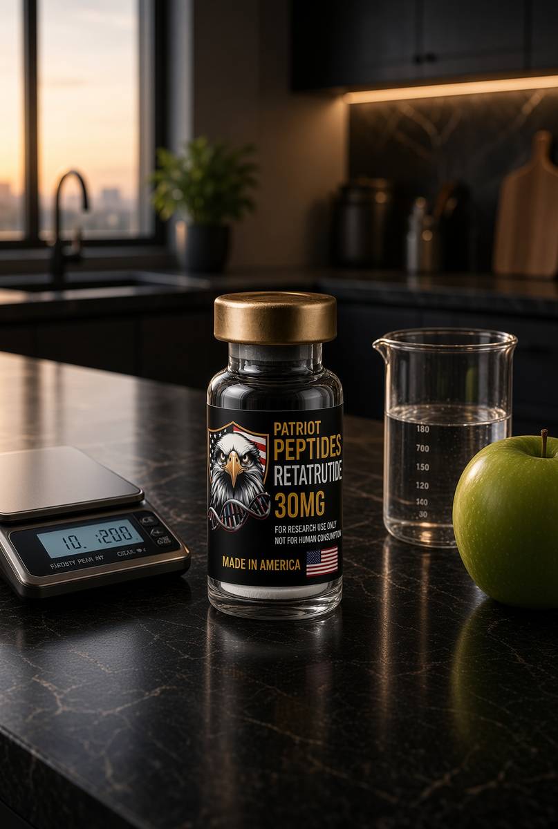
US-made retatrutide 30mg — the published structure LY3437943, HPLC-MS verified.
4.6(609)
74 browsing this now · 5 purchased in the last 24 hours
30mg
£120.00
 Made in USA
Research use only. Not a combined-use instruction.
Read next
51 min · long read · Metabolism
Human metabolism, without the slogan
Calories are bookkeeping. The actual system is a set of hormonally gated fluxes through glycogen, fat and amino acids — with the liver as air-traffic control.
51 min · long read · Metabolism
Reversing type 2 diabetes is a published result, not a slogan
The DiRECT trial put type 2 diabetes into remission by emptying the liver and pancreas of surplus fat. Very-low-calorie and very-low-carbohydrate programmes can both get you there. The mechanism isn't mystical.
46 min · long read · Peptide research
Retatrutide (LY3437943): the published triple-agonist structure
A fatty-acylated unimolecular agonist at GIPR, GLP-1R and GCGR. US-made, HPLC-MS verified, labelled for laboratory research — not a medicine.
48 min · long read · Peptide research
From secretin to retatrutide: a century of gut hormones
Bayliss and Starling named the first hormone in 1902. A hundred and twenty years later a single American-made chain occupies GIP, GLP-1 and glucagon at once. The through-line is the gut talking to the pancreas.
64 min · long read · The living cell
Mitochondria: the bacterium you kept, the genome it kept, the peptides it writes
You turn over 40–60 kg of ATP a day using a 16,569-base genome that still uses a bacterial genetic code. NAD+ is the hydride carrier Complex I spends. MOTS-c is a 16-mer translated from mitochondrial 12S rRNA — Lee, Kim, Cohen, 2015. That last sentence is real, and it is surprising.
70 min · long read · The living cell
How peptides talk to cells: occupancy, amplification, arrestin
A peptide is a ligand. Most of the catalogue binds a GPCR on the cell surface: one occupancy, then enzymes make thousands of second messengers. That amplification is real, and it is not magic. Desensitisation is why more ligand is not more signal forever.
More in this desk

50 min · long read · Metabolism
Fasting, autophagy and the mTOR switch
Eat, and mTOR builds. Fast, and AMPK and ULK1 start recycling the cell. The molecular story is a switch, not a personality.
49 min · long read · Metabolism
Liver fat is the variable type 2 diabetes actually cares about
Ectopic fat in the liver overproduces glucose and VLDL. Empty it — by energy deficit, by carbohydrate restriction, or by both — and first-phase insulin can return. That is the twin-cycle in working clothes.
49 min · long read · Metabolism
Protein, training and muscle protein synthesis
Muscle isn't a mood. It is mechanical tension plus a leucine-gated translational programme. Here is the dose, the threshold, and why the gym still has to happen.
48 min · long read · Metabolism
When insulin falls, the kidney dumps sodium. That is the ‘keto flu’.
Insulin tells the kidney to hold salt. Carbohydrate restriction drops insulin, you diurese, and magnesium and potassium follow. The headache was not a test of character.
Essays describe published research. They are not medical advice and they do not authorise human use of any catalogue item.