

Metabolism · 39 min · 8,581 words
Brown fat, UCP1, and heat as a decision
Brown adipose tissue is a heater with a named protein, UCP1, that spends the mitochondrial proton gradient as warmth instead of ATP. Adults have some; PET-CT found it in 2009. Cannon and Nedergaard wrote the review. Occupancy at the glucagon receptor lives in that neighbourhood. It is not a brown-fat drug.
What this essay actually tells you
- UCP1 is a regulated inner-membrane proton leak. Fatty acids open it; purine nucleotides close it. Heat is the product. ATP synthase is the door the brown adipocyte decided not to use.
- Adults have brown fat. Cypess, van Marken Lichtenbelt, Virtanen, NEJM 2009; Nedergaard pointed at the PET archive in 2007. Tens of grams, cold-recruitable, not a second liver.
- Retatrutide occupies GCGR in the thermogenesis neighbourhood. That is occupancy, not a brown-fat drug. MOTS-c and NAD+ are mitochondrial reagents on the same organelle, different jobs.
What this actually means
White fat stores. Brown fat heats. The difference is a protein in the inner mitochondrial membrane, UCP1, that lets protons slip back into the matrix without making ATP. The energy becomes warmth. Norepinephrine from a sympathetic nerve is the switch: it frees fatty acids inside the cell, UCP1 opens, the chain runs, heat leaves in the blood. Infants have an obvious pad. Adults were supposed not to. 18F-FDG PET-CT, and three New England Journal papers in 2009, showed a supraclavicular remnant that takes up glucose when you are cold and that stains for UCP1 when someone biopsies it. Beige fat is a third object: heater cells that can appear inside white depots. Cannon and Nedergaard's 2004 Physiological Reviews article is still the document. Retatrutide occupies GLP-1, GIP and glucagon receptors; occupancy at GCGR sits in the thermogenesis neighbourhood and is not a brown-fat drug. MOTS-c and NAD+ are mitochondrial reagents in the catalogue, not UCP1 ligands. A tissue, a protein, a scan, and a careful sentence about a third receptor. Research use only on the solids.
A mitochondrion is a battery, and we only need that picture for a moment. Electrons from fuel walk a chain of protein complexes. Protons are pumped the other way. The inner membrane holds a voltage — about 150 millivolts, negative inside — plus a pH difference, matrix alkaline. ATP synthase is the turbine that spends that voltage on a phosphate, and that is how a hepatocyte, a myofibre and a neuron pay the rent. Brown adipose tissue looked at the same battery and installed a second door. Uncoupling protein 1, UCP1, sits in the inner membrane and lets protons return to the matrix without turning the turbine. The energy stored in the gradient becomes heat. That is not a leak the organelle failed to patch. It is a job, encoded as a protein. Barbara Cannon and Jan Nedergaard have been saying this, in reviews the field actually cites, for twenty years: brown fat is the only tissue whose function is to produce heat. Function, not a side-effect. A decision written in amino acids, sitting in a membrane you can isolate.
In short. Brown fat puts UCP1 in its mitochondria so fuel becomes heat instead of ATP. Heat is the product on purpose.
We stock retatrutide because it is the published LY3437943 structure: a unimolecular agonist at GLP-1R, GIPR and the glucagon receptor, GCGR. We stock MOTS-c because it is a 16-mer a mitochondrion translated from its own 12S rRNA. We stock NAD+ because it is the hydride coin Complex I wants oxidised. Those three vials sit near fuel, mitochondria and energy expenditure. Near is not the same as identity. Occupancy at the glucagon receptor is a real sentence about thermogenesis. Calling retatrutide a brown-fat medicine is not. What you are reading is the tissue, the protein, the PET-CT, the beige argument, and the jobs we refuse to mix. It is not a protocol. We do not tell you how long to stand in a cold car park. The groups that ran those cold-room experiments already published how they did it, and you can go there if you are running one. We are here because UCP1 is one of the loveliest decisions a cell ever made, and it deserves to be explained as physiology rather than as a product.
In short. Retatrutide, MOTS-c and NAD+ live near this biology. None of them is a brown-fat medicine. This is physiology, not a how-to.
Brown adipose tissue is the only tissue in the body whose function is to produce heat.— Cannon B, Nedergaard J. Brown adipose tissue: function and physiological significance. Physiol Rev. 2004; 84: 277–359.
Two fats, and they are not a mood
White adipose tissue is a warehouse, and that is a job, not a verdict. One large lipid droplet per cell — unilocular, if you want the histology word — a nucleus shoved to the rim, a thin cytoplasm, a modest mitochondrial census. The work is storage: triglyceride in when insulin is up, triglyceride out when insulin falls and hormone-sensitive lipase is allowed to work. The secretory job is real and we will not pretend otherwise. Leptin, adiponectin, resistin: a hundred other peptides an adipocyte writes so the brain and the liver know the warehouse is stocked. Call it WAT and you have said something anatomical. You have not said bad fat. Visceral and subcutaneous white depots are different organs wearing the same colour, with different venous drainage, different inflammatory tone, different conversation with the liver. An adipocyte is not a moral failure. It is a unilocular cell with a lipid droplet and a receptor sheet, and it is very good at what it does. We need it. We also need to tell it apart from the heater next door, because English hung one syllable on both of them.
In short. White fat stores fuel in one big droplet and talks to the rest of you with hormones. It is a warehouse, not a character flaw.
Brown adipose tissue is a heater, and once you have seen it on a slide you will not mix it up with white. Many small lipid droplets — multilocular is the word — a nucleus that sits more centrally, and mitochondria so dense the tissue looks brown to the naked eye. That colour is cytochromes and iron, a forest of cristae, not a suntan. The vascular bed is rich because heat has to leave in the blood. The innervation is sympathetic and parenchymal: fibres among the adipocytes, not a polite afterthought around a vessel. The lipid is not a long-term store. It is on-site fuel for a mitochondrion that has been told to run without making ATP. You can tell these two cells apart on a decent stain, and you should, because a brochure that treats fat as one tissue is mixing a warehouse with a heater. Two colours, two jobs, two organelles packed at very different densities. Hold that picture and the protein we are about to name will make sense as a decision rather than as a curiosity in a catalogue.
In short. Brown fat is packed with mitochondria and small fat droplets. It burns fuel as heat. The brown colour is the mitochondria.
The lineage split is the sentence most gym accounts skip, and it is worth slowing down for. Classical brown adipocytes come from a Myf5-positive, Pax7-positive progenitor that also makes skeletal muscle. Seale, Kajimura, Spiegelman and colleagues put PRDM16 on that decision: a transcriptional cofactor that, in the right cell, writes brown instead of myocyte. White adipocytes are mostly Myf5-negative. Beige adipocytes — or brite, brown-in-white, Cannon and Nedergaard's word — appear inside white depots when the animal is cold or when a β3-agonist is applied, and they are not simply classical brown cells that took a wrong turning. Different origin, overlapping toolkit, UCP1 when they are on. Treating beige as brown fat you can hack in your love handles is how a Cell paper becomes a tweet, and we can do better than that. Three cells, then: white for storage, classical brown for a constitutive heater, beige for a recruitable one inside a white depot. Same inner-membrane protein when the heater is running. Different developmental story. That is why the next headings keep the names separate.
In short. Classic brown fat is cousins with muscle, not with belly fat. Beige fat can appear in white depots when cold or adrenaline-like signals arrive.
Infants are the case that makes the physiology obvious. A human neonate cannot shiver well and has a surface-to-volume problem: heat leaves a small body faster than a large one. Interscapular, perirenal, around the great vessels — depots of classical brown adipose tissue that keep a small mammal from going cold when the uterus is no longer a climate. Adults were supposed, in the textbooks of the 1980s and 1990s, to have lost it. Residual islands, maybe, of no physiological account. That story died in 2009, and it should have died earlier. Nedergaard, Bengtsson and Cannon had already gone through the oncology 18F-FDG PET literature in 2007 and pointed at the supraclavicular hotspots everyone had been calling muscle uptake or ignoring. Then three New England Journal papers in one year made the adult depot an empirical object instead of a rumour. Babies still have the obvious pad. Adults kept a remnant. The remnant is small, cold-recruitable, and real, and we are going to look at the scans that proved it.
In short. Babies have obvious brown fat because they cannot shiver. Adults were said to have none. PET scans in 2009 showed they do.
- Infant interscapular BAT
- grams, obvious
- Adult supraclavicular BAT
- tens of grams, variable
- UCP1
- ~32 kDa
- Proton-motive force
- ~150 mV
- Classical BAT lineage
- Myf5+
- Beige adipocytes
- inducible in WAT
The textbook depot. A neonate's non-shivering heater.
The PET-CT object. Present, cold-recruitable, not a second liver.
Inner-membrane carrier. The hole the cell installed on purpose.
Same battery as every mitochondrion. BAT spends it as heat.
Shared with skeletal muscle. PRDM16 is the identity cofactor.
UCP1-positive when recruited. Not a love-handle protocol.
UCP1 is a hole you meant
Uncoupling protein 1 is a member of the SLC25 mitochondrial carrier family: six transmembrane helices, about 32 kilodaltons, sitting in the inner membrane of a brown or beige adipocyte. The gene in humans is UCP1. Older literature called the protein thermogenin, which is a better name than most proteins get, because it makes heat. The 1970s and 1980s did the biochemistry. Nicholls, Lindberg, Cannon, Ricquier, Klingenberg — isolation from hamster and rat brown-fat mitochondria, the GDP-binding assay that is still how a laboratory proves the protein is there, the demonstration that the proton leak of those mitochondria is a regulated protein, not a damaged membrane. You can still run that assay. You should, if you claim to have brown fat in a dish, before you write the word thermogenic. A named carrier, a binding assay, a tissue. That is the object. Homology with other mitochondrial carriers is a family resemblance. It is not yet a job description, which is why UCP2 and UCP3 get their own warning in a moment.
In short. UCP1 is a named inner-membrane protein. Labs found it by the way brown-fat mitochondria leak protons, and by a GDP-binding assay that still works.
The mechanism, in working English, is a choice about what protons are for. UCP1 lets protons re-enter the matrix. ATP synthase also lets protons re-enter the matrix. The difference is the product. Synthase couples the return to phosphorylation of ADP. UCP1 couples it to nothing but the thermalisation of the energy that was stored in the gradient. Fatty acids activate the carrier. Purine nucleotides — GDP in the classic binding assay; ATP, ADP and GTP in a living matrix — inhibit it from the cytosolic face. The field has argued for decades about the exact physical picture: a fatty-acid protonophore, a fatty-acid/H+ symport, a conformational proton path through the carrier. Bertholet's recent electrophysiology has not ended the argument so much as made it less cartoonish. You do not need to pick a winner to own the physiology. When UCP1 is on, the chain can run, oxygen is consumed, fuel is oxidised, and ATP is not the point. Heat is. That is non-shivering thermogenesis at the scale of one inner membrane.
In short. UCP1 lets protons back in without making ATP. Fat molecules turn it on. Energy of the gradient becomes warmth.
UCP2 and UCP3 are homologs and they are a trap if you let a family name do a job description. They are mitochondrial carriers. They are not the thermogenic uncoupler of brown fat. UCP3 lives in muscle; UCP2 is widely expressed. Mild uncoupling, fatty-acid anion transport, protection from oxidative stress — pick the paper, pick the assay, do not pick a tweet that says the UCP family equals heat. Knock out UCP1 in a mouse and classical non-shivering thermogenesis is gone. The animal will shiver, it will survive in the cold if you let it, and it will not have a brown-fat depot that can dump the gradient on purpose. That knockout is the reason this protein gets its own heading. Homology is a family tree. A job is what happens when you delete the gene and watch a physiology vanish. We keep the names because the sequences are related. We keep the jobs apart because a shivering mouse has already given us the answer.
In short. UCP2 and UCP3 are relatives, not the same job. Without UCP1, a mouse cannot do classical brown-fat heat. It shivers instead.
Activation is two clocks, and mixing them is how people talk past each other. Acute: free fatty acids liberated by lipolysis occupy UCP1 and the leak opens in seconds to minutes. The same fatty acids are the fuel the chain oxidises. A brown adipocyte is, in that hour, a device that hydrolyses its own droplets and throws the product at a mitochondrion told not to make ATP. Chronic: hours to days of sympathetic drive write more UCP1, more mitochondria, more cells. PGC-1α is the coactivator the endurance-muscle page already named; brown fat uses it too. PPARγ, C/EBPβ, EBF2, ZFP516, and PRDM16 as the identity cofactor. Type 2 deiodinase, DIO2, converts local T4 to T3 so the thyroid signal is amplified inside the depot, which is why a hypothyroid animal is bad at non-shivering thermogenesis and why thyroid is metabolism is a real sentence that still does not license a research peptide as a thyroid. Two clocks. One protein. A transcriptional village around it.
In short. Fat released inside the cell turns UCP1 on right now. Days of cold or nerve drive build more UCP1 and more mitochondria. Thyroid hormone helps the gene stay on.
Diagram
Matrix
- TCA cycle · β-oxidation · mtDNA nucleoids
- NADH produced here. Complex I spends it.
- MOTS-c (MRWQEMGYIFYPRKLR) from 12S rRNA.
Inner membrane
- I → II → III → IV → V (ATP synthase)
- ~150 mV proton-motive force
- ~40–60 kg of ATP turned over per human day
mtDNA is 16,569 bp, 37 genes, 13 proteins of the respiratory chain. Nuclear DNA encodes the other ~1,200 mitochondrial proteins. NAD+ is the hydride carrier between dehydrogenases and Complex I. MOTS-c is a 16-mer translated from 12S rRNA — a peptide the mitochondrion wrote itself.
Proton leak is the physics. Heat is the invoice.
Peter Mitchell's chemiosmotic theory is the reason this page has a diagram, and it is still the most beautiful idea in bioenergetics. Electrons from NADH at Complex I and from succinate at Complex II move to oxygen at Complex IV. Protons are pumped at I, III and IV. The inner membrane holds a proton-motive force: a voltage of about 150 millivolts, negative inside, plus a pH difference, matrix alkaline. ATP synthase is a rotary turbine. Boyer and Walker got the Nobel for the catalytic mechanism; Mitchell got his in 1978 for insisting the gradient was the point. Every mitochondrion in you runs this. A brown-fat mitochondrion runs it with a second, regulated path for the protons. Uncoupling, in the original language, meant uncoupling oxidation from phosphorylation. The chain still oxidises. Phosphorylation is optional. Heat is what conservation of energy does with the difference. Same chain as muscle. Different door home.
In short. Fuel electrons pump protons. Usually those protons make ATP. In brown fat they can slip back through UCP1 and the energy becomes heat.
All mitochondria leak a little, and that sentence has to sit next to UCP1 or people start calling every organelle a heater. Martin Brand spent a career measuring basal proton leak: a background conductance that is not UCP1, present in hepatocytes and myocytes, a non-trivial slice of resting metabolic rate if you trust the whole-body extrapolations. Basal leak is not a decision in the sense this title means. It is a property of a crowded inner membrane, of the adenine nucleotide translocase, of the physics of a thin bilayer with a huge field across it. Inducible leak through UCP1 is the decision. A sympathetic nerve, a β-receptor, a cAMP rise, a lipase, a fatty acid, a carrier that opens. You can turn it up. You can turn it down with nucleotides. You can delete the gene and watch the decision vanish. That is why brown fat is interesting, and why mitochondria make heat as a general claim is a way of not having read Brand or Cannon.
In short. Every mitochondrion leaks a little. UCP1 is a leak you can switch. That switch is what makes brown fat a heater rather than a slightly inefficient battery.
The arithmetic is rude, which is why it is worth doing on a scrap of paper. Oxidation of fuel is a drop in free energy. Capture as ATP is partial in the best inner membrane — slip, leak, the cost of transport, the actual P/O ratio that is messier than the textbook 2.5. Open UCP1 fully and capture as ATP collapses toward zero for those mitochondria, and capture as heat approaches the whole drop. A brown adipocyte can respire at a rate that would look like a working muscle fibre, except the myosin ATPase is not invited. Oxygen in, carbon dioxide out, warmth into the blood leaving the depot. That is non-shivering thermogenesis as a tissue-level fact. Whole-body, in a cold-acclimated rodent, brown fat can account for a startling fraction of energy expenditure. Whole-body, in an adult human at rest in a warm room, the same tissue is a rounding error until you cold-expose it and put it on a PET scanner. Mass is small. Power density, when it is on, is not.
In short. When UCP1 is fully on, those mitochondria make heat instead of ATP. In a cold mouse that can be a lot of the energy budget. In a warm adult it is small until recruited.
- NADH and QH2 feed Complexes I and II. Electrons walk to oxygen. Protons are pumped. A voltage appears.
- ATP synthase is door one: protons return, ADP is phosphorylated. Default in almost every tissue.
- UCP1 is door two: protons return, heat is the product. Default in recruited brown and beige adipocytes.
- Fatty acids open door two. Purine nucleotides lean on it shut. Norepinephrine decides whether the fatty acids arrive.
- Basal leak exists everywhere and is not this decision. Inducible UCP1 leak is.
Diagram
- I. Pumps H⁺. ~45 subunits. The NADH coin is spent here.
- II. TCA entry. No proton pump. FADH₂ neighbourhood.
- Q. Lipid-soluble shuttle in the inner membrane.
- III. Q-cycle. Pumps H⁺. Superoxide leak site.
- c. Intermembrane space. The shuttle everyone has heard of.
- IV. O₂ → H₂O. The reason you breathe.
- V. F₁Fₒ rotary. Protons in, ATP out. ~10²¹ times a second in you.
Mitchell’s chemiosmotic theory (Nobel 1978): the inner membrane is a battery of ~150 mV. NAD+ is the hydride carrier that feeds Complex I. MOTS-c is a 16-mer the mitochondrion translated from 12S rRNA — a different object on the same campus.
Norepinephrine is the switch. The nerve got there first.
Brown fat is innervated, and that is the switch you actually wanted. Not in the polite, vascular way a white depot is innervated. Sympathetic fibres run among the adipocytes themselves. Cold is registered in the skin and in the core, integrated in the preoptic hypothalamus, sent down through brainstem and intermediolateral column, and arrives as norepinephrine at a β-adrenergic receptor on the brown cell. Circulating catecholamines can help. They are not the design. The design is a synapse onto a heater. Cut the nerve, in the old denervation papers, and the depot forgets how to turn on. That is why a cold-shower tweet is a cartoon of a hypothalamic loop, and why the cartoon still gets more traffic than the loop. The loop is still the biology: skin, hypothalamus, cord, nerve, receptor, lipase, UCP1. You can draw it on a napkin. You cannot replace it with a thirty-second splash of tap water and call the experiment done.
In short. Cold talks to the brain. The brain talks to brown fat through nerves that release norepinephrine. The nerve is the switch.
The receptor, in the rodent that built the field, is β3-adrenergic. Gs, adenylate cyclase, cAMP, protein kinase A. PKA phosphorylates perilipin and hormone-sensitive lipase; ATGL, also called PNPLA2, does the first cut on the triglyceride; free fatty acids flood the cytosol and the mitochondrion. Acute UCP1 activation follows. In humans the β-receptor story is less of a β3 monopoly — β1 and β2 are present and functional in human brown fat, and the β3-selective agonists that made mouse inguinal fat go beige have been a disappointment as human drugs, which is information, not a scandal. Mirabegron, a β3 agonist licensed for overactive bladder, has been used as a human brown-fat probe: some FDG-PET studies show recruitment, the effect sizes are not a second thyroid, and nobody careful is calling it a thermogenic medicine for obesity. Receptor occupancy is still receptor occupancy. Species is a variable. We keep having to say that in this journal and we will say it again here, because a mouse inguinal pad is not a human neck.
In short. Norepinephrine hits a β-receptor, cAMP rises, fat is released inside the cell, UCP1 opens. Humans are not mice. A bladder drug that touches β3 is not a fat-loss plan.
Chronic drive is a gene-expression programme, and it is slower than the lipase by design. CREB, ATF2, the MAP kinases that cold and nerve also move, PGC-1α transcription, UCP1 transcription, mitochondrial biogenesis, proliferation of brown precursors, recruitment of beige cells in white depots. PRDM16 stays on the identity. DIO2 keeps the local T3 high so the thyroid receptor on the UCP1 enhancer is occupied. Insulin is allowed to be present — a brown adipocyte still does glucose uptake, which is why FDG-PET works — but insulin is not the on-switch. The on-switch is the nerve. Thyroid hormone is the permission slip. Without it the programme is half-written. With it, and with norepinephrine, you get the tissue Cannon and Nedergaard described, which is not a metabolism booster in the catalogue sense and is a sympathetic, transcriptional, mitochondrial object. Days, not seconds. Genes, not just a leak. That is the second clock, and it is why a weekend of cold air is not the same experiment as a winter in a rodent house.
In short. Days of nerve drive write more heater proteins and more mitochondria. Thyroid hormone has to be on the page. Insulin is not the ignition.
There is a vascular half, and it is as practical as a radiator. Brown fat is a heat exchanger. Blood flow through a recruited depot rises several-fold so the warmth can be exported and so the oxygen can be imported. α-adrenergic tone on the vessels, nitric oxide, a capillary density the white depot does not bother with. A PET hotspot is, among other things, a perfusion hotspot. A cold, pale, poorly perfused island of multilocular cells would be a heater with no chimney, and evolution did not build it that way. The histology already told you this: you cannot miss the vessels unless you are trying to. Heat in the blood leaving the depot is how a few tens of grams can change the temperature of a much larger animal. Oxygen in the blood arriving is how the chain keeps running once UCP1 is open. Two fluxes, one tissue, a reason the scan lights up when the heater is on.
In short. Brown fat has a rich blood supply because heat and oxygen have to move. A PET bright-spot is partly blood flow.
PET-CT, 2009, and the adult who was not supposed to have a heater
Oncology had been looking at it for years without naming it, which is a humbling sentence if you like to think biology waits for the right lab. 18F-fluorodeoxyglucose PET, paired with CT for anatomy, lights up tissues that take up glucose. Tumours do. Brain does. Heart does. And, in a subset of patients, a symmetric, supraclavicular, paravertebral, sometimes mediastinal and perirenal pattern lights up that is not a metastasis and is not trapezius. Radiologists learned to ignore it, or to call it physiological muscle uptake, especially in younger, leaner, female, winter patients — which is already the epidemiology of adult brown fat, sitting in the radiology archive, unnamed. Nedergaard, Bengtsson and Cannon, American Journal of Physiology 2007, named it. Unexpected evidence for active brown adipose tissue in adult humans. They had gone looking in the PET literature on purpose. Sometimes the finding is already in the filing cabinet. You just have to know what colour it should be.
In short. Cancer scans were already lighting up brown fat in adults. Radiologists often ignored it. Cannon and Nedergaard pointed at it in 2007.
2009 was the year the named papers arrived, and it still feels like a plot twist if you were taught the 1990s textbook. Cypess, Lehman, Williams and colleagues, New England Journal of Medicine: retrospective 18F-FDG PET-CT, thousands of scans, a prevalence that tracked age, BMI, sex and outdoor temperature, histology on a subset that was actually brown fat. Van Marken Lichtenbelt, Vanhommerig, Smulders and colleagues, same journal: healthy men, cold protocol, cold-activated brown fat in most of them, inverse relationship with adiposity. Virtanen, Lidell, Orava, Enerbäck and colleagues, same journal: functional brown adipose tissue in healthy adults, biopsy, UCP1, the molecular identity, not just a hotspot. Saito's Diabetes paper the same year. Three NEJM objects and a Diabetes object, one winter of a field. Adult humans have brown fat. It takes up glucose when they are cold. It has UCP1. It is smaller than a rodent's interscapular pad and it is not zero.
In short. In 2009, three NEJM papers showed adult brown fat on PET-CT, with biopsies that found UCP1. Adults have a heater. It is small. It is real.
What the scan is, and is not, matters more than the pretty picture. FDG reports glucose uptake. Brown fat, when it is on, will take glucose — GLUT1, GLUT4, a high hexokinase capacity, insulin helping, cold helping more. The fuel the mitochondrion actually wants, once UCP1 is open, is fatty acid. A glucose-bright depot is a recruited depot. It is not a calorimeter. Oxidative metabolism has been measured with 15O-oxygen PET and with 11C-acetate; those studies exist and they are smaller than the FDG literature. Blood flow, as above, confounds and also informs. Outdoor temperature of the city the scanner sits in is a real covariate. So is the time since the patient last felt cold. A warm waiting room can hide a depot a cold protocol would have shown. Prevalence numbers in the retrospective oncology series are therefore lower bounds on has brown fat that can be recruited, not a census of grams. Read the protocol before you read the percentage.
In short. The common scan tracks sugar uptake, not heat itself. Cold rooms find more brown fat than warm ones. A dark scan does not prove the tissue is absent.
How much is it worth, whole-body? The range is unsatisfying, which is a reason to keep it rather than to round it into a headline. Tens of grams in a typical recruited adult, sometimes more, sometimes a thin ribbon the CT barely resolves. Estimates of cold-induced thermogenesis attributable to brown fat in humans run from almost nothing to a few percent of daily energy expenditure, depending on the protocol, the imaging, and whether you believe the extrapolation from a supraclavicular hotspot to a 24-hour budget. It is not a second liver. It is not nothing. People who stay lean in winter have, in some cohorts, more visible brown fat; causality is a mess because the lean also have less white fat covering the depot, a different thyroid, a different life. Cypess and van Marken Lichtenbelt did not hand us an obesity drug. They handed us a tissue. The tissue is allowed to be interesting without being a product. That permission is rarer than it should be.
In short. Adult brown fat is tens of grams, not kilograms. In the cold it can spend some energy as heat. It is not an obesity cure sitting in your neck.
Unexpected evidence for active brown adipose tissue in adult humans — then three NEJM papers in 2009 that made the unexpected into an object you could biopsy.— Nedergaard, Bengtsson, Cannon, Am J Physiol 2007; Cypess et al., van Marken Lichtenbelt et al., Virtanen et al., N Engl J Med 2009.
Beige is not brown with a tan
Beige adipocytes — Wu, Boström, Spiegelman, Cell 2012, and the brite literature Cannon and Nedergaard preferred — are UCP1-positive, multilocular cells that appear in white depots, classically the inguinal fat of a cold-housed or β3-agonist-treated mouse. They have a molecular signature that is not identical to classical interscapular brown fat, though the signature genes, Tbx1, Tmem26, CD137 in the early papers, have been argued over since. They oxidise, they leak, they make heat. They can disappear when the cold stops, which classical brown fat is less inclined to do. Human adult brown fat, especially the supraclavicular depot, looks on histology and on RNA more like a mix of classical-brown and beige-like cells than like a pure rodent interscapular pad. Jespersen, Lidell, Enerbäck and others have spent a decade saying that, and it is why a sentence that begins human brown fat should admit the mixture. Mix is not a failure. Mix is what the adult neck actually is.
In short. Beige fat is heater cells that show up inside white fat when the animal is cold. Human neck fat is probably a mix of beige-like and classical brown.
Irisin is the cautionary tale this journal was built for, and we can tell it without a sneer. Boström, Wu, Spiegelman, Nature 2012: PGC-1α in muscle induces FNDC5, which is cleaved to irisin, which browns white fat. A beautiful circuit. A subsequent argument about the antibody, the circulating concentration in humans, the cleavage, the replication, and what a commercial ELISA was actually measuring. Jedrychowski and Spiegelman later used mass spectrometry to defend a real, low circulating level. The circuit may well exist. The leap from that paper to exercise makes beige fat via irisin, therefore this supplement, was a category error on the day it was printed. Exercise still wins for mitochondria in muscle, which is a different tissue, a different page, and a PGC-1α story that does not need a cleavage product to be true. Beige recruitment in humans after training is mixed, small, and not a reason to buy a bottle. Beautiful circuits still have to survive an ELISA.
In short. Irisin was proposed as the muscle-to-fat browning signal. The human numbers got messy. Exercise still builds muscle mitochondria. That does not make irisin a supplement.
Other ligands sit in the same neighbourhood and deserve a named sentence each, because a neighbourhood is still worth mapping. FGF21, from liver and from brown fat itself, can recruit beige cells in mice; human translation has been a drug-development story with its own failures and half-successes, not a beige protocol. Natriuretic peptides, ANP and BNP, via cGMP and PKG can lipolyse and, in rodents, brown. BMP7 and BMP8b have developmental and recruitment papers. Capsinoids and TRPV1 agonists have a human brown-fat literature that is real and modest. None of these is UCP1. None of them is norepinephrine. All of them are how a field tries to turn the nerve-and-cold switch into something a clinic could hold. We do not stock them. We do not pretend a catalogue peptide is a quieter version of them. Named ligands, named papers, a heater that still runs on a nerve and a carrier. That is the map. It is allowed to have more than one road without every road becoming a product.
In short. FGF21, heart peptides, bone morphogenetic proteins and chilli-related compounds all have browning papers. They are not UCP1, and they are not this catalogue.
The exercise page on this desk is AMPK, PGC-1α, calcium, mTOR, VO2. Endurance work builds the engines in the fibres you actually used. Heavy work builds the chassis. Brown-fat recruitment is a sympathetic, cold-adjacent programme that may overlap a training week in a person who happens to be outdoors in January, and that overlap is not a mechanism you can put in a shaker bottle. MOTS-c has exercise-mimetic papers in mice — Lee, Kim, Cohen, and the AMPK/folate–methionine follow-ups. Those papers are mitochondrial. They are not UCP1 occupancy. A 16-mer translated from 12S rRNA does not become a beige drug because both sentences contain the word mitochondrion. We stock the 16-mer as HPLC-characterised research material. We do not file it under thermogenesis as a product claim. Shared organelle, different invoice. You can read both papers on the same afternoon. You still have to keep the jobs apart, or the afternoon was wasted.
In short. Training builds muscle engines. Brown fat is a different organ. MOTS-c is a mitochondrial peptide in the catalogue, not a beige-fat activator we claim.
Cannon and Nedergaard already wrote the review
Barbara Cannon and Jan Nedergaard, Stockholm, a physiological review the field treats as statute: Brown adipose tissue: function and physiological significance, Physiological Reviews, 2004, eighty-odd pages, 277 to 359. If you read one document before you speak about this tissue, read that. Function is heat. The cell is a heater. UCP1 is the mechanism. The nerve is the switch. Shivering is muscle and is a different, more expensive, less elegant way to stay warm. Non-shivering thermogenesis is brown fat. Newborns, small mammals, cold-acclimated rodents: the physiology was never in doubt. Adult humans were the argument. They spent the next decade winning it, first with the 2007 PET re-read, then with the 2009 biopsies, then with a run of reviews in 2010, 2013 and 2018 that had to keep saying the same sentence because the obesity industry kept hearing new drug target and not tissue. A tissue first. A target, maybe, later. That order is the whole of their gift to the rest of us.
In short. Cannon and Nedergaard's 2004 review is the document. Brown fat's job is heat. They then spent years proving adults still have some.
What they will not let you do, if you read them, is treat uncoupling as a general mitochondrial lifestyle. A hepatocyte uncoupled by a chemical protonophore — DNP, FCCP — is a poisoned hepatocyte. Dinitrophenol was a weight-loss drug and then a poison; the gradient is not a toy. UCP1 is tolerated because it lives in a cell built around it: droplets on site, vasculature, innervation, a transcriptional identity that does not try to make ATP for a plasma-membrane sodium pump at the same moment. That is why uncouple your mitochondria as a catalogue heading would be a moral and scientific failure, and why this page is about a named protein in a named tissue. We sell reagents. We do not sell holes in the inner membrane. The distinction is not fussiness. It is the difference between a heater with a chimney and a hole punched in the boiler of a cell that still has rent to pay. Cannon and Nedergaard already drew that line. We are only refusing to smudge it.
In short. Forcing leak in the wrong cell is poison — dinitrophenol already taught that. UCP1 is safe in the cell that was built to be a heater. That cell is brown fat.
Shivering versus non-shivering is the other distinction they insist on, and it is kinder than it sounds. Shivering is asynchronous muscle contraction, ATP turned over on purpose so that the inefficiency of the motor and the chain becomes heat. It works. It is miserable. It interferes with movement. Non-shivering thermogenesis is brown fat, and, in the later literature, a set of UCP1-independent futile cycles we will name in a moment. A cold-acclimated rodent stops shivering and keeps the heat. A human in a 16–19 °C room for a research protocol may do a mixture, and the protocol has to say so. If a tweet says activate brown fat and the method was a 30-second cold shower, the actual experiment has not been invited. Cannon and Nedergaard's rodent was housed at 4 °C for weeks, or treated with a β3 agonist, or both. Scale is not optional. Minutes are not weeks. A bathroom is not a cold room. You already knew that. The papers are there to stop us pretending we did not.
In short. Shivering is muscle making heat the hard way. Brown fat is the quieter heater. Lab cold is hours at 16–19 °C, not a quick shower.
UCP1-independent heat is real, and it does not make UCP1 optional
Kazak, Chouchani, Spiegelman, Cell 2015: a creatine futile cycle in beige fat. Creatine kinase, a phosphorylation and hydrolysis that spends ATP as heat without a contraction, a way a beige cell can still thermogenise if UCP1 is missing or insufficient. Follow-up papers extended and argued; the cycle is now a named object, not a rumour. Sarcolipin uncoupling SERCA in muscle — Bal, Periasamy — is another: the calcium pump that spends ATP to put calcium back in the sarcoplasmic reticulum can be told to slip, and heat follows. Lipid cycling, the re-esterification of fatty acids so that lipolysis spends ATP on the way back to triglyceride, is an old hepatic and adipose account. Glycerol-3-phosphate shuttles. None of this is a reason to demote UCP1 in classical brown fat. It is a reason to stop saying thermogenesis as if it had one protein. Name the cycle. Name the tissue. Then talk. Extra heaters exist. The classical depot still runs on the carrier we started with.
In short. Beige fat can also waste ATP on a creatine cycle. Muscle can waste ATP on a leaky calcium pump. Those are extra heaters. Classical brown fat still runs on UCP1.
Futile cycles are catnip for a certain kind of metabolic daydream, because they look like a weight-loss mechanism that does not require a smaller meal. The physics is real. The whole-body integral in an adult human at room temperature is usually not. A cycle that spends milligrams of ATP in a depot that weighs tens of grams will not outrun a biscuit. GCGR agonism, coming in the next heading, can increase hepatic futile cycling — glycogen and glucose, urea, sodium/potassium — as well as whatever it does to brown fat in a rodent. That is energy expenditure as a liver sentence, and it is allowed to be interesting without being UCP1. Mixing those jobs is how you get a thread that says glucagon browns fat from a paper that measured oxygen consumption in a hepatocyte. Liver and brown fat are both allowed to spend energy. They are not the same organ. Keep the receipts on separate spikes and the conversation stays adult.
In short. Wasting ATP can make heat, but a small depot will not outrun a meal. Glucagon-receptor effects on the liver are a different invoice from UCP1.
Glucagon-receptor occupancy is a neighbourhood. It is not a brown-fat drug.
Glucagon is the pancreatic α-cell's broadcast to the liver: glycogenolysis, gluconeogenesis, a rise in hepatic glucose output, a push on β-oxidation and on ureagenesis. The receptor, GCGR, is a class-B GPCR, Gs, cAMP, a hepatocyte's working day. Energy expenditure rises when you give glucagon to a human; Tan, Salem and others have measured it. Some of that energy expenditure is hepatic futile cycling. Some, in rodents, is brown fat — GCGR expression in brown fat is a published observation, and glucagon can increase brown-fat thermogenesis in those animals. Can, in a rodent, on that receptor, in that depot, is a lawful sentence. Glucagon is a brown-fat hormone in the person holding this page is a longer walk, and most of the walk has not been done with the right tracer in the right depot in the right species. Neighbourhood, again. We will keep using that word until a human imaging study makes it an address. Until then, liver first, brown fat maybe, UCP1 not shown.
In short. Glucagon talks mainly to the liver and can raise energy use. In mice it can also talk to brown fat. That is not the same as proving it in you.
Retatrutide is LY3437943, a fatty-acylated unimolecular agonist at GIPR, GLP-1R and GCGR. Coskun and colleagues, Cell Metabolism 2018, for the engineering: the glucagon occupancy was put on the chain as the energy-expenditure and lipid-oxidation arm, against the metabolic adaptation that usually attends large weight loss. Jastreboff and colleagues, New England Journal of Medicine 2023, Phase 2, obesity: 24.2% mean weight loss at 12 mg, 48 weeks. That paper is an investigational-medicine trial. It is not a brown-fat imaging study. It did not show UCP1. It did not show 18F-FDG in a supraclavicular depot. It showed weight, glucose, a side-effect sheet, a dose-response. GLP-1R and GIPR occupancy account for most of the intake suppression. GCGR occupancy is the theoretical energy-expenditure lift. Theoretical, engineered, rodent-supported, human-energy-expenditure-plausible. Not a PET-CT of your neck. Hold those two papers in different hands. Engineering is not a scan. A weight curve is not a UCP1 blot.
In short. Retatrutide occupies GLP-1, GIP and glucagon receptors. The glucagon arm was built to lift energy use. The big trial showed weight loss, not brown-fat scans.
The sentence we will keep using until the literature moves is this: occupancy at GCGR is in the thermogenesis neighbourhood. Retatrutide is not a brown-fat drug. We synthesise the published structure in the United States, HPLC-MS, labelled for research use only. We are not Eli Lilly. We do not have an MHRA marketing authorisation to pretend otherwise. A research vial is a ligand for a tube. A Phase 2 curve is a paper. Adult brown fat is a tissue with UCP1. Those three objects share a reading list. They do not share a mechanism until someone occupies GCGR, images brown fat, and shows the heat is UCP1-dependent in the species you actually care about. That experiment is not this catalogue. Eliding the gap is selling a neighbourhood as an address, and we would rather be slightly boring about a receptor than fluent in a claim the scan has not earned. Neighbourhood. Address. Different words. Worth keeping.
In short. Glucagon-receptor occupancy sits near thermogenesis. That does not make retatrutide a brown-fat medicine. We stock the published structure as a research ligand.
Liver fat is the other GCGR sentence, and it belongs here so it does not get smuggled in as brown fat. Glucagon-receptor tone on a hepatocyte is lipid oxidation, glycogen, glucose output. The twin-cycle page on this desk is ectopic fat in liver and pancreas; DiRECT emptied it with a formula diet; retatrutide's clinical literature empties body weight from the receptor side. A smaller person with a quieter liver is a different NADH supply to a different set of mitochondria. Brown-fat flux may change because the animal changed. That is downstream weather. It is not UCP1 occupancy. The pathophysiology stack still holds: receptor floor, then tissue, then organism. Skip a floor and you write a caption the scan cannot support. Liver and brown fat can both move when body weight moves. Only one of them is the heater this page is about. Keep the hepatocyte on its own spike. It has earned one.
In short. Glucagon-receptor effects on liver fat are a real, separate story. Weight falling can change brown fat later. That is not the same as switching UCP1 on.
MOTS-c and NAD+ sit on the same organelle. Different jobs.
MOTS-c is MRWQEMGYIFYPRKLR, sixteen residues translated from mitochondrial 12S rRNA in a reading frame nobody asked an rRNA to have. Lee, Kim, Cohen, Cell Metabolism 2015. AMPK, the folate–methionine cycle, insulin-sensitivity papers in mice, nuclear translocation under stress, Kim 2018. It is one of the least likely peptides in the catalogue, and it is real chemistry. The organelle that houses UCP1 is the organelle that wrote MOTS-c. That is a shared address, not a shared job. MOTS-c is not a ligand at UCP1. It is not norepinephrine. It is not GCGR. We stock 40 mg, HPLC, lyophilised, for the bench. Exercise-mimetic language in a mouse paper is a phenotype. It is not a thermogenic product claim, and we will not launder it into one. A mitochondrion can write a peptide and still run a heater. Those are two sentences. The 16-mer does not become a browning switch because we said both of them on the same afternoon.
In short. MOTS-c is a short peptide the mitochondrion writes from its own RNA. Same organelle as UCP1, different job. Research sequence, not a heater switch.
NAD+ is the oxidised hydride coin, and we can say that without turning a cofactor into a recruiter. Dehydrogenases mint NADH. Complex I wants NAD+ back. Sirtuins and PARPs spend NAD+ as a substrate, which is why the pool is a budget as well as a redox gauge. SIRT1 deacetylation of PGC-1α is a real paper-trail in metabolic transcription, including in brown fat; SIRT3 in the matrix is a real paper-trail in oxidative metabolism. Those papers do not make a 1000 mg cake of lyophilised β-NAD+ into a brown-fat recruiter. The cofactor does not stroll through plasma membranes into the matrix of a supraclavicular adipocyte because a journal page named both objects. Our vial is a reagent for an assay you control. eLIVEate's intramuscular NAD+ is a clinic appointment on a different till, no commission. Neither is 18F-FDG uptake in a Cypess cohort. Coin, sirtuin papers, heater tissue. Three objects. One organelle in the background. Still not a protocol.
In short. NAD+ is the coin mitochondria use to move electrons. Some sirtuin papers touch brown-fat genes. The vial is still a lab reagent, and the clinic injection is a different product.
Three related peptides on this page, then, and three jobs we can actually name. Retatrutide: class-B GPCR occupancy, intake and a GCGR-shaped energy-expenditure neighbourhood. MOTS-c: a mitochondrial open reading frame with AMPK papers. NAD+: the cofactor the chain and the sirtuins actually spend. Brown fat: UCP1, norepinephrine, a PET-CT object Cannon and Nedergaard spent a career defending. You can read them on the same afternoon. You cannot stack them into a protocol. Research use only on the solids. The tissue will still be a heater in the morning, whether or not anyone opened a vial. That is the point of a physiology page sitting next to a catalogue. The organ is not for sale. The sequences are characterised reagents. The scan is a 2009 fact. Keep those three on the bench and the afternoon was well spent. Mix them into a stack and you have left the papers behind.
In short. Three catalogue items sit near this story. Brown fat remains a tissue with a nerve and a protein. Do not turn the shelf into a protocol.
Heat as a decision the cell already made
Heat as a decision is the title because it is the physics. A brown adipocyte has the same respiratory chain as a myocyte. It imported a nuclear-encoded carrier, put it in the inner membrane, wired a nerve to a lipase, and chose, when the nerve fires, to spend the gradient as warmth. White fat chose storage. Muscle chose work. The liver chose other people's glucose. Lineage and innervation and a transcription factor named PRDM16 are how the choice gets made in development and in the cold. You can recruit beige cells. You can image the adult depot. You can occupy GCGR and measure energy expenditure. You cannot, from the catalogue, sell the decision. We don't write protocols. We don't dose. We don't tell you to shiver on purpose and call it a stack. The cold-room papers that already ran those experiments are in PubMed. Go there if you are running an experiment. Stay here if you wanted the tissue explained. A heater with a name. A protein with a GDP-binding assay. A remnant an adult still has.
In short. Brown fat chose heat. This page explains that choice. It does not tell you how to switch it, dose it, or stack it.
The neighbouring pages are the rest of the map, and they do not need a sales pitch. Human metabolism, without the caption: hormonally gated flux, liver as air-traffic control. Mitochondria, the second genome: 16,569 base pairs, MOTS-c, NAD+ as coin, 40–60 kg of ATP a day. Exercise, mitochondria, PGC-1α: the engines you actually build by working. Liver fat: the depot type 2 cares about. Retatrutide: the triple agonist as published chemistry. This piece is the heater. UCP1 is the hole. Norepinephrine is the switch. PET-CT is why we know adults kept a remnant. Glucagon-receptor occupancy is a neighbourhood on a chain we synthesise in America. Cannon and Nedergaard wrote it down. We put the paper next to the vial so you can see which object you are holding. A tissue, a protein, a scan, a receptor on a different floor. That is enough for one afternoon, and it is more interesting than a protocol would have been.
In short. Read the metabolism, mitochondria and training pages next. This one is the heater, the protein, the scan, and a careful sentence about glucagon-receptor occupancy.
The chain is the same. The door home is not. UCP1 is a decision a cell can make about what a voltage is for. Occupancy at a glucagon receptor is a different decision, on a different floor, about a different protein. We do not staple them together and call it a protocol.
- WAT stores. BAT heats. Beige can be recruited inside white depots. Three cells, not a mood.
- UCP1 is the regulated proton leak. Fatty acids on, nucleotides off. Homologs UCP2/3 are not this job.
- Norepinephrine from a sympathetic nerve is the physiological switch. β-receptors, cAMP, lipolysis, then transcription.
- Adult humans: Cypess, van Marken Lichtenbelt, Virtanen, NEJM 2009. FDG-PET-CT, biopsies, UCP1. Small mass. Real tissue.
- Cannon & Nedergaard, Physiol Rev 2004, is the review. Function is heat. Chemical uncouplers in the wrong cell are poison.
- GCGR occupancy (retatrutide / LY3437943) is the thermogenesis neighbourhood — not a BAT drug, and not the Phase 2 weight curve re-labelled.
- MOTS-c is a 16-mer from 12S rRNA. NAD+ is the hydride coin. Same organelle, different jobs, HPLC reagents, not protocols.
Questions the essay actually answers
- Do adult humans actually have brown fat?
- Yes. Cypess, van Marken Lichtenbelt and Virtanen, New England Journal of Medicine 2009, with Nedergaard, Bengtsson and Cannon pointing at the PET archive in 2007. Supraclavicular, paravertebral, sometimes mediastinal and perirenal. Tens of grams, cold-recruitable, UCP1-positive on biopsy. Not an infant-only leftover and not a second liver.
- Is retatrutide a brown-fat drug?
- No. Retatrutide is published LY3437943: occupancy at GLP-1R, GIPR and GCGR. Glucagon-receptor occupancy sits in the thermogenesis neighbourhood — energy expenditure, lipid oxidation, rodent BAT papers. Jastreboff NEJM 2023 measured weight, not UCP1. We stock the structure as a research ligand. We are not Eli Lilly. Neighbourhood is not identity.
- What does UCP1 actually do?
- It is an inner-membrane carrier that lets protons return to the mitochondrial matrix without going through ATP synthase. Fatty acids activate it; purine nucleotides inhibit it. The energy of the gradient becomes heat. That is non-shivering thermogenesis in classical brown fat. UCP2 and UCP3 are homologs with different jobs.
- Brown, beige, white — what is the difference?
- White is unilocular storage. Classical brown is multilocular, Myf5-positive lineage shared with muscle, constitutively UCP1-equipped, densely innervated. Beige (brite) cells appear in white depots when cold or β-adrenergic drive arrives; UCP1-positive when recruited, a distinct signature, more reversible. Human adult depots are often a mix.
- Can cold showers turn my brown fat on?
- This is not a protocol. The imaging studies used cold rooms, typically around 16–19 °C for an hour or more, sometimes days of acclimation — published cold-room work, not bathroom advice. A brief shower is not that experiment. Cannon and Nedergaard's rodents were housed cold for weeks. Scale is the point.
- Do MOTS-c or NAD+ activate brown fat?
- No product claim of that kind is honest. MOTS-c is a mitochondrial 16-mer with AMPK papers. NAD+ is the hydride coin; sirtuin/PGC-1α literature exists in metabolic transcription. Same organelle as UCP1, different jobs. The 40 mg and 1000 mg listings are HPLC research materials. eLIVEate's NAD+ injection is a different till.
- Why did textbooks say adults had no brown fat?
- Infant interscapular BAT involutes. Without a tracer, the adult remnant is easy to miss. Oncology 18F-FDG PET had been lighting it up as an annoyance. Nedergaard's 2007 read of that archive, then the 2009 biopsies, closed the argument. The textbooks were wrong about 'none', not about 'less than a neonate'.
- If I have more brown fat, will I stay lean?
- Some cohorts show more visible BAT in leaner, younger people. Causality is tangled: less covering white fat, different thyroid, different life, different scanner conditions. Cold-induced BAT thermogenesis in adults is a real, usually small slice of the daily budget. It is not a personality and it is not a substitute for the fluxes in the metabolism essays.
Hypothetical research reconstitution
How these vials are typically mixed
Hypothetical research reconstitution for the named catalogue vial. Not a protocol, not medical advice, not a use instruction. These amounts sit in published and commonly cited laboratory ranges. The vial is labelled for research use only — not for human or veterinary administration.
Retatrutide
30mg
Mix with 3 ml bacteriostatic water → 10 mg/ml
- Hypothetical aliquot
- 1–2 mg to start; published trial arms ran higher by week
- 0.10–0.20 ml · 10–20 units on a U-100 syringe (at 1–2 mg)
- How often
- Once weekly
- The Jastreboff NEJM 2023 arms ran 48 weeks. That is a trial, not a shop protocol.
Bench steps
- Let the vial sit until it is no longer cold to the touch.
- Wipe the stopper with 70% isopropyl alcohol. Let it dry.
- Draw 3 ml bacteriostatic water (0.9% benzyl alcohol).
- Run the water slowly down the inside glass — do not blast the cake.
- Roll between finger and thumb until the cake is gone. Do not shake.
- Label the date. Store the solution at 2–8 °C. Do not freeze. Use within 30 days unless the note below says otherwise.
LY3437943 architecture. Weekly, not daily. Those milligram figures are what the papers used on the investigational medicine — they are not a use instruction for this reagent.
MOTS-c
40mg
Mix with 2 ml bacteriostatic water → 20 mg/ml
- Hypothetical aliquot
- 5–10 mg
- 0.25–0.50 ml · 25–50 units on a U-100 syringe
- How often
- Two or three times per week
- 4–8 weeks
Bench steps
- Let the vial sit until it is no longer cold to the touch.
- Wipe the stopper with 70% isopropyl alcohol. Let it dry.
- Draw 2 ml bacteriostatic water (0.9% benzyl alcohol).
- Run the water slowly down the inside glass — do not blast the cake.
- Roll between finger and thumb until the cake is gone. Do not shake.
- Label the date. Store the solution at 2–8 °C. Do not freeze. Use within 30 days unless the note below says otherwise.
Mitochondrial 16-mer. Fridge. Do not freeze. The 5 mg mark is where most bench notes start.
NAD+
1000mg
Mix with 10 ml bacteriostatic water → 100 mg/ml
- Hypothetical aliquot
- 50–100 mg
- 0.50–1.00 ml · 50–100 units on a U-100 syringe
- How often
- Two or three times per week in published infusion and assay notes
- 4–8 weeks, then a pause
Bench steps
- Let the vial sit until it is no longer cold to the touch.
- Wipe the stopper with 70% isopropyl alcohol. Let it dry.
- Draw 10 ml bacteriostatic water (0.9% benzyl alcohol).
- Run the water slowly down the inside glass — do not blast the cake.
- Roll between finger and thumb until the cake is gone. Do not shake.
- Label the date. Store the solution at 2–8 °C. Do not freeze. Use within 30 days unless the note below says otherwise.
A 1000mg cake wants 10 ml. Protect from light. Solution yellows as it oxidises — that is the cofactor dying, not a flavour. Use promptly.
Bacteriostatic water and sterile syringes ship with peptide orders over £75. Kit details · 10 ml bacteriostatic water
The vials this essay sits on
Named sequences the essay maps — Retatrutide, MOTS-C, NAD+. Hypothetical research neighbourhood, not a protocol, not a medicine. One press puts every in-stock vial in the bag.
 Made in USAOut of stock
Incretin
Retatrutide
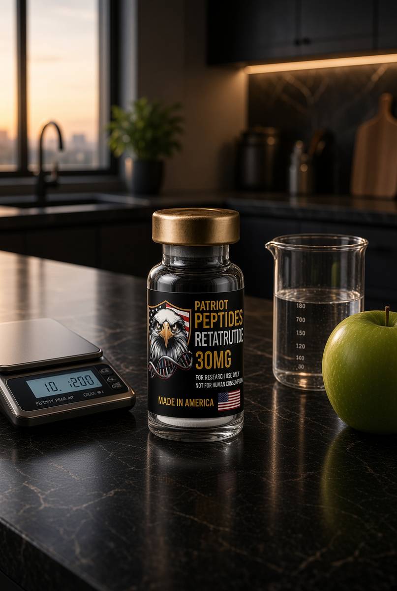
US-made retatrutide 30mg — the published structure LY3437943, HPLC-MS verified.
4.6(609)
114 browsing this now · 5 purchased in the last 24 hours
30mg
£120.00
 Made in USA
 Research only
Research use only. Not a combined-use instruction.
Read next
51 min · long read · Metabolism
Human metabolism, without the slogan
Calories are bookkeeping. The actual system is a set of hormonally gated fluxes through glycogen, fat and amino acids — with the liver as air-traffic control.
64 min · long read · The living cell
Mitochondria: the bacterium you kept, the genome it kept, the peptides it writes
You turn over 40–60 kg of ATP a day using a 16,569-base genome that still uses a bacterial genetic code. NAD+ is the hydride carrier Complex I spends. MOTS-c is a 16-mer translated from mitochondrial 12S rRNA — Lee, Kim, Cohen, 2015. That last sentence is real, and it is surprising.
47 min · long read · Metabolism
Mitochondria, VO2 and why training still wins
Endurance work builds the engines. Heavy work builds the chassis. The molecular story is AMPK, PGC-1α, calcium and mTOR — not a shopping list.
More in this desk
51 min · long read · Metabolism
Reversing type 2 diabetes is a published result, not a slogan
The DiRECT trial put type 2 diabetes into remission by emptying the liver and pancreas of surplus fat. Very-low-calorie and very-low-carbohydrate programmes can both get you there. The mechanism isn't mystical.
50 min · long read · Metabolism
Fasting, autophagy and the mTOR switch
Eat, and mTOR builds. Fast, and AMPK and ULK1 start recycling the cell. The molecular story is a switch, not a personality.
49 min · long read · Metabolism
Liver fat is the variable type 2 diabetes actually cares about
Ectopic fat in the liver overproduces glucose and VLDL. Empty it — by energy deficit, by carbohydrate restriction, or by both — and first-phase insulin can return. That is the twin-cycle in working clothes.
49 min · long read · Metabolism
Protein, training and muscle protein synthesis
Muscle isn't a mood. It is mechanical tension plus a leucine-gated translational programme. Here is the dose, the threshold, and why the gym still has to happen.
Essays describe published research. They are not medical advice and they do not authorise human use of any catalogue item.