

Metabolism · 47 min · 10,339 words
Mitochondria, VO2 and why training still wins
Endurance work builds the engines. Heavy work builds the chassis. The molecular story is AMPK, PGC-1α, calcium and mTOR — not a shopping list.
What this essay actually tells you
- Endurance training increases mitochondrial density and the enzymes of fat oxidation. That's VO2 work at the organelle. You can count the mitochondria. They multiply.
- PGC-1α is the transcriptional coactivator that makes more mitochondria when the cell is repeatedly energy-stressed. Named protein. Repeated stress. More organelles.
- A peptide or a diet that does not change mitochondrial capacity is not a substitute for that stimulus. Training still has a molecular job, and PGC-1α is the one we can name.
What this actually means
If you train, muscle grows more mitochondria and more capillaries, and it gets better at burning fat at a given pace. If you lift, you add contractile protein. The hormones people obsess over sit downstream of those two adaptations. You can study the hormones. You can't skip the work and keep the physiology, which is a sentence we've had to say to more than one person holding a catalogue.
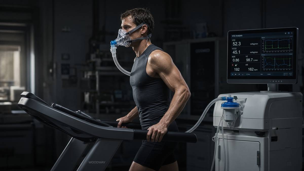
Endurance training increases mitochondrial density and the enzymes of fat oxidation. That's a 1967 result, not a cycling-podcast discovery, and I still get a kick out of how clean the assays were. John Holloszy ran rats, then measured the mitochondrial enzymes of their skeletal muscle, and found cytochrome oxidase, NADH dehydrogenase, succinate dehydrogenase and the β-oxidation spiral all risen. The organelles themselves had multiplied. Hans Hoppeler's electron micrographs, and later the Lundby group's stereology in Zurich, made the census a number: mitochondrial volume density in human vastus lateralis sits around four to six percent of fibre volume in untrained limb muscle and climbs toward eight to eleven percent in people who do the aerobic work. That's VO2 work at the organelle. Whole-body maximal oxygen uptake is the organism-level readout of stroke volume, haemoglobin, capillary density and that mitochondrial capacity, in series. Miss the last term and you have a heart delivering oxygen to fibres that can't take it. The enzymes you can name. The volume you can count.
In short. Endurance training builds more mitochondria and more fat-burning enzymes in muscle. That's the organelle work behind a higher VO2 max.
VO2 max isn't a personality and it isn't a single protein. Bassett and Howley, among others, wrote the series that still holds: pulmonary ventilation and diffusion, cardiac output, haemoglobin, muscle blood flow, capillary-to-fibre ratio, myoglobin, and the mitochondrial machinery that actually consumes the oxygen. Central terms often dominate the first months of a training block because stroke volume and plasma volume move quickly. Peripheral terms dominate the later months, and they dominate the local fatigue that a time-trial actually feels like. Weibel and Taylor's symmorphosis argument — that structures are sized to the flux they carry — is a useful caution, not a law: people run around with a heart that can outrun their cristae, or the reverse, depending on what they trained. Polarised versus pyramidal is a programming argument about how you accumulate those terms. The organelles aren't a programming argument. You either build inner membrane and the fat-oxidation enzymes, or you don't. A session that never energy-stresses the fibre won't write them.
In short. VO2 max is heart, blood and muscle mitochondria in a chain. Training has to build the whole chain, especially the organelles that use the oxygen.
Two programmes share the same fibre and they don't share a kinase. Endurance work raises AMP, calcium and a redox shift; AMPK, CaMKII and p38 MAPK converge on PGC-1α; nuclear and mitochondrial genes for respiratory subunits get written; the census rises. Resistance work deforms the fibre, leucine arrives, mTORC1 opens at the lysosome, and myofibrillar proteins are translated. Concurrent training is the art of not letting one signal chronically cancel the other, and it's solved daily by people who row and squat rather than by people who post about interference. The GH-axis sequences we stock — CJC without DAC, ipamorelin, 191-residue somatropin, IGF-1 LR3 — are how a laboratory occupies the endocrine layer of the same physiology. NAD+ is the hydride coin Complex I wants oxidised. MOTS-c is a 16-mer from mitochondrial 12S rRNA that sits on AMPK. Sharing a shelf isn't a training session. A peptide or a diet that doesn't change mitochondrial capacity isn't a substitute for the stimulus that does.
In short. Endurance work builds mitochondria. Heavy lifting builds contractile protein. Hormones and research peptides sit downstream. They don't do the session.
This page is the organelle as a training adaptation: the fuel-gauge kinases of the bout, the transcriptional coactivator that writes more mitochondria when the cell is repeatedly energy-stressed, the fibre types that decide which motor units even see the work, the zone-2 cartoon of a real fat-oxidation peak, the VO2 intervals that move the heart, and the NAD+ neighbourhood that internet culture keeps filing as a replacement. It isn't a programme. It isn't a clinic appointment written as physiology. Holloszy, Spiegelman, Hardie, Hood, Gibala, Seiler, Lundby, Baar, Hawley, Zierath: those are still the papers I'd start you with. Citrate synthase, an Oroboros chamber, a myosin heavy-chain gel, a PGC-1α blot and a three-minute all-out against a measured VO2 are the machines. The neighbouring mitochondria essay is the organelle as endosymbiont and second genome. The NAD+ essay is the pool as pool. The protein-and-muscle essay is mTORC1 as a meal-and-loading response. This one is why training still wins on the page that people open when they would rather buy a workaround.
In short. This is how muscle builds more mitochondria when you train, and why a peptide or a diet isn't the same stimulus. Named papers, named machines.
You train an organelle, not a slogan
Mitochondrial volume density is a stereological number, not a feeling. Hoppeler's early electron-microscopy in trained and untrained vastus lateralis already showed more inner membrane, more cristae, more organelles packed between the myofibrils of people who ran. Meinild Lundby and colleagues, decades later, with better sampling and the same stubborn counting, put trained endurance muscle in the high single digits to low double digits as a percentage of fibre volume, against untrained values nearer four to six. Type I fibres start richer than type II; both move. The increase isn't a swelling of existing bags. Biogenesis adds protein: nuclear-encoded subunits imported through TOM and TIM, mtDNA-encoded cores written on-site, cardiolipin, the assembly factors, TFAM on the nucleoid. Fusion and fission remodel the network so that a cardiomyocyte looks like a forest and a trained vastus like a denser one. A brochure that says 'boost mitochondria' without a volume-density, a citrate-synthase activity, or an oxygen-consumption trace hasn't yet said anything a proper experiment would recognise.
In short. Training really does pack more mitochondria into muscle fibres. You can count them on electron micrographs. Type I fibres start with more, and both types can gain.
Let's name the fat-oxidation enzymes, because the nicknames skip them and you deserve the real ones. Carnitine palmitoyltransferase-1 on the outer membrane is the malonyl-CoA-gated door that lets acylcarnitines into the matrix. CPT-2 completes the transfer. The acyl-CoA dehydrogenases — VLCAD, LCAD, MCAD — start each turn of the spiral; enoyl-CoA hydratase, 3-hydroxyacyl-CoA dehydrogenase (HADH) and thiolase finish it. Each turn throws off NADH and QH2. CD36 and FABPpm at the sarcolemma, and fatty-acid binding protein in the cytosol, decide how much substrate even reaches CPT-1. Endurance training raises this set. Tunstall, Kiens, Holloway, Spriet, Talanian: the papers that actually blotted FAT/CD36, CPT-1 and HADH after weeks of work, not after a headline. At a given submaximal pace the trained fibre therefore leans on fat and spares glycogen, which is why the same speed later feels cheaper. That isn't a moral category. It's enzyme abundance plus a mitochondrial volume that can take the acetyl-CoA. A diet that empties glycogen can mimic the fuel mix for an afternoon. It doesn't write the enzymes.
In short. Training raises the named enzymes that carry fat into mitochondria and burn it. That's why an easy pace uses more fat after months of work.
Oxygen has to arrive, and we should say how. Krogh put capillaries on the problem a century ago; Andersen and Henriksson, and Ingjer, put them on trained human muscle. Capillary-to-fibre ratio rises with endurance months. Mean diffusion distance falls. Myoglobin, in the fibres that carry it, is a local buffer and a facilitator, not a caption about steak. The point of the plumbing is the organelle: more surface for oxygen and fatty acids to cross, more residence time, less steep a gradient required for the same flux. Weibel's oxygen path from air to cytochrome c oxidase is a series, and training writes several terms at once — haematocrit sometimes, stroke volume almost always, capillaries and mitochondria if the months are real. A VO2 max test that improves in three weeks is mostly blood volume and a more willing heart. A VO2 max that's still climbing at six months has begun to be an organelle story. Both are training. Only one is the census this page is about. Skipping the long work because the first three weeks felt magical is how people stall with a bigger engine on the same chassis.
In short. Training also grows more capillaries around the fibres, so oxygen can reach the new mitochondria. Early VO2 gains are often blood and heart; later gains are the organelles.
Measure it, or you're still in a brochure. Citrate synthase activity on a homogenate is the workhorse marker of mitochondrial content; Larsen and colleagues, 2012, put it against a panel and it survived. Complex IV activity, cardiolipin, mtDNA copy number and OXPHOS immunoblots are the cousins, each with a caveat (mtDNA copy can move without matching function; a blot is abundance, not assembly). Electron microscopy is the gold census if you sample enough fibres. Function is a different invoice: high-resolution respirometry in an Oroboros O2k, or a Seahorse XF on cultured myotubes, with NADH-linked and succinate-linked protocols, ADP, cytochrome c as a quality check, and FCCP to uncouple. Jacobs and Lundby have spent a career insisting that content and function can dissociate — more organelles that leak, or fewer that punch above their weight after intense work. If your training claim is biogenesis, show CS or volume density. If it's respiratory capacity, show oxygen consumption per milligram and per CS. If you've only a well-being score, you have a mood.
In short. Count mitochondria with enzyme activity or microscopy. Measure what they do with oxygen consumption. A feeling after a session isn't a mitochondrial assay.
Capacity is the adaptation a workaround keeps failing to buy. A ketogenic week changes the fuel mix; it doesn't, on its own, raise mitochondrial volume density the way months of contractile energy stress do. A protein-rich diet supplies leucine and essential amino acids for the mTORC1 programme; it doesn't phosphorylate AMPK in the motor units you never recruited. NAD+ precursor trials in humans often move the metabolome and leave VO2 max where it was, which is exactly what you would expect if you filled a cofactor pool without writing more inner membrane. MOTS-c sits on AMPK in the papers; sitting on AMPK in a dish isn't a thousand contractions. Incretin occupancy changes organism-level intake and can secondarily change flux; occupancy is still a GPCR, not Complex IV. The honest sentence, and the one this page will keep: a peptide or a diet that doesn't change mitochondrial capacity isn't a substitute for the stimulus that does. Training still has a molecular job. PGC-1α is the coactivator we can name.
In short. Diet and research peptides can change fuel mix or appetite. They don't, by themselves, build the extra mitochondrial machinery that training writes.
Diagram
Matrix
- TCA cycle · β-oxidation · mtDNA nucleoids
- NADH produced here. Complex I spends it.
- MOTS-c (MRWQEMGYIFYPRKLR) from 12S rRNA.
Inner membrane
- I → II → III → IV → V (ATP synthase)
- ~150 mV proton-motive force
- ~40–60 kg of ATP turned over per human day
mtDNA is 16,569 bp, 37 genes, 13 proteins of the respiratory chain. Nuclear DNA encodes the other ~1,200 mitochondrial proteins. NAD+ is the hydride carrier between dehydrogenases and Complex I. MOTS-c is a 16-mer translated from 12S rRNA — a peptide the mitochondrion wrote itself.
- Mitochondrial volume density
- ~4–6% → 8–11%
- Fatmax
- ~45–65% VO2 max
- PGC-1α mRNA
- hours after a bout
- ATP turnover
- 40–60 kg/day
- AMPK
- α2 / γ3 in muscle
- Fibre types
- MHC-I, IIa, IIx
- Protein under load
- ~1.6–2.2 g/kg/day
- MOTS-c
- 16 residues
Untrained vastus lateralis to trained endurance muscle (Hoppeler; Meinild Lundby). Fraction of fibre volume.
Romijn, Wolfe; Achten, Jeukendrup. Neighbourhood of LT1, which is where the zone-2 cartoon actually lives.
Nuclear protein faster. Citrate synthase and cristae take weeks. A blot at three hours is a signal, not a census.
Rich 2003. Standing pool ~50 g. Training installs more inner membrane. A gram of cofactor is not that mint.
Hardie; Winder; Wojtaszewski. Fuel gauge of the bout. Not a second messenger and not a personality.
Schiaffino and Reggiani 2011. Henneman decides who sees the session. You cannot PGC-1α a unit you did not recruit.
Morton 2018. Bricks for mTORC1. Not a mitochondrial stimulus on its own.
Lee, Cell Metab 2015. AMPK-adjacent. Reynolds 2021: exercise-induced. Neighbourhood, not a 4×4.
AMPK, calcium, and the minutes after the bout
AMP-activated protein kinase is the fuel gauge of the bout, and Grahame Hardie's laboratory is why we can say that without blushing. A heterotrimeric serine/threonine kinase: a catalytic α subunit, a scaffolding β, a nucleotide-sensing γ. Skeletal muscle, in humans, leans on α2 and on a γ3-containing trimer that's enriched in glycolytic fibres; Winder measured the activity going up during exercise in the 1990s; Wojtaszewski and Richter put the α2-containing complexes on human vastus under load. Rising AMP and ADP bind γ; LKB1 phosphorylates α at Thr172; the kinase turns on. Calcium-driven CaMKKβ can do the same phosphorylation when the fibre is firing faster than the adenylate charge has collapsed, which is why some short, hard efforts still light AMPK. Downstream, acetyl-CoA carboxylase is phosphorylated and malonyl-CoA falls, so CPT-1 is less gated. TBC1D1 and TBC1D4 phosphorylation feeds GLUT4 translocation. ULK1 and mitochondrial fission factors see the same gauge. The gauge isn't a personality. It's a kinase that notices the fibre is spending phosphate faster than it's making it.
In short. AMPK is a fuel-gauge kinase that switches on when muscle spends ATP faster than it can remake it. It then opens fat burning and sugar uptake.
Calcium is the other minute-scale messenger, and it arrives whether or not AMP has risen. Every action potential dumps Ca2+ from the sarcoplasmic reticulum onto troponin C; the same pulse hits calmodulin and CaMKII. Ojuka and Holloszy, in contracting myotubes, already showed that a calcium ionophore could raise mitochondrial enzymes without a training montage. Wu, Spiegelman and colleagues put CaMKIV on PGC-1α transcription. NFATc1, calcineurin-dephosphorylated, leans slow-fibre programmes in the fibres that see chronic low-frequency fire — Chin, Olson, Schiaffino, the activity-pattern literature. p38 MAPK is the third kinase of the bout: mechanical and metabolic stress, MKK3/6, then p38α, which can phosphorylate PGC-1α and also hit MEF2. Akimoto, Wright, Hood: the papers that blocked p38 and watched the PGC-1α promoter quieten. Three inputs, one coactivator neighbourhood. You can run a session that's mostly calcium (short, high-frequency, glycolytic fibres recruited hard) or mostly AMP (long, moderate, adenylate charge sagging). Most honest work does both. The fibre doesn't file them as brands.
In short. Calcium from each contraction also tells the fibre to consider making more mitochondria. AMPK, calcium kinases and p38 all feed the same writing programme.
cAMP is a second messenger. AMP is a nucleotide the cell is short of. Confusing them is how a diagram of clouds and kinases gets borrowed for a fuel gauge that works by subtraction. Occupancy at a Gs-coupled receptor — β2-adrenergic, GHRHR, a melanocortin — raises cAMP, PKA, a phosphorylation cascade that's amplification by design. AMPK is the opposite architecture: the γ subunit is a binding site for AMP and ADP, and the kinase turns on because energy charge fell. Calcium is a third architecture again, a 100-nanomolar resting pool that floods to micromolar and is pumped back. The second-messenger diagram on this page is here so you don't steal its language for AMPK. Catecholamines during a session do raise cAMP in the fibre, and PKA does phosphorylate phosphorylase kinase and hormone-sensitive lipase; that's the fight-or-flight overlay, not the biogenesis programme. Biogenesis listens to AMPK, CaMKII, p38, and then to the coactivator those kinases feed. A β2-agonist is a different experiment. A training bout is several messengers at once, which is why a single-kinase knockout never quite abolishes the adaptation.
In short. AMP isn't the same thing as the second messenger cAMP. AMPK notices a fuel shortage. Hormone receptors that raise cAMP are a different system.
NAD+ sits in this neighbourhood and must stay a neighbourhood. Canto, Auwerx and colleagues put AMPK on NAMPT: the kinase raises the salvage bottleneck, the NAD+ pool can rise, SIRT1 has more to spend, PGC-1α is deacetylated as well as phosphorylated, and the mitochondrial programme runs harder. That's a measured coupling, in mice and in cells, and it's why a cofactor essay and a training essay are allowed to share a reading list. It isn't why a 1000 mg cake of lyophilised β-NAD+ is a session. Exercise in humans raises NAMPT in muscle in the papers that measured it; ageing often lowers it. CD38, the age-associated NADase Verdin's group put on the drain, is a different invoice. Filling the pool with nicotinamide riboside or NMN can move the metabolome without writing cristae. Training writes cristae and, along the way, often raises the salvage enzyme. Direction of causation matters. AMPK is the gauge of the bout. NAD+ is the coin Complex I and SIRT3 spend. PGC-1α is the coactivator. Three objects. One fibre. A stack that treats them as interchangeable energy juice hasn't named a kinase, a cofactor or a transcription factor, which is the bit I want you to keep.
In short. Exercise can raise the enzyme that rebuilds NAD+, which helps sirtuins switch on mitochondrial genes. Taking NAD+ isn't the same as doing the exercise.
Acute and chronic AMPK jobs are easy to mix and expensive to mix. In the session, AMPK phosphorylates ACC, TBC1D1, ULK1, and a set of mitochondrial fission and autophagy clients; GLUT4 arrives at the membrane independently of insulin; fat oxidation is disinhibited at CPT-1; a slice of protein synthesis is vetoed, which is the interference conversation in miniature. After the session, the kinase falls as creatine phosphate and ATP recover, and the transcriptional programme — PGC-1α mRNA in the first few hours, protein over days, mitochondrial enzymes over weeks — is what you actually wanted the bout for. Jørgensen, Richter, Wojtaszewski, McConell: the human biopsies that timed this. Metformin occupies a different AMPK story, hepatic and complex-I-adjacent, and isn't an exercise mimetic however many times the sentence is typed. AICAR in a rodent can recruit some of the transcriptional programme; the running still recruits motor units, calcium, strain and a heart. Chronic low-grade AMPK activation isn't a thousand recruited fibres. The training literature that pretends the kinase is the whole adaptation hasn't looked at a CaMKII blot or a capillary count. The kinase is the gauge. The work is the stimulus.
In short. During a session AMPK helps the fibre take up sugar and burn fat. The extra mitochondria appear later, over days and weeks of repeating that stress.
Diagram
× 1
Ligand
One peptide in one pocket. nM–µM. Shape, not a mood.
× 10–10²
G proteins
The occupied GPCR is a GEF. Each Gα is a catalyst.
× 10³–10⁴
cAMP / IP₃ / Ca²⁺
Adenylyl cyclase and PLC do not make one molecule. They make a cloud.
× 10⁴–10⁶
PKA / PKC / CaMK
Kinases phosphorylate many substrates per messenger.
× tissue
Secretion, transcription, motility
The organism-level readout. Still not a protocol.
This is the only magic, and it is not magic. A nanomolar ligand can move a micromolar messenger because enzymes sit between them. Desensitisation (GRK, β-arrestin, endocytosis) is how the cell refuses to let ‘more ligand’ mean ‘more signal’ forever.
PGC-1α writes the census
PGC-1α is peroxisome proliferator-activated receptor-γ coactivator 1-α, and it's the transcriptional coactivator that makes more mitochondria when the cell is repeatedly energy-stressed. Puigserver, Wu, Spiegelman, Cell 1998: cloned from brown fat as a cold-inducible partner of PPAR-γ. Wu, Puigserver, Spiegelman, Nature 1999: expressed in muscle, it induced mitochondrial biogenesis and a slow, oxidative programme. Lin, Spiegelman, Nature 2002: a muscle transgene turned white-ish fibres toward a redder, denser, more respiratory phenotype. The protein doesn't bind DNA as a classical transcription factor. It docks on transcription factors that do — NRF-1, NRF-2/GABP, ERRα, MEF2, PPARs, FOXO1 in some tissues — and it brings histone acetyltransferases and the Mediator complex so those genes actually get written. Nuclear genes for OXPHOS subunits, for import machinery, for TFAM and TFB2M, which then raise mtDNA transcription. One coactivator, two genomes. That's the topology. Named protein. Repeated stress. More organelles — that's the sequence I want you to keep.
In short. PGC-1α is the helper protein that sits on DNA-binding factors and turns on the genes for new mitochondria when the cell keeps running out of energy.
NRF-1 and NRF-2, which is GABP, are Scarpulla's contribution, and they're why a coactivator has something to dock on. Nuclear respiratory factors occupy promoters of cytochrome c, many Complex IV subunits, TOM/TIM components, and — this is the important loop — TFAM and the mitochondrial RNA polymerase partners. TFAM then packs mtDNA as nucleoids and drives transcription of the thirteen cores, the two rRNAs and the twenty-two tRNAs. ERRα, with PGC-1α, writes a large oxidative-phosphorylation and fatty-acid-oxidation set; inverse agonists of ERRα blunt the programme, which is a pharmacological control you can actually buy. PPAR-α and PPAR-δ write the fat-oxidation enzymes we already named. MEF2 sits on slow-fibre genes and on the PGC-1α promoter itself, a feed-forward that calcium and p38 both feed. The diagram on this page is transcription as docking, not as a mysterious inner fire. If you can't name NRF-1, TFAM and ERRα, you aren't yet doing mitochondrial biogenesis. You're doing a caption with a Greek letter in it.
In short. PGC-1α doesn't bind DNA alone. It docks on named factors that turn on nuclear genes, including the one that then transcribes mitochondrial DNA.
Two covalent marks decide whether the coactivator is a passenger or a writer. AMPK phosphorylates PGC-1α; Jäger, Handschin, St-Pierre and Spiegelman, Proceedings of the National Academy of Sciences 2007, is the paper. SIRT1 deacetylates it; Rodgers, Puigserver, Nature 2005, and the Gerhart-Hines follow-up on fatty-acid oxidation. Phosphorylation and deacetylation together lean PGC-1α toward the NRF/ERR neighbourhood and away from a chaperone-bound idle. That's why the NAD+ pool and the AMPK gauge share a sentence without being the same molecule: SIRT1 spends NAD+ to take the acetyls off, AMPK notices the charge. PKA and other kinases add further sites; p38 phosphorylates too. The protein is a hub, which is a polite word for a substrate that accumulates caveats. Acute exercise raises PGC-1α mRNA within hours in human vastus — Pilegaard, Gibala, Little, Hood — and nuclear translocation of existing protein is faster still, which is why a single bout can be a transcriptional event without yet being more cristae. Cristae take weeks. The coactivator is the decision. The membrane is the product, and that's what we're training.
In short. AMPK tags PGC-1α and SIRT1 takes acetyl marks off it. Together those tags switch the coactivator on. Messenger RNA rises in hours; extra mitochondria take weeks.
A single bout is a signal. A census is a history of signals. Perry, Heigenhauser and colleagues timed PGC-1α protein and mitochondrial enzymes across a training week and showed the expected lag: transcriptional burst, then protein, then CS and COX. Little, Safdar, Hood: nuclear PGC-1α rises in human muscle after an acute session, before total protein has moved. You can therefore have a 'PGC-1α result' that's a nuclear-fraction Western at three hours, or a 'PGC-1α result' that's a mitochondrial volume-density at twelve weeks, and they aren't the same experiment. Repeated energy stress is the phrase this page won't let go of. One heroic interval session writes a burst. Three months of sessions write organelles. Detraining writes the reverse on a timescale that's rude to anyone who thought the census was a possession: mitochondria are cheaper to lose than to build, and bed rest plus a leg cast remain the most honest loss-of-function in the field. The coactivator makes more mitochondria when the cell is repeatedly energy-stressed. Take away the stress and the writing stops.
In short. One session switches PGC-1α on for a few hours. Months of sessions are what actually add mitochondria. Stop training and the extra organelles fade.
Knockouts keep the coactivator honest. Handschin, Spiegelman, Journal of Biological Chemistry and the later Cell Metabolism work: muscle-specific PGC-1α deletion impairs the oxidative programme and the training-induced rise in some mitochondrial enzymes, and it doesn't abolish every mitochondrial adaptation, because PGC-1β and PRC (PGC-1-related coactivator) still sit on pieces of the same sheet, and because AMPK, calcium and p38 have clients that aren't PGC-1α. People who write 'PGC-1α is mitochondrial biogenesis' have over-fitted a hub. People who write that it's dispensable haven't read the transgene, the promoter work, or the human mRNA time courses. The honest position is the one the dek already took: it's the transcriptional coactivator we can name, the one endurance biology actually argues about, and it isn't the only writer in the fibre. For a catalogue that also sells a cofactor SIRT1 spends on this protein, the distinction is the whole of not becoming a brochure. Name the coactivator. Name the kinases. Name the weeks. Don't name a vial as the weeks.
In short. Removing PGC-1α in mice weakens the mitochondrial training response but doesn't wipe it out. Other related proteins can cover some of the job.
Spiegelman's laboratory made this protein famous, and the fame is earned in the way Holloszy's enzyme assays were earned: a molecule, a phenotype, a set of promoters. Brown fat was the first address; muscle was the one that changed sport science; liver and heart and brain have their own PGC-1α files, with FOXO, HNF4α and different partners. In muscle the partners that matter for this page are NRF-1, ERRα and MEF2. In brown fat, PPAR-γ and UCP1. Confusing those tissues is how a thermogenesis paper becomes a 5K plan. The neighbouring brown-fat essay is UCP1 as a hole the cell meant. This one is cristae as a surface the fibre meant. Same coactivator family, different job. Exercise raises muscle PGC-1α; cold raises brown-fat PGC-1α; fasting raises the hepatic isoform set. Three stimuli, three organs, one coactivator name. A paper that doesn't say which tissue hasn't started. A shopping list that puts 'PGC-1α activator' on a label hasn't started either. The activator, in muscle, is repeated contractile energy stress — the session you actually did.
In short. PGC-1α was found in brown fat and then shown to build mitochondria in muscle. In muscle the switch is repeated exercise, not a cold-exposure caption.
Diagram
Closed chromatin (H3K27me3, DNA methylation) hides the promoter. Pioneer factors and histone acetyltransferases open it.
PIC: TFIID, TFIIH, Mediator, Pol II. Ser5 phosphorylation of the CTD lets the polymerase leave the promoter.
Elongation ~20–40 nt/s. Capping, splicing, cleavage and polyadenylation happen on the still-growing RNA.
Human genes are islands in 3.1 billion base pairs of mostly noncoding sequence. Promoter, enhancers, chromatin state and the Mediator complex decide whether Pol II is allowed to fire. Epithalon’s literature sits on TERT and pineal clocks — two of the rare promoters anyone bothers to name in a peptide essay.
PGC-1 is a coactivator that, when expressed in muscle, induces mitochondrial biogenesis and a broad programme of oxidative metabolism. The organelles follow the writing.— Wu Z, Puigserver P, Andersson U, Zhang C, Adelmant G, Mootha V, Troy A, Niu Z, Spiegelman BM et al. Mechanisms controlling mitochondrial biogenesis and respiration through the thermogenic coactivator PGC-1. Nature. 1999; 398: 378–395.
Zone 2 is a cartoon of a real thing
Fat oxidation peaks at a modest fraction of VO2 max. Romijn, Wolfe and colleagues, using tracers, put the peak in the neighbourhood of 45–65 percent of VO2 max in healthy people, with trained athletes often sitting a little higher and a little fatter-oxidising at the same relative intensity. Achten and Jeukendrup named the practical test Fatmax. The first lactate threshold, LT1, often lives in the same neighbourhood, which is where the zone-2 literature actually started before it became a brand: an intensity you can hold, with lactate still near baseline, talking rather than gasping, accumulating mitochondrial volume without grinding the sympathetic system into the floor every session. San-Millán and Brooks have argued that lactate handling at these intensities is itself a mitochondrial readout — shuttle, oxidation, a muscle that can take its own spill. That argument has more chemistry than the colour-coded wristband it got turned into. Zone 2 is a cartoon of a real thing. The real thing is Fatmax and LT1 and a fibre that's being energy-stressed enough to write PGC-1α without being destroyed for tomorrow's session.
In short. Fat burning peaks at a moderate effort, near the first rise in lactate. That's the real physiology behind 'zone 2', not a moral category.
Lactate isn't a toxin and it isn't a personality. Brooks' lactate shuttle — production in some fibres, consumption in others, in heart, in liver, in type I neighbours — is still the sentence a first-year gets wrong. The first threshold is the intensity at which appearance begins to outrun disposal in the sampled blood; the second, LT2 or the maximal lactate steady state, is where disposal fails in earnest and the session becomes finite. Mitochondrial capacity in the recruited fibres is a large part of disposal. That's why a well-trained endurance athlete can sit at a pace that would dump lactate in an untrained vastus, and why San-Millán's clinic-adjacent work treats a lactate-power curve as a crude mitochondrial assay. It's crude. It's also more honest than a colour zone named by a watch. Training at LT1 accumulates volume. Training around LT2 and above recruits more type II fibres, drives a larger AMPK and PGC-1α burst per minute, and costs more recovery. Both are tools. Neither is a church. The cartoon becomes a problem only when someone decides the other intensities are a moral failure.
In short. Lactate is fuel that spills when production beats disposal. Better mitochondria dispose of more of it, which is why trained people can hold a faster easy pace.
Intervals that push VO2 max are a different stimulus, and they aren't optional if the organism-level readout is the point. Helgerud and colleagues' 4×4-minute work at 90–95 percent of maximal heart rate, against moderate continuous running, moved stroke volume and VO2 max harder in that cohort. MacInnis and Gibala's reviews, Burgomaster's sprint-interval papers, Weston and Taylor's meta-analyses: short, hard efforts raise mitochondrial enzymes too, sometimes in a fraction of the time, with a larger per-minute PGC-1α burst and a recruitment pattern that includes the higher-threshold motor units zone-2 work barely tickles. Stroke volume, plasma volume and the ability to tolerate a high cardiac output are central terms that easy miles won't fully write. You can accumulate a large mitochondrial volume with a lot of moderate work. You can't honestly claim you've trained VO2 max if you've never asked the heart to sit near it. Polarised programmes put most time below LT1 and a slice above LT2, and they do that on purpose. They aren't a vow of poverty about the middle.
In short. Hard intervals near VO2 max train the heart and recruit fibres that easy work misses. They also raise mitochondrial enzymes. Easy work isn't the only tool.
Polarised versus pyramidal is a programming argument with data, not a culture war. Seiler's work on elite endurance distribution — a lot of time at low intensity, a little at high, not much in the 'threshold' middle — is the polarised cartoon. Pyramidal programmes put more of the middle back in, as many road cyclists and rowers actually live. Esteve-Lanao, Stöggl, Foster, the observational studies and the few randomised comparisons: both distributions can work, outcomes depend on sport, season, and whether the 'low' is actually low. The organelles don't read the blog. They read AMP, calcium, glycogen, the fibres recruited, and whether you repeated the stress. Granata, Jamnick and Bishop, Journal of Physiology and the later reviews, made the useful mess explicit: training intensity and duration both move mitochondrial content and function, and they don't move them identically. Moderate volume is a reliable writer of content. Higher intensity often punches function and the higher-threshold fibres. A programme that uses easy, hard and heavy on purpose is covering the rate-limiters. A programme that picks one and makes a personality out of it's a hobby.
In short. Most elite endurance plans put lots of easy work and some very hard work. Both easy volume and hard intervals can build mitochondria, in different ways.
Time-in-zone isn't a molecular mechanism. It's a logging habit. The mechanism is recruited fibres, a rising AMP/ATP, a calcium integral, glycogen drawn down, PGC-1α written, then weeks of the same. You can get that at LT1 with a long session, or above LT2 with a short one, or with mixed work that the literature calls polarised, pyramidal or 'a week that looked like a week'. Dudley, Abraham and Terjung already showed, in rats, that mitochondrial enzyme adaptation has an intensity-and-duration surface, not a single peak. Gibala's low-volume HIT and Holloszy's long running both land on CS and COX. The practical argument is recoverability, recruitment, and the central terms VO2 max also needs. Zone 2 became a brand because it's holdable, teachable, and kinder to tendons than a diet of 4×4s. Those are features. They aren't a monopoly on PGC-1α. If your easy work never energy-stresses the fibre — too easy, too short, motor units asleep — it isn't zone 2 as physiology. It's a walk with a heart-rate strap. The cartoon is useful until it replaces the organelle.
In short. Easy miles work when they actually stress the fibres you care about. Hard sessions work too. The mechanism is energy stress and PGC-1α, not a colour on a watch.
The chain, the cristae, the fat-oxidation enzymes
Electrons from fuel walk a chain. NADH hands a hydride to Complex I; succinate feeds Complex II; both reduce ubiquinone; Complex III and cytochrome c take the electrons to Complex IV, where oxygen is the terminal acceptor and water is the product. Protons are pumped at I, III and IV. Peter Mitchell's proton-motive force, about 150 millivolts across five nanometres, is the battery. ATP synthase is the turbine that spends it. Training writes more of this machinery: more complexes, more cristae to put them on, tighter supercomplex packing in some papers, more cytochrome c. Hoppeler's micrographs were cristae before they were a brand. A trained type I fibre is a respiratory membrane with some contractile protein packed around it. An untrained type IIx fibre is the reverse bet. You don't need a new physics for this. You need more inner membrane and the assembly factors to occupy it. The neighbouring mitochondria essay is the endosymbiont and the thirteen proteins mtDNA still insists on writing. This page is the training-induced change in how much of that membrane you're carrying around.
In short. Food electrons run along a membrane chain to oxygen, pumping protons that make ATP. Training installs more of that chain on more folded inner membrane.
Complex I wants oxidised NAD+. That's a structural statement, and it's why a cofactor essay keeps walking into a training essay. The flavin of NADH:ubiquinone oxidoreductase oxidises NADH; electrons walk iron-sulphur clusters; ubiquinone is reduced; four protons are pumped. If the NAD+ pool is thin or stuck reduced, dehydrogenases can't mint NADH and Complex I has less to oxidise. Training doesn't primarily work by topping up the pool. Training works by writing more Complex I, more dehydrogenases, more cristae, and often more NAMPT, so the pool and the spend both scale. Peter Rich's whole-body ATP turnover, forty to sixty kilograms a day from a standing pool of about fifty grams, is the scale that keeps a gram of cofactor honest. Almost all of that phosphate is minted on inner membranes. Endurance training is a way of installing more mint. A 1000 mg cake of β-NAD+ is a reagent for the assays that mint sits in. It isn't a second set of cristae. The chain you spend when you run is the chain you've. Weeks of running write a bigger one.
In short. The first machine of the chain spends NADH made from NAD+. Training builds more of those machines. A NAD+ supplement isn't extra membrane.
Malonyl-CoA is the gate the bout opens. AMPK phosphorylates acetyl-CoA carboxylase; malonyl-CoA falls; CPT-1 is disinhibited; long-chain fatty acids enter. Training, over weeks, also raises CPT-1, HADH, the acyl-CoA dehydrogenases, CD36, and the TCA enzymes that take the acetyl-CoA. At the same absolute pace the trained fibre therefore oxidises more fat and spills less lactate, which is the everyday meaning of 'better endurance' long before VO2 max has moved another millilitre. Intramuscular triglyceride, in type I fibres, becomes a real local store rather than a histology curiosity; van Loon and others have shown trained muscle turning that droplet over during work. A low-carbohydrate week can lower malonyl-CoA and raise fat oxidation without those enzyme changes, which is why the fuel mix on a Tuesday isn't the adaptation. Take the diet away and the mix reverts. Take the training away and the enzymes revert too, but they revert from a higher baseline you actually paid for. Capacity is what remains when the acute fuel trick has gone.
In short. During exercise, a gate opens so fat can enter mitochondria. Training also builds more copies of the fat-burning enzymes, which is the lasting change.
Cristae are the point of the volume-density number. Inner membrane, inverted and folded, MICOS complexes and OPA1 holding the junctions, ATP synthase at the rims in rows, supercomplexes in the plane. Surface area is the respiratory job. A trained muscle mitochondrion packs more cristae per organelle as well as more organelles per fibre, which is why function per CS unit can rise even when content is the headline. Picard, Hood, Hoppeler again: morphology isn't a bean. Fusion (MFN1/2, OPA1) and fission (DRP1) remodel the network with the training week and with the fasting day; a fragmented network is a quality-control and a division habit, not automatically a disease. Untrained ageing muscle often shows the worse end of that morphology — fewer cristae, more odd shapes, a census down. Training, including in older adults, pushes it back. The membrane is the adaptation you can photograph. A peptide that doesn't change that photograph isn't this stimulus, however cleverly it sits on AMPK in a dish. Cristae are what PGC-1α is for.
In short. The inner membrane folds into cristae, and training adds folds as well as extra mitochondria. That extra surface is where the chain actually runs.
Content and function are cousins, not twins. Larsen 2012: citrate synthase tracks content. Jacobs, Lundby, Granata, Bishop: six sessions of all-out work can raise respiratory capacity per milligram before CS has caught up; long moderate work can raise CS without an equal jump in flux per mitochondrion. Intensity writes a different mitochondrial proteome — more of some complexes, a different supercomplex pattern, a different lipid — than volume does. That's a reason to train both, and a reason not to treat CS as VO2 max. Gnaiger's Oroboros protocols (the SUIT sequences) exist so you can ask NADH-linked versus succinate-linked respiration, leak, OXPHOS, electron-transport-system capacity, and a cytochrome c check, on the same biopsy. A Seahorse well is the cultured-myotube version. Neither machine is a watch colour. If a paper claims biogenesis and shows only PGC-1α mRNA, it has shown a signal. If it claims a better athlete and shows only CS, it has shown content. The organism-level readout remains a VO2 test, a lactate curve, a time trial. The organelle-level readout is the chamber and the micrograph. Hold both.
In short. More mitochondria and better-working mitochondria are different measurements. Easy volume and hard intervals don't write them in the same ratio.
Diagram
- I. Pumps H⁺. ~45 subunits. The NADH coin is spent here.
- II. TCA entry. No proton pump. FADH₂ neighbourhood.
- Q. Lipid-soluble shuttle in the inner membrane.
- III. Q-cycle. Pumps H⁺. Superoxide leak site.
- c. Intermembrane space. The shuttle everyone has heard of.
- IV. O₂ → H₂O. The reason you breathe.
- V. F₁Fₒ rotary. Protons in, ATP out. ~10²¹ times a second in you.
Mitchell’s chemiosmotic theory (Nobel 1978): the inner membrane is a battery of ~150 mV. NAD+ is the hydride carrier that feeds Complex I. MOTS-c is a 16-mer the mitochondrion translated from 12S rRNA — a different object on the same campus.
Fibre types are myosin, not a personality
Fibre types are myosin heavy-chain isoforms, not tribes. Type I carries MHC-I, is slower to peak tension, more oxidative, richer in mitochondria and capillaries, and recruited first. Type IIa carries MHC-IIa, is faster, still oxidative-capable, and is the fibre endurance training most honestly remodels toward. Type IIx (once called IIb in older human papers, a rodent confusion) carries MHC-IIx, is fastest, most glycolytic, poorest in mitochondria at rest, and is what you use when the weight is heavy or the sprint is real. Schiaffino and Reggiani, Physiological Reviews 2011, is the census. Brooke and Kaiser's ATPase stains were the old microscope; gels and immunohistochemistry are the current one. Hybrid fibres, I/IIa and IIa/IIx, are common and are often the transitional objects in a training block. Human muscle doesn't do a full IIx-to-I conversion on a 5K plan. It does a IIx-to-IIa shift with endurance or with any repeated recruitment, and it keeps a type I population that was mostly set by genetics, usage from childhood, and the motor neuron that innervates the unit.
In short. Slow fibres are already mitochondria-rich. Fast fibres come in a more oxidative kind and a more sprint-like kind. Training can shift the sprint-like ones toward the oxidative kind.
Henneman's size principle still decides who even sees the session. Small, slow motor units, type I, first; larger, faster units as force or speed demand rises; the highest-threshold IIx units only when the task is heavy, fast, or the slower units have failed. A zone-2 run that you could conduct a conversation through is, on this principle, a type I day with some IIa as the miles accumulate or the hill appears. A 4×4 at 90 percent of maximal heart rate recruits deeper. A set of squats to a hard last repetition recruits deeper still, and it recruits for tension rather than for time-at-VO2. You can't PGC-1α a fibre you didn't recruit. That sentence is why fibre type isn't destiny and also why it isn't irrelevant: the untrained IIx pool is a mitochondrial desert with a large headroom, and it won't see a polite walk. Sale's neural-adaptation papers, and every EMG-with-biopsy study since, keep making the same point. Intensity and load are recruitment tools. Duration is how long the recruited units stay energy-stressed. Both go into the coactivator.
In short. Easy work mostly uses slow fibres. Harder or heavier work brings in faster fibres. You only build mitochondria in the fibres you actually use.
What training actually shifts, in humans, is well described and routinely oversold. Howald, Saltin, Gollnick, Henriksson: endurance months raise the oxidative capacity of all recruited types and shift IIx toward IIa. A true IIa-to-I conversion is small, slow, and not the mechanism of your next 10K. Sprint and strength work keep or restore some IIx, which is a myosin story more than a mitochondrial one. Ageing loses type II size first — Lexell, Trappe — which is why the older vastus looks more 'slow' and also weaker, and why heavy work remains a mitochondrial-adjacent job in that decade: keep the fibres that still have headroom. Schiaffino's point, which this page will repeat, is that fibre type is a myosin isoform plus a matching metabolic profile, not a passport. You can have a type I fibre with a poor mitochondrial census if you sat for a year, and a type IIa fibre that looks like a slow fibre's organelle neighbourhood if you trained it. Myosin is the label. The census is the adaptation.
In short. Endurance training makes sprint-like fibres more oxidative and more IIa-like. It doesn't magically turn them all into slow fibres. Use still writes the organelles.
Capillaries follow the fibres you trained, not the ones you photographed on a poster. Ingjer's rowers, Andersen and Henriksson's endurance weeks, the later morphometry: capillary-to-fibre ratio and capillaries around a fibre both rise, more so around the fibres that were recruited. Diffusion distance is a mitochondrial supply chain. Type I fibres already sit in a richer capillary bed; type II fibres have more to gain when you finally recruit them. Myoglobin tracks the same oxidative habit. The practical reading is unglamorous. If you want type II mitochondria, you have to ask type II units to work, which means some of the week has to be hard or heavy. If you want type I mitochondria, duration at a holdable energy stress will do, because those units were on from the first minute. A polarised week is, among other things, a recruitment week: easy for the slow units' volume, hard for the fast units' burst. Fibre type is myosin. Recruitment is Henneman. Capillaries are the plumbing that makes the census usable. Write all three or you're training a poster.
In short. More capillaries grow around the fibres you train. Slow fibres already have more. Fast fibres need harder work before the plumbing and the organelles both move.
Genetics sets the starting mix; it doesn't write the census for you. Twin studies and the old Saltin biopsies put a large heritable slice on percentage type I, which is why some people sit at Fatmax looking bored and others leak lactate at the same relative jog. That slice isn't a life sentence for VO2 max, because VO2 max is the series, and because even a type-I-poor vastus can double its mitochondrial enzymes from an untrained baseline. The untrained IIx fibre is the largest adaptive reserve in the limb. Recruit it, energy-stress it, give it weeks, and HADH and COX will rise, myosin will often shift toward IIa, and the fibre will stop being a pure glycolytic cartoon. People who 'aren't endurance types' are often people who have never recruited those units for long enough, or who have never trained the central terms. Fibre type is a starting hand. PGC-1α is how the hand plays a long campaign. A peptide doesn't change MHC isoform. A session can change what that isoform is sitting on.
In short. How many slow fibres you were born with matters, but untrained fast fibres have the most room to grow extra mitochondria once you actually recruit them.
Heavy work is a different invoice
mTORC1 is a different invoice, and Baar and Esser already put p70S6K phosphorylation on the loaded fibres in 1999. Mechanical tension deforms the fibre; integrin and phosphatidic-acid stories still in play; the lysosome-localised mTORC1 complex opens; S6K1 and 4E-BP1 are phosphorylated; translation of myofibrillar proteins ramps. Bodine, Goodman, Phillips, Schoenfeld: the hypertrophy literature as a tension-plus-amino-acid programme, not as a hormone mood. A meal without tension gives you a few hours of muscle protein synthesis that doesn't accumulate as new sarcomeres. Tension without amino acids is a signal without bricks. That programme doesn't write PGC-1α in the way a two-hour ride does, and a two-hour ride doesn't write sarcomeres in the way a set of squats does. Concurrent training is interesting because the fibre can be asked for both on the same day, and AMPK's veto on mTORC1 is real in the minutes after the endurance bout, and solved in practice by people who eat, wait, and still squat. The chassis and the engines are different jobs. You probably wanted both.
In short. Heavy lifting turns on mTORC1 and adds contractile protein. That's a different programme from the one that adds mitochondria. Most people need both.
Leucine via sestrin2 and the Rag–Ragulator complex is the amino-acid gate, and Atherton, Smith, Phillips have timed it in human legs until the comments-thread version should have given up. Roughly two to three grams of leucine, or 0.3–0.4 grams of high-quality protein per kilogram in a young adult, clears the per-meal threshold; older adults need more because of anabolic resistance. Daily totals around 1.6 to 2.2 grams per kilogram cover almost everyone who is actually training, with Morton's 2018 meta-analysis as the paper that still gets cited. None of that's mitochondrial biogenesis. It's the brick delivery for the mTORC1 programme. A high-protein diet without recruitment is expensive urea. A PGC-1α programme without protein still writes organelles, because the amino-acid demand of extra respiratory subunits is real but modest next to myofibrillar turnover under load. People who treat protein as an endurance drug have mixed two different jobs. People who treat endurance as a hypertrophy drug have mixed them the other way. The neighbouring protein essay is the meal-and-loading page. This page borrows the gate and then returns it, because you still need the meal-and-loading essay.
In short. Protein, especially leucine, is the raw material for building muscle after lifting. It isn't, on its own, a mitochondrial training signal.
Hickson, 1980, is the interference paper everyone cites and few actually read. Ten weeks of combined strength and endurance work blunted strength gains against strength-only, in a design that was deliberately brutal. Coffey and Hawley, Fyfe, Bishop, Baar's later molecular reading: AMPK and calcium-driven programmes can antagonise mTORC1 in a window, glycogen-depleted fibres translate less well, and residual fatigue cuts the tension you can actually apply. The interference is real enough to programme around and not real enough to become a religion. Same-day endurance then lift is the harder order for hypertrophy; separating by hours, lifting first, or putting the hard engine work on different days are the boring solutions that work. Endurance adaptations, in Hickson and since, are less easily blocked by lifting than the reverse, which is a gift if your VO2 is the priority. Concurrent training is the art of covering both rate-limiters without letting one chronically cancel the other. People who row and squat solve it. People who post about interference often don't train either programme hard enough for the kinases to argue.
In short. Doing endurance and lifting in the same period can blunt strength gains if you're careless with timing and fatigue. Endurance itself is harder to block.
The GH axis is the endocrine layer of the same physiology, and it's a different job from skipping the session. Pulsatile growth hormone, nocturnal, amplified by sleep and by a heavy bout, writes IGF-1 in liver and in muscle; IGF-1 occupies a receptor tyrosine kinase; PI3K–Akt feeds, among other things, the mTOR neighbourhood. CJC without DAC is DPP-IV-resistant GRF(1–29) that still pulses at GHRHR. Ipamorelin is a selective GHS-R1a pentapeptide. Recombinant somatropin is the 191-residue ligand itself. IGF-1 LR3 is an analogue with collapsed binding-protein affinity, so a cultured myotube actually sees the kinase occupied. Those are laboratory reagents, HPLC-characterised, how you study the layer. They don't recruit motor units, don't dump calcium onto troponin, don't raise AMP in vastus lateralis, and don't write PGC-1α because a fibre was energy-stressed. Veldhuis already showed the native pulse is a night. A loaded bar already showed the tension. Confusing the endocrine overlay with the contractile stimulus is how a catalogue becomes a workaround. We will sell the named sequences. We won't write them as a 10K.
In short. Growth-hormone peptides are tools for studying the hormone layer of muscle physiology. They don't replace recruitment, tension, or energy stress.
Cover the rate-limiters on purpose. Mitochondrial volume and fat-oxidation enzymes from repeated endurance energy stress. Stroke volume and a VO2-max slice from work that actually approaches VO2 max. Myofibrillar protein from tension that recruits the units you wanted to keep. Protein and sleep as the obvious supports. Fibre type as recruitment, not as a brand. A week that contains holdable aerobic volume, a little high-intensity, and two or three honest strength sessions isn't a personality. It's a map of AMPK, PGC-1α, the heart, and mTORC1. A week that contains a cofactor capsule, a 16-mer, and a GHRH analogue, and no contractions, is a shopping list. The molecular story the dek promised is AMPK, PGC-1α, calcium and mTOR. It was never a shopping list. Heavy work builds the chassis. Endurance work builds the engines. The hormones people obsess over sit downstream of those two adaptations. You can study the hormones. You can't skip the work and keep the physiology.
In short. A complete week trains easy endurance, some very hard efforts, and heavy lifting. That covers mitochondria, the heart, and muscle protein.
Neighbourhood is not a session
NAD+ restoration is a real literature and a poor training replacement. Yoshino, Imai, Klein, the NMN clamp in prediabetic women; Trammell and Brenner on oral nicotinamide riboside and the human metabolome; Martens on a vascular signal; Elhassan on muscle NAD+ metabolome in aged men with mitochondrial function not dramatically remodelled. The pattern is information. You can fill a pool and not own the phenotype the pool sits under. Training, by contrast, writes NAMPT, PGC-1α, CS, volume density and VO2 in the same tissues, which is a larger claim and the one the biopsies keep supporting. SIRT1 deacetylation of PGC-1α is a coupling, not a licence to skip the coupling's upstream kinase. The 1000 mg cake of lyophilised β-NAD+ on the neighbouring listing is a reagent for sirtuin, PARP and isolated-organelle assays. It's the coin Complex I wants oxidised. It isn't a session. If you file it next to zone 2 as an equivalent, you haven't yet put a citrate-synthase assay and a metabolome table on the same bench. Neighbourhood is a reading list. It isn't a substitute.
In short. Raising NAD+ in people often changes metabolites and often doesn't change fitness. Training changes the mitochondrial machinery the NAD+ is spent in.
MOTS-c is the other mitochondrial object on the shelf, and the shelf has to stay honest. MRWQEMGYIFYPRKLR, sixteen residues, translated from an open reading frame in mitochondrial 12S rRNA — Lee, Kim, Cohen, Cell Metabolism 2015. AMPK, the folate–methionine cycle, a metabolic-homoeostasis phenotype in mice. Kim, Lee, Cell Metabolism 2018: nuclear translocation under metabolic stress. Reynolds, 2021: exercise-induced, age-dependent physiology in humans, plasma MOTS-c rising after a bout, a paper that put the 16-mer on a training page without making it a training replacement. Sitting on AMPK is one door from PGC-1α and from NAMPT. It isn't the thousand contractions that raise AMP, dump calcium, and recruit the motor units. We stock the sequence because the papers are real. We won't write it as a 4×4, and we won't write it as NAD+, and we won't write it as zone 2. A 16-mer from 12S rRNA is a surprising peptide. A surprising peptide isn't a mitochondrial census. The census is still PGC-1α, weeks, and the fibres you recruited.
In short. MOTS-c is a short peptide mitochondria can write, and it rises after exercise in some studies. That doesn't make it a replacement for the exercise.
Ageing muscle is the control that should shut the workaround up. Short, Nair and colleagues showed aerobic training in older adults raising mitochondrial enzymes and VO2; Robinson, Dasari, Konopka, Nair, Cell Metabolism 2017, put high-intensity intervals and resistance work on the proteome of older muscle and watched mitochondrial proteins move. Cartee, Hepple, Bamman, Zierath: exercise remains the most honest geroprotector the muscle literature has, not because it's a sirtuin brand, but because it writes the census in the tissue that's losing it. Lanza and Nair's reviews of mitochondrial ageing are the loss function: fewer cristae, lower CS, more ROS leak, a PGC-1α programme that has gone quiet because the fibre is no longer asked. CD38 rising and NAMPT falling are the NAD+ chapter of the same decade. Filling the chapter from a capsule without asking the fibre to contract is a supply-side cartoon. Asking the fibre to contract, in a seventy-year-old, still works. That's the rude, hopeful finding. Training still wins in the decade people most want a workaround for, which is why we keep saying train.
In short. Older muscle loses mitochondria, and training still builds them back. That's one of the most reliable findings in ageing physiology.
A peptide or a diet that doesn't change mitochondrial capacity isn't a substitute for that stimulus. Write it again, because the search bar won't. Ketosis changes the fuel mix. A high-protein plate feeds mTORC1 if tension was there. An incretin agonist occupies GLP-1, GIP and, in retatrutide's case, glucagon receptors, and organism-level intake falls; mitochondrial flux will follow because flux follows fuel, not because a triple agonist is Complex IV. NAD+ precursors fill a pool. MOTS-c sits on AMPK in a dish and in a mouse. GH-axis secretagogues occupy pituitary receptors. All of those are real chemistry. They aren't Holloszy's result, Hoppeler's volume density, or Spiegelman's coactivator written by a thousand contractions. Capacity is CS, cristae, HADH, CPT-1, a respiratory chamber, a VO2 number. If your intervention didn't move those, it didn't do the job this page is about. It may have done a different job. Different jobs are allowed. Substitutes aren't.
In short. If a peptide or a diet didn't increase mitochondrial capacity, it didn't replace training. It may still be interesting chemistry. It's a different job.
Design the assay as if you had to convince a careful reader who wasn't in the room. Pre-post VO2 max on a calibrated ergometer, not a watch estimate. A lactate curve if you're going to say zone 2. Citrate synthase or volume density if you claim biogenesis. Oroboros or an equivalent chamber if you claim function. PGC-1α mRNA at three hours is a signal; PGC-1α protein and CS at twelve weeks is an adaptation. Fibre type by MHC isoform if you claim a shift. NAMPT, NAD+ metabolome and a SIRT1 client if you're in the cofactor neighbourhood, with the drain named. Training log that can say which motor units you think you recruited. Time of last meal, because glycogen and AMPK talk. Time of last caffeine, because it's an adenosine antagonist and a confounder. If this sounds like a lot of work, that's because mitochondrial training adaptations are a lot of work. A kit, a capsule and a testimonial are the opposite design. The machines already exist. Use them.
In short. If you claim more mitochondria, measure enzymes or microscopy and oxygen use, not a mood. Fitness tests and muscle samples are how the claim is earned.
- Name the stimulus: duration at energy stress, intensity that recruits, load that deforms. A watch colour is not a kinase.
- Name the coactivator: PGC-1α mRNA is a signal; protein and CS at weeks are an adaptation.
- Name the census: volume density or citrate synthase. Function is an Oroboros trace, not a mood.
- Name the fibre: MHC isoform and whether Henneman even recruited it.
- Name the neighbourhood: NAD+ pool, NAMPT, MOTS-c, GH-axis occupancy. Adjacent chemistry is not the bout.
- Write the weeks. One session is a burst. The membrane is a history.
Close: the coactivator, the organelle, the reagent
The reading list is short enough to actually read. Holloszy, Journal of Biological Chemistry 1967, the enzyme assays that started the field. Wu, Puigserver, Spiegelman, Nature 1999, PGC-1α in muscle. Lin, Spiegelman, Nature 2002, the transgene. Jäger, Spiegelman, PNAS 2007, AMPK on PGC-1α. Rodgers, Puigserver, Nature 2005, SIRT1 on PGC-1α. Scarpulla's NRF and TFAM reviews. Egan and Zierath, Cell Metabolism 2013, and Hawley, Hargreaves, Joyner, Zierath, Cell 2014, the integrative physiology. Granata, Bishop, the intensity-and-function papers. MacInnis and Gibala on interval training. Seiler on distribution. Larsen 2012 on CS as content. Jacobs and Lundby on function versus content. Schiaffino and Reggiani 2011 on myosin. Hickson 1980 and Coffey and Hawley on concurrent work. Robinson, Nair, Cell Metabolism 2017, older muscle still adapts. Lee, Cohen, Cell Metabolism 2015, MOTS-c, so the neighbourhood stays named. Rich 2003, so the ATP scale stays honest. That's a fortnight of evenings, not a guru. The shopping lists will still be there when you come back, and they'll look smaller.
In short. A short stack of named papers covers the enzymes, the coactivator, the kinases, the training studies and the ageing controls. Read those before a shopping list.
What you should leave with is a map, not a plan. Endurance training increases mitochondrial density and fat-oxidation enzymes: VO2 work at the organelle, countable. PGC-1α is the transcriptional coactivator that makes more mitochondria when the cell is repeatedly energy-stressed: AMPK, calcium and p38 feed it, SIRT1 deacetylates it, NRF-1 and ERRα and TFAM are the writers it docks on. Zone 2 is a cartoon of Fatmax and LT1; VO2 intervals write the central terms and recruit the fibres easy work misses. Fibre types are myosin isoforms plus a matching census; Henneman decides who sees the bout. Heavy work is mTORC1 and sarcomeres, a different invoice. NAD+ and MOTS-c live on the same campus and aren't the campus. A peptide or a diet that doesn't change mitochondrial capacity isn't a substitute for that stimulus. Training still wins because it's the only intervention that reliably writes the membrane. The rest is chemistry you can study, and should, without confusing it for the work.
In short. Leave with the map: training writes mitochondria via PGC-1α, easy and hard work both count, lifting is a separate job, and a peptide isn't a session.
Research-use-only. Not for human consumption / not a medicine. The NAD+, MOTS-c and GH-axis listings this page has named are laboratory reagents, HPLC-characterised, labelled for in-vitro work: a sirtuin tube, an AMPK blot, a GHRHR assay, a cultured myotube whose oxygen consumption you actually record. The physiology in the paragraphs above is public, cited, and older than those vials. Use it to design the experiment you have the controls for, with the fibre named, the kinase named, the coactivator named, and the weeks written down. Read Holloszy, read Spiegelman, read Hardie, then recruit the motor units. We'll sell you the named sequences. We won't tell you they're a mitochondrial census you can deposit in a vein and draw as VO2 max. Cellular endurance is a set of rates in a recruited fibre. This rate you can measure, in a chamber, with a chromatogram on the bench beside it if the question is a cofactor. The session still has to happen in the muscle.
In short. The chemistry named here is for experiments, not a medicine and not a shortcut. The biology is public. Training still has to happen in the muscle.
Questions the essay actually answers
- Can a peptide replace training?
- No. Mitochondrial biogenesis and myofibrillar protein synthesis need the activity. PGC-1α is written by repeated energy stress in recruited fibres. The GH-axis peptides, NAD+ and MOTS-c we carry are how you study layers of that physiology in a dish. They don't do the session for you.
- What is PGC-1α?
- Peroxisome proliferator-activated receptor-γ coactivator 1-α, a transcriptional coactivator (Wu, Spiegelman, Nature 1999). It docks on NRF-1, ERRα and others, writes nuclear OXPHOS genes and TFAM, and so raises mitochondrial biogenesis when the fibre is repeatedly energy-stressed. AMPK phosphorylates it; SIRT1 deacetylates it.
- What does AMPK do during exercise?
- It's the fuel-gauge kinase of the bout (Hardie; Winder; Wojtaszewski). Rising AMP/ADP turns it on. It phosphorylates ACC so fat can enter mitochondria, helps GLUT4 to the membrane, and feeds PGC-1α. It isn't cAMP, and it isn't an exercise mimetic in a tablet.
- Is zone 2 the only way to build mitochondria?
- No. Zone 2 is a cartoon of Fatmax and the first lactate threshold: holdable work that accumulates volume. Intervals near VO2 max raise mitochondrial enzymes too, recruit higher-threshold fibres, and move stroke volume. Granata and Bishop: intensity and duration both write the organelle, not identically.
- How does VO2 max relate to mitochondria?
- VO2 max is the organism-level readout of a series: heart, blood, capillaries, then mitochondrial capacity. Early gains are often stroke volume and plasma volume. Later gains, and local fatigue, are more the organelle. Skip the last term and the heart delivers oxygen to fibres that can't take it.
- Do fibre types decide whether training works?
- They decide who sees the session. Type I units are recruited first and already carry more mitochondria. Type IIx units have more headroom and need harder or heavier work (Henneman). Endurance shifts IIx toward IIa. Myosin is the label. The census is the adaptation.
- Are NAD+ or MOTS-c training replacements?
- No. NAD+ is the hydride coin Complex I and SIRT3 spend; human precursor trials often move the metabolome and leave VO2 where it was. MOTS-c is a 16-mer from mitochondrial 12S rRNA that sits on AMPK (Lee 2015; Reynolds 2021). Neighbourhood, not a session.
- Does lifting build mitochondria?
- Not as its main job. Heavy work opens mTORC1 and adds myofibrils. Some mitochondrial signal accompanies recruitment, especially in type II fibres that endurance never touched. Concurrent training can cover both rate-limiters if you programme around AMPK's short veto on mTORC1. Different jobs.
- How should a mitochondrial training adaptation be measured?
- Citrate synthase or electron-microscopy volume density for content (Larsen 2012). High-resolution respirometry for function. PGC-1α mRNA at hours is a signal; CS at weeks is an adaptation. VO2 max and a lactate curve are the organism readouts. A well-being score isn't a mitochondrion.
- Is this medical advice?
- No. It's a physiology essay: AMPK, PGC-1α, fibre types, and why training still writes the membrane. Nothing in it's a protocol, a dose, or a reason to put a research peptide into a training week.
Hypothetical research reconstitution
How these vials are typically mixed
Hypothetical research reconstitution for the named catalogue vial. Not a protocol, not medical advice, not a use instruction. These amounts sit in published and commonly cited laboratory ranges. The vial is labelled for research use only — not for human or veterinary administration.
NAD+
1000mg
Mix with 10 ml bacteriostatic water → 100 mg/ml
- Hypothetical aliquot
- 50–100 mg
- 0.50–1.00 ml · 50–100 units on a U-100 syringe
- How often
- Two or three times per week in published infusion and assay notes
- 4–8 weeks, then a pause
Bench steps
- Let the vial sit until it is no longer cold to the touch.
- Wipe the stopper with 70% isopropyl alcohol. Let it dry.
- Draw 10 ml bacteriostatic water (0.9% benzyl alcohol).
- Run the water slowly down the inside glass — do not blast the cake.
- Roll between finger and thumb until the cake is gone. Do not shake.
- Label the date. Store the solution at 2–8 °C. Do not freeze. Use within 30 days unless the note below says otherwise.
A 1000mg cake wants 10 ml. Protect from light. Solution yellows as it oxidises — that is the cofactor dying, not a flavour. Use promptly.
MOTS-c
40mg
Mix with 2 ml bacteriostatic water → 20 mg/ml
- Hypothetical aliquot
- 5–10 mg
- 0.25–0.50 ml · 25–50 units on a U-100 syringe
- How often
- Two or three times per week
- 4–8 weeks
Bench steps
- Let the vial sit until it is no longer cold to the touch.
- Wipe the stopper with 70% isopropyl alcohol. Let it dry.
- Draw 2 ml bacteriostatic water (0.9% benzyl alcohol).
- Run the water slowly down the inside glass — do not blast the cake.
- Roll between finger and thumb until the cake is gone. Do not shake.
- Label the date. Store the solution at 2–8 °C. Do not freeze. Use within 30 days unless the note below says otherwise.
Mitochondrial 16-mer. Fridge. Do not freeze. The 5 mg mark is where most bench notes start.
CJC-1295 (no DAC)
10mg
Mix with 2 ml bacteriostatic water → 5 mg/ml · 5,000 mcg/ml
- Hypothetical aliquot
- 100–300 mcg
- 0.02–0.06 ml · 2–6 units on a U-100 syringe
- How often
- Once daily, often with ipamorelin in the same window
- 8–12 weeks
Bench steps
- Let the vial sit until it is no longer cold to the touch.
- Wipe the stopper with 70% isopropyl alcohol. Let it dry.
- Draw 2 ml bacteriostatic water (0.9% benzyl alcohol).
- Run the water slowly down the inside glass — do not blast the cake.
- Roll between finger and thumb until the cake is gone. Do not shake.
- Label the date. Store the solution at 2–8 °C. Do not freeze. Use within 30 days unless the note below says otherwise.
No DAC — the pulse, not the drip. This is not CJC with DAC. Fridge. Often paired with the ipamorelin listing or the 10/10 blend.
Ipamorelin
10mg
Mix with 2 ml bacteriostatic water → 5 mg/ml · 5,000 mcg/ml
- Hypothetical aliquot
- 200–300 mcg
- 0.04–0.06 ml · 4–6 units on a U-100 syringe
- How often
- Once or twice daily (morning and/or evening)
- 8–12 weeks
Bench steps
- Let the vial sit until it is no longer cold to the touch.
- Wipe the stopper with 70% isopropyl alcohol. Let it dry.
- Draw 2 ml bacteriostatic water (0.9% benzyl alcohol).
- Run the water slowly down the inside glass — do not blast the cake.
- Roll between finger and thumb until the cake is gone. Do not shake.
- Label the date. Store the solution at 2–8 °C. Do not freeze. Use within 30 days unless the note below says otherwise.
GHS-R1a hexapeptide. The 200 mcg mark is the usual starting aliquot. Stacks with CJC-1295 no DAC in the papers that run both.
IGF-1 LR3
1000mcg
Mix with 1 ml bacteriostatic water → 1,000 mcg/ml
- Hypothetical aliquot
- 20–50 mcg
- 0.02–0.05 ml · 2–5 units on a U-100 syringe
- How often
- Once daily
- 4–6 weeks, then a pause
Bench steps
- Let the vial sit until it is no longer cold to the touch.
- Wipe the stopper with 70% isopropyl alcohol. Let it dry.
- Draw 1 ml bacteriostatic water (0.9% benzyl alcohol).
- Run the water slowly down the inside glass — do not blast the cake.
- Roll between finger and thumb until the cake is gone. Do not shake.
- Label the date. Store the solution at 2–8 °C. Do not freeze. Use within 30 days unless the note below says otherwise.
A thousand micrograms, not milligrams. 50 mcg is 5 units. Over-mixing the cake with a large water volume makes the marks unreadable — 1 ml is the point.
Bacteriostatic water and sterile syringes ship with peptide orders over £75. Kit details · 10 ml bacteriostatic water
The vials this essay sits on
Named sequences the essay maps — NAD+, MOTS-C, CJC without DAC, Ipamorelin, IGF-1 LR3. Hypothetical research neighbourhood, not a protocol, not a medicine. One press puts every in-stock vial in the bag.
 Research only
 Made in USA
 Research only
Growth axis
CJC without DAC
10 mg CJC without DAC — a GHRH pulse, not a weekly drip.
4.6(620)
91 browsing this now · 4 purchased in the last 24 hours
10mg · In stock
£30.00
 Research only
Growth axis
Ipamorelin
10 mg ipamorelin. The clean ghrelin-receptor pentapeptide.
4.7(457)
98 browsing this now · 4 purchased in the last 24 hours
10mg · In stock
£30.00
 Research only
Research use only. Not a combined-use instruction.
Read next
64 min · long read · The living cell
Mitochondria: the bacterium you kept, the genome it kept, the peptides it writes
You turn over 40–60 kg of ATP a day using a 16,569-base genome that still uses a bacterial genetic code. NAD+ is the hydride carrier Complex I spends. MOTS-c is a 16-mer translated from mitochondrial 12S rRNA — Lee, Kim, Cohen, 2015. That last sentence is real, and it is surprising.

49 min · long read · Peptide research
NAD+: the currency of cellular time
NAD+ is nicotinamide adenine dinucleotide, the hydride coin Complex I wants oxidised and the stoichiometric substrate sirtuins, PARP1 and CD38 spend. Salvage through NAMPT is the kinetic bottleneck; the pool falls with age. Restoration in mice moves muscle, endothelium and stem-cell assays; human NR and NMN trials more often move the metabolome than a hard clinical endpoint. This essay is that topology, the named papers, and why a 1000 mg cake of lyophilised β-NAD+ is a laboratory reagent rather than an infusion.

49 min · long read · Metabolism
Protein, training and muscle protein synthesis
Muscle isn't a mood. It is mechanical tension plus a leucine-gated translational programme. Here is the dose, the threshold, and why the gym still has to happen.
51 min · long read · Metabolism
Human metabolism, without the slogan
Calories are bookkeeping. The actual system is a set of hormonally gated fluxes through glycogen, fat and amino acids — with the liver as air-traffic control.

50 min · long read · Peptide research
Sirtuins, caloric restriction and the NAD+ budget
Yeast SIR2, worm daf-16, mouse SIRT1 — a conserved story about spending NAD+ to deacylate the proteins that decide whether a cell is in famine or feast. The cofactor is the budget. We stock it at 1000mg.
52 min · long read · Peptide research
Fatigue, cellular energy and the NAD+ / MOTS-c neighbourhood
NAD+ is the rechargeable chip every cell spends on fuel and DNA repair — and the pool shrinks with age. MOTS-c is a mitochondrial 16-mer sent out under metabolic stress. Two answers to 'I have no energy' that are not coffee.
More in this desk
51 min · long read · Metabolism
Reversing type 2 diabetes is a published result, not a slogan
The DiRECT trial put type 2 diabetes into remission by emptying the liver and pancreas of surplus fat. Very-low-calorie and very-low-carbohydrate programmes can both get you there. The mechanism isn't mystical.

50 min · long read · Metabolism
Fasting, autophagy and the mTOR switch
Eat, and mTOR builds. Fast, and AMPK and ULK1 start recycling the cell. The molecular story is a switch, not a personality.
49 min · long read · Metabolism
Liver fat is the variable type 2 diabetes actually cares about
Ectopic fat in the liver overproduces glucose and VLDL. Empty it — by energy deficit, by carbohydrate restriction, or by both — and first-phase insulin can return. That is the twin-cycle in working clothes.
49 min · long read · Metabolism
Regulating blood sugar: the control system
Glucose is a tightly bound variable. Here is the hardware — pancreas, liver, muscle, incretins — and the places it fails.
Essays describe published research. They are not medical advice and they do not authorise human use of any catalogue item.