

Frontier biology · 46 min · 10,228 words
AlphaFold solved the protein-folding problem and then won a Nobel
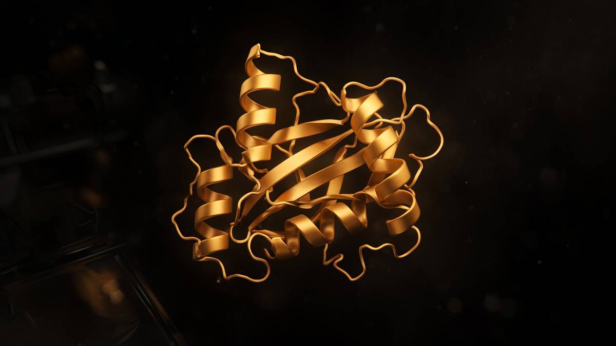
Fifty years of physical chemistry, then a neural net. Hassabis, Jumper and Baker shared the 2024 chemistry prize. Every peptide in we now has a predicted structure you can actually look at.
What this essay actually tells you
- AlphaFold 2 (Jumper et al., Nature 2021) predicted protein structures from sequence at accuracy that retired a 50-year grand challenge. That's the sentence, and it still sounds like a joke.
- CASP14 was the public exam. The Nobel Prize in Chemistry 2024 went to Hassabis, Jumper and Baker for this class of work. Three names. One retired problem.
- A predicted fold is a hypothesis you can dock a ligand against. It isn't a cryo-EM map. Wet validation still has a job, and anyone skipping it will find out why.
What this actually means
A protein is a string of amino acids that folds into a 3D machine. Predicting that shape from the string used to take a PhD, a synchrotron and luck. In 2020 DeepMind's AlphaFold2 started handing out structures that matched crystals. In 2024 Demis Hassabis and John Jumper shared half a Nobel with David Baker, who designs proteins that never existed. AlphaFold3 now also guesses how proteins hold DNA, RNA and small molecules. The fold is no longer the bottleneck. The experiment is. We say that last bit a lot, because the browser can make you forget it.
Let's start with a tube, because that's where the dare was born. Christian Anfinsen denatured bovine ribonuclease A in urea, reduced the four disulphides, then removed the denaturant and let air reoxidise the thiols. The enzyme came back. Sequence, given a physiological solvent, was enough to find the native fold. That 1960s result, and the 1972 chemistry Nobel that followed it, became a dare the physical chemists spent fifty years failing to cash at scale. A chain of a hundred residues, three or so rotatable states per peptide bond, is on the order of 5 × 10^47 shapes if you count the way Cyrus Levinthal did in 1969. Proteins fold in milliseconds to seconds. A random walk of that space would outlast the universe. Force fields missed. Fragment libraries helped. Homology to a solved cousin was the only method a structural biologist actually trusted on a Tuesday. Then a group at DeepMind that had already solved Go walked into CASP14 with a network trained on the Protein Data Bank and on evolutionary couplings, and the median accuracy crossed the line experimentalists treat as a model they would start from.
In short. A protein’s amino-acid string contains the instructions for its shape. For fifty years, computers couldn't read those instructions well. Then one could.
A protein is a chain of amino acids that folds into a three-dimensional machine. Twenty side chains, a repeating backbone of nitrogen, alpha-carbon and carbonyl, a partial double-bond character that keeps each peptide unit planar. The degrees of freedom that actually search are the backbone dihedrals, phi and psi, plus the chi angles of the side chains. Secondary structure is local: alpha helix, beta strand, turns, the hydrogen-bond patterns Pauling and Corey drew before anyone had a crystal of myoglobin. Tertiary structure is the pack: hydrophobic core, salt bridges, disulphides, the particular 3D machine that a sequence prefers in water. Quaternary structure is how chains sit on one another. That hierarchy is first-year biochemistry. The unsolved bit, for most of a career, was going from a fasta file to the coordinates without a beamline, a freezer of homologues, or a year of crystal trials. AlphaFold 2, Jumper and colleagues, Nature 2021, is the paper that made that jump ordinary. The 2024 chemistry Nobel, shared with David Baker, made it official. We watched that announcement in the lab like it was a football match.
In short. Proteins are chains that fold into helices, sheets and packed cores. The hard problem was guessing that 3D shape from the letters alone.
Why the problem ate a field isn't mysterious. Mechanism lives in geometry: a catalytic triad is three side chains in space, not a catchphrase on a gene card. A disease mutation is often a packing error, a lost salt bridge, a register shift in a strand. A ligand binds a pocket whose shape you can't invent from hydropathy plots. For decades the Protein Data Bank grew by crystallography, then by cryo-electron microscopy, at a rate that left most of UniProt untouched. Membrane proteins lagged. Orphan sequences lagged. Alternative spliced isoforms lagged. Anyone docking a peptide against a receptor was docking against a homology model, a distant cousin, or a wish. The grand challenge, as CASP framed it, was to take a sequence whose structure had been solved but not yet published and predict the coordinates well enough that a sceptical experimentalist would use the model. That's a public exam with a known answer, which is why it could retire a problem rather than a press release. CASP14 was that exam. AlphaFold 2 sat it.
In short. Shape is how enzymes work, how mutations fail, and how drugs sit. Most proteins had no experimental map. That was the bottleneck.
If you're coming to this cold, you need names and numbers rather than a mood about artificial intelligence. Anfinsen, Nobel 1972: sequence determines structure under physiological conditions. Levinthal, 1969: the naive conformational count can't be searched. Moult, Fidelis, Kryshtafovych: CASP, every two years from 1994, a blind test. Senior, DeepMind, Nature 2020: AlphaFold 1, already first at CASP13, not yet the retirement. Jumper, Evans, Pritzel, Green, Figurnov, Ronneberger, Tunyasuvunakool, Bates, Žídek, Potapenko, Bridgland, Meyer, Kohl, Ballard, Cowie, Romera-Paredes, Nikolov, Jain, Adler, Back, Petersen, Reiman, Clancy, Zielinski, Steinegger, Pacholska, Berghammer, Bodenstein, Silver, Vinyals, Senior, Kavukcuoglu, Kohli, Hassabis, Nature 15 July 2021: AlphaFold 2, median GDT-TS 92.4 at CASP14. Tunyasuvunakool, Nature 2021: the human proteome, predicted. Abramson and colleagues, Nature 2024: AlphaFold 3, a diffusion model over atom coordinates, complexes and ligands. Baker, Rosetta, RosettaFold, RFdiffusion: the generative half of the same Nobel. That's the reading list. A predicted fold is a hypothesis you can dock a ligand against. It isn't a cryo-EM map. Wet validation still has a job. What follows is that map.
In short. What follows is the map: why the problem was hard, what the 2021 network actually does, what the colour bar means, and why the wet lab isn't optional.
Sequence was supposed to be enough
Ribonuclease A is a small, tough, disulphide-rich enzyme that was already a reagent when Anfinsen picked it. Eight cysteines, four native pairings. Urea unfolds the chain. A reductant opens the disulphides. In the unfolded soup the cysteines can, in principle, pair 105 ways. Anfinsen’s point wasn't that folding is magic. It was that the native pairing, and the catalytic activity, returned spontaneously once the solvent was again water and the thiols were allowed to find each other, with a bit of help from the protein-disulphide isomerase neighbourhood in the cell and from air in the tube. The thermodynamic hypothesis, as he wrote it, is that the native structure is the free-energy minimum of the chain plus solvent under physiological conditions. No extra genetic instruction is required beyond the sequence. Chaperones, we later learned, keep chains out of trouble and out of aggregation; they don't generally rewrite the native minimum. Anfinsen’s Nobel lecture is still the cleanest statement of the dare. Predict the minimum from the sequence. The next half-century was the bill.
In short. Anfinsen unfolded an enzyme and watched it fold itself again. The shape was already in the sequence. Nobody needed a separate instruction booklet.
Thermodynamics isn't a folding pathway. That distinction ate careers. Levinthal’s argument, often flattened into a paradox, is a counting argument against exhaustive search, not a claim that proteins violate physics. If each residue has even three coarse backbone states, a 100-residue chain has 3^100 conformations, about 5 × 10^47. Sample one per picosecond and you're still looking at times that make the age of the universe look prompt. Proteins don't sample that space. They fold along funnels, as Bryngelson, Onuchic, Wolynes and the energy-landscape school later put it: a biased landscape in which native-like contacts are, on average, lower in energy than non-native ones, so that the chain is guided rather than lost. Dill’s hydrophobic-core picture, Baldwin’s hydrogen-exchange work, Englander’s foldons, the phi-value analysis of Fersht: those are maps of how a particular chain actually travels. They aren't a general algorithm that takes an arbitrary fasta and returns coordinates. The landscape explains why Levinthal’s number is the wrong search. It doesn't compute the fold.
In short. Proteins don't try every shape. They slide down a biased energy landscape. Knowing that doesn't, by itself, give you the coordinates.
Physics-based methods tried to compute it anyway, and they weren't foolish. Molecular dynamics integrates Newton’s laws on atoms with a force field: bonds, angles, torsions, van der Waals, electrostatics, a water model. CHARMM, AMBER, GROMOS, OPLS: named parameter sets, named water models, decades of tuning against small-molecule thermodynamics and against the PDB. On a microsecond, with a specialised machine, a small protein can be seen to fold, as Shaw’s group showed for villin and a handful of others. That's a triumph for those sequences and a warning about the rest. Force fields still mis-rank decoys. Explicit solvent is expensive. A 400-residue multidomain protein, a membrane protein in a lipid belt, a complex with a ligand and a glycan, aren't villin. Replica-exchange, coarse-graining, implicit solvent, and the whole Monte Carlo literature were attempts to buy back time. They produced papers. They didn't produce a method you would use instead of a crystal on a sequence you had never seen. The energy function was never quite right, and the search was never quite cheap, and both sentences were true at once.
In short. Simulating every atom is possible for tiny proteins and still too slow and too approximate for most of the ones you actually care about.
What working groups actually used, when they needed a model on a Tuesday, was homology. If a cousin in the PDB shares 30 percent identity or more, you can align, thread, rebuild loops, and get a backbone that's often right in the core and wrong in the places you cared about. SWISS-MODEL, Modeller, the Phyre2 server: those were the tools. Below 20 percent identity you were in the twilight, and twilight is where most of a novel bacterial genome still lives. Comparative modelling is honest about its premise. It copies. It can't invent a fold the PDB has never seen. That's why CASP split targets into template-based and free-modelling categories, and why the free-modelling category was the public humiliation engine for the field. Very good people spent careers inside that engine. We aren't going to sneer at them. They built the PDB the network later read. Without those structures there's no training set, and without the training set there's no AlphaFold. Interpolation in a space experimentalists spent sixty years filling is still interpolation. It's also, as of 2021, astonishingly good.
In short. If a similar protein had already been solved, you could copy its shape. If it hadn't, you were mostly guessing. That was the daily method.
The native conformation is determined by the totality of interatomic interactions and hence by the amino acid sequence, in a given environment.— Christian B. Anfinsen, Nobel lecture, 1972 — the thermodynamic hypothesis, in one sentence
CASP was the public exam
John Moult’s bet, in 1994, was that a field talking to itself about progress needed a blind test. Crystallographers and NMR groups would solve structures and hold the coordinates until predictors had submitted models of the same sequences. Then the models would be scored. GDT-TS — global distance test, total score — became the currency: the percentage of C-alpha atoms that can be superimposed within a set of distance cut-offs, 1, 2, 4 and 8 Å, a number that sits between 0 and 100. A GDT-TS of 90 is, as a rule of thumb the community actually used, a backbone you could mistake for a low-resolution experimental model. A GDT-TS of 50 is a roughly correct topology with the details wrong. RMSD on the core, lDDT, and a drawer of local scores sat beside it. The point of the machinery wasn't a league table for its own sake. It was to stop a methods paper from scoring itself on the proteins it already knew how to do. CASP is rude in the way a methods section should be rude. You don't get to pick the exam question after you have seen the answer.
In short. Every two years, groups tried to predict shapes that had been solved in secret. A simple score said how close the guess sat to the real coordinates.
Fragment assembly was the best idea the 2000s had, and David Baker’s laboratory at the University of Washington was the place that industrialised it. Rosetta takes short fragments of the PDB that match local sequence, assembles them under a knowledge-based energy function, and clusters. It isn't a first-principles force field. It's a vote of the database, plus a score that likes buried hydrophobics and unclashed side chains. On a good day, for a small globular domain, Rosetta could find a fold. On a typical CASP free-modelling target it couldn't, not at the accuracy an experimentalist would start from. Baker kept at it anyway, because the same machinery that almost predicted natural folds could be run backwards: specify a shape, search for a sequence that would fold into it. That inversion is protein design, and it's the other half of the 2024 Nobel, and it was already working when structure prediction was still a stubborn almost. Top7, Kuhlman, Baker, Science 2003, is a de novo fold that doesn't exist in nature. Prediction and design are cousins. One reads a sequence. The other writes one. Baker was writing while the reading was still hard.
In short. Baker’s software built shapes from short pieces of known proteins, and later designed new proteins. Prediction was still the weaker of those two jobs.
The other pre-AlphaFold idea that actually contained the later trick was coevolution. If two residues sit next to each other in the fold, mutations at one site often get compensated at the other, across a deep alignment of homologues. A statistical coupling, once you have disentangled the transitive ones, is a contact. Marks, Sander, the EVfold work, Morcos, Onuchic, the whole DCA literature: take a multiple-sequence alignment, infer a contact map, fold from the map. When the alignment was deep — thousands of diverse homologues — the contact map was often good enough to see the topology. When the protein was an orphan, the method went quiet. That dependence on evolutionary depth is still inside AlphaFold 2. The network doesn't magic a fold from a single sequence as its preferred mode of action. It reads a stack of related sequences, some of them from metagenomes you would never have aligned by hand, and treats the couplings as a noisy contact map it can refine. An orphan with no homologues is still a harder AlphaFold job than a well-populated Pfam family. The method didn't abolish evolutionary biology. It industrialised it.
In short. If two parts of a protein always mutate together across species, they probably sit together in space. Deep alignments made that signal usable.
CASP13, 2018, was the first time DeepMind showed up as more than a rumour. AlphaFold 1, Senior, Evans, Jumper, Kirkpatrick, Sifre, Green, Qin, Žídek, Nelson, Bridgland, Penedones, Petersen, Simonyan, Crossan, Kohli, Jones, Silver, Kavukcuoglu, Hassabis, Nature January 2020, used coevolved contacts and a neural network to score fragments, and it won the free-modelling category by a margin the field noticed. It didn't retire anything. Median accuracy was better than Rosetta and not yet at the experimentalists’ line. The paper is worth reading as the prototype: DeepMind had decided that protein structure was a problem of the same kind as Go, a large structured space with a public score, and that a group which had already beaten Lee Sedol might have something to say. Hassabis has been blunt that the protein problem was on the list early. Jumper, a physicist who had done protein simulation, joined and rebuilt the architecture. Two years later the rebuild sat CASP14. The jump between those two CASPs is the jump the Nobel citation is about. AlphaFold 1 was a very good methods paper. AlphaFold 2 was a change of era.
In short. DeepMind’s first version already won a 2018 contest. It was better than the rest, and still not good enough that experimentalists would trust it as a starting model.
CASP14 was the public exam AlphaFold 2 sat
CASP14 ran in 2020. Predictors received sequences. Experimentalists held the structures. Scoring was the usual GDT-TS and its cousins. AlphaFold 2’s median GDT-TS across the assessment was 92.4. For a large fraction of domains, including some that had no useful template, the backbone sat within the envelope people describe as experimental quality. The next-best groups, on the hard free-modelling targets, weren't close. That sentence is the one that retired the grand challenge, and it has to be held at the size of a median on a particular exam, not as a claim that every protein is now a solved object. Disordered regions were still disordered. Some domains packed wrongly relative to one another. A few targets were missed. The jump was still of a kind the field hadn't seen in twenty-six years of CASP. Kryshtafovych, Schwede, Topf, Fidelis, Moult, in the assessment papers, didn't reach for understatement. Experimentalists who had spent careers on a single fold looked at models of proteins they hadn't crystallised and recognised the core. That recognition, not a press officer’s adjective, is what “retired” means here.
In short. In 2020 the network sat a blind test and got most backbones right, including proteins with no close cousin in the database. That's the result that ended the old challenge.
Jumper, Hassabis and the DeepMind team published the method on 15 July 2021, Nature 596: 583–589, with a methods dump large enough that other groups could reimplement it. The headline architecture has three parts, and a methods section that can't name them isn't yet talking about AlphaFold 2. First, a multiple-sequence alignment of the query against sequence databases, plus optional templates from the PDB. Second, the Evoformer, a stack of attention blocks that mix information within sequences and within residue pairs, updating a pair representation that's, in effect, a learned contact map with geometry. Third, a structure module that turns that representation into 3D frames: one rigid body per residue, backbone atoms, then side-chain chi angles, recycled so that the network can look at its own output and revise. End-to-end, differentiable, trained on PDB structures with dates that respect CASP cut-offs so the exam stayed blind. The loss includes a frame-aligned point error, a distogram, and a per-residue confidence head that became pLDDT. Those are the named pieces. Attention isn't a synonym for magic. It's a way of letting every residue talk to every other residue, weighted by what the alignment suggests.
In short. The 2021 method lines up related sequences, lets every residue talk to every other residue, and then builds a 3D chain, scoring its own confidence as it goes.
pLDDT is predicted local distance difference test, a number from 0 to 100 on each residue. It's the colour bar on every AlphaFold figure you have seen, blue to orange to red in the public palette, and it's the most abused output of the method. Above 90, the backbone and many side chains are, empirically, as trustworthy as a good experimental model in that neighbourhood. Between 70 and 90 the backbone is usually right and the side chains want a look. Between 50 and 70 you're looking at a possibility. Below 50 the residue is, more often than not, disordered in the cell as well, or at least not a single well-defined conformer in the training distribution. AlphaFold 2 was trained on folded chains. It's allowed to say “I don't know” by painting a tail red, and when it does that it's often telling the truth about intrinsically disordered regions. Treating a red loop as a docked helix is how a first-year computational paper gets written and then retracted by a blot. The colour bar is the result. A ribbon without it's a decoration.
In short. Each residue gets a confidence score from 0 to 100. High means the local shape is trustworthy. Low often means that stretch doesn't have one fixed shape.
The other number that has to sit next to pLDDT is PAE, predicted aligned error: a matrix of how confidently the network places residue i relative to residue j. A domain can be blue all over, locally correct, and still be packed against its neighbour with a yellow PAE, which means the orientation is a guess. Multidomain proteins, linkers, and complexes are where this matrix earns its keep. AlphaFold-Multimer, Evans and colleagues, 2021–2022, extended the same machinery to chains sitting on chains, with ipTM as the interface-confidence cousin of pTM. A confident monomer with an unconfident interface is a pair of folded domains whose docking you haven't yet earned. If you drops a two-chain prediction into a grant figure without the PAE plot hasn't finished the experiment. The network will always give you coordinates. Coordinates aren't a result. Confidence-conditioned coordinates are the result, and even those are a hypothesis, which is a sentence we'll keep paying until the close.
In short. A second plot says whether two parts of the protein are confidently placed relative to each other. A confident piece can still be attached at a guessed angle.
Diagram
- Amino acid~110 DaTwenty side chains. The alphabet.
- Peptide bondamide, planarCarboxyl carbon to the next nitrogen. Resonance holds it flat.
- Oligopeptide< ~20 residuesMost hormones and fragments. GHK is three. KPV is three.
- Polypeptide20–50+Insulin 51. GLP-1 31. Retatrutide is a designed chain in this band.
- Proteinfolded machineHaemoglobin, a GPCR, lysyl oxidase. Tertiary structure worth drawing.
Insulin (Banting and Best, 1921) was the first peptide anyone bothered calling a medicine. A collagen hydrolysate is food. A named sequence with a mass and a chromatogram is a research peptide. The shared word is the accident.
- Levinthal 100-mer
- ~5 × 10⁴⁷ shapes
- CASP14 median GDT-TS
- 92.4
- pLDDT
- 0–100 per residue
- PDB in 2021
- ~180,000 experimental
- AlphaFold DB
- >200 million
- AlphaFold 2 paper
- Nature, 15 July 2021
- Nobel Chemistry
- 9 October 2024
- AlphaFold 3
- Nature 2024
Three coarse states per residue, 1969 counting. Proteins do not walk that space. They fold in milliseconds to seconds.
AlphaFold 2, 2020 exam. GDT-TS of 90 is the rule-of-thumb experimental-quality backbone.
≥90 high; 70–90 backbone usually right; <50 often disordered. The colour bar is the result.
The training library. No PDB, no AlphaFold. Interpolation, astonishingly good.
UniProt-scale predictions, EMBL-EBI. Human proteome first, Tunyasuvunakool, Nature 2021.
Jumper et al. 596: 583–589. MSA, Evoformer, structure module, pLDDT.
Hassabis and Jumper, prediction. Baker, design. One prize, two jobs.
Abramson et al. Diffusion over atoms. Complexes, ligands, nucleic acids, PTMs.
What AlphaFold 2 actually is
The multiple-sequence alignment is the dataset the network was born wanting. JackHMMER and HHblits against UniRef, BFD, MGnify: named tools, named metagenomic piles, a stack that can be thousands of sequences deep for a popular domain and nearly empty for a recent orphan. Each column of the alignment is a residue; each row is a homologue. Couplings live in that matrix. AlphaFold 2 doesn't throw the alignment away and look only at the query. It embeds the stack, lets attention run along sequences and across residue pairs, and builds a pair representation that has learned, from the PDB, what a contact looks like when the statistics are noisy. Templates from the PDB, if they exist, are an extra hint, not a requirement; the CASP14 result that mattered was the free-modelling one. ColabFold, Mirdita, Schütze, Moriwaki, Heo, Ovchinnikov, Steinegger, Nature Methods 2022, replaced the slow DeepMind MSA stack with MMseqs2 and put the whole inference on a notebook that a graduate student could run on a Friday. That's how the method left the company and entered the lab. A method that only runs in Mountain View is a demonstration. A method that runs in Colab is a reagent.
In short. The network reads a stack of related sequences from many species, not just the one you typed. A public notebook put that job on an ordinary computer.
The Evoformer is the named block, forty-eight layers in the published configuration, and it's where the pair representation gets geometry. Triangle multiplicative updates and triangle attention are the bits the paper had to invent: if residues i and j are close and j and k are close, i and k shouldn't be far, and a network that doesn't know that will draw impossible maps. The triangle is the transitivity of Euclidean space, written as a layer. Sequence attention and pair attention talk to each other, so a coupling that looks like a contact can be confirmed or killed by the rest of the chain. This is still not physics. It's a learned surrogate of physics plus evolution, trained on the folds experimentalists had already deposited. The honesty of that sentence is why a genuinely new fold, a designed protein far from the PDB, or a chain whose homologues are all disordered, is a different request. Baker’s generative models later filled some of that gap from the other direction. AlphaFold 2, as published, is a reader of the natural distribution. It's a very good reader.
In short. The core network enforces the simple fact that space is three-dimensional: if A sits near B and B near C, A can't sit far from C.
The structure module is the bit that finally emits coordinates. Each residue is a rigid frame, an origin and a rotation, from which the backbone N, C-alpha, C and O are written by chemistry rather than by a free atom soup. Side-chain chi angles are predicted on that backbone. Invariant point attention lets the module look at other residues in a way that doesn't depend on how you rotated the whole protein in the box, which is a computer-vision idea Jumper’s group made structural. Recycling — typically three iterations in the published inference — feeds the current coordinates back into the pair representation so the network can notice a clash or a register error and revise. That loop is why a first pass can look merely good and a recycled pass look finished. It's also why a bad MSA can still be polished into a confident-looking wrong answer. Garbage in, blue ribbon out, is a failure mode. Look at the alignment depth. Look at the PAE. Then look at the ribbon.
In short. The last stage places each amino acid as a small rigid body, builds the backbone from chemistry, and revises the shape a few times before it stops.
Open-sourcing the weights and the inference code is the industrial half of the 2021 paper, and it's the half a methods section should thank out loud. DeepMind released AlphaFold 2 under a licence a lab could run. EMBL-EBI, with DeepMind, stood up the AlphaFold Protein Structure Database: first the human proteome and twenty-one model organisms, Tunyasuvunakool, Nature 2021, then a 2022 drop that covered more than 200 million UniProt entries, essentially a predicted fold for every sequence the databases then treated as a protein. Varadi, Velankar and colleagues documented the resource in Nucleic Acids Research. A graduate student can now type a UniProt accession and download a coordinate file with a pLDDT column. That sentence was science fiction in 2019. It's a URL in 2026. The PDB didn't shrink. Experimental depositions continued, and AlphaFold models are now themselves used as molecular-replacement search models to phase new crystals, which is a pleasing recursion: the network trained on the PDB now helps the PDB grow. We still deposit the map. We just start closer to a fold that's probably right.
In short. The weights were published. A public database then offered predicted shapes for more than two hundred million proteins, which is most of the ones with a name.
Cousins arrived immediately, which is how you know a method is real. Baek, DiMaio, Anishchenko, Dauparas, Ovchinnikov, Lee, Grishin, Baker, Science 2021: RoseTTAFold, a three-track network from the Baker laboratory, less accurate than AlphaFold 2 on CASP14 and independently invented, and the seed of the generative tools that followed. OpenFold, Ahdritz, O’Donnell, AlQuraishi and colleagues, reimplemented AlphaFold 2 in PyTorch so the training, not only the inference, could be inspected. ESMFold, Lin, Akin, Rao, Hie, Zhu, Lu, dos Santos Costa, Fazel-Zarandi, Sercu, Rives, Science 2023, skipped the MSA and read a single sequence with a language model trained on UniRef, faster, shallower on orphans in the other direction, a different trade. None of those papers undid Jumper. All of them are what a field looks like when a bottleneck has moved. The scarce step is no longer a backbone for a globular domain with homologues. The scarce step is the state you didn't train on, the ligand pose, the membrane, the alternative conformer, and the experiment that still has to say yes.
In short. Other groups rebuilt the idea, sometimes without the sequence stack, sometimes as a designer rather than a predictor. The backbone of an ordinary folded protein is no longer the scarce step.
Confidence is a colour bar, not a certificate
Intrinsically disordered regions are the first place a confident-looking movie goes wrong. A large fraction of eukaryotic proteins carry stretches that don't adopt a single folded structure in isolation: linkers, activation loops, tails that only fold on a partner, low-complexity domains that phase-separate. AlphaFold 2 paints many of those stretches red, pLDDT below 50, and that paint is often the correct biological statement. The network was trained on the PDB, which is a museum of folds that could be crystallised or stuffed into a cryo-EM particle. Disorder doesn't live there except as missing density. When the model hands you a long red tail, the honest move is to treat it as disordered, or as a region that will only become a helix in a complex you haven't specified. The dishonest move is to let a graphics program ribbon it and then dock a ligand into the ribbon. Construct design is the practical gift: drop the red tails from the expression construct, keep the blue core, and your crystallisation trial just got cleaner. That's wet validation using the colour bar as a filter, which is the right use.
In short. Stretchy, floppy parts of proteins often get a low score, and that's frequently correct. Don't pretend a floppy tail is a neat helix just because the software drew one.
Alternative states are the second abuse. A GPCR has inactive and active-like ensembles. A kinase has DFG-in and DFG-out. A transporter cycles. AlphaFold 2, given a sequence, emits one structure, typically a state over-represented in the PDB for that family, which is a point estimate rather than a molecular-dynamics trajectory or a Boltzmann ensemble. Methods that condition on a state — AlphaFold with templates from an active-like cousin, or later specialised tools — can bias the output, and they're still sampling a point, not a landscape. If you write “the AlphaFold structure of the receptor” as if there were one has skipped a two-state enzyme. The 2021 paper didn't claim dynamics. The coverage did. A predicted fold is a hypothesis about a populated conformer. If your biology lives in the other conformer, you have the wrong hypothesis, with excellent local geometry. That's a more dangerous error than a messy homology model, because the ribbon looks finished. Finished-looking and true are different adjectives. Keep them apart.
In short. Many proteins adopt more than one shape. The network usually returns one, often the shape most common in the training set. The other shape may be the one your experiment needs.
Orphans, antibodies, and designed sequences are the third. An orphan with a thin alignment has less coevolutionary signal; pLDDT falls, PAE worsens, and you should believe the fall. Antibodies have a conserved framework and hypervariable loops that don't coevolve like a globular enzyme; CDR H3 remains a known failure mode, better in later tools, not a solved object in 2021. Sequences far from natural proteins — a de novo design, a heavily mutated enzyme, a concatenation that never saw a ribosome — are out of the training distribution. Baker’s networks are the ones you ask about those, and even those you still crystallise, or you still put on a cryo-EM grid, if the claim matters. AlphaFold 2 is a reader of natural globular proteins with homologues. That's a vast and important class. It isn't the set of all polymers of amino acids. A methods section that can't say which class it's in has borrowed a ribbon from the wrong paper.
In short. Proteins with few relatives, antibody loops, and wholly invented chains are harder. A confident-looking picture of those is the moment to get suspicious.
What the colour bar won't tell you is whether a side chain that matters for catalysis is rotameric, whether a predicted pocket will bind your ligand, or whether a phosphorylation will flip a loop. Those are experimental questions with experimental machines: a crystal, a cryo-EM reconstruction, an NMR ensemble, a hydrogen-deuterium exchange time course, a chemical cross-link, a mutational scan, a thermal shift, an activity assay. Molecular replacement against an AlphaFold model has become a standard way to phase a crystal, which is the network paying the PDB back. It isn't a reason to skip the crystal if the claim is a ligand pose or a catalytic geometry. The 2021 paper is scrupulous about this. The browsers that followed were less so. We'll stay with the paper. High pLDDT means “this local geometry is probably right.” It doesn't mean “this protein is understood.” Understanding is a blot, a map, a rate, and a sentence you can defend in front of a colleague who doesn't like you.
In short. A high score says the local shape is probably right. It doesn't say the protein is understood, and it doesn't place a drug in the pocket.
Diagram
Closed chromatin (H3K27me3, DNA methylation) hides the promoter. Pioneer factors and histone acetyltransferases open it.
PIC: TFIID, TFIIH, Mediator, Pol II. Ser5 phosphorylation of the CTD lets the polymerase leave the promoter.
Elongation ~20–40 nt/s. Capping, splicing, cleavage and polyadenylation happen on the still-growing RNA.
Human genes are islands in 3.1 billion base pairs of mostly noncoding sequence. Promoter, enhancers, chromatin state and the Mediator complex decide whether Pol II is allowed to fire. Epithalon’s literature sits on TERT and pineal clocks — two of the rare promoters anyone bothers to name in a peptide essay.
AlphaFold 3, Baker, and the 2024 prize
Abramson, Adler, Dunger, Evans, Green, Pritzel, Ronneberger, Willmore, Ballard, Bambrick, Bodenstein, Evans, Hung, O’Neill, Reiman, Tunyasuvunakool, Wu, Žemgulytė, Arvaniti, Beattie, Bertolli, Bridgland, Cherepanov, Congreve, Cowen-Rivers, Cowie, Figurnov, Fuchs, Gladman, Jain, Khan, Low, Perlin, Potapenko, Savy, Singh, Stecula, Thillaisundaram, Tong, Yakneen, Zhong, Zielinski, Žídek, Bapst, Kohli, Jaderberg, Hassabis, Jumper, Nature 2024, is AlphaFold 3. The architecture changed. Pairformer plus a diffusion module over atom coordinates, not a residue-frame structure module. The object changed with it. AlphaFold 3 is asked to place proteins against proteins, proteins against DNA and RNA, proteins against small-molecule ligands, ions, and some post-translational modifications, in one forward pass. That's a different dare from CASP14. CASP14 was a monomer or a domain. The 2024 paper is a complex. PoseBusters and a set of ligand benchmarks are the scoring language, and they're already being argued with, which is how you know the claim is in the adult literature rather than in a keynote. Isomorphic Labs, the DeepMind spin-out, is the industrial reading. The scientific reading is that the atom, not the residue frame, became the unit, so that a ligand could sit in the same object as a side chain.
In short. The 2024 version places proteins against other proteins, DNA, RNA and small molecules in one go. It works at the level of atoms, not just amino-acid frames.
A ligand pose from a network is still a pose: a hypothesis about where a compound might sit, without a co-crystal, without density for the compound, and without a binding constant attached. AlphaFold 3 improved protein–ligand geometry relative to the docking-into-an-apo-pocket workflow many of us grew up with, and it still hallucinates, still misplaces waters, still gets a tautomer wrong, still can't see a covalent warhead as chemistry. The honest Tuesday use is to generate a hypothesis you'll dock, then score, then soak, then collect on. The dishonest Tuesday use is to publish a figure of a drug in a pocket and call it a structure. Wet validation still has a job, and the job has a name: the experiment that measures the pose, the affinity, or the activity. Protein–nucleic-acid complexes are the same sentence with a different polymer. AlphaFold 3 can draw a plausible DNA-binding interface. A footprinting experiment, a mutation in the recognition helix, or a map is how you find out whether it drew the right one. We'll take the plausible drawing. We won't skip the footprint.
In short. A predicted pose for a drug is a guess you can test. It isn't the same object as a crystal of the protein holding that drug.
David Baker’s half of the 2024 chemistry Nobel is the generative inverse of prediction, and it wasn't a consolation prize. The Institute for Protein Design in Seattle spent thirty years writing sequences that would fold into shapes nature hadn't bothered to invent. Rosetta was the energy function and the search. Top7 was the existence proof. Repeat proteins, designed enzymes, designed cages, designed transmembrane barrels: a literature, not a stunt. RoseTTAFold, 2021, was their reader. RFdiffusion, Watson, Juergens, Bennett, Trippe, Yim, Eisenach, Ahern, Borst, Ragotte, Milles, Wicky, Courbet, Feldman, Kollman, Baker, Nature 2023, is a diffusion model that hallucinates backbones conditioned on a motif, a pocket, a symmetry. ProteinMPNN writes a sequence onto that backbone. Binders to influenza haemagglutinin, to PD-1, to rattlesnake toxin, arrived as designed proteins you could express and, in the papers that mattered, crystallise or put on a grid. Hassabis and Jumper predicted what evolution already wrote. Baker wrote new ones. The Nobel committee split the prize along that axis and, for once, a split prize was the right taxonomy.
In short. Baker’s prize was for inventing proteins that never evolved, including binders and enzymes. Predicting a natural fold and writing a new one are different jobs.
The announcement came on 9 October 2024. Demis Hassabis and John Jumper, DeepMind, London, shared one half for protein structure prediction. David Baker, University of Washington, shared the other half for computational protein design. The committee’s line was that they cracked the code for proteins’ amazing structures. Codes, in chemistry, are usually a bit less cracked than the citation. The working version is that a 50-year grand challenge in predicting the native coordinates of a natural globular protein, as CASP defined the challenge, is over as a bottleneck, and that designing a binder is now a computation you follow with an expression trial rather than a computation you follow with a shrug. The microscope still has a queue. The blot still has a job. Both halves of the prize changed what you do on the Tuesday before you book either. We've sat with that Tuesday. It's better. It's still a Tuesday in a lab.
In short. The 2024 chemistry Nobel went to the two DeepMind scientists who predicted folds and to Baker who designed new ones. The microscope still has a queue.
They cracked the code for proteins’ amazing structures.— Nobel Committee for Chemistry, 2024, on Hassabis and Jumper — the citation, at the size of a citation
A predicted fold is a hypothesis you can dock against
A cryo-EM map is a density. Particles on a grid, electrons, a reconstruction, a resolution in ångströms that you have to argue about, a model built into that density and deposited with the map so that anyone can see where the model is a guess. A crystal structure is a set of structure factors, phases you earned by experiment or by molecular replacement, a model refined against amplitudes, a PDB file with occupancies and B-factors. An AlphaFold coordinate file is a hypothesis sampled from a trained distribution, with a confidence tag on each residue. Those three objects share a graphics program. They don't share an epistemology. Docking a ligand against an AlphaFold pocket is a perfectly reasonable experiment, the way docking against a homology model was a perfectly reasonable experiment: you're asking whether a shape that's probably right can host a compound you can then test. You aren't reporting a structure of the complex. The coverage blurs those sentences because the ribbon is pretty. Pretty isn't a map. We'll keep the map, the crystal, and the hypothesis in separate clauses until the close, because that's the whole remaining adult problem.
In short. A microscope map, a crystal model, and a predicted set of coordinates look similar on a screen. They aren't the same kind of evidence.
Wet validation has a named kit, and the kit didn't go into storage in 2021. X-ray crystallography still solves ligands, still catches covalent adducts, still sees waters the network invents. Cryo-EM, Henderson, Frank, Nogales, and a decade of cheap detectors, still solves the complexes that won't crystallise, membrane proteins in nanodiscs, large assemblies. NMR still sees dynamics and disordered states the PDB under-samples. Hydrogen-deuterium exchange mass spectrometry still reports which peptides are protected. Chemical crosslinking and XL-MS still report which lysines sat near which lysines in solution. SAXS still reports a shape in solution that a single crystal might have compressed. Deep mutational scanning still reports which residues the function actually needed. Thermal shift, SPR, ITC, a functional assay: those report binding and activity, which a ribbon never will. AlphaFold changed the order of operations. You start with a model. You still end with one of those machines if the claim is going to leave the computer. If you write the remaining work as a rounding error has skipped a ligand that refused to soak.
In short. Crystals, electron-microscope maps, magnetic-resonance ensembles and binding assays still decide whether a prediction was true. The network changed the starting point, not the finish.
Molecular replacement is the quiet revolution inside the crystallography core, and it's the one we actually like. Phaser, MOLREP, a search model, a rotation and translation that phases the dataset. For twenty years the search model had to be a homologue. Now it's often an AlphaFold monomer, sometimes a trimmed one with the red tails cut off, and a structure that would have sat unsolved in a drawer phases on a Thursday. That's the network as a reagent in a pipeline that still ends in a deposited map. Cryo-EM model building has the same flavour: start with the prediction, rigid-body fit into density, rebuild where the density disagrees, and the disagreement is the result. A prediction that fights the density is a wrong prediction, and you keep the density. A prediction that fits is a hypothesis that just got promoted. Promotion is earned. It isn't the default. We'd rather have that discipline than a browser that never loses.
In short. Crystallographers now often start from a predicted model to solve the first phases. They still finish by fitting that model to measured data.
Failure modes, listed, because a methods section that only celebrates GDT-TS is a brochure. Thin MSAs. Multidomain packing. Alternative conformers. Ligand-induced rearrangements the apo training set never saw. Prosthetic groups, glycans, lipids in a belt. Coiled coils that look right locally and wrong in register. Homooligomers with the wrong copy number. Antibodies, as already named. Proteins that only fold on DNA. Proteins that only fold in a condensate. A confidence score is a correlation with correctness on the distribution the network was trained on. It isn't a guarantee on yours. The adult move is to treat every downstream claim — this pocket binds, this interface is the interface, this loop is a helix in the complex — as a claim that still needs a measurement matched to it. Dock, then soak. Predict an interface, then cross-link. Drop a tail, then crystallise. The hypothesis is cheap. The measurement is the job. That pairing is the remaining grand challenge, and it isn't a neural-network problem. It's a laboratory one.
In short. Wrong alignments, wrong packing of domains, missing partners, and shapes that only appear when a ligand binds are still ordinary ways to be confidently wrong.
Membrane proteins are the special case a peptide-receptor essay actually lives in. A GPCR is a seven-helix bundle in a lipid bilayer, glycosylated, often bound to a G protein, with loops that only become ordered on a ligand. The PDB of 2021 already held a GPCR boom — inactive inverse-agonist crystals, then active-like cryo-EM with Gs — but the majority of human membrane proteins were still a hydropathy plot plus a wish. AlphaFold 2 produces plausible helical bundles for many of them, and the bundles are often right in the core and wrong in the loops you wanted to dock against. Lipid, if it's there at all, is implied rather than placed. A predicted 7TM fold is a starting model for a construct, a molecular-replacement search, or a docking hypothesis, drawn as if the bilayer were optional. Nanodiscs, SMALPs, a detergent screen, and a grid remain how you find out whether the loop that binds your peptide is the loop the network drew. Class-B receptors, the incretin family this catalogue actually touches, were lucky: experimental maps arrived in the same decade as the network. Most of the membrane proteome wasn't that lucky.
In short. Membrane proteins fold in fat, not in water. The network often gets the helix bundle and misses the loops, the lipids, and the partner that orders them.
Diagram
- 0.1 nmHydrogen atomA proton and an electron. Chemistry starts here.
- 0.3 nmWater molecule70% of a cell by mass. The solvent life is.
- 1 nmAmino acidTwenty kinds. Peptide bonds string them.
- 2–4 nmResearch peptideA named chain. BPC-157 is 1.4 kDa, 15 residues.
- 4–10 nmGlobular proteinHaemoglobin, a GPCR’s extracellular face.
- 25 nmRibosomeThe factory that reads mRNA into protein.
- 5 nmMembraneA lipid bilayer. Every compartment starts here.
- 0.5–1 µmMitochondrionA bacterium the cell swallowed and kept.
- 6–10 µmNucleusTwo metres of DNA folded into a sphere.
- 10–30 µmTypical cellA city. 10¹⁰ proteins. One genome.
- 1 mmTissue grainA thousand cells talking across ECM.
- 1.7 mYou~36 trillion human cells. Most of them are red blood cells.
Lengths are characteristic, not exact. A research peptide is closer in size to a water molecule than to the cell that assays it — which is why a 15-mer can occupy a receptor pocket a small-molecule drug also wants.
What you can do on a Tuesday now
Paste a sequence. Get a fold. Get a confidence map. That's the Tuesday, and it's new. You can then dock a ligand if AlphaFold 3 will have you, or if a classical docking package will take the pocket. You can design a binder with RFdiffusion and ProteinMPNN if you're hungrier. You can trim a construct, drop the red termini, and order a synthetic gene that's more likely to express a crystallisable core. You can look at a mutation from a patient, see whether it sits in a packed core or on a blue surface, and decide whether a stability assay is worth the protein prep. You can take a peptide ligand you actually have on the bench and ask where, on a now-public receptor model, it might sit, then go and measure that. None of those sentences is a structure. All of them are better Tuesdays than 2019, when the same questions started with a BLAST against the PDB and a shrug when the cousin was 18 percent identical. The fold is no longer the bottleneck. The experiment is. We say that last bit a lot, because the browser can make you forget it.
In short. You can now paste a sequence on a Tuesday and get a starting shape the same morning. The experiment that tests the shape is still the slow step.
Class-B GPCRs are the reason this page sits next to a vial, and the honesty has to stay intact. GLP-1 receptor, GIP receptor, glucagon receptor — the three receptors retatrutide occupies — are no longer mysterious ribbons. They have experimental cryo-EM complexes from the 2017–2023 GPCR boom, active-like, peptide-bound, Gs-coupled, deposited maps you can actually open. AlphaFold didn't uniquely reveal them. What AlphaFold did was make the long tail of the rest of the proteome a public object, fill loops the maps left noisy, and make an apo or alternative-state hypothesis cheap enough to dock against before you book time on a microscope. A house that stocks the published LY3437943 triple agonist is stocking a ligand whose targets now have both experimental maps and high-quality predicted coordinates. That isn't a marketing line. It's a description of what 2024 did to receptor biology: the receptor is no longer the scarce structural object. The pose of a particular analogue in a particular state, in a particular membrane, still is. We won't write those two sentences as one.
In short. The three gut-hormone receptors one catalogue peptide occupies already have microscope maps. Predicted structures made everything around them easier to look at.
The rest of the peptide neighbourhood folds at different difficulties, which is why neighbourhood isn't identity. Somatropin is 191 residues of growth hormone, a four-helix bundle the PDB has held for decades; AlphaFold agrees, as it should. IGF-1 LR3 is a small, disulphide-rich hormone with a long-known fold; the prediction is a convenience, not a discovery. GHK is a tripeptide. It doesn't have a fold in the Anfinsen sense. It has a copper and a preferred coordination, and a predicted protein receptor for it's a different object from the ligand. BPC-157 is a pentadecapeptide; any “structure” of it in water is an ensemble, not a ribbon you should trust at pLDDT 95. The catalogue is full of ligands. Some of them are long enough to be domains. Some of them are motifs. AlphaFold made the fold of the targets a public object. It didn't turn a 15-mer into a globular protein, and it didn't turn a copper-binding tripeptide into a structure-based drug-design campaign. A reading list can sit them together. A methods section can't.
In short. Some catalogue peptides are small folded hormones. Some are short motifs with no single shape. Predicted structures help most at the receptor, not at a three-residue ligand.
What you shouldn't do with a prediction is the shorter list, and it's the one a sceptical colleague will hold you to. Don't report a ligand pose as a co-crystal. Don't silently omit the pLDDT colouring in a figure that will be screenshotted. Don't treat a yellow PAE as a solved domain orientation. Don't design a primer around a red loop and then act surprised when the loop is a protease snack. Don't take an AlphaFold 3 complex of a peptide and a GPCR, in vacuo, without lipid, without glycan, without the G protein, and call it the bound state of retatrutide. Don't skip the activity assay. The sci-fi is that the hypothesis used to take a beamline and now takes a browser. The true sentence is that the hypothesis is cheap and the measurement is still the measurement. We try not to get lazy about the experiment that still has to follow. Laziness is the failure mode of a solved bottleneck. The field is already producing it. We'd rather not.
In short. Don't dress a prediction up as a finished experimental structure, and don't hide the confidence colouring. The cheap guess still has to be measured.
Diagram
| Node | Catalogue | Conversation |
|---|---|---|
| GPCR | Ipamorelin, MT2, PT-141, retatrutide, CJC | Second messengers, secretion, appetite, pigment |
| RTK / IGF1R | IGF-1 LR3 | IRS–PI3K–Akt–mTOR and Shc–ERK |
| Cytokine receptor | Somatropin (HGH) | GHR–JAK2–STAT5b, hepatic IGF-1 |
| Cofactor | NAD+ | Sirtuins, PARPs, CD38, redox |
| Actin buffer | TB-500 / Tβ4 motif | G-actin sequestration, motility |
| Growth-factor-like | BPC-157 | VEGFR2 / FAK / eNOS neighbourhood |
| Copper ligand | GHK-Cu | Transcriptome shift in fibroblasts |
| MC fragment | KPV | NF-κB, PepT1, no pigment |
| Nuclear / pineal | Epithalon (AEDG) | TERT and melatonin literatures |
| mtORF peptide | MOTS-c | AMPK, folate–methionine cycle |
Each row is a different kind of molecular conversation. The catalogue peptides bind at these nodes; they are not interchangeable, and stacking them because a forum did mixes unrelated literatures.
How to use a prediction without lying to yourself
Always look at the colour bar before you look at the ribbon. That's the whole methods-line, and everything else is a corollary. Download the mmCIF or the PDB, load pLDDT as B-factor, colour it, and ask which stretches you're about to believe. Then open the PAE plot and ask which domains you're about to treat as a single rigid body. Then look at the MSA depth, or at the AlphaFold DB page that reports it, and ask whether this protein was a well-populated family or an orphan. Then ask whether a structure of a close homologue already exists in the PDB, because if it does you should be looking at that map as well, not instead. Then, if the claim is a pocket, decide in advance what measurement will count as a yes: a soak, a shift, a Kd, a mutant that kills the function. A prediction without a pre-registered measurement is a picture. Pictures are allowed. They aren't results. The machines that make them results have names, and the names are in the previous heading.
In short. Colour the model by confidence, check how the parts sit relative to each other, and decide beforehand what experiment will count as a yes.
Construct design is the place the colour bar has already earned its keep in wet labs that will never dock a drug. Cut the low-pLDDT termini. Keep a domain boundary that the PAE says is a domain. Move a purification tag to the end the model says is disordered. That isn't structure determination. That's using a hypothesis as a filter on a cloning plan, and it's why expression cores that used to take three constructs now often take one. Molecular replacement, already named, is the crystallographic version of the same filter. Interface prediction with AlphaFold-Multimer or AlphaFold 3, followed by a single cysteine pair for a disulphide lock or a cross-link, is the biochemical version. In each case the network proposes, the bench disposes. A lab that has started to skip the disposal step is a lab that will spend 2027 retracting a pocket. We'd rather spend 2027 collecting on one.
In short. The practical gift is often dull: trim floppy ends, pick domain boundaries, and clone a piece that's more likely to behave. That's already worth the tool.
Comparisons, named, because “we ran AlphaFold” isn't a methods line. If an experimental structure exists, superpose, report RMSD on the core, and point at the loops that disagree; those loops are either a state change or a wrong prediction, and you don't yet know which. If you used AlphaFold 3 on a ligand, run a classical docking package on the same pocket and say whether they agreed. If you used a predicted interface, say the ipTM, show the PAE, and name the residues you'll mutate. If you used a design from RFdiffusion, say that it's a design, express it, and measure binding; the 2023 papers did. Software versions, database dates, and whether templates were allowed belong in the methods the way a lot number belongs on a vial. A prediction from 2021 weights and a prediction from 2024 weights are different objects. Write which. You'll be asked. The answer should be in the paper, not in an email after review.
In short. Write the software version, show how the prediction compares with any existing experimental structure, and name the residues you'll actually test.
- Colour by pLDDT before you trust a ribbon. Below 50 is often disorder. Do not dock into it.
- Open the PAE. A blue domain with a yellow packing is two hypotheses, not one protein.
- Name the MSA depth, the software version, and whether templates were on.
- If a crystal or a cryo-EM map exists, superpose and report the disagreement. Keep the map.
- A ligand pose is a hypothesis. Soak, shift, mutate, or measure a Kd. The pose is not the Kd.
- Trim red termini from constructs. That is a cloning decision the colour bar is allowed to make.
Close: the fold is cheap; the experiment is not
The node is conserved, which is the only reason a 1960s ribonuclease tube, a 1969 counting argument, a 1994 blind exam, a 2021 attention architecture and a 2024 diffusion model can sit in one essay without being a collage. Peptide bonds join amino acids. The chain finds a minimum in water. Evolution writes couplings into alignments. Experimentalists write folds into the PDB. A network trained on those two libraries now emits coordinates with a colour bar. You can walk this argument from Anfinsen’s urea to a UniProt accession and the peptide unit will still be the chemistry. Conservation isn't a licence to treat a blue ribbon as a co-crystal. It's a licence to take the geometry seriously enough to measure it, in the protein you have, with the machine the claim requires. The popular story got loud because the node is central. The work got easier at the start and not at the end, for the same reason. A solved bottleneck moves the queue. It doesn't close the laboratory.
In short. From a 1960s refolding tube to a 2021 network, the chemistry is still a chain of amino acids in water. The guess is now good. The measurement is still the measurement.
The public papers are the reading list, and they're short enough to actually read. Anfinsen, Science 1973, the thermodynamic hypothesis. Levinthal, 1969, the counting argument. Moult, Proteins 1995, the exam. Senior, Nature 2020, AlphaFold 1. Jumper, Nature 2021, AlphaFold 2, the document this page is for. Tunyasuvunakool, Nature 2021, the human proteome. Mirdita, Nature Methods 2022, ColabFold. Baek, Science 2021, RoseTTAFold. Lin, Science 2023, ESMFold. Abramson, Nature 2024, AlphaFold 3. Watson, Nature 2023, RFdiffusion. Kuhlman, Baker, Science 2003, Top7, so the design half has an origin story older than the network. The Nobel citation, 9 October 2024, Hassabis, Jumper, Baker. That's a fortnight of evenings, not a guru. The solved-biology headlines will still be there when you come back, and they will look smaller. They should. A median GDT-TS of 92.4 is a stunning number. It doesn't place a ligand, map a folding pathway, or spare you the blot.
In short. A short stack of named papers covers the dare, the exam, the 2021 method, the 2024 complexes, and the design tools. Read those before any headline.
Here's the map we'd like you to take home, rather than a catchphrase. Anfinsen was right: sequence determines structure, under conditions. Levinthal was right: you can't search the naive count. CASP was the public exam. AlphaFold 2, Jumper et al., Nature 2021, sat CASP14 at a median GDT-TS of 92.4 and retired the 50-year grand challenge as CASP had defined it. The method is an MSA, an Evoformer, a structure module, and a colour bar called pLDDT. AlphaFold 3, Abramson et al., Nature 2024, extends the object to complexes, nucleic acids and ligands, as a diffusion over atoms. Baker’s Rosetta, RoseTTAFold and RFdiffusion write sequences that never evolved. A predicted fold is a hypothesis you can dock a ligand against. It isn't a cryo-EM map. Wet validation still has a job. The fold of a natural globular protein with homologues is no longer the scarce step. The state, the pose, the dynamics and the measurement are. If your experiment needs a starting model, take the blue core and show the PAE. If it needs a pose, measure it. If it needs a medicine, this catalogue doesn't sell one.
In short. Leave with the map: sequence, exam, 2021 network, colour bar, 2024 complexes, Baker’s designs. A predicted shape is a hypothesis. The wet lab still has a job.
We'd rather have the caveats and the working tool. The caveats are disorder, alternative states, thin alignments, ligand poses that are still poses, and a coverage that will keep saying solved when the right word is hypothesised. The working tool is a network that took a 50-year physical-chemistry problem and made a high-quality first model ordinary, then a database of more than 200 million of those models, then a 2024 extension into complexes, then a Nobel that also recognised the man who had been building proteins while the rest of us were still failing to predict them. The title of this page spent a solved on purpose, because CASP14 earned it for the problem CASP was invented to score. The word is hereby scoped. What remains is a fasta file, a colour bar, a pocket, a ligand you might actually have in a vial, and an experiment. Use them in that order. Read Jumper before the headline. Read the PAE before the ribbon. Read a density, if you can get one, before the claim.
In short. The old challenge is over as a bottleneck. The caveats are real. The tool works. A pretty ribbon is still the start of an experiment, not the end of one.
Research-use-only, for the peptides this journal sits next to. The lyophilised chains on those listings are laboratory reagents: HPLC-characterised sequences for a tube, a receptor assay, a blot you actually run. They aren't a deposited structure, a licensed medicine, or a substitute for the measurement the colour bar is asking you to go and do. Retatrutide, where it appears in this neighbourhood, is a published triple-agonist sequence whose three class-B targets now have maps and models; the vial is still the ligand, labelled for in-vitro work. GHK-Cu, IGF-1 LR3, somatropin: same legal class, different folds, different questions. The physiology in the paragraphs above is public, cited, and older than the browser. Use the coordinates to design the experiment you have the controls for, with the confidence named, the state named, and the machine written down. We'll sell you the chain. We won't tell you the ribbon is the result.
In short. The peptides next to this page are laboratory chemicals, not medicines and not structures. Use the predicted shape to plan the measurement. Then make the measurement.
Questions the essay actually answers
- Did AI replace crystallography?
- No. It made a high-quality first model ordinary. Hard structures, ligands and dynamics still need experiments. The queue at the synchrotron got more interesting, not shorter, which is the version of 'replaced' we'll accept.
- What did David Baker win for?
- Computational protein design: building sequences that fold into shapes nature didn't bother to invent, including binders and enzymes. The other half of the 2024 chemistry prize. Hassabis and Jumper predicted. Baker built.
- What was CASP14?
- The 2020 round of the Critical Assessment of Structure Prediction, a blind biennial exam run since 1994. AlphaFold 2’s median GDT-TS was 92.4. That's the public exam that retired the grand challenge as CASP had defined it.
- What is pLDDT?
- Predicted local distance difference test, a per-residue confidence from 0 to 100. Above 90 is high. Below 50 is often disorder. The colour bar on an AlphaFold figure is this number. A ribbon without it's a decoration.
- What did Jumper et al., Nature 2021, actually do?
- AlphaFold 2: a multiple-sequence alignment, an Evoformer attention stack, a structure module that emits backbone frames and side chains, and a confidence head. Trained on the PDB. Open-sourced. That's the paper.
- What is AlphaFold 3?
- Abramson et al., Nature 2024. A diffusion model over atom coordinates that handles proteins, nucleic acids, small-molecule ligands, ions and some post-translational modifications as one object. A ligand pose from it's still a pose, not a co-crystal.
- Can I dock a ligand against an AlphaFold model?
- Yes, as a hypothesis. That's a reasonable Tuesday experiment, the way docking into a homology model was. It isn't a structure of the complex. Soak, shift, mutate, or measure a Kd if the claim has to leave the computer.
- What about disordered proteins?
- AlphaFold 2 often paints them red (pLDDT below 50), and that's frequently the correct biological statement. Don't ribbon a red tail and dock into it. Trim it from a construct if you want a crystallisable core.
- How does this sit next to retatrutide?
- GLP-1R, GIPR and GCGR now have experimental cryo-EM complexes and public predicted coordinates. The catalogue peptide is the ligand. The fold of the target is no longer the scarce object. The pose of a particular analogue in a particular state still is.
- Is a predicted structure as good as a crystal?
- For many globular domains with homologues, the backbone is in the same quality band as a low-resolution experimental model, which is the CASP14 result. Ligands, alternative states, and the places the colour bar is yellow or red aren't. Use both. Keep the map.
Hypothetical research reconstitution
How this vial is typically mixed
Hypothetical research reconstitution for the named catalogue vial. Not a protocol, not medical advice, not a use instruction. These amounts sit in published and commonly cited laboratory ranges. The vial is labelled for research use only — not for human or veterinary administration.
Retatrutide
30mg
Mix with 3 ml bacteriostatic water → 10 mg/ml
- Hypothetical aliquot
- 1–2 mg to start; published trial arms ran higher by week
- 0.10–0.20 ml · 10–20 units on a U-100 syringe (at 1–2 mg)
- How often
- Once weekly
- The Jastreboff NEJM 2023 arms ran 48 weeks. That is a trial, not a shop protocol.
Bench steps
- Let the vial sit until it is no longer cold to the touch.
- Wipe the stopper with 70% isopropyl alcohol. Let it dry.
- Draw 3 ml bacteriostatic water (0.9% benzyl alcohol).
- Run the water slowly down the inside glass — do not blast the cake.
- Roll between finger and thumb until the cake is gone. Do not shake.
- Label the date. Store the solution at 2–8 °C. Do not freeze. Use within 30 days unless the note below says otherwise.
LY3437943 architecture. Weekly, not daily. Those milligram figures are what the papers used on the investigational medicine — they are not a use instruction for this reagent.
Bacteriostatic water and sterile syringes ship with peptide orders over £75. Kit details · 10 ml bacteriostatic water
The American-made molecule
Identical to Eli Lilly’s LY3437943. Synthesised in the United States. HPLC-characterised.
 Made in USAOut of stock
Incretin
Retatrutide
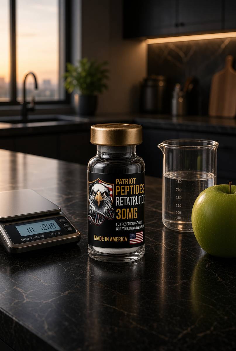
US-made retatrutide 30mg — the published structure LY3437943, HPLC-MS verified.
4.6(609)
121 browsing this now · 5 purchased in the last 24 hours
30mg
£120.00
Read next
50 min · long read · Frontier biology
We can rewrite a genome the way a word processor rewrites a sentence
CRISPR cut DNA. Base editors change one letter without a double-strand break. Prime editors write arbitrary edits from an RNA template. The machinery is no longer hypothetical.
46 min · long read · Peptide research
Retatrutide (LY3437943): the published triple-agonist structure
A fatty-acylated unimolecular agonist at GIPR, GLP-1R and GCGR. US-made, HPLC-MS verified, labelled for laboratory research — not a medicine.
45 min · long read · Peptide research
GHK-Cu: a copper tripeptide that rewrites a spreadsheet of genes
Pickart isolated GHK from plasma fractions that made old liver tissue synthesise proteins like young tissue. The tripeptide holds copper. The live claim is a transcriptome, not a moisturiser.
More in this desk
51 min · long read · Frontier biology
Tardigrades taught a human protein how to ignore radiation
Dsup, a disordered DNA-binding protein from a water bear, protects cultured human cells from X-rays. The animal that dries to a tun and lives through vacuum brought a transferable shield.
50 min · long read · Frontier biology
Xenobots: frog cells that became a new kind of machine
No genome was rewritten. Skin and heart cells from Xenopus were sculpted — first by hand, then by an evolutionary algorithm — into millimetre-scale organisms that walk, heal, and assemble copies of themselves.
49 min · long read · Frontier biology
We have the Neanderthal genome. Some of it is still in you.
Svante Pääbo pulled a genome out of bone powder, won the 2022 Nobel, and found that most people outside Africa carry a percent or two of an extinct human. Palaeogenomics is not only mammoths.

49 min · long read · Frontier biology
The woolly mouse is the mammoth’s twenty-day dress rehearsal
Colossal edited seven coat-and-metabolism genes into laboratory mice and got golden, shaggy, cold-curious animals. Elephant gestation is 22 months. A mouse tells you in three weeks whether the edit was worth the wait.
Essays describe published research. They are not medical advice and they do not authorise human use of any catalogue item.