

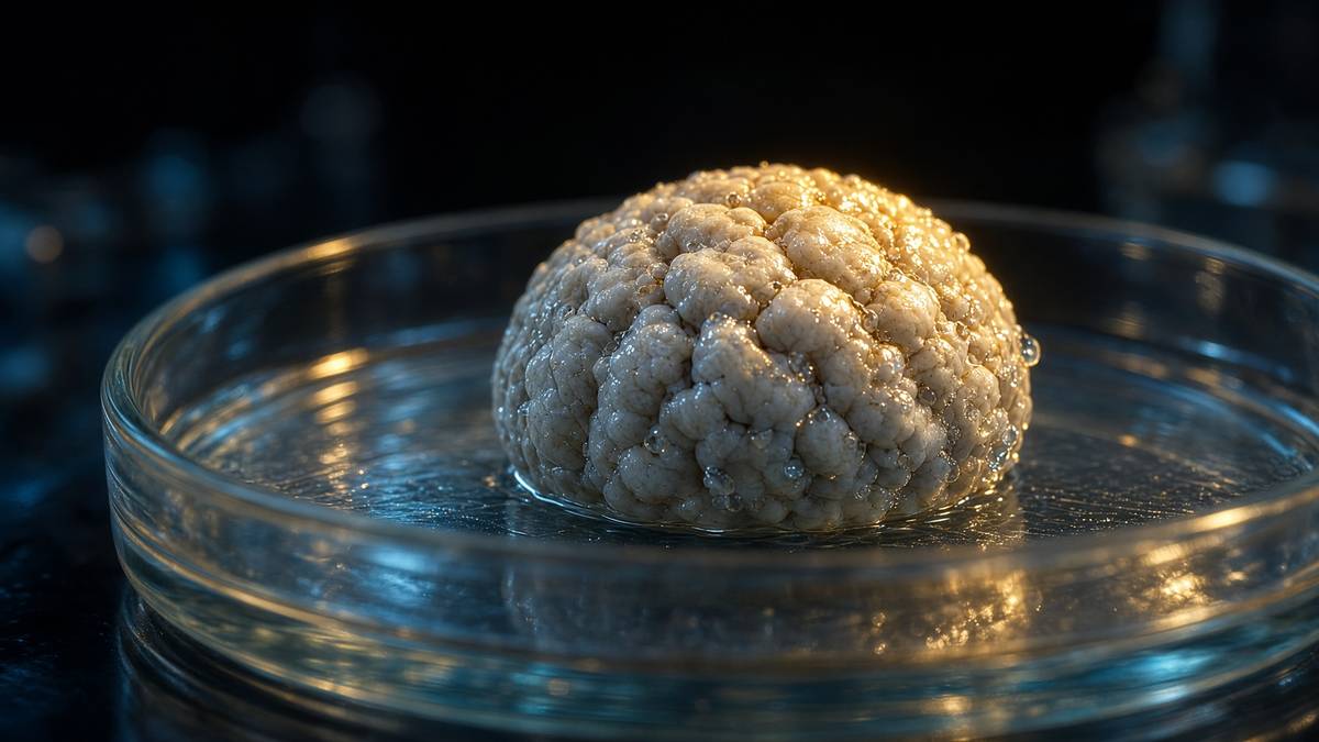
Frontier biology · 48 min · 10,465 words
Brain organoids: a cortex the size of a lentil
Stem cells, left to their own patterning, build layered neural tissue in a dish. They are not minds. They are the most honest model of human development we have ever had.
What this essay actually tells you
- Lancaster and Knoblich's cerebral organoids (Nature 2013) self-organise from stem cells into layered tissue the size of a lentil. Tissue. Not a mind. Lentil.
- They've been used to study lissencephaly, Zika microcephaly and, with caution, network activity. A model. Nobody's keeping a person in a dish, and the ethics section is not optional.
- Vascularisation and microglia are still the missing pieces. A cortex without blood supply is a short experiment, which is why the pretty pictures have a time limit.
What this actually means
Give human pluripotent stem cells the right growth factors and a bit of Matrigel, and they will try to become a brain. Not a person. A millimetre-scale ball with layers that look like cortex, with electrical activity, sometimes with a retina-like patch. Madeline Lancaster's 2013 cerebral organoids made the method famous. Sergiu Pașca's assembloids stick two regionalised pieces together so you can watch a connection form. Patient-derived organoids are how some labs now study autism, epilepsy and lissencephaly without waiting for a post-mortem. We find that last use the most defensible, and the ethics conversation around it the most necessary.
Madeline Lancaster and Jürgen Knoblich, Nature 2013, is still the paper you start from if you want to grow a lentil of brain. Human pluripotent stem cells, left to aggregate, then dropped into Matrigel and spun in a bioreactor, self-organised into three-dimensional neural tissue with ventricle-like lumens, a ventricular-zone-like progenitor belt, and a cortical-plate-like neuronal layer. The balls reached a few millimetres — the size of a lentil — and they weren't a mind. They were tissue. A patient-derived line carrying a CDK5RAP2 microcephaly allele made smaller organoids with premature neuronal differentiation, which is the sentence that took the method out of a tricks folder and into a disease folder. Yoshiki Sasai's laboratory had already shown that human and mouse stem cells would build an optic cup and a cortical neuroepithelium if you stopped flattening them onto plastic. Lancaster's contribution was rudeness of protocol: less morphogen choreography, more self-patterning, a spinning flask, and a photograph a neuropathologist could annotate. We still think that's one of the more astonishing sentences in twenty-first-century methods.
In short. In 2013, stem cells in a gel were left to build a tiny ball of layered brain tissue, about the size of a lentil. It was tissue, not a person thinking in a dish.
A mouse isn't a human cortex. The radial-glial scaffold, the outer radial glia that humans run in numbers a mouse barely bothers with, the gestational calendar, the transcriptional timing of SATB2 against BCL11B, the diseases that don't exist in Mus: lissencephaly of the Miller-Dieker sort, Zika microcephaly as a human-public-health event. A monolayer of iPSC neurons isn't a cortex either. It has human cells and a MEA if you pay for one, and it has no layers, no ventricle, no progenitor zone, no inside. Organoids are the awkward, powerful middle: human cells, three dimensions, layers, electrical activity, and a size and vascular failure that keep them from being a mind. That last clause isn't a disappointment. It's the reason the ethics conversation is tractable and the reason the model is a model. If you wanted a homunculus, look at the dying core. If you wanted only a mouse, look at a human outer radial glia.
In short. A mouse brain is the wrong species. A flat dish of human neurons has no layers. Organoids sit in between: human, three-dimensional, and still far too small and starved to be a mind.
The field is a short list of laboratories and a long list of protocols. Sasai, RIKEN, the SFEBq method and the optic cup, died in 2014 and left a generation of people who had watched epithelium fold itself. Lancaster, then at IMBA in Vienna with Knoblich, now at the MRC Laboratory of Molecular Biology in Cambridge, the unguided cerebral organoid. Lorenz Studer, Sloan Kettering, dual-SMAD inhibition in 2009 with Chambers, the neural-induction recipe almost everyone still starts from even when they claim to be hands-off. Sergiu Pașca, Stanford, guided regionalised spheroids and then assembloids, the second-generation trick of sticking two known identities together so you can watch a connection form. Paola Arlotta, Harvard, cell-type censuses and the long-culture networks. Guo-li Ming and Hongjun Song, then at Johns Hopkins, the mini-bioreactor and the Zika papers. Fred Gage, Salk, the transplant into a mouse cortex that brought host vessels in. That's the reading list. What follows is the map: how a lentil is built, what it can be asked, what it can't, and why the ethics section isn't a coda you tack on for a reviewer.
In short. A handful of labs, over a decade, turned a curious ball of tissue into a way to study human brain development and some of its diseases. What follows is the map of that method.
If you're coming to this cold, you need names and numbers rather than a mood about mini-brains. Chambers, Fasano, Papagiannouli, Tomishima, Sadelain, Studer, Nature Biotechnology 2009: Noggin plus SB431542, dual-SMAD, neural induction of human embryonic stem cells in two dimensions. Eiraku, Sasai, Nature 2011 and Nakano, Sasai, Cell Stem Cell 2012: the optic cup. Lancaster, Renner, Martin, Wenzel, Bicknell, Hurles, Homfray, Penninger, Jackson, Knoblich, Nature 2013: cerebral organoids, microcephaly. Kadoshima, Sasai, Proceedings of the National Academy of Sciences 2013: self-organised cortical tissue, a parallel Japanese sentence. Pașca, Sloan, Nature Methods 2015: human cortical spheroids. Birey, Pașca, Nature 2017: assembloids, Timothy syndrome, interneuron migration. Quadrato, Arlotta, Nature 2017: long-term organoids, photosensitive cells, active networks. Cugola, Beltrão-Braga, Science 2016, and Garcez, Rehen, Science 2016: Zika. Iefremova, Ladewig, Cell Reports 2017, and Bershteyn, Kriegstein, Cell Stem Cell 2017: Miller-Dieker lissencephaly. Bhaduri, Kriegstein, Nature 2020: stress, missing cell types, the honesty paper. Those are a fortnight of evenings. The headlines will still be there when you come back, and they will look smaller.
In short. The method rests on a short stack of named papers. Read those before any headline about a brain in a jar.
Cerebral organoids can be used to model disorders of neural development, such as microcephaly. What they cannot be used as is a mind. The 2013 paper already knew the first sentence. The coverage has spent a decade forgetting the second.— Lancaster et al., Nature 2013 — the finding, and the distinction the finding requires
Dual-SMAD, then leave them alone
Neural induction, in a dish, is a subtraction. BMP and TGF-β/Activin/Nodal signalling hold human pluripotent cells in a non-neural fate; block both with Noggin (or LDN193189) and with SB431542, and the cells default to neuroectoderm. Chambers and Studer named that dual-SMAD inhibition in 2009, and it remains the usual first move even in laboratories that then brag about self-patterning. SMAD is the transcription-factor family that carries those two pathways into the nucleus. Dual inhibition is therefore a transcriptional decision before it's a morphological one: the cells stop writing the mesendoderm programme and start writing SOX1, PAX6, a neuroepithelial identity. You can do this in two dimensions and plate a carpet of neural progenitors. You can do it, or a gentler version of it, in a three-dimensional aggregate and then stop telling the tissue what to be. The second choice is the organoid. The first choice is the monolayer everyone already had. Organoids exist because someone was willing to stop flattening the carpet.
In short. To make brain tissue, you first block two chemical signals that would push stem cells toward other body parts. The cells then take a neural path on their own.
Sasai's SFEBq method — serum-free floating culture of embryoid-body-like aggregates — was the demonstration that the epithelium would do the rest. Quick aggregation of dissociated stem cells in a low-adhesion well, a few days, a neuroepithelial cyst with apical polarity facing a lumen, then spontaneous folding. The optic-cup papers are the ones people remember because a cup is a photograph: retinal pigment epithelium and neural retina, invagination, a structure you could have stolen from an embryology atlas. The cortical papers from the same laboratory showed a stratified neuroepithelium with radial glia and deep- then upper-layer neurons, arriving in the right order. Self-organisation is the word, and it isn't a metaphor. Morphogen gradients that the tissue itself secretes, mechanical buckling of a polarised epithelium, a lumen that sets an inside, cadherin-mediated packing: those are the parts. A protocol that adds WNT, SHH or BMP later is choosing a region. A protocol that doesn't is asking the aggregate to choose. Lancaster asked. Sasai, in parallel, often specified. Both balls are organoids. They aren't the same experiment.
In short. Once the cells are neural, they can fold themselves into layered tissue, complete with an inside and an outside. Some recipes then steer the region; the famous 2013 recipe mostly didn't.
Lancaster's original cerebral-organoid protocol is almost rude in its simplicity, and the rudeness is the point. Embryoid bodies from human embryonic stem cells or iPSCs, a neural-induction medium, embedding in a twenty-microlitre Matrigel droplet, then a spinning bioreactor so that nutrients and oxygen move. Matrigel is a basement-membrane extract from an Engelbreth-Holm-Swarm mouse sarcoma: laminin, collagen IV, entactin, a fog of growth factors you didn't order individually. It isn't a defined matrix, and everyone knows, and everyone still uses it because neuroepithelium wants a basal lamina to polarise against. The spin isn't setting. Static culture lets a millimetre-scale ball starve at the core on a shorter clock; a stirred flask delays that clock. Over weeks the droplet grows buds of neuroepithelium that surround ventricle-like cavities. By two months you have a few millimetres of tissue with regional identities that include, in the unguided version, dorsal forebrain, ventral forebrain, choroid-plexus-like epithelium, and sometimes a retina-like patch. The tissue decided. The protocol got out of the way, which is a more disciplined sentence than it sounds.
In short. The 2013 recipe put cell clumps in a droplet of gel and spun them so food and oxygen could reach the surface. Over weeks the clumps grew buds and layers by themselves.
Name what a neuropathologist actually sees, because headlines skip the histology. A ventricle-like lumen, apical. Around it, a dense belt of SOX2-positive radial glia whose nuclei migrate with the cell cycle — interkinetic nuclear migration, the same motion an embryonic ventricular zone runs. Basal to that, an intermediate zone with TBR2/EOMES-positive intermediate progenitors. Further out, a cortical-plate-like band of neurons that stain for deep-layer markers first (TBR1, BCL11B/CTIP2) and upper-layer markers later (SATB2, CUX1), which is the inside-out order the foetal cortex already knew. Outer radial glia, the basal, unipolar progenitors that humans run in numbers mice don't, sit in a subventricular-zone-like territory and are one of the reasons this model exists at all. Phosphohistone-H3 at the apical surface. A mitotic figure a student can find. None of this is six-layer adult isocortex. None of it has a thalamus to talk to. It's first-trimester-ish laminated tissue, and a neuropathologist can read it, and that's already a large fact.
In short. Under the microscope you see a hollow, a zone of dividing cells, then layers of neurons arriving in the same order they do in a foetal cortex. That's layered tissue, not a finished brain.
Unguided is a feature and a bug, and the field spent the next decade arguing about which. An unguided organoid will sometimes grow a choroid plexus next to a cortical plate next to a retinal pigment patch, because the aggregate is a small embryo without a body to tell it where anterior is. Batch-to-batch variability is therefore not a scandal. It's the method admitting it handed patterning back to the tissue. Guided protocols — dual-SMAD, then a timed WNT, a timed SHH, a timed retinoic acid — specify pallium or subpallium or midbrain or spinal and give you a more homogeneous spheroid, which is what you want if the question is a named cell type or a fusion. Pașca's cortical spheroids are guided. Lancaster's cerebral organoids, in the original, weren't. Both words should stay in the literature. Calling everything a mini-brain is how you lose the difference between a self-patterned mosaic and a specified cortical plate. We'll keep the difference, because the next heading is size, and size doesn't care which protocol you used if the core is still dying.
In short. If you don't steer the tissue, it may grow several brain regions at once and no two balls look identical. Later recipes steer on purpose, so experiments can be compared.
A lentil is a measurement
A typical cerebral organoid, after one or two months, is two to four millimetres across. A lentil is four to six. That's the photograph. Oxygen and glucose in a tissue without capillaries diffuse on the order of a hundred to two hundred micrometres before cells go hypoxic; a millimetre-scale ball therefore has a rim that lives and a core that's, on a long enough clock, dying. Haematoxylin will show it. A TUNEL stain will show it. If you has sectioned an organoid older than a few weeks has seen the cavity or the debris at the centre and has, if they were honest, put it in the supplement. The spinning flask delays the clock. An air-liquid interface, Giandomenico and Lancaster, Nature Neuroscience 2019, slices the ball and feeds it from the surface, which is how you get axon tracts and more mature physiology at the price of no longer having a ball. Vascularisation, when it works, is the only way to keep a true three-dimensional interior. Until then, a cortex without blood supply is a short experiment. The size limit isn't a branding problem. It's Fick's law, and it's why these objects stay models.
In short. These tissues grow to a few millimetres. Without blood vessels, oxygen can't reach the middle, so the core dies. That size limit is why they remain models.
Put the lentil next to the organ it's named after, because scale is the argument this journal keeps having to have. A human brain at birth is already on the order of three hundred and fifty grams; an adult brain is about one thousand three hundred to one thousand four hundred grams. Suzana Herculano-Houzel's census put the adult complement at roughly eighty-six billion neurons, sixteen billion of them in the cortex. Six layers in isocortex, a thalamus, a contralateral hemisphere joined by a corpus callosum, meninges, cerebrospinal fluid in a ventricular system that actually circulates, a body that sends sensory afferents and takes motor efferents. Gestation is nine months before any of that's even a starting brain, and synaptic and myelination programmes then run for two decades. An organoid has none of the body, none of the thalamus as a default, a few hundred thousand to a few million cells if you're being generous about a mature specimen, and a gene-expression age that even at many months in culture still reads as first-trimester-to-early-second. The word cortex in 'cortical organoid' is a histological courtesy. It isn't a transplant of the adult object.
In short. A human brain weighs more than a kilogram and holds tens of billions of nerve cells. An organoid is millions of cells at most, early-foetal in age, with no body attached.
Time is the other axis on the same diagram. Human corticogenesis runs over months; layer identity is a calendar, not a switch. Deep-layer neurons are born first, upper-layer neurons later, astrocytes later still, oligodendrocytes later than that, and microglia arrive from the yolk sac on a separate timetable the ectodermal organoid doesn't possess. In a dish you can keep an organoid for six, nine, twelve months, and people have. You don't thereby obtain a two-year-old cortex. You obtain a long-cultured foetal-ish tissue with more astrocytes than it had at week six, more synapses, more electrical bursts, and a core that has been negotiating hypoxia the whole time. Quadrato and Arlotta's nine-month organoids grew photosensitive cells and networks; they didn't grow childhood. Anyone quoting a culture time as if it were a postnatal age hasn't looked at the RNA. The developmental clock, in human cells, is stubborn. That stubbornness is why a mouse cortex is a poor stand-in and why an organoid still can't be rushed into adulthood with a cocktail, however much the cocktail literature wishes otherwise.
In short. Keeping a ball of tissue in a dish for months doesn't make it as mature as a child's brain. The cells still look and act like early foetal cells, only older and more stressed.
Diagram
- 0.1 nmHydrogen atomA proton and an electron. Chemistry starts here.
- 0.3 nmWater molecule70% of a cell by mass. The solvent life is.
- 1 nmAmino acidTwenty kinds. Peptide bonds string them.
- 2–4 nmResearch peptideA named chain. BPC-157 is 1.4 kDa, 15 residues.
- 4–10 nmGlobular proteinHaemoglobin, a GPCR’s extracellular face.
- 25 nmRibosomeThe factory that reads mRNA into protein.
- 5 nmMembraneA lipid bilayer. Every compartment starts here.
- 0.5–1 µmMitochondrionA bacterium the cell swallowed and kept.
- 6–10 µmNucleusTwo metres of DNA folded into a sphere.
- 10–30 µmTypical cellA city. 10¹⁰ proteins. One genome.
- 1 mmTissue grainA thousand cells talking across ECM.
- 1.7 mYou~36 trillion human cells. Most of them are red blood cells.
Lengths are characteristic, not exact. A research peptide is closer in size to a water molecule than to the cell that assays it — which is why a 15-mer can occupy a receptor pocket a small-molecule drug also wants.
- Lancaster & Knoblich
- Nature 2013
- Typical diameter
- 2–4 mm
- Oxygen diffusion
- ~100–200 μm
- Human adult brain
- ~1.3–1.4 kg
- Organoid census
- 10⁵–10⁶ cells
- Transcriptional age
- first trimester-ish
- Microcephaly assay
- CDK5RAP2
- Assembloid
- Birey, Pașca 2017
Self-patterning cerebral organoids. The methods paper the coverage still under-cites.
A lentil is 4–6 mm. Beyond that, the core starves without vessels.
Fick, not branding. A millimetre-scale ball has a living rim and a dying centre.
Herculano-Houzel: ~86 billion neurons. Sixteen billion in cortex.
Generous, mature specimens. Not a cortex. A tissue.
Even at many months in culture. The developmental clock is stubborn.
Lancaster 2013, patient iPSCs, smaller organoids, premature differentiation.
Two regionalised spheroids fused. A connection you can watch, not a mind you can ask.
Lancaster would, and does, wince at mini-brain. So should you. The phrase does work that the histology hasn't earned: it implies a unified organ, a homunculus, a thing that might be about to notice you. What you have is self-patterning neural tissue with layers a neuropathologist can read. Learn that distinction and the rest of the field makes sense. Variability between organoids is then a methods fact rather than a scandal. A retina-like patch on a cerebral organoid is a leftover of unguided patterning rather than a creature that can see. Electrical bursts are a network in a millimetre, not an experience. The wince isn't prudishness. It's the same wince a crystallographer gives when a predicted fold is called the structure. Courtesy toward the object — it's human tissue, it came from a donated cell, it deserves a sentence of respect — is compatible with refusing the fairy tale. We hold both of those, on purpose, and the ethics heading will hold them again. Size is the first reason the fairy tale fails. Missing cell types are the second.
In short. Calling these tissues mini-brains sells a story the microscope doesn't support. They're layered human neural tissue. That's already remarkable, and it's the accurate name.
Transcriptional identity is the method
Regionalisation is a morphogen conversation that becomes a transcription-factor address. After dual-SMAD has put the aggregate into neuroectoderm, WNT (often via CHIR99021, a GSK3 inhibitor that stabilises β-catenin) dorsalises or posteriorises depending on dose and timing; SHH ventralises; BMP can push toward choroid plexus or non-neural; retinoic acid caudalises. Pallium versus subpallium is the fork the cortical people care about. High WNT, low SHH, and you're in FOXG1-positive, PAX6-positive, EMX1-positive dorsal forebrain territory, glutamatergic projection neurons ahead. SHH on, and you're in NKX2.1-positive, GSH2-positive ventral forebrain, the ganglionic-eminence programme that mints GABAergic interneurons. Midbrain is LMX1A and FOXA2 if you went further caudal with WNT. Spinal is retinoic acid plus SHH, motor neurons if you're greedy. A guided spheroid is a claim that this conversation was had on purpose. An unguided organoid is a claim that the tissue had it with itself, noisily. Either way, the identity you think you have is a set of transcription factors you have to stain for. A photograph of a ball isn't a pallium.
In short. Chemical signals tell the tissue whether to become the outer cortex, the inner cells that make inhibitors, or something further down the nervous system. You check that identity with stains, not with a photograph.
The cortical cascade is named, and the names are the assay. PAX6 and SOX2 in the progenitor belt. FOXG1 to confirm forebrain rather than a midbrain tourist. TBR2/EOMES in the intermediate progenitors that radial glia hand off to. TBR1 and BCL11B/CTIP2 in deep-layer neurons, the ones that in a real cortex would head for thalamus and subcortical targets. SATB2 and CUX1 in upper-layer neurons, the callosal and cortico-cortical population, arriving later. RELN in Cajal-Retzius-like cells if the preplate has organised. Then, if you're asking a human-specific question, HOPX and PTPRZ1 in outer radial glia, the basal progenitors that sit away from the ventricle and that a mouse cortex runs sparsely. Single-cell RNA sequencing — 10x Genomics, a Parse or a Smart-seq2 if you're still in a boutique — is how a 2019 paper earns the word census. Velasco, Arlotta, Nature 2019: dorsal forebrain organoids can, under a tight protocol, make a reproducible set of those identities. Reproducible is doing a lot of work. It doesn't mean every laboratory's lentil is the same lentil. It means a specified protocol can land the same cell types more than once, which is the floor a disease paper requires.
In short. Each layer of the developing cortex has named marker genes. Counting those markers, cell by cell, is how you show the tissue became what you claimed, rather than a random ball of nerve cells.
Diagram
Closed chromatin (H3K27me3, DNA methylation) hides the promoter. Pioneer factors and histone acetyltransferases open it.
PIC: TFIID, TFIIH, Mediator, Pol II. Ser5 phosphorylation of the CTD lets the polymerase leave the promoter.
Elongation ~20–40 nt/s. Capping, splicing, cleavage and polyadenylation happen on the still-growing RNA.
Human genes are islands in 3.1 billion base pairs of mostly noncoding sequence. Promoter, enhancers, chromatin state and the Mediator complex decide whether Pol II is allowed to fire. Epithalon’s literature sits on TERT and pineal clocks — two of the rare promoters anyone bothers to name in a peptide essay.
Outer radial glia are a large part of why a human organoid isn't a mouse reaggregate. In the human foetal cortex, a population of basal, unipolar radial glia in the outer subventricular zone divides and contributes to the upper-layer boom that a lissencephalic mouse doesn't bother with. They're the cells ARHGAP11B — a human-specific gene duplicate — acts on, the Pollen, Kriegstein, Cell 2019 neighbourhood, the reason a chimpanzee organoid and a human organoid aren't the same experiment. Interkinetic nuclear migration still happens at the apical surface; outer radial glia have moved the factory out into the subventricular zone and keep a basal process. Mitotic delay in those cells is one of the lissencephaly phenotypes. Abundance of them is one of the human-ape phenotypes. A protocol that doesn't produce HOPX-positive outer radial glia is a protocol that hasn't yet earned a human-cortical boast. A protocol that does has a cell type you can't ethically sample from a living foetus except as discarded tissue, which is the moral arithmetic that makes the model worth the hassle.
In short. Humans grow an extra kind of dividing cell in the developing cortex that mice barely have. Organoids can make that cell type. That's one of the strongest reasons to use them.
Great-ape organoids are the comparative experiment this method uniquely allows, and they belong in a transcriptional heading because the readout is a census, not a skull. Kanton, Treutlein, Nature 2019: human, chimpanzee and macaque iPSC organoids, delayed maturation on the human line, a set of genes whose timing had shifted. Pollen, Bhaduri, Kriegstein, Cell 2019: chimpanzee and human, organoid and primary foetal tissue, ARHGAP11B and a human-specific progenitor programme. Benito-Kwiecinski, Lancaster, Cell 2021: gorilla, chimpanzee and human, a morphogenetic difference — human neuroepithelium stays longer in a progenitor-expanding shape before neurogenesis. Those papers are why 'human-specific developmental biology' stopped meaning a just-so story about FOXP2 and started meaning a differential expression table you could stain for. They're also why an organoid is already, before anyone mentions disease, an ethical object: you're building ape neural tissue in a dish and asking it questions a living ape cortex wouldn't be offered. The transcriptional machine doesn't care. The committee should.
In short. Growing matched tissues from human, chimpanzee and gorilla cells has shown that the human cortex delays its birth of neurons and expands its progenitor pool. That comparison is almost impossible in living tissue.
Assembloids are how you watch a connection
Pașca's move was to stop asking one ball to be the whole forebrain. Make a cortical glutamatergic spheroid, make a subpallial GABAergic spheroid, put them next to each other in a well, and over weeks the ventral piece sends interneurons that migrate into the dorsal piece, the way ganglionic-eminence derivatives migrate into embryonic cortex. Birey, Li, Pașca, Nature 2017 is the paper: human assembloids, Timothy syndrome (CACNA1C), a migration phenotype you could see and a calcium-channel phenotype you could measure. Guided identity plus fusion is a different object from Lancaster's unguided mosaic. You know what each half was. You can optogenetically tag one half. You can ask whether a patient mutation breaks the journey rather than the destination. The word assembloid is useful because it refuses the mini-brain. It says: two tissues, a boundary, a projection, a question. Subsequent fusions have stacked more parts — cortico-striatal, cortico-thalamic, cortico-spinal — and the naming has struggled to keep up. The method hasn't. Specify, fuse, wait, record.
In short. A later trick is to grow two known pieces and stick them together, then watch one send fibres into the other. That's a connection, not a whole brain.
Interneuron migration is the first question the fusion was built for, and it's a human-relevant question a mouse only partly answers. Cortical GABAergic interneurons are born in the ventral forebrain and travel tangentially; their arrival, integration and maturation run on a calendar that, in humans, stretches late. Timothy syndrome, a gain-of-function in the L-type calcium channel CaV1.2, delayed that migration in the 2017 assembloids and gave the laboratory a phenotype that patient-derived cells, not a hypothesised mouse allele, actually showed. Saltatory movement, a leading process, a soma that follows, a failure of that saltation: those are the movies. You can rescue with a channel blocker in the dish. You can't, from that rescue, write a paediatric dosing schedule, and the Pașca group has been more careful about that leap than the coverage has. What you can write is: a human mutation, in human cells, in a three-dimensional journey that a monolayer doesn't run. That sentence didn't exist in 2012. It exists now, and it's why assembloids aren't a rebrand of organoids. They're a different experiment.
In short. Some inhibitory nerve cells are born in one region and crawl into another. Fusing two lab-grown pieces lets you watch that journey, and see it go wrong in cells from patients.
Andersen, Pașca, Cell 2020, is the greedier fusion: cortical spheroid, spinal spheroid, skeletal-muscle spheroids, a cortico-motor assembloid in which human corticofugal axons find motor neurons that find muscle, and you can watch a twitch. That paper is either a circus or a methods landmark depending on how hungrily you read a twitch. We read it as a landmark with a circus attached. A three-part circuit, human, in a dish, with a contractile readout, is a tool for asking whether a motor-neuron disease allele breaks the upper motor neuron, the lower, the neuromuscular junction, or the muscle. It's also a photograph that will be stolen by every 'lab-grown nervous system' headline for the rest of the decade. The twitch is a calcium-and-actin event in a few millimetres of tissue. It isn't reaching for a toy. Hold the millimetres. Subsequent cortico-striatal and diencephalic fusions from the same and neighbouring laboratories are the quieter, more useful cousins: a projection you can slice, a synapse you can stain for VGLUT1 and PSD-95, a live-cell calcium transient you can count. Greed in an assembloid is how you get a circus. Restraint is how you get a paper a sceptic will believe.
In short. Some groups now fuse cortex-like, spinal and muscle tissues so that a human nerve pathway can make a scrap of muscle twitch. That's a useful assay. It isn't a body moving on purpose.
Specify two identities, fuse them, and watch a human projection form. That is a developmental question you can ask without an invasive human experiment. It is also still two millimetres of tissue in a well.— The assembloid rationale, as the field has had to keep restating it
Machines, named, because 'the organoid was active' isn't a methods line. A multi-electrode array — Maxwell Biosystems, Axion, a CMOS chip if you're fancy — reports spikes and bursts from the face of the tissue that sat on the electrodes, which is a sampled surface, not a mind. Two-photon calcium imaging with GCaMP6 or jGCaMP7 reports a fluorescent proxy of calcium in the cells you can see, which at millimetre depth isn't many cells unless you have cleared or sliced. Whole-cell patch clamp on a sliced organoid is still the gold standard for a current, a potential, a synaptic event; it's also slow, and it asks one neuron at a time. Optogenetics — channelrhodopsin-2, a 470-nanometre LED, a labelled projection — is how you ask whether the fusion actually wired. Single-cell RNA-seq is how you ask whether the cells you recorded were the cells you thought. None of that's glamorous. All of it's how you stop an electrical essay becoming a ghost story. If the claim is a network, show the array, the raster, the calcium, and the cell type. If the claim is a mind, you don't have the machine.
In short. Spikes, calcium flashes and a twitch are measured with named instruments sitting on a millimetre of tissue. Those readings aren't a mind. They're how you show a circuit is actually there.
Lissencephaly, Zika, and the questions a tissue can take
Lissencephaly is a smooth-brain phenotype the mouse doesn't honestly have, because a mouse cortex is already smooth. Miller-Dieker syndrome, a 17p13.3 deletion that takes LIS1/PAFAH1B1 and often YWHAE, is the human disease the organoid literature reached for first. Iefremova, Ladewig, Cell Reports 2017: patient iPSC organoids, a radial-glial scaffold that was poorly organised, N-cadherin and Wnt/β-catenin in the story, a phenotype you could see in the cytoarchitecture. Bershteyn, Kriegstein, Cell Stem Cell 2017: mitotic delay in outer radial glia, a cell-type-specific failure a monolayer would have smeared. Klaus, Knoblich, Nature Cell Biology 2019: LIS1 and NDE1, asymmetric division of human radial glia, a spindle-orientation argument that only a three-dimensional progenitor zone can run. Those papers are why the heading exists. You can't ask a lissencephaly question of a mouse without pretending the gyrified human cortex is optional. You can ask it of an organoid, with the patient's alleles, and you can ask it again after you put the missing gene back. The organoid is still not gyrified the way a third-trimester human cortex is. It's gyrification-adjacent enough, in its progenitor dynamics, to be the least-bad assay the species has ever had.
In short. Some human brain disorders never look right in a mouse, because a mouse cortex is already smooth. Organoids from patient cells have shown how the dividing cells fail in those conditions.
Zika virus, 2015–2016, was the public-health test of the method, and the method held. Cugola, Beltrão-Braga, Russo, Science 2016: Brazilian ZIKV in mice and in human organoids, microcephaly-like reductions, a virus tropism for progenitors. Garcez, Rehen, Science 2016, the same week in spirit: human neural stem cells, neurospheres, organoids, a productive infection, a smaller ball. Qian, Ming, Song, Cell 2016: forebrain organoids in a cheap mini-bioreactor, layer-specific thinning, a practical methods gift to laboratories that didn't own a spinning flask. Dang, Tang, Cell Stem Cell 2016, and a scramble of follow-ups: TLR3, centrosomes, a list of host factors that may or may not have survived the next year of papers. What survived, and what we'll keep, is the logic. A virus that was causing microcephaly in human foetuses could be put onto human cortical tissue in a dish, and the tissue got smaller, and the progenitors died or exited the cycle too early, and that was a result you could have in weeks rather than in a primate pregnancy. Caution, still: an organoid isn't a placenta, not an immune system, not a third-trimester cortex. It was the right emergency model. It remains a model.
In short. During the Zika outbreak, laboratories infected these human tissues and watched them shrink, as infected foetal brains had. That gave answers in weeks that animal pregnancies couldn't have given in time.
Timothy syndrome we've already named. Autism-spectrum and epilepsy alleles have followed, with the usual quality gradient. Samarasinghe, Novitch, Nature Neuroscience 2021: fused cortical and ganglionic-eminence organoids, a Rett- and a tuberous-sclerosis-adjacent conversation, network bursts you could count, a fusion that had a seizure-like phenotype rather than only a cell-fate phenotype. Pașca's laboratory and a dozen others have put patient iPSCs through the same guided spheroid, the same fusion, the same array, and asked whether a named variant changes migration, synapse density, or burst statistics. Some of those papers are beautiful. Some are a variant of unknown significance plus a principal-component plot. The honest move is the isogenic pair: CRISPR a correction into the patient line, or CRISPR the variant into a control line, and show the phenotype moves with the letter. That's now ordinary. It was science fiction in 2012. Ordinary is the achievement. A principal-component plot of three patient lines against three controls, with no isogenic, is how this literature gets sloppy, and the sloppiness is optional.
In short. Patient stem cells, grown into these tissues, are now used to study autism, epilepsy and rare channel diseases. The convincing experiments compare the patient cells with a CRISPR-corrected copy of the same cells.
Casgevy's cousins belong in this heading as a tool, not as a therapy. A CRISPR–Cas9 edit in an iPSC, followed by an organoid, is a human-developmental genetic screen you can run without an embryo past the fourteen-day rule and without a paediatric biopsy. Knock out a microcephaly gene. Base-edit a lissencephaly allele. Prime-edit a correction and ask whether the progenitor zone is rescued. The enzyme family is the one the neighbouring CRISPR essay spent a week on; the payload here is a tissue that self-organises rather than a haematopoietic stem cell that engrafts. Ethical payload, again, isn't shared. One is a licensed autologous medicine. The other is a dish. What is shared is the reason both exist in the same decade: a programmable nuclease plus a human cell you're allowed to grow. Anyone writing an organoid paper without an isogenic CRISPR control, in 2026, should be able to say why the letter wasn't the experiment. Anyone writing a CRISPR screen without a three-dimensional readout, for a cortical-development gene, should be able to say why a monolayer was enough. Sometimes it's. Often it isn't.
In short. Gene editing of the stem cells, then growing the tissue, lets you test a human disease letter directly. That's a dish assay, not a treatment.
What you still can't ask is the list that keeps a disease paper honest. You can't ask a third-trimester folding question of a four-millimetre ball that doesn't fold like a gyrencephalic cortex; some organoids wrinkle, and the wrinkles aren't gyri. You can't ask a circuit-level cognition question, because there's no thalamus, no long-range white matter worth the name, no body. You can't ask an immune question unless you put microglia in, and then you have a co-culture with its own artefacts. You can't ask a pharmacokinetic question, because there's no blood. A smaller organoid is evidence that a virus can damage human cortical progenitors, not proof that it causes microcephaly on its own. Bhaduri, Kriegstein, Nature 2020, is the honesty paper the field needed: organoids activate stress pathways, some cell types are missing or mis-specified relative to primary foetal cortex, and a disease claim that ignores that stress is a disease claim about a stressed tissue. Cite it in the limitation paragraph. Then, if the phenotype is large, the isogenic pair is clean, and the cell type is one the organoid actually makes, publish.
In short. These tissues can't model a folded adult cortex, a whole-brain circuit, or a drug in the bloodstream. They can model early human cell behaviour in a disease, if the controls are tight.
Spikes are not a mind
Electrical activity in an organoid is real, and it has been real for years, and it's still not experience as far as any measurement we trust can tell. Neurons fire. Synapses form. Bursts travel a millimetre. Quadrato, Arlotta, Nature 2017: organoids cultured for more than nine months, a surprising diversity of cell types including photoreceptor-like cells that could respond to light, spontaneously active neuronal networks. Trujillo, Muotri, Cell Stem Cell 2019: nested oscillatory waves on a multi-electrode array, a statistical resemblance to preterm electroencephalograms that the paper was careful-ish about and the press wasn't. A preterm EEG is a recording from a skull with a thalamus, a brainstem, a blood supply and a body. An organoid oscillation is a recording from a millimetre of unvascularised tissue on a chip. Resemblance is a statistic. It isn't an identity. We'll keep saying that until the analogy is retired, because the analogy is how a network paper becomes a ghost story, and the ghost story is how the ethics conversation gets both louder and stupider than the tissue warrants.
In short. Nerve cells in these balls do fire in groups, and that activity can look a little like a very premature baby's brain waves. Looking alike isn't the same as being a mind.
What a network claim needs, and what it often doesn't get, is the unglamorous panel. A raster plot with a scale bar. A spike-sorting method named. A burst-detection threshold you could reproduce. A cell-type census of the recorded region, because a photoreceptor-like cell and a deep-layer projection neuron aren't the same oscillator. A pharmacological dissection: tetrodotoxin silences, AP5 and CNQX take the synaptic component, a GABA blocker tells you whether inhibition is in the mix. If you fused two spheroids, a demonstration that the spikes in one half can be driven from the other. If you claim preterm-EEG kinship, a quantification that survives contact with a neonatal neurologist. Trujillo's oscillations were a real observation. The kinship language was the part that needed a colder sentence, and a fraction of the subsequent literature spent its energy on the kinship rather than on the pharmacology. We'll take the pharmacology. Criticality, 'organoid intelligence', computing with wetware: those are laboratories running ahead of the controls. Ahead is allowed in a grant. It isn't allowed in a results section.
In short. A serious activity paper names the recording kit, silences the tissue with known drugs, and shows which cells were firing. Fancy comparisons with baby brain waves are the optional extra, and often the shaky one.
Transplant is the experiment that adds blood, microglia and a body, and it's also the experiment that drags the ethics from a dish onto a living animal. Mansour, Gage, Nature Biotechnology 2018: human organoids into a mouse cortex, host vasculature invading, host microglia infiltrating, human axons projecting, a graft that survived and matured past what the dish had allowed. Revah, Pașca, Nature 2022: human cortical organoids into newborn rats, more mature cell types, sensory-evoked activity, a rat that could, in a reward assay, show a behavioural bias the authors tied to the graft. That last clause is the one a committee reads twice. A chimaeric rodent with a few millimetres of human cortical tissue isn't a human mind in a rat skull. It's also not a dish. ISSCR 2021 asked for specialised review of such grafts, and for a prohibition on experiments that might plausibly alter an animal's cognition in a human-like direction. The Pașca rat paper sat on the allowed side of that line. Vascularisation worked. Maturation moved. The animal was still a rat. The line will move again when the grafts get larger. That's why the line was written down in advance.
In short. Putting these tissues into a mouse or rat lets blood vessels and immune cells move in, and the human cells mature further. The animal doesn't become a person.
A cortex without blood is a short experiment
Vascularisation and microglia are still the missing pieces, and they're missing for developmental reasons, not because nobody has tried. Cerebral organoids, in the standard recipes, are ectodermal. Blood vessels are mesodermal. Microglia are yolk-sac-derived myeloid cells that enter the brain on a separate calendar. An ectodermal ball won't mint a capillary bed or a microglial network as a default, any more than a cortical plate will mint a liver. The living rim and the dying core are the physiological consequence. Hypoxia-inducible factors fire at the centre; glycolysis takes over; mitochondrial membrane potential collapses in the cells that have run out of oxygen; necrosis and apoptosis both show up on the stain. A cortex, in an animal, is an expensive, oxidative tissue with a capillary a few tens of micrometres from every neuron. A cortex in a dish, beyond the diffusion limit, is a short experiment dressed as a long one. Every methods talk eventually admits this. The talks that don't are selling a photograph.
In short. These tissues come from the cell layer that makes brain, not blood vessels or brain immune cells. Without those, the middle of the ball runs out of oxygen and dies.
Mitochondria notice first, which is why a respiratory-chain diagram belongs here as well as in an energy essay. Neurons and radial glia at the rim are still running oxidative phosphorylation; Complex I still wants NADH; oxygen is still the terminal acceptor at Complex IV. A hundred micrometres in, the partial pressure has fallen. The same cell type, a few cell diameters closer to the centre, is now a glycolytic, HIF-high, membrane-potential-poor object, and then not an object. That gradient is a confound every metabolic or electrophysiological claim has to live with. A burst that looks like a network may be a rim talking. A progenitor defect that looks like a disease may be a core starving. Fractionate the observation by depth, or slice, or vascularise, or admit the confound. Isolated mitochondria from an organoid homogenate are a soup of rim and core and debris. A Seahorse well of dissociated organoid cells is a soup that has already lost the gradient. The honest metabolic object is a section with a hypoxia probe, a TMRM image with a depth, or a graft that finally has blood. Until then the organelle is telling you about oxygen.
In short. The energy-making structures inside cells fail first when oxygen runs out. In an organoid that failure starts in the middle, so a disease claim has to show it isn't just a starved core.
Diagram
Matrix
- TCA cycle · β-oxidation · mtDNA nucleoids
- NADH produced here. Complex I spends it.
- MOTS-c (MRWQEMGYIFYPRKLR) from 12S rRNA.
Inner membrane
- I → II → III → IV → V (ATP synthase)
- ~150 mV proton-motive force
- ~40–60 kg of ATP turned over per human day
mtDNA is 16,569 bp, 37 genes, 13 proteins of the respiratory chain. Nuclear DNA encodes the other ~1,200 mitochondrial proteins. NAD+ is the hydride carrier between dehydrogenases and Complex I. MOTS-c is a 16-mer translated from 12S rRNA — a peptide the mitochondrion wrote itself.
Attempts to put blood in are now a subfield, and they come in two flavours. Bottom-up: Cakir, Park, Nature Methods 2019, and neighbours, ETV2 overexpression to push endothelial-like networks inside the organoid, a vascular-like tree that isn't a connected circulation to a heart but is already better than nothing. Co-culture with iPSC-derived endothelial cells, pericytes, a microfluidic chip that actually perfuses: the organ-on-chip version, more plumbing than biology, sometimes more honest about the plumbing. Top-down: the transplant, already named, in which a mouse or a rat donates the vessels and the circulating cells. Mansour's invading host vasculature is still the cleanest demonstration that the organoid was vessel-hungry rather than vessel-incapable. Perfusion in vitro that matches a capillary bed, with human endothelium, with a blood-brain barrier worth the name, isn't a product you can order. It's a paper in progress in several cities. Until it's a protocol, the lentil remains a lentil. We'd rather a well-characterised lentil than a claimed circulation that doesn't carry a red cell.
In short. Labs have tried to grow vessel-like networks inside the balls, or let a mouse's vessels grow in after transplant. True circulating blood in a dish is still not standard.
Microglia are the other missing crew, and the literature has three ways of not quite solving them. Ormel, Hol, Nature Communications 2018: some unguided organoids, if you let mesodermal progenitors hang around, will mint microglia-like cells from inside the ball — an accident of mixed germ layers that the original ectodermal boast had skipped. Co-culture: iPSC-derived microglia, from the Muffat, Jaenisch and Abud, Blurton-Jones recipes, added to a cortical organoid at a chosen week, a defined input, a defined artefact. Transplant, again: host microglia invade, which is the wrong species sitting in a human tissue, useful for survival, confounding for a human-immune question. Microglia tile, prune synapses, eat debris, and set a cytokine tone; a cortex without them is a cortex without its innate immune system, which is a problem if your disease is Alzheimer's, a neurodevelopmental inflammatory insult, or anything that claims a complement phenotype. It's less of a problem if your disease is a radial-glial spindle. Name whether they're in. If they aren't, don't write a synapse-pruning paragraph.
In short. The brain's immune cells come from outside the brain lineage, so standard organoids lack them unless you add them. Many disease questions need those cells. Some don't.
The rest of the missing list is how you keep a methods section from becoming a brochure. Myelination is late and sparse; oligodendrocytes appear in long culture and in some specified protocols (Marton, Pașca, and the Schwann-cell-adjacent spinal work) and don't wrap like a two-year-old white-matter tract. A thalamus isn't present unless you made one and fused it. Six-layer adult isocortex, with a true layer IV and a barrel, isn't present. Meninges and a circulating cerebrospinal fluid aren't present. A sensory periphery isn't present. Bhaduri's stress signature is present more often than people stain for it. Velasco's reproducibility is present in the laboratories that actually run the tight protocol and not in the laboratories that bought a kit and a headline. Giandomenico's air-liquid interface is present if you were willing to slice the ball and give up the photograph. The missing pieces aren't a reason to throw the model away. They're the reason a model is a model. A cortex without blood supply is a short experiment. A field that says so out loud is a field we'll keep reading.
In short. Still missing, in the standard ball: a proper myelin wrap, a thalamus, adult layers, membranes and fluid, and a body. Naming those gaps is how the method stays honest, not how it fails.
Ethics is not a coda
The ISSCR 2021 guidelines are the adult document, and they should be on the bench next to the protocol. Specialised review for neural organoids cultured into periods of more complex activity, and for organoid-animal chimaeras. A prohibition on experiments that would plausibly give an animal human-like cognition, and on breeding chimaeras in which human cells might contribute to the germline. The fourteen-day rule on intact human embryos is a neighbouring conversation, not the same one: an organoid isn't an embryo, it lacks extraembryonic tissues and a body axis as a default, and stretching fourteen-day language onto a lentil is how you get a catchphrase rather than a review. Insoo Hyun, Nita Farahany, Hank Greely, Jeantine Lunshof, the Nuffield Council: the people who have been writing this down while the photographs got prettier. Farahany, Nature 2018, 'The ethics of experimenting with human brain organoids', is still the piece to hand a student. Precaution without mysticism. Oversight that scales with connectivity, vascularisation and chimaerism, not with the scariness of the word brain. That's a policy you can run a laboratory under. A ban on the model isn't, and a shrug isn't either.
In short. International stem-cell rules already ask for extra review as these tissues get more connected, and especially when they're put into animals. An organoid isn't an embryo, and it isn't nothing.
There's no evidence of unified experience, memory or perception in a millimetre-scale unvascularised spheroid. Electrical activity isn't a mind. Pain requires, at minimum, a nociceptive pathway and a report; the organoid has neither. Consciousness, in the sense a neonatal intensive-care unit argues about, sits in a body with a brainstem and a thalamus. We aren't going to pretend a burst on a Maxwell chip has crossed that line. We're also not going to pretend the line is fixed. Vascularised, multi-region, long-cultured, transplanted tissues are moving toward more of the anatomical ingredients people have used as proxies, and the proxies were always proxies. The honest ethical position isn't 'never' and not 'already'. It's: current objects, on current evidence, are tissues; next objects need a review that has read the methods, not the headline; sentience claims require a measurement, and we don't have one we trust. That position will look conservative to a philosopher in a hurry and reckless to a campaigner in a hurry. It's the position a methods section can defend.
In short. Nothing we can currently measure says these millimetre tissues are aware, in pain, or thinking. As they improve, ask again with better tests, not louder headlines.
Organoid intelligence — Hartung, Smirnova, a 2023 manifesto about wetware computing and sentient-adjacent biocomputers — is the sentence this heading has to refuse. A cultured network that can be trained to play a simplified game is a cultured network. It isn't a reason to write a rights charter before you have written a tetrodotoxin panel. The manifesto's useful half is: these objects will get more complex, and it's better to have a governance conversation early. The unuseful half is the branding. Intelligence is a word that, applied to a lentil, does the same work mini-brain did, and we already wince at that. Chimaeras are the more serious adjacent object. A human organoid in a rat, contributing to a reward bias, is a living experiment with a committee, a veterinary protocol and a stopping rule. A human organoid in a dish, oscillating, is a tissue with a consent form attached to the original donation of the iPSC. Both deserve oversight. They don't deserve the same oversight. Collapsing them is how a campaign eats a research programme, and also how a research programme ducks a campaign it should face. Keep the objects separate. Review them separately.
In short. Talk of lab-grown intelligence, and of human tissue inside animal brains, shouldn't be lumped together. The first is mostly branding. The second is a real animal experiment and already has rules.
Lancaster would (and does) wince at mini-brain. What you've got is self-patterning neural tissue with layers a neuropathologist can read. Learn that distinction and the rest of the field makes sense.
Respect without mysticism is the tone this tissue actually warrants. The starting cells were donated — an embryo that wouldn't become a person, or a skin biopsy reprogrammed, or a banked iPSC with a consent form that didn't mention lentil-sized cortices because the form is older than the method. That consent is a real ethical object and should be updated as the method moves, which several biobanks have now done. The tissue is human. It's alive in the sense a culture is alive. It can be hypoxic, depolarised, dying, and those are reasons to treat it well as a specimen, to minimise pointless harvest, to publish the negative organoids rather than only the pretty ones. Dual-use is a thin worry if the worry is a weapon, and a thicker one if the worry is commercialisation of unreviewed chimaeras. Donor privacy, in an age of cheap sequencing, is the unglamorous half. We'll keep the lentil on the side of tissue. We'll keep the committee on the side of the next millimetre. That's a stopping rule that can move when the data do.
In short. The cells came from a donation and deserve care as living human tissue. They aren't a patient or a child. Consent and committees are the grown-up version of the argument.
Neighbourhood is not identity
Connect this to the rest of what we write, without stirring it into a broth. CRISPR in a dish — Casgevy's cousins, base editors, prime editors — plus an organoid is a human-developmental CRISPR screen, already named. Casgevy itself is an autologous haematopoietic medicine under a licence, an enhancer cut in BCL11A, a patient. The enzyme family is shared. The ethical payload isn't. A lissencephaly correction in an organoid doesn't licence a paediatric cortical edit, and anyone who writes that leap has skipped delivery, mosaicism, or the fact that a developing cortex isn't a CD34-positive cell you can infuse. De-extinction, two shelves over, is rewriting genomes of animals that used to walk. Organoids are rebuilding tissues that still do, from cells that forgot they were a body. Xenobots, the neighbouring frontier essay, are the opposite dare: leave the frog genome alone and rewrite the anatomy. Organoids leave the human genome (mostly) alone and let anatomy self-organise, then, in the disease papers, rewrite one letter and see. Same century. Same toolkit. Different ghosts. We like the ghosts that let you run a control.
In short. Gene editing, de-extinction and these dish tissues share a decade and some tools. They aren't the same job. An organoid experiment doesn't licence an edit in a child's brain.
Xenobots are worth the extra sentence because they fail the same public test from the other direction. Michael Levin's group, with Bongard and Blackiston, assembled frog skin and heart cells into millimetre-scale walkers that heal and, in one geometry, pile loose cells into new walkers. Wild-type Xenopus genome. Unheard-of anatomy. Organoids are heard-of anatomy — a cortical plate is a cortical plate — from a human genome, sitting still in a well, firing. The public will call both mini-creatures. A methods section won't. One is morphogenetic plasticity in an amphibian cell that was never asked to be a tadpole. The other is developmental self-organisation in a human cell that was asked, by a protocol, to be neuroectoderm. Intelligence, sentience, robot, brain: the vocabulary keeps trying to cover both and covers neither. We'll keep xenobots in the frog-and-geometry essay and organoids in the human-development essay, and we'll point at both from here so a reader who came for one doesn't miss the other. Anatomy is more programmable than a twentieth-century textbook wanted. That sentence is true twice, for two different values of programmable.
In short. Frog-cell walkers and human brain organoids are both millimetre living objects from this decade. One is a new shape with ordinary frog DNA. The other is an old shape with human DNA.
Diagram
| Node | Catalogue | Conversation |
|---|---|---|
| GPCR | Ipamorelin, MT2, PT-141, retatrutide, CJC | Second messengers, secretion, appetite, pigment |
| RTK / IGF1R | IGF-1 LR3 | IRS–PI3K–Akt–mTOR and Shc–ERK |
| Cytokine receptor | Somatropin (HGH) | GHR–JAK2–STAT5b, hepatic IGF-1 |
| Cofactor | NAD+ | Sirtuins, PARPs, CD38, redox |
| Actin buffer | TB-500 / Tβ4 motif | G-actin sequestration, motility |
| Growth-factor-like | BPC-157 | VEGFR2 / FAK / eNOS neighbourhood |
| Copper ligand | GHK-Cu | Transcriptome shift in fibroblasts |
| MC fragment | KPV | NF-κB, PepT1, no pigment |
| Nuclear / pineal | Epithalon (AEDG) | TERT and melatonin literatures |
| mtORF peptide | MOTS-c | AMPK, folate–methionine cycle |
Each row is a different kind of molecular conversation. The catalogue peptides bind at these nodes; they are not interchangeable, and stacking them because a forum did mixes unrelated literatures.
A peptide that claims a neuronal phenotype can, in principle, be asked in an organoid instead of a rumour. That's the only catalogue-adjacent sentence this page needs, and it won't be dressed as a protocol. Semax, a melanocortin fragment, a nootropic claim; a BDNF-adjacent story; a calcium transient someone reported in a cell line. If the claim is a human developmental or network phenotype, an organoid or an assembloid is a stricter assay than a PC12 well, and a still-loose assay compared with a human. MOTS-c, NAD+, a mitochondrial neighbourhood: the core-hypoxia problem above is why a metabolic peptide claim in an unvascularised organoid is a claim about a gradient until proven otherwise. Retatrutide occupies three class-B GPCRs in metabolic tissue and isn't a cortical ligand. Neighbourhood, on this journal, is a courtesy on a reading list. It isn't a combination claim. We'll sell characterised research reagents where we stock them. We won't tell you to drip them on a lentil and write a cognition paper. The physiology in the paragraphs above doesn't depend on a vial. The vial, if it appears on a neighbouring page, doesn't inherit a mind.
In short. If a research chemical is said to change nerve cells, these tissues are a tougher test than a simple cell line. That isn't a recipe, and it doesn't make the chemical a medicine.
Close: a tissue, a model, not a mind
Here's the map we'd like you to take home, rather than a ghost story. Lancaster and Knoblich, Nature 2013: human pluripotent cells, Matrigel, a spinning flask, self-patterned layered neural tissue the size of a lentil. Dual-SMAD inhibition is the usual first move; unguided patterning is the original trick; guided spheroids and assembloids are how you watch a named connection. Lissencephaly and Zika are the disease sentences the model has already earned; autism and epilepsy are the sentences it's earning, with isogenic CRISPR as the control that makes a paper a paper. Spikes are real. Experience isn't in evidence. Vascularisation and microglia are still the missing pieces; a cortex without blood supply is a short experiment; mitochondria at the core notice first. Ethics isn't a coda: ISSCR 2021, specialised review, no measurement of sentience we trust, a line that will move when the grafts get larger. The genome is human. The anatomy is self-organised, incomplete, foetal. The object is a tissue. A mind-in-a-jar claim skips the dying core. Dismissing the model skips a Miller-Dieker radial glia.
In short. Leave with the map: a lentil of layered human tissue, useful for early development and some diseases, electrically active, not a mind, still missing blood and brain immune cells, already worth taking seriously.
The public papers are short enough to actually read. Chambers, Studer, Nature Biotechnology 2009, dual-SMAD. Lancaster, Knoblich, Nature 2013, the lentil. Birey, Pașca, Nature 2017, the assembloid and Timothy syndrome. Iefremova 2017 and Bershteyn 2017, lissencephaly. Cugola and Garcez, Science 2016, Zika. Quadrato, Arlotta, Nature 2017, long culture and networks. Trujillo, Muotri, 2019, oscillations, with the kinship language held at arm's length. Bhaduri, Kriegstein, Nature 2020, stress and fidelity, the honesty paper. Mansour 2018 and Revah 2022, transplant, vessels, a rat with a human graft and a committee. Farahany, Nature 2018, and the ISSCR 2021 guidelines, so the ethics has documents rather than a mood. The documents are the point. Velasco, Arlotta, 2019, so reproducibility has a figure. Giandomenico, Lancaster, 2019, so the air-liquid interface has a methods home. That's a fortnight of evenings, not a guru. The photographs will still be there when you come back, and they will look more like a histology and less like a person.
In short. A short stack of named papers covers the recipe, the disease models, the recordings, the honesty about stress, the animal grafts and the ethics rules. Read those before any jar-brain headline.
They're already changing how neurodevelopmental disease is studied, which is the only scoreboard that finally matters. A microcephaly allele that makes a smaller organoid. A lissencephaly allele that delays a mitosis in an outer radial glia. A Zika isolate that thins a progenitor zone in weeks. A Timothy-syndrome fusion in which interneurons miss their saltation. An isogenic CRISPR pair that moves the phenotype with the letter. Those are results you couldn't honestly get from a mouse, and couldn't ethically get from a living human foetus, and can get, with asterisks, from a lentil. The asterisks are vascularisation, microglia, stress, foetal age, missing gyri, no thalamus, no body. The results are still results. We'll keep this page at the size of the histology. When someone publishes a perfused, microglial, multi-region human tissue whose network claims survive tetrodotoxin, depth and a neurologist, the size line will move, and not before. Until then the honest object is the one Lancaster handed us in 2013, improved, fused, transplanted, argued over, and still not a mind.
In short. The model has already earned a few disease results a mouse couldn't give. The gaps remain large. Update the story when someone actually supplies blood and a recording that survives doubt.
This page isn't a catalogue listing. Research-use-only is the wrong legal class for a tissue grown from a donated cell under a stem-cell protocol. The objects here are organoids, assembloids, arrays, grafts, guidelines and papers. A neighbouring CRISPR medicine is licensed for two blood diseases. A neighbouring peptide vial, if you came from that shelf, is a characterised laboratory solid and isn't a cortex. Confusing those classes is how a reader ends up with a protocol they shouldn't have. The physiology in the paragraphs above is public, cited, and already in dishes in Cambridge, Stanford, Harvard, San Diego, São Paulo and a hundred other rooms. Use it to read the next Nature paper with the papers in front of you rather than the press release. Name the protocol. Name the stains. Name the millimetres. Name the missing vessels. Name the isogenic pair. Name the fact that no measurement we trust has found a mind. Then, if you're a developmental biologist or a committee member or a person who came here from a headline, argue about the next millimetre, which is the adult argument. We won't sell you a brain. We'll tell you what the lentil is.
In short. This isn't a product page. It's a map of a real, unfinished developmental method. Read the next paper against the stains, the size and the missing blood supply, not against a catchphrase.
- Name the protocol: unguided cerebral organoid, guided spheroid, or assembloid. They are not the same experiment.
- Name the stains: PAX6, FOXG1, TBR2, SATB2, BCL11B, HOPX. A photograph of a ball is not a pallium.
- Name the millimetres and the core. Diffusion is ~100–200 μm. A necrotic centre is a methods fact.
- Name the disease control: patient versus isogenic CRISPR pair. Three unrelated lines are not that pair.
- Name the missing pieces: vessels, microglia, myelin, thalamus, a body. Spikes are not a mind.
- Name the ethics document: ISSCR 2021, specialised review as connectivity and chimaerism rise. Update when the grafts get larger, not when the headline does.
Questions the essay actually answers
- Can an organoid think?
- There's no evidence of unified experience, memory or perception in a millimetre-scale unvascularised spheroid. Electrical activity isn't a mind. The ethics still deserve attention as the models get bigger and better, and we'd rather have that conversation early than late.
- How big do they get?
- Usually millimetres. Without a blood supply the core starves. That size limit is why they stay models, and why anyone claiming a jar-brain has skipped the protocol.
- What did Lancaster and Knoblich actually make in 2013?
- Cerebral organoids: human pluripotent stem cells, aggregated, embedded in Matrigel, grown in a spinning bioreactor, self-organised into layered neural tissue with ventricle-like lumens and a cortical-plate-like neuronal band, a few millimetres across. Tissue, not a mind. A CDK5RAP2 microcephaly line made smaller organoids. Nature 2013; 501: 373–379.
- What is an assembloid?
- Two or more independently patterned spheroids fused so that a projection can form. Birey, Pașca, Nature 2017: a cortical glutamatergic piece meeting a subpallial GABAergic piece, interneuron migration, a Timothy-syndrome phenotype. Specify, fuse, wait, record. It's a connection you can watch, not a brain you can ask.
- Which diseases have organoids actually been used to study?
- Microcephaly (Lancaster 2013, CDK5RAP2). Lissencephaly and Miller-Dieker syndrome (Iefremova 2017; Bershteyn 2017). Zika microcephaly (Cugola, Garcez, Qian, 2016). Timothy syndrome, and a growing autism and epilepsy literature, with isogenic CRISPR as the control that makes a paper a paper. They're developmental assays, not a substitute for a patient.
- Why don't they have blood vessels or microglia?
- Standard cerebral organoids are ectodermal. Endothelium is mesodermal; microglia are yolk-sac myeloid cells. Without capillaries, oxygen diffuses ~100–200 μm and the core dies — a cortex without blood supply is a short experiment. Vascularisation (Cakir 2019; transplant, Mansour 2018, Revah 2022) and microglial co-culture or innateness (Ormel 2018) are still the missing pieces.
- Do they have proper cortical layers?
- They have ventricular-zone-like progenitors, intermediate progenitors, and a cortical-plate-like band that produces deep- then upper-layer neurons in the right order. They don't have six-layer adult isocortex, a true gyrification programme, or a thalamus. First-trimester-ish lamination is the honest description, even after many months in culture.
- What do the ethics guidelines actually say?
- ISSCR 2021 asks for specialised review of long-cultured neural organoids and of organoid-animal chimaeras, and prohibits experiments that would plausibly give an animal human-like cognition. An organoid isn't an embryo and isn't covered by the fourteen-day rule as such. Farahany, Nature 2018, is still the piece to hand a student. Precaution without mysticism.
- How does this connect to CRISPR or Casgevy?
- A CRISPR edit in an iPSC followed by an organoid is a human-developmental genetic screen. Casgevy is a licensed autologous haematopoietic medicine, an enhancer cut in BCL11A. The enzyme family is shared. The ethical payload isn't. An organoid correction doesn't licence a paediatric cortical edit.
- Are organoids related to xenobots?
- Only as neighbours in a decade that learned anatomy is more programmable than the textbooks wanted. Xenobots are wild-type frog cells in an unheard-of body plan. Organoids are human cells in a heard-of, incomplete, foetal body plan. Different genomes, different questions, both millimetre-scale, neither a mind.
Read next
50 min · long read · Frontier biology
Xenobots: frog cells that became a new kind of machine
No genome was rewritten. Skin and heart cells from Xenopus were sculpted — first by hand, then by an evolutionary algorithm — into millimetre-scale organisms that walk, heal, and assemble copies of themselves.
46 min · long read · Frontier biology
The axolotl does not scar. It rebuilds the limb.
A salamander with a 32-gigabase genome can amputate a leg and grow a perfect one back — bone, muscle, nerve, skin. The blastema is the most interesting factory in vertebrate biology.
48 min · long read · Frontier biology
CRISPR is no longer a paper. It is a medicine.
In 2023 the UK and then the US licensed exagamglogene autotemcel — Casgevy — the first CRISPR-based therapy. Sickle-cell and β-thalassaemia patients had their own stem cells rewritten at BCL11A. The sci-fi crossed the pharmacy counter.
More in this desk
51 min · long read · Frontier biology
Tardigrades taught a human protein how to ignore radiation
Dsup, a disordered DNA-binding protein from a water bear, protects cultured human cells from X-rays. The animal that dries to a tun and lives through vacuum brought a transferable shield.
50 min · long read · Frontier biology
We can rewrite a genome the way a word processor rewrites a sentence
CRISPR cut DNA. Base editors change one letter without a double-strand break. Prime editors write arbitrary edits from an RNA template. The machinery is no longer hypothetical.
49 min · long read · Frontier biology
We have the Neanderthal genome. Some of it is still in you.
Svante Pääbo pulled a genome out of bone powder, won the 2022 Nobel, and found that most people outside Africa carry a percent or two of an extinct human. Palaeogenomics is not only mammoths.
49 min · long read · Frontier biology
The woolly mouse is the mammoth’s twenty-day dress rehearsal
Colossal edited seven coat-and-metabolism genes into laboratory mice and got golden, shaggy, cold-curious animals. Elephant gestation is 22 months. A mouse tells you in three weeks whether the edit was worth the wait.
Essays describe published research. They are not medical advice and they do not authorise human use of any catalogue item.