

Frontier biology · 46 min · 10,159 words
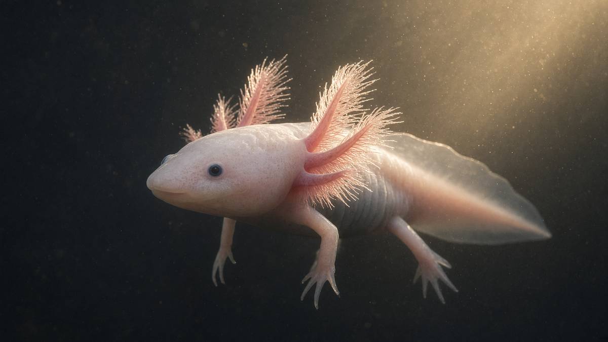
The axolotl does not scar. It rebuilds the limb.
A salamander with a 32-gigabase genome can amputate a leg and grow a perfect one back — bone, muscle, nerve, skin. The blastema is the most interesting factory in vertebrate biology.
What this essay actually tells you
- Ambystoma mexicanum can regenerate a complete limb, jaw, and portions of heart and spinal cord without a scar. Complete. Without a scar. Mammals cannot, which is the whole comparison.
- A blastema of progenitor cells rebuilds the pattern. Positional identity along the limb axis is the unsolved software. We can name the cells. We still can't write the code.
- Mammals scar instead. That's why axolotl single-cell atlases are now a regeneration library rather than a curiosity you show undergraduates.
What this actually means
Cut the leg off an axolotl and it grows another one. Not a stump. A limb, with the right number of fingers, the right bones, the right nerves. A blastema (a mound of lineage-restricted progenitors that remember where they're on the body) does the building. Mammals mostly scar. The axolotl mostly doesn't. Its genome is ten times the size of ours and was only fully assembled in 2018, because a 32-billion-base salamander is a sequencing headache. Repair peptides in this catalogue are probes of mammalian programmes. The axolotl is what a vertebrate looks like when the programme still includes 'replace the whole part'. We keep this animal next to BPC-157 on purpose.
Ambystoma mexicanum is a neotenic salamander that will, after amputation, rebuild a complete limb: bone, muscle, nerve, vasculature, and skin without a scar. The same animal regenerates a jaw, a tail, portions of the ventricle, and stretches of spinal cord. That's a laboratory fact older than molecular biology, sitting in tanks from the Ambystoma Genetic Stock Center in Kentucky to Vienna, Dresden and Mexico City, and it's the existence proof that a tetrapod body plan isn't obliged to scar. Mammals, ourselves included, mostly are. Cut a mouse at the stylopod and you get a fibrotic stump. Cut an axolotl at the same anatomical level and, over weeks, you get a limb with the right number of digits. The difference is a developmental programme mammals parked, plus an immune and fibrotic tone mammals didn't. We remain slightly furious about the parking. What follows is the factory — a blastema of lineage-restricted progenitors — the unsolved software of positional identity, the thirty-two-gigabase genome that made the factory hard to read until 2018, and the reason a catalogue that already talks about gastric cytoprotection and actin buffering keeps this animal on the same desk.
In short. This salamander can grow back a whole leg, a jaw, and parts of its heart and spinal cord, without a scar. Mammals can't.
Most of what we call repair, in a mammal, is a negotiation with a scar. Platelets arrive, a clot forms, neutrophils and then macrophages patrol, fibroblasts become myofibroblasts, transforming growth factor-β drives collagen I and III into a bundle, and the original geometry doesn't return. Distal fingertip injuries in children, liver, endometrium, and a modest amount of neonatal heart are the exceptions a methods section is expected to name, and they're spatially or temporally restricted. Ambystoma skipped that deal. Distal to a cut, a wound epidermis signals, a blastema accumulates, and over weeks a limb is redrawn from the cut surface outward, autopod last, like development happening again with a memory of the missing piece. Magic is the wrong word. It's reuse of the limb-bud toolkit — FGFs, SHH, WNTs, BMPs, retinoic acid — inside an adult animal that didn't trade that toolkit for a fibrotic default. A human who loses a hand still gets a scar. The distance between those two sentences is the research programme, and it's a long one.
In short. In a mammal, a deep cut usually becomes a scar and the original shape doesn't return. The salamander redraws the missing part from the cut surface.
The factory is a blastema: a mound of proliferating, largely lineage-restricted progenitors that assemble under a specialised wound epidermis and rebuild the missing pattern. The 20th-century romance was a soup of totipotent cells. The 21st-century measurement is ruder and more interesting. Muscle progenitors make muscle, dermal fibroblasts make dermis and, crucially, carry much of the positional map, Schwann cells remain Schwann, epidermis remains epidermis. Kragl, Tanaka and colleagues, Nature 2009, is still the paper a colleague from another lab should read first: cells keep a memory of their tissue origin. What they don't automatically keep, or rather what they have to be told again, is where they sit on the proximal–distal, anterior–posterior and dorsal–ventral axes of the limb. That positional identity is the unsolved software. Hardware — who is allowed to become what — is now a single-cell atlas problem, and the atlases exist. Software — how a wrist blastema makes a hand and a shoulder blastema makes an arm — is still a wiring diagram with missing pages. Keep those two apart. The rest of this piece is commentary on it.
In short. A mound of building cells remakes the missing piece. Those cells remember what tissue they came from. The unsolved puzzle is how they know where they're on the limb.
Why this page sits next to BPC-157 and TB-500: those peptides are mammalian probes of endothelium, nitric oxide, actin and migration, real programmes, characterised, in the catalogue. Body protection compound-157 is a fifteen-residue gastric fragment, GEPPPGKPADDAGLV, that the Zagreb papers and the cleaner endothelial papers put against lesion area and VEGFR2 phosphorylation. TB-500 orbits thymosin β4, a G-actin sequesterer whose LKKTETQ motif is the actin-binding idea a scratch assay actually tests. Neither molecule is a bottled blastema. Anybody who writes a closed scratch in a fibroblast monolayer as a regenerated limb has collapsed a millimetre of crawl into a developmental field, and the collapse is how repair talk gets sloppy. The axolotl is the existence proof that a tetrapod can go further than a probe. It can redraw the part. We stock the probes because the mammalian papers are real. We keep the salamander on the reading list because anybody who writes about repair without mentioning this animal is telling half the story, and we got tired of the half.
In short. Two research peptides on this shelf probe how mammal cells move and how vessels behave. Neither is a bottled version of the salamander’s rebuild.
Wound epidermis, then a mound
Amputation is the assay. Within hours the cut surface is covered by a wound epidermis, a specialised epithelium that isn't ordinary skin and isn't yet the apical epithelial cap it will become. Mescher's older histology, Satoh, Gardiner, Bryant on neurotrophic regulation of epidermal dedifferentiation, and a stack of Tanaka reviews, all say a version of the same sentence: without that epithelium, a blastema doesn't form. Dermal collagen is degraded rather than deposited as a scar. Underlying cells — connective-tissue fibroblasts above all — accumulate, re-enter the cell cycle, and build a mound. The apical epithelial cap, the amphibian analogue of the embryonic apical ectodermal ridge, then sits on that mound and supplies FGFs and other factors the blastema wants. Denervate below a threshold and the cap and the mound both fail; Marcus Singer quantified that nerve requirement in the 1950s, and it hasn't gone away. A mammal, facing the same cut, writes a provisional matrix and then a scar. The first fork in the road is already here: epithelium that instructs, versus epithelium that seals.
In short. Right after the cut, a special skin covers the wound and building cells gather underneath. If the nerves are gone, that gathering fails. A mammal tends to seal and scar instead.
The blastema isn't an embryo and it isn't a tumour. It's a proliferating field with a proximal stump as its reference. Over days to weeks, depending on animal size and on whether you cut at the wrist or the shoulder, the mound grows, patterns, and differentiates in a proximal-to-distal sequence: stylopod, then zeugopod, then autopod. Roensch, Tazaki, Chara, Tanaka, Science 2013, argued for progressive specification of segments rather than a pure intercalation of a missing middle, which is a live argument with the older polar-coordinate and intercalation literature of French, Bryant, Stocum. Both pictures can sit in one methods section if you let them. Distal cells know they're distal. A wrist blastema makes a hand. A mid-humerus blastema makes everything distal to the cut. Graft a distal blastema onto a proximal stump and, in the classical experiments, extra structure appears where identities fail to match. That's pattern regulation, named, and it's why 'the blastema rebuilds the limb' is true and incomplete. The blastema rebuilds the limb from the identities it has, toward the identities it still needs.
In short. The mound grows from the cut outward, making upper arm, then forearm, then hand. A wrist mound makes a hand. A shoulder mound makes a whole arm.
Nerve dependence is the sentence that still surprises people who think regeneration is a property of the skin. Singer showed that a limb needs a threshold of innervation; below it, regeneration aborts and a stump results. Kumar, Godwin, Gates, Garza-Garcia, Brockes, Science 2007, named a secreted factor in newts — nAG, newt anterior gradient, a ligand for Prod1 — that's supplied by Schwann cells after the nerve is cut and that can, in the newt, rescue a denervated blastema. Axolotls aren't newts, and the exact orthologue story is messier than a review slide, which is a reason to keep the species names in the citations. The broader fact holds across salamanders: the nerve isn't just a cable the new limb will need later. It's an early trophic and patterning input. Accessory-limb experiments, in which a wound plus a rerouted nerve plus a skin graft from the opposite side of the limb can induce a supernumerary limb, are the existence proof that the ingredients are local and combinatorial. A mammal has nerves too. What it doesn't have, after a proximal amputation, is this conversation between nerve, wound epidermis and positional skin.
In short. The new limb needs nerves from the start, not only at the end. Rerouting a nerve to a wound can even start an extra limb if the skin identities disagree.
MARCKS-like protein is the initiating molecule the field can actually name. Sugiura, Wang, Barsacchi, Simon, Tanaka, Nature 2016: an extracellular signal, detected in a screen, sufficient in the experiments they ran to induce the early proliferative response in axolotl appendage regeneration. Myristoylated alanine-rich C-kinase substrate is a family most of us met, if at all, as an actin-binding PKC substrate in a mammalian cytoplasm. The salamander version, after injury, is a cue. That's a more interesting sentence than a growth-factor catchphrase, because it sits at the fork between regeneration and scarring: something has to tell the stump to build a blastema rather than a fibrotic cap. Transforming growth factor-β, FGFs, WNT, thrombin-derived peptides, the complement literature, macrophages as required residents (Godwin, Pinto, Rosenthal, PNAS 2013): the neighbourhood is crowded, and a single named protein isn't a complete circuit. It's a foothold. If you're about to claim an initiating molecule in a dish, the Sugiura assay is the one a sceptical colleague will ask you to match. A mammalian peptide that closes a scratch hasn't matched it.
In short. One named protein released after injury helps start the rebuild. Many other signals sit around it. Closing a scratch in a dish isn't the same test.
Proliferation in the mound is a growth-control problem as well as a patterning problem, and mTOR sits on it. The blastema has to make mass: cells, matrix, a volume that will become a limb. Mechanistic target of rapamycin complex 1 reads amino acids, energy and growth-factor tone and then licenses translation. Zhulyn, Whited and colleagues, in work that put an evolutionarily divergent mTOR against the regenerating translatome, showed that this node isn't a background kinase in the axolotl; it's remodelled, and the remodelled translatome is part of how a tissue that should scar instead grows. Rapamycin, in the experiments where it has been used, shrinks the proliferative response. That's a tool, not a protocol for a person. A mammal has mTORC1 too — every growing cell does — and still scars at a proximal amputation. The difference isn't the presence of the kinase. It's which translatome, in which cell type, under which immune tone, the kinase is allowed to write. Name the node. Then name the cell. A homogenate phospho-S6 blot of a whole blastema is a scout, not a paper.
In short. The mound has to grow, and a well-known growth switch in cells is part of that. Blocking the switch shrinks the rebuild. Mammals have the same switch and still scar.
Cells keep a memory of their tissue origin during axolotl limb regeneration. Muscle makes muscle. Dermis makes dermis. The blastema is a coalition, not a totipotent soup.— Kragl M, Knapp D, Nacu E, Khattak S, Maden M, Epperlein HH, Tanaka EM. Cells keep a memory of their tissue origin during axolotl limb regeneration. Nature. 2009; 460: 60–65.
Lineage-restricted, not totipotent
Kragl's 2009 grafting and transgenesis paper is the document that retired the totipotent-blastema romance, and it deserves to be read rather than cited from a slide. They labelled tissues, amputated, and asked what those tissues became in the regenerate. Skeletal muscle didn't make epidermis. Schwann cells didn't make muscle. Dermal fibroblasts made dermis and, importantly, could contribute to skeletal elements — connective tissue is the plastic lineage, not a free-for-all. Cartilage made cartilage. The blastema is therefore a mosaic of progenitors that remember their germinal job. That's ruder than the 20th-century picture and much more useful, because it tells you where to look for the map. If muscle cells can't become cartilage, then the proximal–distal pattern of the skeleton isn't being drawn by muscle. It's being drawn by the connective-tissue cohort. Gerber, Murawala, Knapp, Tanaka, Treutlein, Science 2018, then took that logic into single cells and found lineage segregation and an adult connective-tissue stem-cell neighbourhood that's the current address of positional identity. Pattern is a fibroblast problem. Myogenesis is a myogenic problem. Stop writing 'blastema cell' as if it were one thing.
In short. Building cells don't forget what they were. Muscle stays muscle. Skin stays skin. Connective-tissue cells are the ones that carry much of the map of the limb.
Dedifferentiation is a word that needs a species tag. In newts, multinucleate skeletal-muscle fibres can fragment, cell-cycle re-enter, and contribute progenitors to the blastema; that's the classical picture Brockes and Kumar spent years on. In axolotls, Sandoval-Guzmán, Wang, Simon, Tanaka, Nature 2014, watched live and found that skeletal muscle in the regenerate comes from PAX7-positive satellite cells, the same resident stem-cell pool a mammalian muscle uses after a strain, not from fibre fragmentation. Two salamanders, two recruitment strategies, one word in the reviews. A methods section that says 'the axolotl dedifferentiates muscle' without that 2014 paper is a methods section from a different animal. Connective-tissue cells do loosen their differentiated programme and migrate into the mound; that's a real, imaged event, and it's the event that matters for pattern. Satellite-cell recruitment is the event that matters for muscle mass. Both happen. They aren't the same experiment. Live imaging, Cre lineages, and a willingness to say 'newt' when you mean newt, are how this heading stays honest.
In short. Newts and axolotls don't rebuild muscle in quite the same way. Axolotls lean on the same resident muscle stem cells a mammal uses after a strain.
Connective tissue is the patterning tissue, and that sentence is the one a peptide assay can't reach. McCusker, Gardiner, the Bryant laboratory's skin-graft work, and then Gerber's single-cell atlas, all converge: fibroblasts from different coordinates of the limb carry different positional identities, and those identities are what a blastema intercalates or progressively specifies. Swap skin from the opposite side of the limb, add a nerve, and you can induce a supernumerary limb because you have created a positional discontinuity the system tries to fill. Muscle from a wrist, grafted into a shoulder blastema, doesn't rewrite the skeleton as a hand. The skeleton listens to connective tissue. That's why a mammalian fibroblast scratch — even a clever one, even one with a thymosin-β4 analogue in the medium — is a migration assay and not a pattern assay. Migration is necessary. Pattern is a coordinate system. The unsolved software lives in the fibroblast cohort, in a code that retinoic acid can proximalise and that Prod1 has been proposed to hold, and that a single-cell atlas can now name as a transcriptional state even while the wiring remains incomplete.
In short. The map of the limb lives mainly in connective-tissue cells. Moving those cells, or mixing skin from opposite sides, can change what gets built. A scratch in a dish can't.
Epidermis, endothelium and Schwann cells have their own jobs and their own limits. Wound epidermis becomes the apical cap and has to stay specialised for weeks; if it reverts to ordinary skin too soon, the mound fails. Endothelium has to vascularise a growing blastema without laying down the granulation tissue a mammal would recognise as the road to a scar; nitric-oxide tone, VEGF, and a calmer macrophage conversation are in that neighbourhood, which is why a gastric pentadecapeptide with endothelial papers sits on the same desk without being a blastema factor. Schwann cells remyelinate the axons that will innervate the new limb, and they're one source of the nerve-derived cues the mound wants. Lineage restriction isn't a failure of plasticity. It's how you rebuild a three-dimensional organ without scrambling cell types. A totipotent soup would be a tumour with a memory problem. A coalition of restricted progenitors with a shared coordinate system is a limb. The atlas papers are how that sentence stopped being a hope.
In short. Skin, blood vessels and nerve-support cells each keep their jobs. That restriction is how you rebuild a limb rather than a confused lump of tissue.
Positional identity is the unsolved software
A limb has three axes, and a blastema has to know its address on all three. Proximal–distal runs shoulder to fingertip: stylopod, zeugopod, autopod, the nested segments a tetrapod already used in the embryo. Anterior–posterior runs thumb-side to little-finger-side, the axis sonic hedgehog patterns from the zone of polarising activity in a bud. Dorsal–ventral is the back of the hand versus the palm, a Wnt7a neighbourhood in the embryo and a less tidy literature in the regenerate. Wolpert's positional-information idea — a cell knows where it's, and that knowledge decides what it becomes — was written with hydra and with chick limbs, and salamander regeneration is still the adult test of it. The hardware of lineage says what a cell is allowed to become. The software of position says which part of the allowed repertoire it should run. We've the hardware in atlases. We don't yet have a complete address code. That isn't a failure of the field. It's the interesting problem. If you write 'the axolotl just knows' has skipped a supernumerary limb, a retinoic-acid-proximalised blastema, or a Prod1 blot.
In short. A rebuilding limb has to know shoulder versus fingertip, thumb versus little finger, and back versus palm. Cells remember those addresses. We still don't have the full code.
Retinoic acid is the pharmacological handle the field actually has, and it's a handle, not a master key. Treat a distal blastema with RA and you proximalise it: a wrist cut starts making upper-arm structures, a result Maden, Stocum and Brockes established before anyone had a genome. The interpretation is that RA raises a proximal identity, in part by raising Prod1, a GPI-anchored cell-surface protein whose newt orthologue da Silva, Gates and Brockes put on the proximal–distal map in Developmental Cell, 2002. Prod1 binds nAG in the newt story. Whether the axolotl orthologue does the same job at the same address is a sentence with more clauses than a press release wants. What isn't in dispute is that a vitamin-A metabolite can move the identity of a blastema along one axis. That's rare. Most of pattern is a conversation you can't pipette. RA you can. Dose, stage, and the fact that RA is also a teratogen in a mammalian limb bud, are why this handle is a laboratory object and not a clinic object. It's also why any 'regenerative' peptide that can't move positional identity isn't doing this job, whatever it does to a scratch.
In short. A vitamin-A relative can make a hand-level rebuild start making upper-arm parts instead. That's one of the few drugs that can shift the address of the mound.
Anterior–posterior identity has a cleaner embryonic grammar and a messier regenerative one. Nacu, Gromberg, Oliveira, Drechsel, Tanaka, Nature 2016: FGF8 and SHH can substitute for anterior–posterior tissue interactions that a regenerating axolotl limb normally needs. Anterior and posterior connective tissues have to talk; if they don't, the mound doesn't pattern an autopod. Supply the two ligands and you can, in the experiments they ran, bypass some of that conversation. That paper is the reason a methods section can now say 'the bud toolkit is reused' without hand-waving. Sonic hedgehog from posterior mesenchyme, FGFs from the cap, a feedback loop a chick embryologist would recognise. Reuse isn't identity. An adult blastema is innervated, immune-patrolled, and sitting on a stump that already has a history. The same ligands, a different context, a different clock. Accessory limbs, extra digits, mirror-image duplications when you scramble the conversation: those are the phenotypes that tell you the software is real. They're also the phenotypes that tell you it can crash.
In short. Thumb-to-little-finger pattern needs a conversation between opposite sides of the limb. Two well-known developmental signals can, in experiments, stand in for some of that talk.
Intercalation is the older software metaphor, and it still earns its keep. French, Bryant and Bryant's polar-coordinate model said that when two cells with non-adjacent positional values meet, the system fills in the missing values. Graft a left blastema onto a right stump, or anterior skin onto a posterior wound, and extra limbs or extra segments appear where the discontinuity was. Stocum's intercalary-regeneration experiments — a distal regenerate on a proximal stump filling the gap — are the salamander version of the same idea. Tanaka's progressive-specification paper is the competing picture: the blastema lays down segments in order from the cut, rather than computing a missing middle all at once. The data haven't given one picture a knockout. A colleague from another lab should hold both. What neither picture yet supplies is the molecule that's the coordinate. Prod1 is a candidate on one axis. Hox genes are transcribed in regenerating limbs, as they're in buds, and they aren't a full address book. Until someone can read a fibroblast's position as a number, and write a new number, and get a predicted segment, the software is unsolved. That's the sentence this heading is for.
In short. When cells from mismatched addresses meet, extra parts can grow to fill the gap. We still don't know the molecular number those cells are reading.
Hox transcription, chromatin, and the slow floor under a ligand: an edit or a peptide that doesn't change what a fibroblast transcribes won't change what a blastema builds. HOXA13 and HOXD13 mark distal identity in buds; more proximal Hox paralogues mark stylopod and zeugopod. Regenerating axolotl limbs re-deploy that code, and the 32-gigabase genome made even that sentence hard to write until the 2018 assembly gave the Hox clusters a map. Promoters, enhancers, the apical-cap FGF input, retinoic-acid response elements: Pol II has to find the locus in a nucleus that holds ten times more DNA than ours. Closed chromatin at a Hox gene is a failed segment, whatever the blastema's cell census says. This is why the transcription diagram belongs in a regeneration essay and not only in a peptide-receptor essay. Pattern is a transcriptional state in a connective-tissue cell. Migration is actin. Growth is mTOR and a cell cycle. Three floors. A closed scratch is floor two, sometimes. A limb is all three, plus the address.
In short. Limb-identity genes must be switched on in the right cells at the right time. If those genes stay off, the mound can't build the right part, however many cells it has.
Diagram
Closed chromatin (H3K27me3, DNA methylation) hides the promoter. Pioneer factors and histone acetyltransferases open it.
PIC: TFIID, TFIIH, Mediator, Pol II. Ser5 phosphorylation of the CTD lets the polymerase leave the promoter.
Elongation ~20–40 nt/s. Capping, splicing, cleavage and polyadenylation happen on the still-growing RNA.
Human genes are islands in 3.1 billion base pairs of mostly noncoding sequence. Promoter, enhancers, chromatin state and the Mediator complex decide whether Pol II is allowed to fire. Epithalon’s literature sits on TERT and pineal clocks — two of the rare promoters anyone bothers to name in a peptide essay.
Mammals scar. Axolotls largely do not.
A mammalian wound that reaches dermis has a default: fibrosis. Platelets and a fibrin clot, neutrophils, then macrophages that often stay in a pro-inflammatory register, fibroblasts that become α-smooth-muscle-actin-positive myofibroblasts, TGF-β1 as the cytokine the fibrosis literature can't stop naming, collagen I and III bundled by lysyl oxidase, and a scar that contracts. The original architecture of hair follicles, glands and a dermal papilla doesn't return in human skin. In a limb, the original architecture of a growth plate, a muscle belly and a patterned autopod doesn't return at all. That default isn't incompetence. It's a strategy: close the barrier fast, stop bleeding, keep infection out, live to reproduce. Salamanders, facing a wet, cold, pathogen-rich pond, somehow kept a different strategy: rebuild. The immune system isn't absent in the axolotl; Godwin's macrophage-depletion experiment produced scarring, which is the sentence that retired the 'they have no immune system' cartoon. Macrophages are required. What they don't do, in this animal, is drive the myofibroblast programme to a permanent collagen plug. Tone, not presence, is the variable.
In short. Mammals usually close a deep wound with a collagen scar. The salamander needs immune cells to rebuild, but those cells don't push the tissue into a permanent scar.
TGF-β is the fork, and both animals have it. In a mammal, TGF-β1 from platelets and from macrophages phosphorylates SMADs in fibroblasts, writes collagen and α-SMA, and locks a myofibroblast state that's hard to leave. In axolotl regeneration, TGF-β signalling is present and, in several reports, required for the early events; block it and the regenerate can fail. Lévesque, Roy, and the amphibian TGF-β literature are the place a mammalian fibrosis person should start before declaring a contradiction. The resolution isn't that axolotls lack TGF-β. It's that the dose, the isoform, the duration, and the partners — macrophages that resolve, a wound epidermis that doesn't become a fibrotic cap, a matrix that's degraded rather than bundled — write a regenerative conversation instead of a scarring one. A single cytokine doesn't own the phenotype. The network does. Anyone offering a TGF-β inhibitor as a bottled blastema has skipped the amphibian papers, in which the same pathway is a necessity, not a villain. Context is the whole story. Context is also why a peptide that moves nitric-oxide tone in a rat vessel hasn't, thereby, moved this fork.
In short. The same scar-linked signal exists in the salamander and is even needed early on. The difference is how long it runs, and what other cells are saying at the same time.
Children's fingertips are the mammalian echo, and they belong in this heading so that 'mammals can't' doesn't become a cartoon. Illingworth, Journal of Pediatric Surgery, 1974: distal fingertip amputations in children, treated conservatively, can regenerate a usable tip with nail, pulp and a surprising amount of shape, provided the cut is distal to the nail matrix. Muneoka, Simkin, Han, and the mouse P3 literature then put that echo on a laboratory animal: amputate through the terminal phalanx, keep the nail organ, and a mouse will regenerate a digit tip; amputate more proximally, through P2, and it won't. The nail epithelium is doing some of the work an apical cap does. The limitation is spatial. A child doesn't regenerate a hand. A mouse doesn't regenerate a paw from the wrist. The programme, if it's the same programme, has been spatially restricted to a millimetre of distal anatomy that still has a specialised epithelium and a relatively modest fibrotic response. We mention the children because that echo is the closest we get, and because it's already a paediatric fact rather than a salamander documentary.
In short. Young children can regrow a fingertip if the cut is far enough forward and the nail bed remains. They don't regrow a hand. That's the nearest mammalian echo.
Acomys, the African spiny mouse, and the MRL mouse are the other mammalian exceptions a regeneration essay is expected to name, with their sizes kept honest. Seifert, Maden, Nature 2012: Acomys heals large skin wounds by regeneration of hair follicles and dermis rather than by a dense scar, a phenotype that made the animal a celebrity and then a serious model. Ear-hole closure in MRL/MpJ mice was billed, for a while, as mammalian epimorphic regeneration; the follow-up has been more modest, strain-dependent, and not a limb. Neither rodent rebuilds a proximal limb. Neither has a blastema that will give you a stylopod. They're evidence that scarring isn't an iron law of the mammalian body plan, and they aren't axolotls. Acomys is the better of the two for skin. Digit-tip mice are the better for a true, spatially restricted blastema. Axolotls remain the tetrapod that kept the whole-limb programme. Stacking those three sentences is how you avoid both despair and a supplement label. The research programme is to find which pieces of the salamander conversation a mammal still has, and which it has parked.
In short. A few unusual mice heal skin or a digit tip better than we do. None of them grows back a whole leg. The salamander is still the animal that kept the full programme.
Heart is the organ that makes the mammalian limit personal. A neonatal mouse can regenerate myocardium for a few days after birth; after that window, ligation or infarction is a scar, a thinned wall, a failing pump. Adult zebrafish and adult axolotls will, after resection or cryoinjury, replace ventricular muscle with new cardiomyocytes, with a much calmer fibrotic transient. Oberpriller's older newt work, Cano-Martínez, Witman, the more recent axolotl ventricle papers: cell-cycle re-entry of cardiomyocytes, a less rigid extracellular matrix, an immune tone that doesn't lock a collagen wall. Human ischaemic heart disease is the scar. Peptides that occupy endothelial nitric-oxide neighbourhoods, or that buffer actin in a migrating cell, aren't going to reopen a neonatal regenerative window by themselves, and this journal won't write them as if they were. They're probes of programmes that sit next to the scar. The axolotl is the existence proof that an adult tetrapod ventricle can choose the other fork. Proof of possibility isn't a medicine. It's the reason the medicine is worth looking for, in the species that actually gets the infarct.
In short. Newborn mice can repair heart muscle for a few days; adults scar. Adult axolotls replace lost heart muscle. That possibility isn't a treatment, but it's why the search continues.
A 32-gigabase genome and a single-cell library
The axolotl genome is about thirty-two billion bases, ten times a human diploid haploid-equivalent, one of the largest genomes ever assembled, and the reason this animal was a developmental-biology hero long before it was a genomics one. Nowoshilow, Schloissnig, Fei, Tanaka, Hiller, Myers and colleagues, Nature 2018, is the landmark: a chromosome-scale assembly, gene models, and the slightly deflating finding that the gene count isn't correspondingly monstrous. The inflation is repetitive DNA — transposable elements, vast introns, a genome that's mostly not genes. Sequencing it was a decade-class headache because short reads can't span repeats, and because a 32-gigabase project doesn't fit in the memory of a casual assembler. PacBio long reads, Chicago and Hi-C scaffolding, and a community that wanted the Hox clusters badly enough to fund the pain, are how 2018 happened. Gene count roughly ordinary; junk, if it's junk, extraordinary. If you write that the axolotl regenerates because it has more genes hasn't looked at the assembly paper. It regenerates with a standard vertebrate gene kit sitting in a palatial non-coding landscape.
In short. The salamander’s DNA is about ten times longer than ours, mostly because of repeated padding, not extra genes. Assembling it took until 2018.
Why a palatial genome matters for regeneration is still an open, adult question, and we won't pretend it's closed. Long introns slow transcription; a gene with a 100-kilobase intron takes longer to transcribe than a compact mammalian orthologue, which has been offered as a cell-cycle and developmental-rate argument in other giant genomes. Transposable-element activity is a mutagenesis and a regulatory resource. The Ambystoma Genetic Stock Center's white and wild-type strains, Voss's linkage maps, and the 2018 gene models together let you now design CRISPR in this animal — Fei, Tanaka and others have already done so — rather than only graft and hope. Before the assembly, 'the gene for positional identity' was a phrase you could say in a seminar and not have to point at a locus. After the assembly, you can point, knock, and be wrong in a more useful way. Large genomes aren't a superpower. They're a methods tax that this community paid, and the receipt is a FASTA file a mammalian lab can BLAST. That's the quiet revolution underneath the cute pink animal.
In short. A huge genome isn't why regeneration works. It did make the genes hard to find. Now they can be mapped and, in some cases, edited.
Single-cell atlases turned the blastema from a histology noun into a library. Gerber, Treutlein, Tanaka, Science 2018: thousands of cells, lineage segregation, a connective-tissue cluster that's the address of much of the pattern. Leigh, Dunlap, Whited, Nature Communications 2018: the blastema niche at single-cell resolution, a transcriptomic landscape rather than a handful of markers. Subsequent atlases from the Whited, Monaghan, Tanaka and Murawala groups have added time, injury type, and comparisons to uninjured limb. You can now ask, in a browser, which fibroblasts are proximal, which macrophages are resolving, which satellite cells are cycling, and which wound-epidermis cluster is writing FGFs. That's a regeneration library. It's also a mammalian comparison library: take a scarring mouse wound, take an Acomys wound, take an axolotl blastema, and look at the same cell classes. The genes that differ are the candidate list a CRISPR screen, a drug screen, or a sceptical peptide assay would actually want. A library isn't a medicine. It's how you stop guessing which cell you were talking about.
In short. Single-cell surveys now catalogue which cells sit in the rebuilding mound and what they're switching on. That catalogue is how you compare a salamander to a scarring mammal.
Transcription programmes, not a mystery organ, are what those atlases actually contain. Connective-tissue fibroblasts in a blastema write a limb-bud-like state: HOX, SHH-pathway listeners, FGF receptors, retinoic-acid machinery, a set of extracellular-matrix genes that look more like morphogenesis than like a scar. Macrophages write a resolution-leaning set rather than a chronic TGF-β set. Wound epidermis writes a cap programme, not a stratified-squamous programme. Muscle progenitors write PAX7 and the myogenic regulatory factors. That's cell-type-specific transcription, the slow floor under every ligand this catalogue sells and under every growth factor the salamander already makes. A peptide in a well can, at best, occupy one receptor and move one of those programmes in one cell type. An atlas can tell you whether you occupied the cell that carries the map. Most mammalian repair papers never ask. The axolotl library is why they now can, even if the assay stays a mouse. Comparative single-cell biology is the method. The pink animal is the reference.
In short. Each cell type in the mound runs a different gene programme: map-holders, immune cells, skin, muscle. A drug or peptide, at best, nudges one programme in one cell type.
Scale is the honesty test this journal uses when a word like regeneration starts doing too much work. A covalent bond is a tenth of a nanometre. BPC-157 is a fifteen-residue chain, about a nanometre and a half, 1419 daltons. Thymosin β4 is forty-three residues, a few nanometres, a stoichiometric G-actin partner at tens to hundreds of micromolar. A fibroblast is a city of ten billion proteins and two metres of DNA — thirty-two gigabases of it, in this animal, folded into a nucleus that still has to find a Hox gene. A blastema is millimetres. A regenerated limb is centimetres. A person is seventy kilograms of fibrotic default. The salamander is being asked, in the popular version of this subject, to donate a medicine. The work is happening at the scale of a transcriptional state in a connective-tissue cell, a macrophage's cytokine tone, and an apical epithelium's FGF output. You can't pipette a blastema into a tendon. You can pipette a characterised ligand into a well, and you can read the atlas to see whether that well had the right cell. The ruler belongs on every sentence that jumps from a 15-mer to a hand.
In short. A research peptide is tiny. A rebuilding limb is made of many cells reading many genes. Jumping from a vial to a hand skips most of the sizes that matter.
Diagram
- 0.1 nmHydrogen atomA proton and an electron. Chemistry starts here.
- 0.3 nmWater molecule70% of a cell by mass. The solvent life is.
- 1 nmAmino acidTwenty kinds. Peptide bonds string them.
- 2–4 nmResearch peptideA named chain. BPC-157 is 1.4 kDa, 15 residues.
- 4–10 nmGlobular proteinHaemoglobin, a GPCR’s extracellular face.
- 25 nmRibosomeThe factory that reads mRNA into protein.
- 5 nmMembraneA lipid bilayer. Every compartment starts here.
- 0.5–1 µmMitochondrionA bacterium the cell swallowed and kept.
- 6–10 µmNucleusTwo metres of DNA folded into a sphere.
- 10–30 µmTypical cellA city. 10¹⁰ proteins. One genome.
- 1 mmTissue grainA thousand cells talking across ECM.
- 1.7 mYou~36 trillion human cells. Most of them are red blood cells.
Lengths are characteristic, not exact. A research peptide is closer in size to a water molecule than to the cell that assays it — which is why a 15-mer can occupy a receptor pocket a small-molecule drug also wants.
- Axolotl genome
- 32 Gb
- Human haploid genome
- ~3.2 Gb
- Blastema
- days to mound, weeks to limb
- Lineage
- restricted progenitors
- Initiating cue
- MARCKS-like protein
- Mammalian echo
- child P3 / mouse P3
- BPC-157 neighbour
- 15-mer, ~1.4 kDa
- TB-500 neighbour
- actin motif / Tβ4 orbit
Nowoshilow et al., Nature 2018. Repeats and giant introns. Gene count roughly ordinary.
Tenfold smaller. The regenerative difference is not a gene census.
Clock depends on animal size and amputation level. Autopod last.
Kragl, Nature 2009. Connective tissue carries much of the map (Gerber, Science 2018).
Sugiura, Nature 2016. Macrophages required (Godwin, PNAS 2013).
Illingworth 1974; Muneoka digit-tip. Spatially restricted. Not a hand.
Gastric fragment, endothelial and nitric-oxide papers. Not a blastema.
G-actin buffer, migration assays. A crawl is not a pattern.
The cytoskeleton of a rebuilding limb
A blastema cell has to crawl before it can pattern. Connective-tissue fibroblasts loosen from the stump, polarise, and move into the mound; endothelial cells sprout; macrophages patrol; myogenic progenitors take up their positions. The motor of that crawl is actin. Globular actin polymerises onto the barbed end of a filament, depolymerises from the pointed end, and the filament treadmill is how a leading edge advances. Profilin charges monomers with ATP, Arp2/3 nucleates branches, cofilin severs, thymosin β4 holds a reserve of G-actin so the cell isn't polymerised into a static gel. That's the same physics a mammalian fibroblast uses to close a scratch, and the same physics a catalogue peptide in the thymosin-β4 orbit is asking about. Concentration regimes matter. Intracellular Tβ4 is a stoichiometric buffer, tens to hundreds of micromolar. A growth factor is picomolar to nanomolar at a receptor. Writing those two as one 'healing' sentence is how a mechanism becomes a caption. The axolotl doesn't have a different actin. It has a different conversation around the crawl: a wound epidermis that instructs, a macrophage that resolves, a fibroblast that's also a coordinate. The motor is shared. The destination isn't.
In short. Cells must crawl into the rebuilding mound, and they crawl by assembling and taking apart actin filaments. Mammals use the same motor. They don't use it to redraw a limb.
Diagram
BPC-157: Pro-rich, acid-stable, Sikiric corpus. VEGFR2 internalisation, FAK–paxillin, eNOS-dependent NO tone. A cytoprotection story that escaped the stomach.
TB-500: cytoskeletal buffer. Injury releases Tβ4 extracellularly; VEGF, MMPs and keratinocyte migration follow. SDKP is a separate N-terminal anti-fibrotic pharmacophore. Two literatures, two jobs.
Thymosin β4 is the principal G-actin sequestering peptide. TB-500 is built around the LKKTETQ motif. BPC-157 is a gastric 15-mer (GEPPPGKPADDAGLV) that talks to VEGFR2 and focal adhesions. Related in folklore. Unrelated in mechanism.
TB-500, in this neighbourhood, is a mammalian probe of that motor, and the neighbourhood has to stay a neighbourhood. Thymosin β4 is a 43-residue intrinsically disordered protein, the principal G-actin sequesterer in many cytoplasms; LKKTETQ is the actin-binding motif the mutagenesis papers named. The analogue some catalogues sell as TB-500 isn't the 43-mer, and the 43-mer isn't Ac-SDKP, the anti-fibrotic N-terminal fragment a prolyl oligopeptidase can bite off. Three pharmacophores, three experiments, a mess if you let a trade name collapse them. A pyrene-actin polymerisation curve, a G-actin binding measurement, a scratch with a stated cell type and a stated nanomolar-to-micromolar dose: those are the assays. None of them is a blastema. None of them moves Prod1. A closed scratch in eight hours is a millimetre of actin treadmill. An axolotl limb in eight weeks is a coordinate system that used a treadmill along the way. We stock the characterised chain because the actin papers are real. We won't write it as a salamander in a vial. The diagram above is the motor. The salamander is the destination the motor, in a mammal, doesn't reach.
In short. The actin peptide on this shelf tests the crawling motor in mammal cells. It isn't the salamander’s address code, and a closed scratch isn't a new limb.
Endothelium is the other mammalian programme this desk already stocks, and it sits one door along. A blastema has to be vascularised; ischaemic mesenchyme doesn't pattern, it necroses. VEGF, VEGFR2, nitric-oxide tone, endothelial migration and a calmer clotting conversation at the new capillaries are the mammalian language for that job. BPC-157, GEPPPGKPADDAGLV, entered the literature as a gastric cytoprotectant and then accumulated endothelial phosphorylation papers: VEGFR2 internalisation, FAK, paxillin, an NO-system argument the Zagreb corpus framed as a reset. Independent labs can blot some of that; some of the wider injury-model corpus they can't. Either way, a 15-mer that a vessel cell notices is a vessel-cell reagent. It isn't an apical-epithelial-cap factor, and it isn't MARCKS-like protein. Putting it next to the axolotl is a reading-list courtesy: both conversations touch repair, both touch endothelium, and only one of them redraws a limb. A methods section that treats them as interchangeable healing juice hasn't named a receptor, a lineage, or a coordinate. We'll sell you the named 15-mer. We won't design the regenerate.
In short. New tissue needs a blood supply. A stomach-derived research peptide on this shelf talks to vessel cells. That's next door to a blastema, not the same room.
Nitric oxide, actin and a macrophage: three mammalian handles, none of them a positional code. eNOS makes NO in endothelium; NO relaxes smooth muscle and, at signalling doses, S-nitrosylates a neighbourhood of proteins. Actin is the crawl. Macrophages, in Godwin's depletion, are the required immune residents without which the axolotl scars like a mammal. A peptide that moves NO tone, a peptide that buffers G-actin, a drug that shifts macrophage polarisation: those are lawful, named, assayable objects in a mouse. They're also the objects people most often smuggle into a 'regeneration' sentence. Smuggling is the error. The axolotl uses all three and then does something a mammal doesn't: it keeps fibroblasts in a morphogenetic transcriptional state instead of a myofibroblast state, under an epithelium that stays a cap, with a coordinate system RA can still move. Occupy the handles if your assay is a vessel or a scratch. Don't write the occupancy as a hand. The peptide map that follows is a reminder that neighbourhood of cell biology isn't identity of invoice.
In short. Blood-vessel signals, the crawling motor, and immune cells are all real mammalian handles. The salamander uses them and then still has to run a map. A handle isn't a limb.
Diagram
| Node | Catalogue | Conversation |
|---|---|---|
| GPCR | Ipamorelin, MT2, PT-141, retatrutide, CJC | Second messengers, secretion, appetite, pigment |
| RTK / IGF1R | IGF-1 LR3 | IRS–PI3K–Akt–mTOR and Shc–ERK |
| Cytokine receptor | Somatropin (HGH) | GHR–JAK2–STAT5b, hepatic IGF-1 |
| Cofactor | NAD+ | Sirtuins, PARPs, CD38, redox |
| Actin buffer | TB-500 / Tβ4 motif | G-actin sequestration, motility |
| Growth-factor-like | BPC-157 | VEGFR2 / FAK / eNOS neighbourhood |
| Copper ligand | GHK-Cu | Transcriptome shift in fibroblasts |
| MC fragment | KPV | NF-κB, PepT1, no pigment |
| Nuclear / pineal | Epithalon (AEDG) | TERT and melatonin literatures |
| mtORF peptide | MOTS-c | AMPK, folate–methionine cycle |
Each row is a different kind of molecular conversation. The catalogue peptides bind at these nodes; they are not interchangeable, and stacking them because a forum did mixes unrelated literatures.
Heart, cord, jaw — the rest of the programme
Limb is the celebrity, and it isn't the whole animal. Resect a piece of axolotl ventricle and cardiomyocytes re-enter the cell cycle, the matrix stays a scaffold rather than a wall, and over weeks the chamber is restored with a much smaller scar than a mouse would keep. Cryoinjury models, closer to an infarct than a clean resection, still resolve in this species in a way adult mammalian myocardium doesn't. The comparison that matters isn't axolotl versus zebrafish — both adult regenerators — but axolotl versus postnatal mouse, where the regenerative window closes in days. Human cardiology lives on the mouse side of that comparison. Whatever you think about a gastric 15-mer and a nitric-oxide blot, the ventricular fact is why a peptide house that already talks about endothelium isn't wasting a page on a pond salamander. Adult tetrapod heart muscle can cycle. Ours mostly doesn't. The distance is a research programme in cardiomyocyte cell-cycle, matrix stiffness and immune tone, not a reconstitution protocol.
In short. Cut a piece out of this salamander’s heart and the muscle cells divide and fill the gap. Adult human heart muscle mostly doesn't. That gap is the research programme.
Spinal cord is the sentence that should be impossible, and in this animal it isn't. A complete transection, in an axolotl, is followed by bridging, by neurogenesis, and by a recovery of swimming that a mammalian cord injury laboratory wouldn't dare to write as an endpoint. Tazaki, Tanaka, Monaghan, Hui: ependymal cells, a less inhibitory matrix, a macrophage conversation that doesn't lock a glial scar of the mammalian kind. Axons cross. Neurons are born. The cord isn't a limb blastema, and the lineages aren't the same, and we won't collapse them. It will say that the same animal that redraws a hand also refuses the glial-scar default that makes human spinal-cord injury a permanent geography. Portions of the CNS, the dek said. Portions is doing honest work: this isn't a whole-brain rebuild, and it isn't a licence to imply a person. It's a vertebrate CNS that, after a cut, chooses morphogenesis over a permanent plug. Mammalian spinal-cord research has been trying to negotiate with that plug for decades. The salamander doesn't negotiate. It rebuilds.
In short. This salamander can reconnect a cut spinal cord well enough to swim again. A mammal, after the same kind of cut, is left with a scar that axons struggle to cross.
Jaw, lens, tail, gills: the list is long because the programme isn't a limb special. A resected lower jaw is rebuilt with bone and teeth in a geometry a maxillofacial surgeon would recognise as the original, not as a graft. Larval axolotls, like newts, can regenerate a lens; Wolffian regeneration from iris pigmented epithelium is the classical newt version, and the axolotl larval version is a reminder that the eye isn't exempt. The tail is a spinal-cord-plus-fin-plus-muscle regenerate, a second celebrity organ, the one a lot of live imaging has actually used because it's optically kinder than a limb. Neoteny — sexual maturity in a larval body, gills kept, metamorphosis optional — is why this species is a laboratory animal. You can breed it without a land phase. You can see through some of it. You can amputate and watch. Ambystoma tigrinum, the tiger salamander, metamorphoses and is a different husbandry. mexicanum stayed wet, stayed larval, and became the tetrapod a regeneration laboratory can actually staff. Xochimilco, the wild lakes, is where the wild type is dying. The laboratory is where the library is being written. Both facts belong in a methods section that uses the animal.
In short. Jaw, lens and tail also come back. The species stays larval into adulthood, which is why a laboratory can breed it and watch. Wild populations are in trouble.
Xenobots sit on the neighbouring frontier page, and the neighbourhood is morphogenetic plasticity, not a shared method. Levin, Blackiston, Bongard: wild-type frog cells, sculpted by an algorithm and by hand, walking as a body plan no tadpole used. No genome edit. Anatomy as a software layer. The axolotl is the opposite dare in one sense and the same dare in another. Opposite: the genome is enormous, the body plan is the one the embryo already had, and the trick is to rerun it after injury. Same: cells contain more anatomy than the default fate map admits. A fibroblast that can be a scar or a coordinate is a morphogenetic fact. A frog cell that can be a tadpole epidermis or a millimetre-scale walker is a morphogenetic fact. De-extinction rewrites hardware. Xenobots rewrite the chassis without touching the genome. Axolotl regeneration reruns the chassis from a stump, genome intact, positional software still not fully read. Three frontier essays, one decade, one lesson a peptide assay should steal: anatomy isn't only DNA, and a ligand isn't a body plan. We'll keep stealing it.
In short. Frog cells can be shaped into tiny walkers without changing their DNA. The salamander reruns its own body plan from a stump. Both say anatomy is more flexible than a fate map admits.
What a mammalian assay can actually ask
An honest mammalian assay in this neighbourhood names a tissue, an insult, a clock and a readout, and then stops before the word limb. Gastric lesion area after indomethacin, at a stated hour, is an assay. Load-to-failure of a tendon-to-bone junction at six weeks is an assay. Tenocyte outgrowth from an explant, endothelial migration across a scratch in eight hours, a pyrene-actin curve, VEGFR2 phosphorylation at a stated minute, a macrophage-polarisation panel: assays. A regenerated stylopod isn't among them, in a mouse, in a person, or in a well. The axolotl atlas is a comparison library for those assays, not a substitute for them. If your fibroblast, after a ligand, writes a more blastema-like transcriptome and a less myofibroblast-like one, you have a result a Tanaka-group reviewer might actually read. If your fibroblast closed a scratch and you wrote 'regeneration', you have a caption. We'll keep the first. We won't print the second as physiology. The salamander doesn't make the mouse assay uninteresting. It makes the overclaim obvious.
In short. In mammal work, say the tissue, the injury, the time and the measurement. A closed scratch isn't a new limb. The salamander papers are the comparison, not a free upgrade to the claim.
Cell-type choice is a control, not a convenience. A HEK293 well will tell you whether a construct and a ligand move a blot. It won't tell you what a positional fibroblast does with retinoic acid, and it won't tell you what a Godwin-type macrophage does to a neighbour's matrix. Primary tenocytes aren't blastema connective tissue. Dermal fibroblasts from a human scar aren't Acomys dermis and aren't axolotl stump fibroblasts. If the claim is anti-fibrotic, show α-SMA, collagen I, a SMAD phosphorylation, and a matrix-remodelling panel, and say the species. If the claim is angiogenic, show endothelium, a VEGF-pathway blot, a tube or a scratch, and don't write a limb. If the claim is actin, show G-actin binding or a pyrene curve, not only a pretty phalloidin picture. Aged tissue isn't a late-passage dish. A late-passage dish isn't a blastema. The axolotl work succeeded because the animal is the right cell type, in three dimensions, with a nerve and an epithelium. Borrow the standard. Don't borrow the noun.
In short. Use the cell that has the job you're claiming. A kidney-cell line isn't a limb fibroblast, and a pretty stain isn't a binding curve. Name the species.
Clocks, named, because a blastema and a scratch don't share one. Endothelial migration is hours. A gastric lesion in a rat is hours to a few days. A tendon-to-bone junction is weeks. An axolotl wrist is weeks; an axolotl shoulder, in a large animal, can be months. Transcription of a Hox gene is a different clock from phosphorylation of VEGFR2, which is a different clock from collagen crosslinking by lysyl oxidase. Mixing those clocks is how a four-hour FSR becomes a twelve-week phenotype in the neighbouring muscle essays, and how an eight-hour scratch becomes a regenerate here. Write the time. Write what had actually happened by then. A ligand that moves a phospho-blot at ten minutes hasn't moved a coordinate system at ten days unless you showed the transcriptome and the anatomy. The salamander literature is good on clocks; the regenerative stages are named, photographed, and argumentative. Mammalian peptide papers that skip the clock are asking to be filed as captions. File them as captions. Run the time course if you want the other shelf.
In short. Crawling in a dish takes hours. A new salamander limb takes weeks. A ten-minute blot isn't a ten-day body-plan change unless you show both.
What you can't ask, and shouldn't write as if you had: a bottled blastema, a positional-identity drop, a human limb, a spinal-cord cure from a 15-mer, a heart regenerate from an actin analogue. Those sentences are the popular version of this subject, and they're the version this desk exists to refuse. What you can ask: whether a named ligand moves a named mammalian programme that the axolotl also uses — endothelium, actin treadmill, macrophage tone, TGF-β duration, mTORC1, a fibroblast transcriptome — and whether that move is real in the assay you actually have the controls for. Comparative biology is the method. The pink animal is the reference, not the product. Xenopus, zebrafish, Acomys, P3 mice, organoids, and a human digit-tip clinic are the other references. Stack them. Don't blend them. A reading list that holds Kragl, Godwin, Sugiura, Nowoshilow, Gerber, Nacu, Illingworth, Seifert, and Hsieh's VEGFR2 paper is a grown-up reading list. A caption that holds all of those as one healing story isn't.
In short. You can't bottle the salamander’s rebuild. You can ask whether a named molecule moves a named mammal process the salamander also uses, and then stop at the size of that result.
- Name the tissue and the insult: gastric lesion, tendon-to-bone, infarct, scratch, P3 digit. Not 'injury'.
- Name the cell: endothelium, macrophage, positional fibroblast, satellite cell, cardiomyocyte. A mixed dish is a scout.
- Name the floor: actin crawl, endothelial phosphorylation, macrophage tone, fibroblast transcriptome, anatomy. A limb is all of them.
- Name the clock: minutes for a blot, hours for a scratch, weeks for a regenerate. Do not file one as the other.
- Put the axolotl atlas next to the mammalian blot if the claim is regenerative. Marker overlap is a result. A closed scratch is not.
- The peptide is a probe of one mammalian programme. The salamander is the existence proof. Keep them apart.
Close: existence proof, unsolved software, laboratory probes
Here's the map we'd like you to take home, rather than a mascot. Ambystoma mexicanum regenerates limbs, jaw, tail, heart muscle and stretches of spinal cord without a scar. The factory is a blastema of lineage-restricted progenitors under a specialised wound epidermis; connective-tissue fibroblasts carry much of the map; muscle, in this species, comes largely from satellite cells. Positional identity along three axes is the unsolved software: retinoic acid proximalises, Prod1 and nAG are the newt-adjacent handles, FGF8 and SHH can stand in for some anterior–posterior talk, and nobody can yet read a fibroblast's address as a number and write a new one. Mammals scar because fibrosis is the default; macrophages are still required in the salamander, so the difference is tone, not absence. Children's P3 digit tips and Acomys skin are spatially restricted echoes. The 32-gigabase genome, assembled in 2018, is repeats, not a secret gene kit. Single-cell atlases are now a library. BPC-157 and TB-500 are mammalian probes of endothelium and actin on the same desk. Neighbourhood isn't identity.
In short. Leave with the map: a rebuild factory of cells that remember their tissue, an unsolved address code, a mammal that scars, and two research peptides that probe side programmes, not the factory itself.
The public papers are the reading list, and they're short enough to actually read. Kragl, Tanaka, Nature 2009, lineage memory. Sandoval-Guzmán, Simon, Tanaka, Nature 2014, satellite cells not fibre fragmentation. Gerber, Treutlein, Tanaka, Science 2018, the single-cell coalition. Sugiura, Tanaka, Nature 2016, MARCKS-like protein. Godwin, Rosenthal, PNAS 2013, macrophages required. Nacu, Tanaka, Nature 2016, FGF8 and SHH. Roensch, Tanaka, Science 2013, progressive specification. da Silva, Brockes, Dev Cell 2002, Prod1. Kumar, Brockes, Science 2007, nAG in the newt. Nowoshilow, Nature 2018, 32 Gb. Leigh, Whited, Nat Commun 2018, blastema niche. Illingworth 1974 and the Muneoka digit-tip papers, the mammalian echo. Seifert, Nature 2012, Acomys. Zhulyn, Whited, mTOR and the translatome. Hsieh on VEGFR2 if your bench is the 15-mer; the Hannappel and Safer actin papers if it's the 43-mer. That's a fortnight of evenings, not a guru. The healing headlines will still be there when you come back, and they will look smaller.
In short. A short stack of named papers covers the cell types, the start signal, the immune cells, the genome and the faint human echo. Read those before any headline about growing a limb.
The scoreboard, kept boring on purpose. Complete limb, jaw, portions of heart and spinal cord: true in this species, false in adult humans, spatially restricted at a child's fingertip. Blastema as totipotent soup: retired, 2009. Blastema as lineage-restricted coalition with connective tissue holding the map: current, 2018 atlas onward. Positional software: unsolved, handles named. Genome: assembled, 2018, 32 Gb, gene count ordinary. CRISPR in axolotl: possible, done in a handful of loci, not a factory. Single-cell library: public, growing. Bottled blastema: doesn't exist. Mammalian probes on this desk: a gastric 15-mer and an actin-orbit peptide, certificates on the listings, assays in their own essays. Xenobots, on the neighbouring page: a different morphogenetic dare, wild-type frog cells, no genome rewrite. The popular story got loud because a pink salamander can grow a hand. The work got hard because the hand is an address, not a growth factor. We'll stay with the work.
In short. The animal does grow a hand. The cell types are known. The address code isn't. No one has bottled that rebuild. The peptides on the shelf are separate tools.
Ageing, fibrosis, a missing hand, a transected cord, an infarct: those are mammalian bills, and they won't be paid by a salamander photograph. They might, eventually, be paid in part by a comparative programme that takes the atlas seriously — which fibroblast state, which macrophage tone, which epithelial cap factor, which mTOR translatome — and asks which of those states a human cell can still enter. That programme runs on HPLC-characterised reagents, on mice whose injury you state, on organoids and on P3 digits, and on a willingness to publish the assay that didn't move the anatomy. We'll keep the 15-mer and the actin-orbit peptide on the shelf at serious aliquots because those mammalian programmes are real. We'll keep this animal on the journal because the existence proof is real and because the software is still unsolved. We won't write your discussion section. Regeneration, in a tetrapod, is a developmental programme that can be parked. This species didn't park it. Ours did. The distance is measurable, now, in cells and in transcripts, which is a newer sentence than the tanks are, and the only sentence this desk is for.
In short. Human scars, heart attacks and cut cords won't be fixed by a photograph of a salamander. They might be better studied now that the salamander’s cells are a public library.
Research-use-only. Not for human consumption / not a medicine. The BPC-157 and TB-500 listings that sit next to this page are laboratory reagents, HPLC-characterised, labelled for in-vitro work: a vessel blot, a pyrene-actin curve, a scratch whose cell type you name. The physiology in the paragraphs above is public, cited, and older than those vials. The axolotl is an animal, not a product. Use the papers to design the experiment you have the controls for, with the lineage named, the floor named, and the clock written down. Read Kragl, read Godwin, read Nowoshilow, read Gerber, then weigh the peptide if your assay needs one. We'll sell you the named chain. We won't tell you it's a blastema you can deposit in a tendon and draw as a hand. A limb is a coordinate system. This coordinate system you can watch, in a tank, with an atlas open beside it.
In short. The vials are research chemicals for named mammal assays, not medicines and not a bottled rebuild. The salamander biology is public. Watch it, and keep the claim the size of the assay.
Questions the essay actually answers
- Can humans regenerate anything?
- Distal fingertips in children (Illingworth 1974; the nail organ has to remain), liver, endometrium, and a modest amount of neonatal heart. Not a limb, not an adult ventricle after infarct, not a transected cord. The axolotl is the existence proof that the tetrapod body plan can, in principle, go further. In principle is doing a lot of work in that sentence.
- Why is the axolotl genome so large?
- About 32 billion bases (Nowoshilow et al., Nature 2018), inflated by repetitive DNA and giant introns, not by a wild excess of genes. Gene count is roughly ordinary. It made sequencing a decade-class problem and the 2018 assembly a landmark. The junk, if it's junk, isn't a secret regeneration kit.
- What is a blastema?
- A mound of proliferating, largely lineage-restricted progenitors that forms under a specialised wound epidermis after amputation and rebuilds the missing pattern, proximal to distal. It isn't a totipotent soup (Kragl, Nature 2009) and it isn't a tumour. Connective-tissue fibroblasts carry much of the positional map (Gerber, Science 2018).
- Do blastema cells become anything they like?
- No. Muscle progenitors make muscle, epidermis makes epidermis, Schwann cells remain Schwann. Dermal connective tissue is the plastic lineage and the one that contributes to skeleton and carries positional identity. The 20th-century totipotent picture was retired by grafting; the 21st-century atlas confirmed the coalition.
- What is positional memory?
- A cell 'knowing' it's from the upper arm rather than the wrist, or from thumb-side rather than little-finger-side. That's why a wrist blastema makes a hand and a shoulder blastema makes an arm. Retinoic acid can proximalise the identity. Prod1/nAG and FGF8/SHH are named handles. The full address code is the unsolved software.
- Why do mammals scar instead?
- Fibrosis is the default: TGF-β, myofibroblasts, collagen bundled into a plug, barrier closed fast. Axolotls still need macrophages (Godwin, PNAS 2013); deplete them and they scar too. The difference is immune and fibrotic tone, plus a wound epidermis that stays an instructive cap, not the absence of an immune system.
- How is this related to BPC-157 and TB-500?
- Neighbourhood, not identity. BPC-157 is a gastric 15-mer with endothelial and nitric-oxide papers. TB-500 orbits thymosin β4, a G-actin buffer, and a migration assay. The axolotl uses vessels, actin and macrophages and then runs a positional programme those peptides don't write. Two listings, one animal, three different jobs.
- Has anyone bottled a blastema?
- No. MARCKS-like protein is an initiating cue (Sugiura, Nature 2016), not a medicine. Retinoic acid moves one axis in a salamander blastema and is a teratogen in a mammalian bud. A closed scratch isn't a limb. A bottled regenerate is a caption, not a result.
- What did the single-cell atlases show?
- Lineage segregation, a connective-tissue cluster that holds much of the map, a wound-epidermis cap programme, resolving macrophages, cycling satellite cells (Gerber, Science 2018; Leigh, Nat Commun 2018, and later time-resolved atlases). The blastema is now a library you can compare to a scarring mouse wound. A library isn't a medicine.
- Can axolotls regenerate heart and spinal cord?
- Portions, yes. Ventricular muscle can be replaced after resection or cryoinjury, with a much smaller scar than a mouse keeps. A transected cord can bridge, make neurons, and restore swimming. That isn't a whole-brain rebuild and it isn't a human protocol. It's why the dek says portions, and why the animal is the outlier among tetrapods.
Hypothetical research reconstitution
How these vials are typically mixed
Hypothetical research reconstitution for the named catalogue vial. Not a protocol, not medical advice, not a use instruction. These amounts sit in published and commonly cited laboratory ranges. The vial is labelled for research use only — not for human or veterinary administration.
BPC-157
10mg
Mix with 2 ml bacteriostatic water → 5 mg/ml · 5,000 mcg/ml
- Hypothetical aliquot
- 250 mcg
- 0.05 ml · 5 units on a U-100 syringe
- How often
- Once or twice daily
- 2–4 weeks in the papers that actually run a course
Bench steps
- Let the vial sit until it is no longer cold to the touch.
- Wipe the stopper with 70% isopropyl alcohol. Let it dry.
- Draw 2 ml bacteriostatic water (0.9% benzyl alcohol).
- Run the water slowly down the inside glass — do not blast the cake.
- Roll between finger and thumb until the cake is gone. Do not shake.
- Label the date. Store the solution at 2–8 °C. Do not freeze. Use within 30 days unless the note below says otherwise.
Stable in bacteriostatic water in the fridge. 500 mcg is the upper end of what most bench notes call a daily aliquot; 250 mcg is the usual starting mark.
TB-500
10mg
Mix with 2 ml bacteriostatic water → 5 mg/ml
- Hypothetical aliquot
- 2 mg
- 0.40 ml · 40 units on a U-100 syringe
- How often
- Twice weekly for four weeks, then once weekly
- 4–6 weeks loading, then a weekly hold if the assay continues
Bench steps
- Let the vial sit until it is no longer cold to the touch.
- Wipe the stopper with 70% isopropyl alcohol. Let it dry.
- Draw 2 ml bacteriostatic water (0.9% benzyl alcohol).
- Run the water slowly down the inside glass — do not blast the cake.
- Roll between finger and thumb until the cake is gone. Do not shake.
- Label the date. Store the solution at 2–8 °C. Do not freeze. Use within 30 days unless the note below says otherwise.
Thymosin β4 fragment. The literature uses milligrams, not micrograms — do not treat it like BPC-157. Same fridge rule.
Bacteriostatic water and sterile syringes ship with peptide orders over £75. Kit details · 10 ml bacteriostatic water
The vials this essay sits on
Named sequences the essay maps — BPC-157, TB-500. Hypothetical research neighbourhood, not a protocol, not a medicine. One press puts every in-stock vial in the bag.
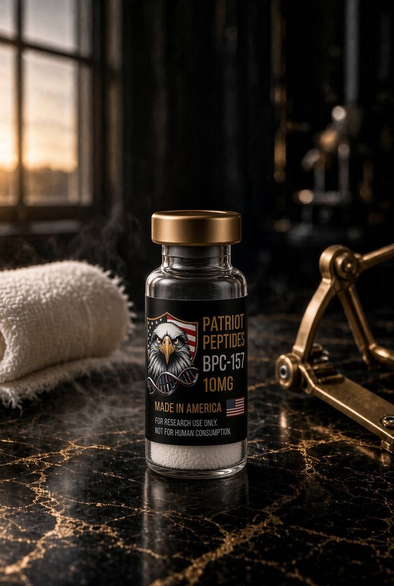
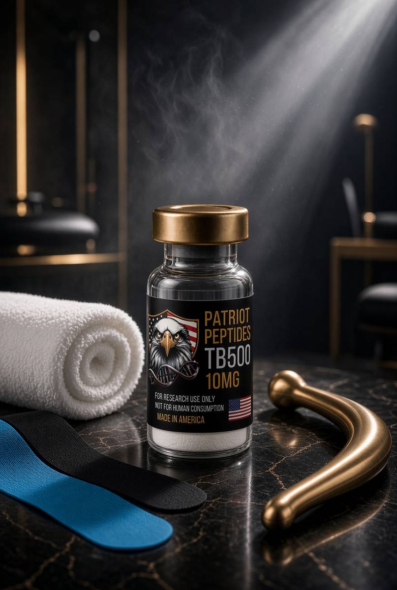
Research use only. Not a combined-use instruction.
Read next
45 min · long read · Peptide research
BPC-157: the pentadecapeptide that survived the stomach
A 15-residue fragment of a gastric protein, stable in acid, with a large preclinical literature on endothelium, tendon and nitric oxide. Here is the molecule, the papers, and what they actually mean.
49 min · long read · Peptide research
TB-500 and thymosin β4: repair is a cytoskeletal programme
Cells crawl by parking and releasing actin monomers. Thymosin β4 is the buffer. TB-500 is the research analogue built around that idea — characterised, named, on the bench.
50 min · long read · Frontier biology
Xenobots: frog cells that became a new kind of machine
No genome was rewritten. Skin and heart cells from Xenopus were sculpted — first by hand, then by an evolutionary algorithm — into millimetre-scale organisms that walk, heal, and assemble copies of themselves.
More in this desk
51 min · long read · Frontier biology
Tardigrades taught a human protein how to ignore radiation
Dsup, a disordered DNA-binding protein from a water bear, protects cultured human cells from X-rays. The animal that dries to a tun and lives through vacuum brought a transferable shield.
50 min · long read · Frontier biology
We can rewrite a genome the way a word processor rewrites a sentence
CRISPR cut DNA. Base editors change one letter without a double-strand break. Prime editors write arbitrary edits from an RNA template. The machinery is no longer hypothetical.
49 min · long read · Frontier biology
We have the Neanderthal genome. Some of it is still in you.
Svante Pääbo pulled a genome out of bone powder, won the 2022 Nobel, and found that most people outside Africa carry a percent or two of an extinct human. Palaeogenomics is not only mammoths.
49 min · long read · Frontier biology
The woolly mouse is the mammoth’s twenty-day dress rehearsal
Colossal edited seven coat-and-metabolism genes into laboratory mice and got golden, shaggy, cold-curious animals. Elephant gestation is 22 months. A mouse tells you in three weeks whether the edit was worth the wait.
Essays describe published research. They are not medical advice and they do not authorise human use of any catalogue item.