

Bodybuilding · 14 min · 3,128 words
Actin and myosin: the motor inside the fibre
The sarcomere, the thin rope, the thick motor, the eight-nanometre stroke, and the calcium switch that lets a nerve turn all of it on. One ATP per pull.
What this essay actually tells you
- A sarcomere shortens by sliding, not folding. The A-band stays the same length. Huxley and Niedergerke, and Huxley and Hanson, 1954.
- One ATP releases the myosin head, hydrolysis cocks the lever, phosphate leaving accompanies an ~8 nm stroke and a few piconewtons. Only a fraction of heads are attached at once. No ATP, and the head stays stuck: rigor.
- Calcium from the sarcoplasmic reticulum, opened mechanically by the T-tubule voltage sensor, moves tropomyosin off actin. SERCA spends ATP putting the calcium back. MYH7, MYH2 and MYH1 are the same motor with different timers.
What this actually means
Muscle shortens because myosin heads pull on actin filaments and then let go. Calcium uncovers the actin so the heads are allowed to bind. One ATP releases the head and cocks it for the next pull, about eight nanometres. Fast and slow fibres use different versions of the same motor. Getting stronger is more of these motors, not a different stroke.
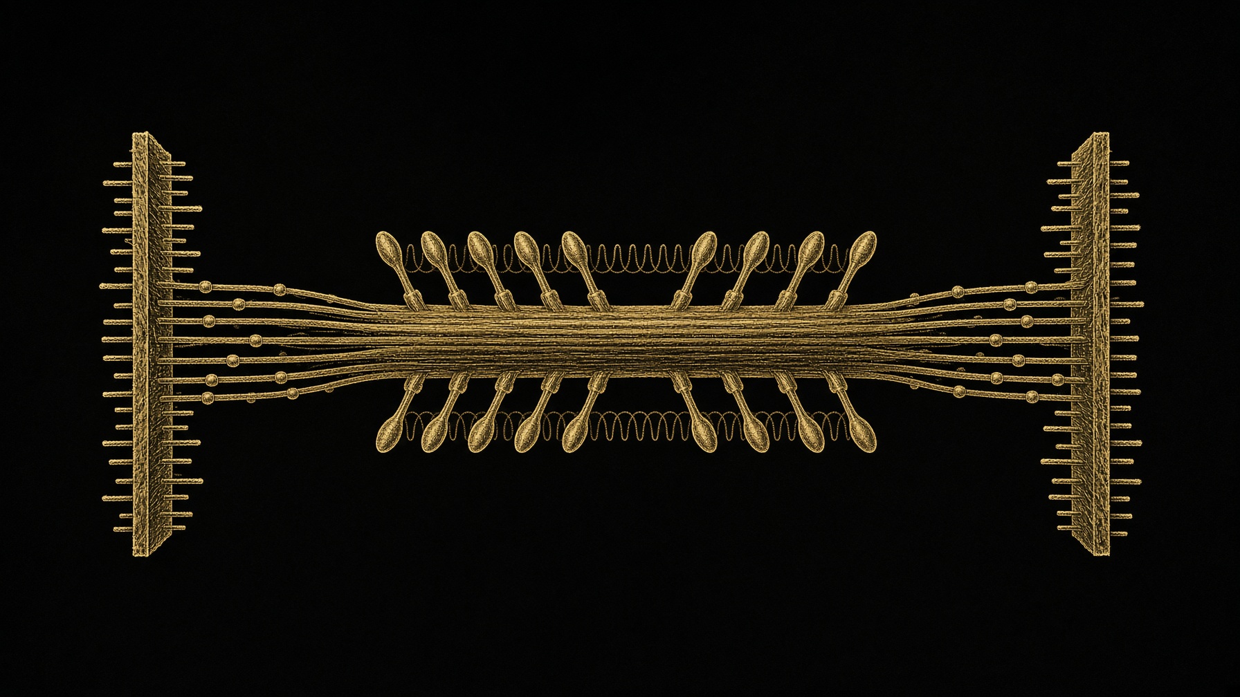
A fibre shortens because two proteins row past each other. Actin is the thin filament, a helical rope. Myosin is the thick filament, a bundle of tails with heads that grab the rope, pull, and let go. The repeating unit they live in is the sarcomere, Z-line to Z-line, about two micrometres long at rest in the fibres the classic experiments used, a bit longer in human muscle. A single biceps fibre contains thousands of these units end to end, and thousands of myofibrils side by side. Force is the sum of heads that happen to be attached at that instant. Speed is how fast those heads let go. Everything else in this series — fibre type, a hard set, a protein meal, an androgen — is a way of changing how many of these motors you have, or how long they stay on.
In short. Muscle shortens because myosin heads pull on actin ropes and then release them. A fibre is thousands of those units, end to end and side by side.
The proof that the filaments slide, rather than crumple, is from 1954, two papers in the same week. Huxley and Niedergerke, and Huxley and Hanson. Under the microscope the A-band — the region of the thick filament — stayed the same length while the muscle shortened. The I-band, the pale region where only thin filaments live, got shorter. The H-zone in the middle of the thick filament, where no actin reaches, also got shorter. If the proteins themselves had folded up, the A-band would have shrunk. It did not. The ropes slid over each other. That is still the picture. A pump in the gym is water. A contraction is this sliding.
In short. When a muscle shortens, the thick-filament band stays the same length and the gaps shrink. The proteins are sliding past each other, not scrunching up.
Actin: the rope
The unit of the rope is G-actin, a globular protein of about 42 kilodaltons. It polymerises into F-actin, a filament that looks like two strings wound together. The ends are not the same. The barbed end, the plus end, points at the Z-line and is where actin prefers to grow. The pointed end, the minus end, points at the middle of the sarcomere. In a working muscle those ends are capped so the rope does not fall apart mid-set: CapZ at the barbed end, tropomodulin at the pointed end. Nebulin, a long thin protein, runs along the actin and is the ruler that sets how long the rope is allowed to be. Mutations in nebulin are one cause of nemaline myopathy, a disease of weak, poorly organised sarcomeres. The ruler is not decorative.
In short. Actin is a chain of small globular proteins, capped at both ends so it stays a stable rope. A long partner protein decides how long that rope is.
Bare actin would be grabbed by myosin all the time, including at rest, which is rigor, the stiffness of a muscle with no ATP. The switch that prevents that is two more proteins lying in the groove of the helix. Tropomyosin is a coiled coil that covers seven actins in a row. Troponin sits every seventh actin and is itself three pieces. Troponin T binds tropomyosin. Troponin I is the inhibitory piece that, at rest, helps hold tropomyosin over the sites myosin wants. Troponin C binds calcium. When calcium arrives, troponin C changes shape, troponin I lets go, and tropomyosin rolls deeper into the groove. The myosin-binding sites open. Geeves described this as three positions, not two: blocked, closed, and open. Myosin itself, once a few heads get on, helps push tropomyosin the rest of the way. The rope is not a passive stick. The first heads make it easier for the next ones.
In short. At rest, a cover protein hides the sites myosin wants. Calcium moves that cover. The first myosin heads to bind help uncover the rest.
Myosin: the motor
Muscle myosin is myosin II. Each molecule is a hexamer: two heavy chains and four light chains. The heavy chain is about 220 kilodaltons. Its tail is a long coiled coil. Hundreds of those tails pack side by side, bipolar, so that heads stick out at both ends of the thick filament and a bare zone remains in the middle. That bare zone is why the H-zone looks empty. Each head, the part enzymologists call S1, is the motor. It has an actin-binding face, a pocket that hydrolyses ATP, and a neck that acts as a lever. Two light chains stiffen that neck: the essential light chain and the regulatory light chain. The lever is the piece that swings. The head does not crawl. It rocks, lets go, and rocks again.
In short. Myosin is two heads on a coiled tail. The tails bundle into the thick filament. The head is the engine, and a neck on the head is the lever that actually moves.
Which heavy chain you express is the fibre-type essay at the level of one molecule. MYH7 is the slow myosin. MYH2 is the fast oxidative one. MYH1 is the fastest human skeletal myosin. They are the same shape and a different timer. The slow head holds on to actin longer and splits ATP more slowly, so the filament slides slowly and cheaply. The fast head lets go sooner and splits ATP faster, so the filament slides faster and the bill is higher. Bottinelli's single-fibre work is the measurement: power and velocity change much more than the force of one millimetre of fibre. A type IIx fibre is not a type I fibre that tried harder. It is a different isoform of the same motor, burning ATP on a shorter duty cycle.
In short. Slow and fast fibres use different versions of the same motor. The fast one lets go sooner and spends more ATP. That is why it is powerful and why it tires.
The regulatory light chain is a small extra dial. Myosin light-chain kinase, switched on by the calcium-calmodulin complex, phosphorylates it. In fast fibres that phosphorylation makes the heads more likely to swing out toward actin, so the next twitch is a little stronger. This is post-tetanic potentiation: a muscle that has just fired hard produces more force on the next twitch than a quiet muscle did. It is not hypertrophy. It fades. It is the reason a few heavier reps can make the next set feel crisper, and it is not a reason to confuse a warm-up with growth.
In short. A small chemical tag on myosin's neck makes the next twitch stronger for a little while. That is a warm muscle, not a bigger one.
The stroke, one ATP at a time
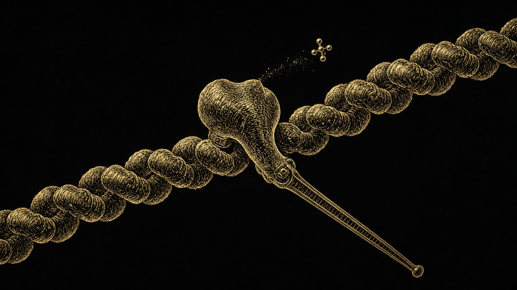
The cycle is short enough to memorise, and it is the whole of contraction. Start with the head bound tightly to actin and holding no nucleotide. That is the rigor configuration, strong and stuck. ATP binds in the pocket. The head immediately loses its grip on actin. The ATP is then hydrolysed to ADP and phosphate, and the energy of that split cocks the lever into the pre-power-stroke position. The head can now bind actin again, weakly at first. If the sites are open and the geometry is right, the binding becomes strong, phosphate leaves, and the lever swings through roughly seventy degrees. Actin is pushed along the filament by about eight nanometres. That number comes from the optical trap, a single molecule held between beads, Finer, Simmons and Spudich. ADP then leaves. The head is back in rigor, waiting for the next ATP to prise it off. One ATP, one stroke, a few piconewtons of force. A fibre produces kilograms because an absurd number of heads are doing this in parallel, and only a small fraction of them are attached at any instant. Muscle myosin is not a processive walker. If every head held on, the filament could not slide.
In short. ATP makes the head let go, then cocks it. The head rebinds, phosphate leaves, the lever swings about eight nanometres, and a new ATP releases it. Millions of those strokes are one lift.
Without ATP the cycle stops at the stuck step. That is rigor mortis: the calcium switch has leaked open as pumps fail, heads bind, and there is no ATP to release them. In a living fibre the opposite problem is more interesting. During an isometric hold — you pushing against a pin, the bar not moving — the filaments are barely sliding and the heads are still cycling. You pay ATP to produce force that goes nowhere. That is why a paused squat burns. The creatine kinase sitting on the myofibril recharges ADP to ATP on the spot from phosphocreatine, which is why the creatine essay exists and why the first seconds of a heavy rep feel different from the twentieth. After that, glycolysis and the mitochondria have to keep up. A fast myosin, with a higher ATPase, empties that account sooner. The fibre-type split is an ATP-split.
In short. Even when the bar is not moving, the heads are still spending ATP to hold force. That is a paused squat. The fast motor spends that ATP faster, so it fades first.
Calcium: how a nerve turns the motors on
The nerve does not touch actin. An action potential runs along the sarcolemma and dives into T-tubules, which are invaginations of the membrane aimed at every sarcomere. In the T-tubule membrane sits the dihydropyridine receptor, CaV1.1, a voltage sensor. Directly opposite it, on the sarcoplasmic reticulum, sits the ryanodine receptor, RyR1, the calcium-release channel. In skeletal muscle those two proteins are mechanically coupled. The voltage sensor moves, and it pulls the release channel open. This is not the cardiac trick, where a little calcium entering the cell triggers a lot more to leave the store. Skeletal muscle does not wait for that. One action potential opens the store.
In short. The nerve's electrical signal dives into the fibre and physically opens a calcium gate. Skeletal muscle does not wait for calcium to trigger more calcium, the way heart muscle does.
Resting calcium in the cytosol is about a hundred nanomolar. During a tetanus it rises toward ten micromolar in the regions that matter, troponin C binds it, tropomyosin moves, and the heads are allowed to work. Relaxation is the expensive reverse. SERCA, the sarcoplasmic reticulum calcium ATPase, pumps two calcium ions back into the store per ATP. Fast fibres use SERCA1, which is quick. Slow fibres lean on SERCA2. Calsequestrin inside the store soaks the calcium up so the free concentration in the reticulum does not fight the pump. Parvalbumin in fast fibres binds calcium in the cytosol and helps the twitch end crisply. A slow relaxation and a fast relaxation are different pumps and different buffers, not a difference in willpower. A cramp, or a muscle that will not let go, is this switch failing to close, not the myosin 'forgetting' to stop.
In short. Calcium uncovers the actin. Pumps spend ATP to hide it again. Fast fibres have a faster pump, which is why their twitches end sooner.
Length and speed
Force depends on how much overlap the filaments have, because only overlapped heads can bind. The classic curve, Gordon, Huxley and Julian on frog fibres, is flat between about 2.0 and 2.2 micrometres, where every head that can reach an actin does. Stretch further and you walk down the descending limb: fewer heads reach, force falls, until the filaments no longer overlap and active force is gone. Shorten too far, onto the ascending limb, and thin filaments collide in the middle and interfere. Human thin filaments are longer than the frog's, nebulin's ruler set differently, so the human plateau sits further out, nearer 2.6 to 2.8 micrometres. The shape is the lesson, not the frog's exact number. A joint angle that puts a muscle on a useless part of this curve is a lighter muscle, whatever the mirror says. The long-length finding in the growth essay is this curve plus something else: sarcomeres grown in series shift where on the curve a given joint angle sits.
In short. A fibre is strongest at the length where myosin heads can just reach actin. Too stretched, they miss. Too short, the ropes crash into each other. Joint angle is this curve.
Speed runs the other way. Hill's curve: as shortening speed rises, force falls, because heads are given less time to attach and the ones that do are dragged. Power, which is force times velocity, peaks at an intermediate speed. A one-rep max is the slow, high-force end. A throw is nearer the power peak. An unloaded limb is the high-speed end, where force is nearly nothing. Slow myosin shifts the whole curve leftward. Fast myosin shifts it rightward. You cannot rep your way from MYH7 kinetics to MYH1 kinetics. You can hypertrophy whichever isoform you recruit, which adds heads in parallel and raises the force end of the curve without turning a slow motor into a fast one.
In short. The faster a muscle is shortening, the less force it holds, because the heads cannot grab in time. Slow and fast myosins sit on different versions of that curve.
Titin is the spring that the sliding-filament story left unnamed for a while. It is the largest protein in the body, three to four megadaltons, gene TTN, one molecule running from the Z-line to the M-line. Immunoglobulin domains and a floppy PEVK region unfold under stretch and refold when you let go. Passive tension — the tightness you feel at the end of a range before the heads have done anything — is mostly titin, plus a little collagen. On the descending limb, titin recentres the thick filament so the next contraction is not lopsided. There is a live argument, Herzog and others, that calcium and active myosin make titin stiffer still, which would add force during a lengthening contraction beyond what the heads provide. It is not settled. What is settled is that lowering a heavy bar loads this spring as well as the heads, and that a sarcomere stretched until titin fails does not come back as the same sarcomere.
In short. Titin is a giant spring from end to end of the sarcomere. It is the tightness at the end of a stretch. A lengthening rep loads it. Pull it apart and that unit is done.
Where the force actually goes
The heads pull on actin. Actin is anchored at the Z-line by α-actinin. The Z-line is tied to the membrane by costameres, which are the dystrophin-associated complex and the integrins. The membrane is tied to the extracellular matrix, and the matrix becomes tendon. Duchenne muscular dystrophy is what it looks like when dystrophin is missing: the motors still fire, the membrane tears, and the force never arrives as a clean pull on the bone. That path is why a bigger myosin does not automatically mean a tougher lifter. The heads can be ready before the costamere and the tendon are. The bill essay is that gap at the scale of a person. This is the same gap at the scale of one Z-line.
In short. Myosin pulls actin, actin is nailed to the Z-line, and the Z-line is tied through the membrane to the tendon. If that tie is missing, the motors still run and the fibre tears.
Growth, when it is real, is more of this machine, not a different machine. Sarcomeres added in parallel are more heads side by side, which is a thicker fibre and a higher force. Sarcomeres added in series are more of these units end to end, which is a longer fibre and a joint angle that has moved. An androgen increases the transcription of this machinery and recruits nuclei to serve it. It does not invent a third filament. A protein meal supplies the amino acids the ribosome needs to finish another myosin heavy chain, which is a long protein and a hungry one. Creatine does not touch the lever. It keeps ATP in the pocket for the next release. Read the fibre-type essay for which isoform you are mostly building. Read the growth essay for how the ledger of synthesis stays ahead for long enough that a new sarcomere appears. This page was the motor those essays have been pointing at.
In short. Getting bigger means more of these same motors, side by side or end to end. Hormones and food change how many you can build. They do not change the stroke.
The A-band did not shorten. The filaments slid.— Huxley and Niedergerke, and Huxley and Hanson, 1954. The observation that still organises every set you do.
- ATP binds
- the head lets go
- ATP splits
- the lever cocks
- Phosphate leaves
- the lever swings
- New ATP
- release, and again
No ATP, and the head stays stuck. That is rigor.
ADP and phosphate on the head. Weak binding, ready.
About eight nanometres of slide. A few piconewtons.
Only a fraction of heads are attached at once. That is how the rope can move.
Questions the essay actually answers
- What do actin and myosin actually do?
- Actin is the rope. Myosin is a bundle of motors whose heads grab the rope, pull about eight nanometres, and let go when a new ATP binds. Thousands of those pulls, side by side, are a contraction.
- What is a sarcomere?
- The repeating unit of a fibre, from one Z-line to the next. Thin actin filaments project in from each Z-line. A bipolar myosin filament sits in the middle. Titin runs the length as a spring. Muscle shortens because these units shorten in series.
- How does calcium start a contraction?
- A nerve impulse travels into T-tubules and mechanically opens ryanodine receptors on the calcium store. Calcium binds troponin C, tropomyosin moves off the myosin-binding sites on actin, and the heads are allowed to work. Pumps then spend ATP putting the calcium back.
- Why does a muscle go stiff after death?
- Rigor mortis. Calcium has leaked out and there is no ATP left to make myosin let go of actin. The heads stay bound.
- Why are fast fibres more powerful and quicker to tire?
- They express a faster myosin heavy chain. It detaches sooner and splits ATP faster, so the filament slides faster and the energy bill is higher. The shape of the motor is the same. The timer is not.
- Does this change how I should train?
- The stroke does not change. What changes is how many motors you have and whether the joint angle puts them on a useful overlap. Heavier or longer-range work is more heads, or heads at a better length. It is not a different chemistry, and it is not a drug protocol.
Read next
10 min · Bodybuilding
Type I and type II: the two fibres a muscle actually is
Slow fibres last. Fast fibres throw the force. Diet, drugs and workouts all land on that split — so the muscle series starts with the myosin, not the vial.
8 min · Bodybuilding
How a fibre grows: tension, time, and the protein ledger
Synthesis minus breakdown, for weeks. Why soreness is the wrong score, why the pump is water, and why long ranges of motion keep showing up in the trials.
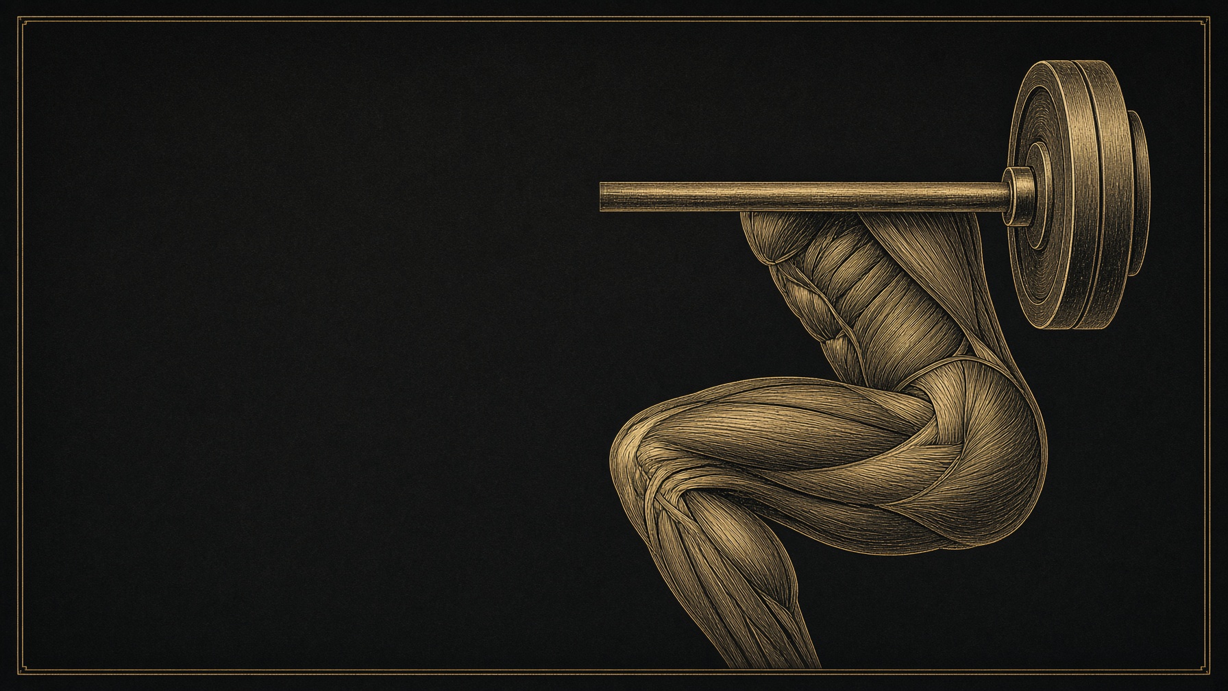
9 min · Bodybuilding
What the set is doing: failure, volume, rest, range
Why the last reps matter more than the first, why ten hard sets is a floor and not a personality, and why a short rest quietly deletes the tension you came for.
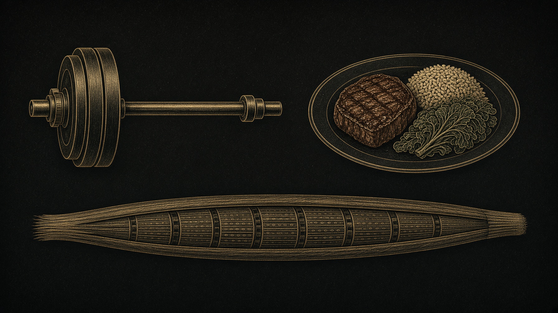
9 min · Bodybuilding
Food and training: how to spend the fibre you have
The practical half of the muscle series. Recruit the fast fibres, feed them, sleep, and stop asking a drug to do a week's work.
More in this desk
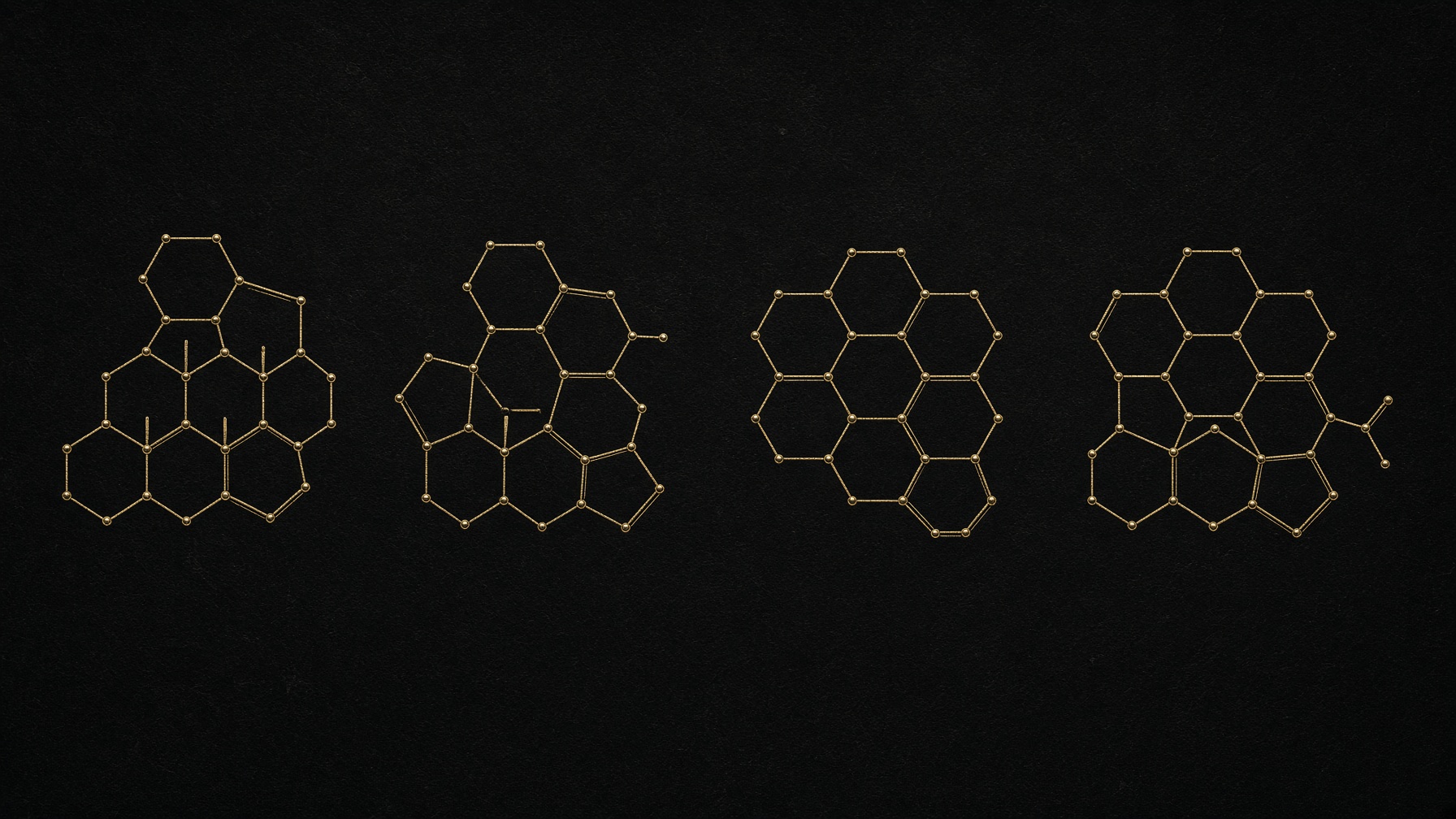
10 min · Bodybuilding
Steroid families: four chemistries, one receptor
Testosterone, the 19-nors, the DHT-derived, and the orals that rent the liver. Wet and dry are oestrogen and water, not two kinds of muscle.
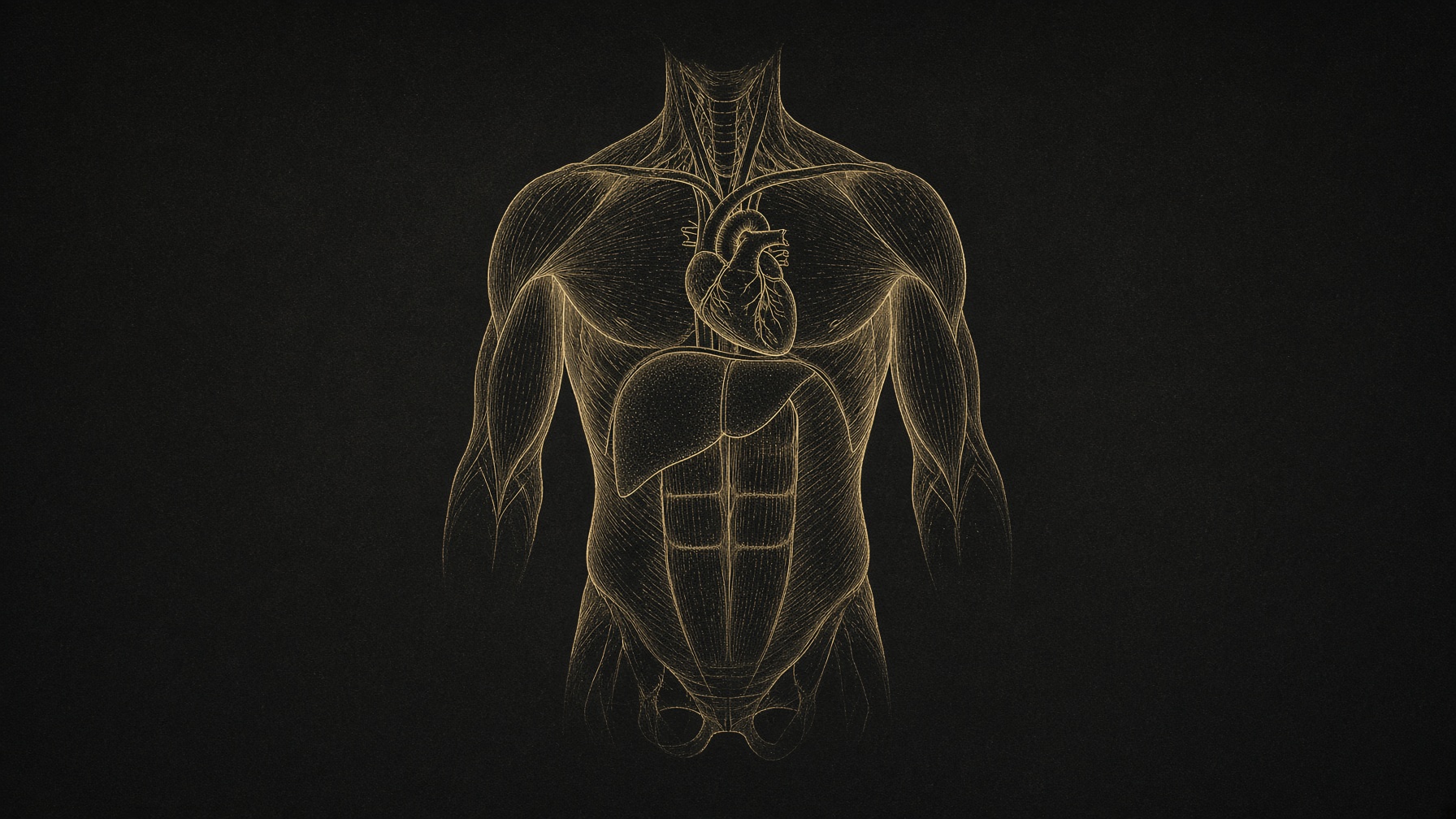
9 min · Bodybuilding
The bill outside the fibre: axis, blood, liver, heart
The androgen receptor is in more places than the biceps. Fertility, HDL, haematocrit, the left ventricle, the voice, the tendon. A map of the tissues. Not a course.
8 min · Bodybuilding
Androgens: the hormone that talks to the muscle nucleus
Testosterone binds a receptor in the myonucleus, adds nuclei, and asks for food. It is not growth hormone, not a cycle written as an essay, and not a fibre you can buy.
8 min · Bodybuilding
Growth hormone, insulin, myostatin: the other levers on a fibre
After testosterone, the rest of the muscle-drug list is different receptors: a liver hormone, a storage hormone, and a brake. Food decides which of those signals becomes tissue.
Essays describe published research. They are not medical advice and they do not authorise human use of any catalogue item.