

Clinic · 47 min · 10,398 words
Dermalux LED phototherapy: light as a mitochondrial instruction
Dermalux LED in Buckinghamshire. Blue 415 nm hits acne bacteria; red 633 nm and near-infrared 830 nm talk to cytochrome c oxidase. A course of six is how repair accumulates.
· updated
What this essay actually tells you
- Red (~633 nm) and near-infrared (~830 nm) light are absorbed by cytochrome c oxidase (complex IV). Working model: NO photodissociation, then a rise in ATP.
- Blue (~415 nm) hits porphyrins in Cutibacterium acnes. Different wavelength, different job. Blemish versus mitochondria, on the same head because that's convenient.
- A single session can change how skin looks that afternoon. A course of six is how photobiomodulation is supposed to accumulate, which is why diaries sell courses.
What this actually means
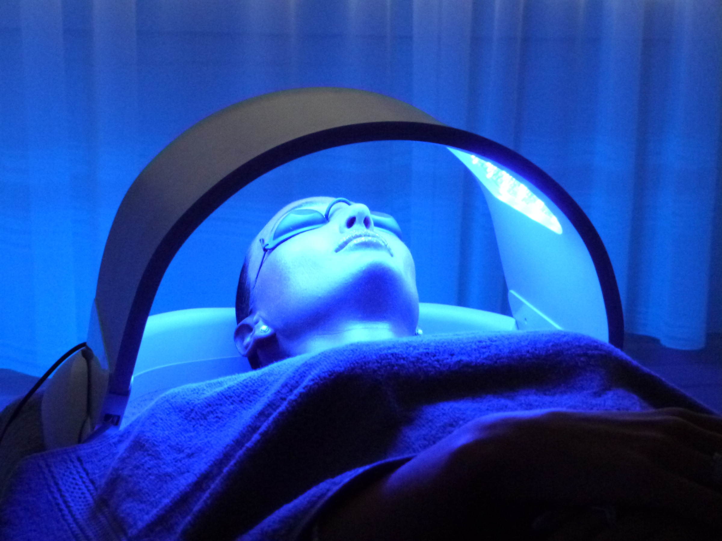
Dermalux isn't a heat lamp. It's three well-chosen wavelengths doing three jobs, and the clinic in Great Missenden runs them as a course because transcription doesn't clock in for a single afternoon. Blue at 415 nm is absorbed by porphyrins inside Cutibacterium acnes; the bacteria make ROS and die. That's the blemish tool. Red at 633 nm and near-infrared at 830 nm are absorbed, in part, by cytochrome c oxidase, complex IV of the respiratory chain. Tiina Karu spent a career showing that those photons can photodissociate inhibitory nitric oxide from the copper centres, let electron transport resume, and raise ATP. A brief ROS blip then acts as a signal, not a bleach. Fibroblasts, over repeated sessions, transcribe more collagen. Wunsch and Matuschka (2014) measured that in human skin with red/NIR LEDs: more collagen, more elastin, less MMP-1. One session can change how a complexion looks that afternoon. A course of six is how a transcriptional programme accumulates. One afternoon is allowed to look different. The collagen change was measured after a course.
Dermalux is a tri-wave LED phototherapy head, and the colours are the whole argument: 415 nanometres, 633 nanometres and 830 nanometres, delivered in a thirty-minute sitting at eLIVEate Me in Great Missenden. Photobiomodulation is the name the literature prefers, because it doesn't pretend the light is warmth or a laser. Photons at those three wavelengths are absorbed by three different molecular stories, and the clinic diary sells both a single treatment and a course of six because those are different experiments on the same dermis. A complexion can look different the afternoon you walk back onto the High Street. Collagen I transcription in a dermal fibroblast is a repeated-cue programme, and Wunsch and Matuschka measured the matrix change after a course, not after a single glow. We will keep those clocks apart. The device is CE-marked and doesn't need a laser chromophore; it also doesn't denature collagen at 60–70 °C the way nitrogen plasma does. Photons at a copper-haem enzyme are a different physics from a thermal fibril. JP owns the head, the fluence and the consent.
In short. The lamp is three precise colours, not a heater. One visit can change how the face looks that day. Lasting repair is built across a set of visits.
A CO₂ laser at 10,600 nanometres is absorbed by water. An alexandrite at 755 nanometres is absorbed by melanin. Those beams are chromophore hunters: miss the target colour and the energy is a waste; hit too much of it and the epidermis pays on the way in. An LED phototherapy head is doing something ruder and more interesting. The red and near-infrared diodes are aimed at an enzyme you already own, cytochrome c oxidase, complex IV of the mitochondrial respiratory chain. The blue diodes are aimed at porphyrins inside Cutibacterium acnes. Neither job is coagulation. Neither job is a tan. The working model, which Tiina Karu spent a career making respectable and which Michael Hamblin's reviews still carry, is photochemistry at named metal centres, then a change in ATP and a brief reactive-oxygen signal, then transcription. That sequence is why the treatment can be cold and still do work. You aren't cooking a fibril. You're talking to the last complex in the chain that hands electrons to oxygen, and, on a different diode, to a bacterium that made the mistake of synthesising a pigment that absorbs at 415 nanometres.
In short. Lasers hunt a colour in the tissue and dump heat. These diodes talk to a named energy enzyme and to a skin bacterium, without cooking the surface.
A single session is allowed to be a glow, and that glow is real physiology, not a trick of the waiting room. Blood flow in the papillary dermis can change on a timescale of minutes; scattering and surface optics follow; inflammatory tone can quiet enough that a face looks less busy by late afternoon. That's a real, short-clock physiology, and it isn't a fraud. It's also not photobiomodulation as the papers use the word. The field’s claim is accumulation: repeated fluence at cytochrome c oxidase, repeated ATP and redox cues, a fibroblast that keeps hearing the instruction and eventually writes more collagen I, more elastin, less MMP-1. Wunsch and Matuschka, Photomedicine and Laser Surgery 2014, measured those matrix changes in human skin after a course of red and near-infrared LED exposures, not after one sitting. Diaries sell six sessions for the same reason a transcriptional programme isn't a same-day event. One afternoon is cosmetics with a mechanism underneath. Six is how the mechanism was supposed to be tested. Confusing the two is how a serious enzyme gets talked about as if it were a facial glow.
In short. One session can make skin look better that afternoon. A run of six is how the deeper repair, the collagen change, is meant to add up.
BPC-157 and TB-500 sit on the same shelf because they're repair-biology probes in other models, not because anyone is reconstituting them into the head. The 15-mer is a gastric fragment with a nitric-oxide and VEGFR2 literature. The thymosin analogue orbits an actin-binding motif and a migration literature. Photons at complex IV are a third invoice. Neighbourhood isn't identity, and it isn't a protocol. eLIVEate is a separate company. Patriot Peptides doesn't take a commission on the booking, doesn't write the appointment as a peptide facial, and doesn't pretend a lyophilised cake inherits a thirty-minute LED sitting. Adults can want a paper and an appointment. Bundling them is how a research reagent becomes a medical claim it isn't allowed to be. The physiology below doesn't depend on it. The label does. The rest of the page is the photoacceptor, named, with the numbers you'd want before designing the dish you actually have the controls for.
In short. Two research peptides sit nearby in the library because they also turn up in repair papers. They're not in the lamp, and the lamp isn't a vial.
The photoacceptor is an enzyme you already own
Cytochrome c oxidase is complex IV, EC 1.9.3.1 — the last enzyme of the mitochondrial electron-transport chain, and the one that reduces molecular oxygen to water. In mammals it's a thirteen-subunit assembly. Three of those subunits — COI, COII, COIII — are still written on mitochondrial DNA, which is why this organelle keeps a genome. The metal centres are the point. Copper A is a mixed-valence binuclear copper site in COII, the first acceptor from cytochrome c. Haem a is a six-coordinate iron that funnels electrons onward. Haem a3 and copper B form the binuclear centre in COI where oxygen actually binds and is reduced. Those coppers and haems are why the enzyme is green-brown and why it's a respectable absorber of red and near-infrared light. Karu’s action spectra put usable peaks near 620, 680, 760 and 820 nanometres, which is the neighbourhood 633 and 830 live in. You don't need a dye. You don't need melanin. You already brought the chromophore with you, in every mitochondrion of every keratinocyte and fibroblast the light can reach.
In short. The catcher for the red light is the last enzyme of the cell’s energy chain. It already contains copper and iron, so it can absorb those colours without any dye.
Tiina Karu, Institute of Laser and Information Technologies, Troitsk, spent the 1980s and 1990s doing the unfashionable work of measuring action spectra in cells rather than photographing a complexion. Primary photoacceptors, secondary signalling, a distinction the field still has to relearn every time a new diode is marketed. Her argument, now the working model, is that cytochrome c oxidase is the absorber for the red and near-infrared window that actually reaches mitochondria in tissue, and that the first chemical event isn't heat and isn't a covalent change to the protein. It's the photodissociation of inhibitory nitric oxide from the copper and haem centres, after which electron transport, which NO had been pausing, resumes. ATP rises. A small pulse of mitochondrial reactive oxygen leaves the organelle and is read as a signal, not as a bleach. Downstream, transcription changes: NF-κB modulation, TGF-β, collagen I in dermal fibroblasts, a list Hamblin’s reviews have kept honest by refusing to pretend one photon does everything. That's a mechanism a paper can name. It's also a mechanism a bathroom bulb doesn't get to borrow.
In short. A physicist named Karu showed that red and near-infrared light can knock a pause-signal off that energy enzyme, so fuel burning restarts and the cell writes new instructions.
Nitric oxide binds cytochrome c oxidase reversibly and competes with oxygen at the binuclear centre — a pause button on the last complex of the chain. Brown and Cooper, FEBS Letters 1994, put nanomolar NO against synaptosomal respiration and watched oxygen consumption fall; Sarti, Giuffrè and colleagues mapped the same brake onto CuB and haem a3 in the purified enzyme. In a hypoxic or inflamed tissue the brake isn't a curiosity. It's how a cell stops wasting oxygen it can't finish reducing. Photons at 633 or 830 nanometres supply enough energy, at those metal centres, to kick NO off. Electron transport resumes. The protons Complex IV wasn't pumping start to move again. ATP synthase sees a fuller gradient. That's the working model in one paragraph, and it's why a nitric-oxide donor in the dish can blunt a photobiomodulation phenotype, and why a scavenger can sometimes mimic it. The model isn't closed in the way a crystal structure of the photodissociated state would close it. It's closed enough that writing ‘red light boosts energy’ without naming NO and complex IV has left the chemistry.
In short. Nitric oxide can sit on the energy enzyme and slow it. The red and near-infrared light kick that gas off. Energy production then rises. That's the working idea.
ATP is the first cellular readout people reach for, and it's a real one, which I find quietly cheering. Isolated mitochondria, permeabilised cells, and intact fibroblasts have all been reported to raise ATP after a red or near-infrared fluence that sits on the useful shoulder of the dose curve. The numbers in the older Karu papers and in the Hamblin compilations run from modest to a doubling, depending on the cell, the starting redox state, and whether the enzyme was NO-bound to begin with. A cell that wasn't paused will move less. A cell whose complex IV was already throttled will move more. That isn't mysticism. It's occupancy of a reversible inhibitor, then chemistry. The ATP is then spent on the unglamorous work a dermis actually does: ion pumping, protein synthesis, the charging of aminoacyl-tRNAs, the folding budget in the endoplasmic reticulum where collagen is born. A brief reactive-oxygen blip rides along. Superoxide and hydrogen peroxide at that scale are second messengers, the same way they're in growth-factor signalling. At a higher fluence they become the damage you meant to avoid. The biphasic curve in the next heading is that sentence drawn as a U.
In short. After the pause-gas leaves, the cell can make more of its energy molecule. A small burst of reactive oxygen is then used as a message, not as a bleach.
The nucleus is the slow room, and I like that about it. Mitochondrial reactive oxygen and a changed NADH/NAD+ ratio both talk to transcription factors; NF-κB is the one the photobiomodulation reviews name most often, with TGF-β and a collagen-I programme in dermal fibroblasts as the tissue-level consequence people actually booked the appointment for. None of that's a same-day event. RNA polymerase II has to find COL1A1 and COL1A2, the message has to be spliced, procollagen has to be hydroxylated and packed, lysyl oxidase has to cross-link the secreted helix. Hours to days for the message. Weeks for a dermal band a histologist will still see. That clock is why a course exists, and why a single 30-minute sitting is allowed to change how a face looks without being asked to remodel a dermis. Heat-shock protein 47, the collagen chaperone, and the MMP/TIMP balance sit on the same reading list as the thermal-plasma essay; the input here is photons at complex IV, not a 65 °C fibril. Two ways to ask a fibroblast to work. Don't write them as one.
In short. Making new collagen is slow. The cell has to write the gene, fold the protein and stitch it outside. That's why a course of visits exists.
Diagram
- I. Pumps H⁺. ~45 subunits. The NADH coin is spent here.
- II. TCA entry. No proton pump. FADH₂ neighbourhood.
- Q. Lipid-soluble shuttle in the inner membrane.
- III. Q-cycle. Pumps H⁺. Superoxide leak site.
- c. Intermembrane space. The shuttle everyone has heard of.
- IV. O₂ → H₂O. The reason you breathe.
- V. F₁Fₒ rotary. Protons in, ATP out. ~10²¹ times a second in you.
Mitchell’s chemiosmotic theory (Nobel 1978): the inner membrane is a battery of ~150 mV. NAD+ is the hydride carrier that feeds Complex I. MOTS-c is a 16-mer the mitochondrion translated from 12S rRNA — a different object on the same campus.
Two wavelengths, one enzyme, different depths
Tissue isn't a cuvette, which is the unglamorous half of the problem. Melanin, haemoglobin, water and scattering decide which photons still exist a millimetre down. The so-called optical window of tissue, roughly 600 to 900 nanometres, is the band where those absorbers are collectively kind enough that a useful fluence still reaches dermis and, at the long end, some subcutis. Blue 415 nanometres isn't in that window. It's stopped in epidermis, which is exactly where Cutibacterium acnes lives and exactly where you would want an antibacterial photon to stop. Red 633 nanometres reaches papillary and upper reticular dermis, a few millimetres in fair skin, less in a melanised epidermis. Near-infrared 830 nanometres travels further: lower scattering, weaker melanin absorption, the wavelength you pick when the mitochondrion you care about sits deeper. Karu’s action spectrum said both 633 and 830 can occupy cytochrome c oxidase. Anatomy says they don't occupy the same depth. Combining them on one head is therefore not a mood-lighting choice. It's two sampling depths of the same enzyme, plus a third diode that's a different molecule entirely.
In short. Blue light stops near the surface, where the acne bacterium lives. Red reaches the upper dermis. Near-infrared goes deeper still. Same energy enzyme, different depths.
Karu tabulated four peaks in the red to near-infrared for the oxidised enzyme: around 620, 680, 760 and 820 nanometres. Dermalux’s 633 and 830 sit on two of those shoulders. They aren't arbitrary spa numbers. They're also not the only numbers that would have worked; 660 and 810 are common in other devices and in the NASA LED papers, and they're the same conversation. What wouldn't have worked is a broad tungsten heater, a 940-nanometre diode that's already sliding toward water absorption, or a green that haemoglobin will eat before a mitochondrion sees it. Narrow-band LEDs exist so that the spectral width sits on the photoacceptor rather than on everything else in the skin. Bandwidth, peak wavelength, and the milliwatts per square centimetre at the stated working distance are the three numbers a paper has to write. ‘Red light’ isn't a wavelength. 633 nanometres, 10–20 nanometres full width, a named irradiance at the face, is a wavelength. The clinic head is built to make that sentence true. A bathroom bulb is built to make a room look warm.
In short. 633 and 830 nanometres were chosen because they match peaks of that energy enzyme. A wide household bulb doesn't match those peaks and it isn't the same treatment.
Other chromophores still sit in the path, and pretending they don't is how you write a cartoon. Melanin absorbs more strongly toward the blue and still takes a bite at 633; darker phototypes therefore deposit more of the red in epidermis and less in the fibroblast you meant to instruct. Haemoglobin has peaks in the blue-green and a weaker tail through the red; a flushed or telangiectatic dermis is a different optical object from a pale one. Water is almost irrelevant at 633 and only begins to matter as you climb past 900. Scattering, not absorption, is often the larger loss at these wavelengths: photons take a drunkard’s walk through collagen fibre and cell boundaries, which is why a milliwatt measured at the diode isn't a milliwatt at a mitochondrion two millimetres in. Optical properties are the unglamorous half of photobiomodulation and the half device companies sometimes skip. Fitzpatrick phototype still belongs in the consult, even though the plasma essay next door is the one that lives or dies on pigment. Here pigment is a filter, not a target.
In short. Skin colour, blood and scatter still steal some of the light on the way in. Darker skin filters more of the red before it reaches the deeper cells.
Dermalux Tri-Wave puts 415, 633 and 830 on one head so a clinician can run them singly or together without swapping devices. That's a clinical convenience, not evidence that one photon does everything. Blue remains the porphyrin channel. Red and near-infrared remain the complex-IV channel at two depths. After a nitrogen-plasma pulse or a spicule facial, JP will often want the red/near-infrared pair as a settling tool: more ATP for a tissue that just ran a wound programme, a quieter inflammatory tone, no extra thermal insult. Blue stays the blemish lever. Choose the diode that matches the photoacceptor you actually want in that sitting. It's worth being grateful when the wavelengths actually map onto named photoacceptors rather than onto a colour wheel. Three diodes, three jobs, one thirty-minute diary slot. The physics doesn't merge because the plastic housing did. Hold that number; we'll need it.
In short. The clinic lamp can fire one colour or all three. Combining them is handy, not proof that one beam does every job.
Diagram
Matrix
- TCA cycle · β-oxidation · mtDNA nucleoids
- NADH produced here. Complex I spends it.
- MOTS-c (MRWQEMGYIFYPRKLR) from 12S rRNA.
Inner membrane
- I → II → III → IV → V (ATP synthase)
- ~150 mV proton-motive force
- ~40–60 kg of ATP turned over per human day
mtDNA is 16,569 bp, 37 genes, 13 proteins of the respiratory chain. Nuclear DNA encodes the other ~1,200 mitochondrial proteins. NAD+ is the hydride carrier between dehydrogenases and Complex I. MOTS-c is a 16-mer translated from 12S rRNA — a peptide the mitochondrion wrote itself.
Blue is a different weapon
Cutibacterium acnes — the former Propionibacterium acnes — is an anaerobic-to-aerotolerant Gram-positive rod that lives in the pilosebaceous unit and makes coproporphyrin III as a metabolic by-product. That porphyrin is a tetrapyrrole with a Soret band, a strong absorption in the violet-blue around 400 to 420 nanometres. 415 nanometres isn't a lucky guess. It's the Soret overlap, the same optical fact a haematologist uses when a haemoprotein is sitting in a spectrophotometer. Photons at that band raise the porphyrin to an excited state. Energy then transfers to molecular oxygen, or the excited porphyrin itself starts radical chemistry, and the bacterium is treated to a burst of reactive oxygen it did not budget for. Lipid membranes, proteins, the organism’s own DNA: those are the targets. The host mitochondrion, at 415 nanometres, is a much poorer target, both because the light doesn't travel far and because mammalian cytochrome c oxidase isn't a Soret-peak porphyrin sitting in a bacterium. Different wavelength. Different job. The sentence is the whole antibacterial channel of the head.
In short. Acne-related bacteria make a pigment that soaks up blue light. The light then poisons them with a burst of reactive oxygen. That's a different job from the red light.
Photodynamic killing of C. acnes with blue light is older than Dermalux and older than the word photobiomodulation as a brand. Dermatology used 415-nanometre sources for acne long before anyone put them on the same board as 830. The evidence is a mixture of randomised device trials, before-and-after lesion counts, and the biochemistry of coproporphyrin, which is better than a testimonial and worse than a Phase III drug study. Lesion counts move. Not always by the amount a leaflet would like. Inflammatory papules often move more than comedones, which is what you would expect if the organism and the inflammatory tone around it were the target rather than a keratin plug. Combining blue with red on the same course is a common clinic pattern: blue for the organism, red for the host repair and for the inflammatory quieting that photobiomodulation also claims. That combination is a protocol a clinician owns. It isn't proof that 415 nanometres occupies complex IV. It does not.
In short. Blue light has been used against acne bacteria for years. It often helps inflamed spots more than blocked pores. It still doesn't do the red light’s energy-enzyme job.
Why not just use blue for everything, if reactive oxygen is the currency? Because the host isn't C. acnes. A keratinocyte and a dermal fibroblast run cytochrome c oxidase as a living, and they don't thank you for a Soret-band fluence aimed at their own haems. Blue at therapeutic acne doses is meant to stop in the follicle. Push it, and you have a phototoxic epidermis, pigment incontinence in darker phototypes, a different consult. Red and near-infrared at the Karu bands are meant to reach mitochondria without that porphyrin photochemistry. The two channels can be run in one sitting because the head can switch diodes, not because the photochemistry merged. After a plasma crust or a spicule facial, blue is often the wrong settling tool. The tissue has already had an insult; it wants ATP and a quieter NF-κB, not another ROS burst in the follicle. JP’s protocol knows that. Device copy sometimes skips that step. Wavelength is a job description. Treat it as one.
In short. Blue light is a surface weapon against a bacterium. Used too hard it can irritate the person's own skin. After a stronger clinic treatment, the red pair is usually the calmer choice.
Naming the organism matters because the taxonomy moved and a lot of copy did not. Propionibacterium acnes became Cutibacterium acnes in 2016; the porphyrin did not change. Phylotypes differ in how much coproporphyrin they make and in how inflammatory they are, which is already a reason a blue-light course isn't a universal acne therapy. Resistant C. acnes, biofilm in the follicle, a retinoid already on the surface: covariates a clinician has to own. A journal essay can name the Soret band and stop. What it shouldn't do is write blue light as a mitochondrial therapy that happens to be a pretty colour. 415 nanometres is a porphyrin weapon. 633 and 830 are a copper-haem instruction. Dermalux’s contribution is putting both on one CE-marked head so the consult can choose. Choice is the clinical act. The photochemistry was already in the papers. Wavelength is a job, not a colour wheel.
In short. The bacterium changed its official name; its blue-light pigment did not. Blue remains a pigment weapon. Red remains an energy-enzyme instruction.
Red and near-infrared LED phototherapy increased collagen density and elastin and reduced MMP-1 in human skin. That is a dermal readout of a mitochondrial instruction, measured after a course, not after a single glow.— Wunsch A, Matuschka K. Photomed Laser Surg. 2014; 32: 93–100.
Dose is a curve, not a slogan
The Arndt–Schulz rule, borrowed from an older pharmacology, is the biphasic pattern photobiomodulation inherited and then had to re-learn in every tissue. Too little fluence, nothing happens: not enough NO photodissociated, not enough ATP, not enough of a redox blip to talk to the nucleus. Too much, and you inhibit the same enzyme you meant to help, or you generate a reactive-oxygen load that's damage rather than a message, or you photobleach the photoacceptor. The useful shoulder is a band of joules per square centimetre that depends on the wavelength, the irradiance, the tissue, and whether the cells were already NO-paused. Hamblin’s reviews have been drawing that U for a decade. Clinic devices exist so that the face sits on the useful shoulder rather than in a bathroom-bulb anecdote or in a burn. Fluence is the time-integral of irradiance. Irradiance is milliwatts per square centimetre at the working distance, not at the diode in the manufacturer's picture. Distance matters because intensity falls, and because the field at the cheek isn't the field at the inner canthus.
In short. Too little light does nothing. Too much can slow the same enzyme you meant to help. Clinic devices are built to sit in the useful middle of that curve.
Write the units, because the field has spent twenty years drowning in unspecified ‘treatments’. Irradiance in mW/cm². Time in seconds. Fluence in J/cm², which is irradiance times time, with the honesty that a pulsed source and a continuous source at the same average fluence aren't always the same biology. Spot size and number of diodes decide whether the number on the screen is the number on the skin. A 30-minute Dermalux sitting is a time. It isn't a fluence until someone has measured milliwatts at the face. We won't invent a joule figure the manufacturer’s file did not put in this journal. We will insist that a paper which can't say J/cm² hasn't yet done photobiomodulation; it has done lighting. The same pedantry applies in a dish. Height of the LED array above the monolayer, temperature of the well (LEDs do dump some heat; a heat-matched dark control isn't optional), attenuation by phenol red in the medium, which is itself a dye. Skip those and you have a colour photograph of a plate.
In short. The useful numbers are brightness at the skin and how long it sits there. Minutes on the diary aren't those numbers until someone has measured them.
A bathroom bulb is warmth and a broad spectrum. An infrared heater is water absorption and a rise in tissue temperature that can, on its own, change blood flow and make a face look different. Photobiomodulation at 633 and 830 is allowed to be thermally quiet. The point of the LED, rather than a heat lamp, is to occupy the photoacceptor without a confounding temperature jump. If your dish warmed by two degrees, you have a heat experiment plus a light experiment, and you won't know which phenotype belongs to which. If the clinic room is comfortable and the head is a few centimetres off the face, the thermal confound is small, which is why the treatment can be sold as having no downtime and no heat. Small isn't zero. A fair paper still says so. The contrast with NEOGEN, next door on the same diary, is the whole physics lesson: that device wants 60–70 °C in the dermis. This one wants a copper centre to let go of nitric oxide. Don't file them as ‘energy treatments’. Energy isn't a mechanism.
In short. A heat lamp warms water in the tissue. These diodes are meant to work without a real temperature jump. That's a different physics from plasma tightening.
Thirty minutes, once, is a sitting. Six times, spaced over weeks, is a course. The diary at eLIVEate prices them as different objects because they're different objects: £50 for one, a course of six for the accumulation the papers actually measured. Spacing matters as much as session count. Stack six on six days and you have a different integral from six over six weeks; the fibroblast's transcriptional programme and the epidermis's turnover (about a month on the face) are the clocks those spacings are trying to sit on. There is no large randomised literature that makes six the magic integer rather than eight or twelve. There is a practical integer a clinic can book, and a set of LED studies that used courses rather than one-off exposures when they wanted a matrix endpoint. That's enough to take a course seriously, and not enough to treat six as a law of photochemistry. JP owns the spacing. The enzyme owns the biphasic curve. Neither belongs to a colour wheel.
In short. One visit and a set of six are different treatments. The number six is a practical clinic course, not a law of physics, and the gaps between visits matter.
Collagen is the slow readout
Wunsch and Matuschka, 2014, is the human-skin paper this page will keep citing because it measured matrix rather than mood. Volunteers, red (611–650 nm) or red-plus-near-infrared (570–850 nm) LED phototherapy, twice a week for thirty sessions in the full protocol, with ultrasonographic collagen density, a blinded wrinkle assessment, and, on biopsies in a subset, more collagen, more elastin, less MMP-1. Photomedicine and Laser Surgery 32: 93–100. It isn't a Dermalux-brand trial, and pretending it's would be a category error. It's a red/near-infrared LED trial in human skin with a dermal readout, which is the neighbourhood the 633/830 pair lives in. Sample size, device, and the fact that thirty sessions isn't six, all belong in the caveat. The direction of the matrix change is the fact. Collagen up, MMP-1 down, after a course. That's a fibroblast doing its job under a repeated mitochondrial cue, not a lamp depositing collagen onto a face.
In short. A 2014 human study found more collagen and elastin and less of a collagen-chewing enzyme, after a course of red and near-infrared light. That was a course, not one visit.
A dermal fibroblast is a secretory cell whose day job is type I and type III collagen, a little elastin, proteoglycans, and the MMP/TIMP balance that decides whether the warehouse is built or chewed. Give it ATP, a TGF-β cue, and a quieter inflammatory cytokine field, and it will, over weeks, lay more organised matrix. That sentence is also the NEOGEN sentence, and the microneedling sentence, which is why three different clinic physics rhyme at week six without being the same appointment. Photobiomodulation’s version of the cue is the complex-IV instruction. It doesn't denature the existing fibril, so there is no bronze eschar and no 90-day necrotic-epidermis story. The remodelling is quieter and slower and, on the published numbers, smaller than a thermal denaturation. Plasma-scale tightening from an LED course isn't what the histology shows. A glow-only story isn't what Wunsch measured either. The useful claim is a modest matrix shift after repeated fluence. Modest, in a dermis, is still a lot of fibrils.
In short. Skin fibroblasts build and chew collagen for a living. Repeated red light gives them a nudge. The change is real and smaller than a heat-tightening treatment.
MMP-1 is interstitial collagenase, the enzyme that nicks intact type I collagen and so starts the turnover cascade. Photoaged dermis is, among other things, an MMP-1 problem: ultraviolet transcription of the protease, fragmented fibrils, a wrecked mechanical environment that tells the fibroblast, via integrins, to behave worse. A treatment that lowers MMP-1 and raises collagen is pushing both sides of that ledger. Wunsch reported both. Elastin is the bonus and the caution: adult dermis makes almost no new elastin under ordinary conditions, so an elastin rise on a biopsy is interesting and needs to be read as fibre organisation and tropoelastin transcription, not as a childhood elastic-fibre factory switched back on. We won't write ‘restores elastin’ as a caption. We will write that a course of red/near-infrared LED phototherapy has been measured to move collagen, elastin and MMP-1 in human skin, in one well-cited trial, with the session count that trial actually used. Then we will remember that Dermalux’s six is a clinic course, not a replication of thirty.
In short. The enzyme that cuts intact collagen fell in that study and collagen rose. Adult skin hardly makes new elastin, so that part of the result should be read carefully.
Six is how photobiomodulation is supposed to accumulate, and that's a physiological claim rather than a loyalty card. Each sitting is a pulse of ATP and a redox message. Between sittings the fibroblast transcribes, secretes, and begins to cross-link. The next sitting lands on a slightly different dermis. Transcriptional programmes in matrix cells aren't digital; they're a drumbeat. Miss the drumbeat and you have an afternoon. Keep it up and you have a chance at a band a microscope will still see. Epidermal turnover on the face is on the order of four weeks, so a course that spans that window is at least sitting on the right biological clock. There is no trial that proves six is the unique integer at 633 and 830 on this exact head. There is a field that stopped taking single-exposure complexion photographs as evidence of remodelling. The diary followed the field. That's the right way round.
In short. Each visit is a short energy pulse. The cells keep working between visits. A row of six is meant to catch that drumbeat, which a single afternoon cannot.
Harry Whelan’s NASA LED papers treated photobiomodulation as a wound-healing aid for astronauts and for diabetic ulcers: near-infrared diodes, fibroblast and epithelial models, then animal wounds, a programme that put 670 and 880 nanometre LEDs into the same CCO conversation in a different tissue. J Clin Laser Med Surg, early 2000s. Military and space medicine wanted a non-thermal way to talk to mitochondria when a wound was slow. Dermatology inherited the diodes. The wavelengths shifted a little; the enzyme did not. Collagen transcription in a diabetic ulcer and collagen transcription in a photodamaged cheek aren't the same paper, and treating Whelan as a face trial is how a footnote becomes a testimonial. Treating Whelan as irrelevant is how you forget that the photoacceptor was already being asked to work in a wound bed, at fluences a physicist had measured, before anyone put a tri-wave head over a Buckinghamshire consult chair. Same complex IV. Different body. The peptide literature that sits on the same shelf is repair through ligands rather than photons. Two doors. One tissue.
In short. NASA studied similar red and near-infrared lights for slow wounds, not for faces. Same energy enzyme, different body. That history is a footnote, not a face trial.
Nitric oxide sits on both doors
Nitric oxide is a gasotransmitter and a respiratory brake, and photobiomodulation uses both of those facts — often without saying so. At complex IV it's the reversible inhibitor the photon kicks off. In endothelium and in a wound bed it's the product of nitric-oxide synthases, a vasodilator, an anti-adhesive signal, a modulator of platelet and leukocyte behaviour. Raising ATP by de-inhibiting complex IV, and changing local NO tone because some of the photodissociated NO is now free to act as a signal, are two consequences of one photochemical event. They shouldn't be collapsed into ‘increases circulation’, although circulation does often rise, and an afternoon glow is partly that. eNOS phosphorylation, organ-bath relaxation, cutaneous Doppler: those are the measurements if you claim the vascular half. An ATP kit is the measurement if you claim the mitochondrial half. A paper that claims both with neither assay has claimed a colour.
In short. The same gas that pauses the energy enzyme is also a blood-vessel signal. Knocking it off the enzyme can therefore change both fuel burning and local blood flow.
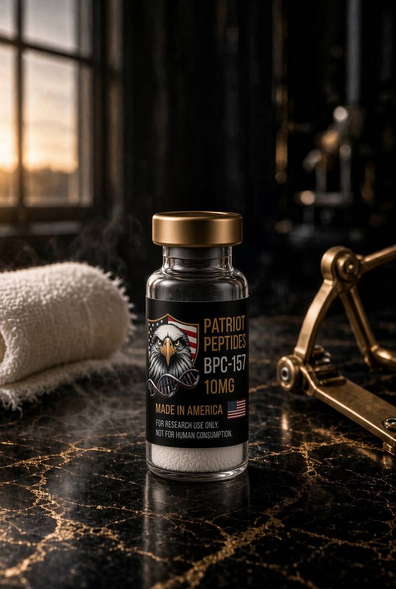
BPC-157, GEPPPGKPADDAGLV, 1419.5 daltons, entered this neighbourhood through nitric oxide, not through a lamp. Sikiric’s Zagreb programme reported that the 15-mer nudges NO tone back toward a set-point: L-NAME makes lesions worse, L-arginine can make them better, and the peptide, in their hands, restores the middle from either side. Independent benches have been firmer on the endothelial half — VEGFR2 internalisation and phosphorylation, focal-adhesion kinase, endothelial migration — than on the set-point claim. Hsieh 2017 and Chang 2011 are the nodes a new lab can actually run. That's a ligand literature. It rhymes with photobiomodulation because both stories spend time on NO and on a tissue that's trying to repair, and rhyme isn't a receptor. Photons occupy copper centres of complex IV. The 15-mer, on the cleaner papers, talks to a receptor tyrosine kinase and to FAK. You can't substitute one for the other in a paper. You can file them on the same shelf so a colleague walking from the lamp to the peptide catalogue doesn't get lost.
In short. A stomach-derived research peptide turns up in nitric-oxide and blood-vessel papers. That's a neighbour on the reading list, not a stand-in for the lamp.
TB-500 orbits the LKKTETQ actin-binding motif of thymosin β4, the principal G-actin sequestering peptide of animal cells. Goldstein isolated thymosin; Safer identified the actin job. A crawling cell — a keratinocyte closing epidermis, a fibroblast filling a defect, an endothelial cell making a sprout — needs a lamellipodium, and a lamellipodium needs a controlled G/F-actin equilibrium. That's a cytoskeletal invoice. It's silent on what 830 nanometres did to complex IV, and it's silent on whether NO was bound to CuB this morning. Repair folklore stacked the 15-mer and the actin analogue because both showed up in injury models and both were short enough to lyophilise. Photobiomodulation gets stacked with them because a wound bed also likes ATP. Three useful objects. Three mechanisms. A map is how you stop the stack becoming a protocol. We stock the two ligands as characterised sequences. eLIVEate owns the head. Nobody is mixing a cake into a diode, and the diode doesn't become a pentadecapeptide by sharing a paragraph.
In short. A second research peptide helps cells crawl by parking spare actin. That's another repair neighbour. It doesn't explain what the red light did to the energy enzyme.
Two doors, one tissue: that's the topology. Door one: photons at 633 and 830 nanometres, cytochrome c oxidase, NO photodissociation, ATP, a redox blip, transcription, a course of six. Door two: defined ligands with papers in endothelium, actin, gastric mucosa, tendon explants, whichever model the certificate is actually for. The tissue is dermis, or a wound, or a mucosa, depending on the paper. The mistake is walking the doors as if they were the same corridor — treating an LED course as a reason not to read the 15-mer, or treating a research vial as a home version of the lamp. We won't write that corridor. We will write the map, and we will keep the legal objects apart. The appointment is a CE-marked phototherapy sitting on a clinic diary in Buckinghamshire. The vials are HPLC-characterised sequences for a bench. Partner clinic. No commission on the booking. Different rooms, different paperwork, and they stay that way because that's how the two legal objects stay themselves.
In short. Light at the energy enzyme and research peptides in a dish are two ways into repair biology. They share a tissue in the library, not a recipe in a bowl.
eLIVEate is a separate company in Great Missenden. JP sets the diodes, the combination, the spacing and the consent. Patriot Peptides doesn't take a cut of that diary, doesn't write a post-LED peptide protocol, and doesn't staff the recovery chair. Where the journal files the lamp next to BPC-157, it's because a colleague who has just watched complex IV let go of NO may want to know where else nitric oxide sits in this catalogue, and because a colleague who has just read a VEGFR2 blot may want to know that a clinic down the road occupies the same gas from the other direction. Filing is a courtesy. It isn't a bundle, not a SKU, and not a facial. Adults can book the head and read the 15-mer in the same week. We would rather they knew they had done two different things. That's enough splitting of rooms. The enzyme doesn't care who owns the head.
In short. The clinic and the peptide catalogue are different companies. Sitting their papers next to each other is a reading list, not a combined treatment.
Diagram
| Node | Catalogue | Conversation |
|---|---|---|
| GPCR | Ipamorelin, MT2, PT-141, retatrutide, CJC | Second messengers, secretion, appetite, pigment |
| RTK / IGF1R | IGF-1 LR3 | IRS–PI3K–Akt–mTOR and Shc–ERK |
| Cytokine receptor | Somatropin (HGH) | GHR–JAK2–STAT5b, hepatic IGF-1 |
| Cofactor | NAD+ | Sirtuins, PARPs, CD38, redox |
| Actin buffer | TB-500 / Tβ4 motif | G-actin sequestration, motility |
| Growth-factor-like | BPC-157 | VEGFR2 / FAK / eNOS neighbourhood |
| Copper ligand | GHK-Cu | Transcriptome shift in fibroblasts |
| MC fragment | KPV | NF-κB, PepT1, no pigment |
| Nuclear / pineal | Epithalon (AEDG) | TERT and melatonin literatures |
| mtORF peptide | MOTS-c | AMPK, folate–methionine cycle |
Each row is a different kind of molecular conversation. The catalogue peptides bind at these nodes; they are not interchangeable, and stacking them because a forum did mixes unrelated literatures.
Copper is a different collagen sentence
Lysyl oxidase, LOX, oxidatively deaminates lysine and hydroxylysine in collagen telopeptides so the helix can become a load-bearing cable. The enzyme carries copper and a lysine tyrosylquinone cofactor that's built in place once the metal is bound. No copper, no cofactor, no aldehyde, no cross-link. A culture deprived of copper doesn't make a dermis; it makes a weak gel. Loren Pickart’s GHK-Cu — glycine-histidine-lysine holding Cu²⁺ in a square-planar complex — is a plasma tripeptide that delivers that metal without dumping free copper, a Fenton reagent, into the medium. Maquart, Borel and colleagues showed in 1988 that the complex stimulated collagen synthesis in fibroblast cultures. Pickart and Margolina’s later arrays put collagen, decorin and TIMPs up and some MMPs down, a spreadsheet that's still waiting for a 2020s RNA-seq. Copper delivery to LOX is already enough to be interesting. The transcriptome-reset is a hypothesis with an outline. Either way, the ligand is a metal chaperone, not a photon.
In short. Collagen has to be stitched with a copper enzyme or it stays a weak gel. A small blood peptide carries that copper. That's chemistry, not light.
The rhyme with the lamp is transcriptional and tissue-level, not mechanistic. Photobiomodulation asks a fibroblast for more collagen by talking to complex IV. GHK-Cu asks a fibroblast for more collagen by delivering copper toward the enzyme that finishes the helix, and perhaps by being recognised as a ligand in its own right. Both sentences can be true in the same dermis. They don't become one sentence because a journal filed them next to each other. A copper-matched control, a GHK-without-copper arm, and a wavelength-and-fluence log are three different methods lines. Run them if you're going to write a paper that mentions both objects. Don't run a lamp and then tip a research tripeptide into a moisturiser and call it translational. Cosmetic copper-peptide lotions have used Pickart as a reading list for decades; that's a cosmetic regulatory object. The vial on a research shelf is Gly-His-Lys·Cu²⁺, mass about 340.7, a chromatogram, a certificate. The head on JP’s wall is a CE-marked LED array. Two legal classes. One tissue. Keep the jobs.
In short. The lamp and the copper peptide can both end up asking skin cells for more collagen. They still do it by different routes and they aren't one product.
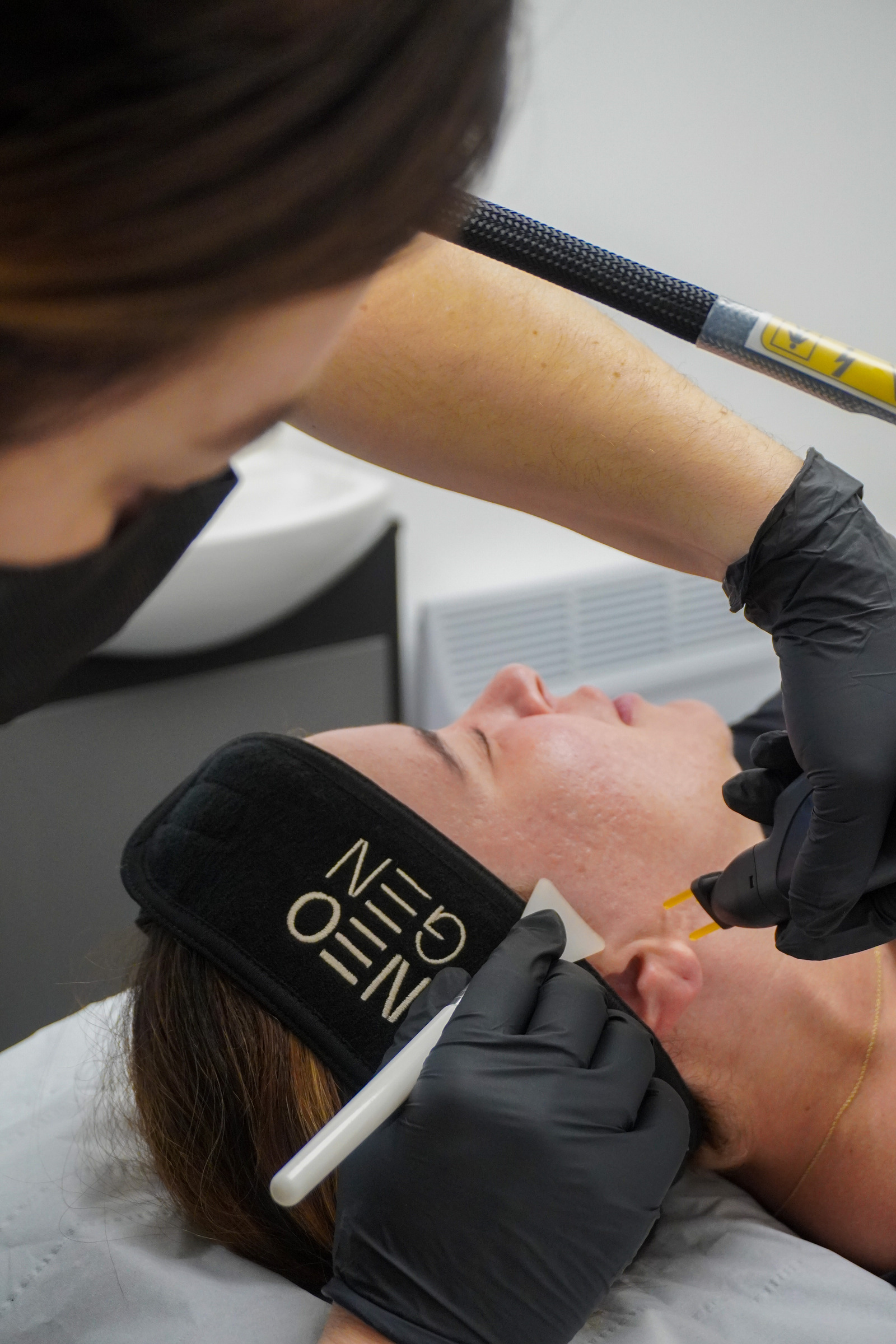
NEOGEN, on the same diary, is the thermal version of the collagen ask: a nitrogen-plasma pulse that denatures type I at 60–70 °C, a bronze eschar, ninety days of fibroblast rebuild. CELL STORY is the micro-injury version: siliceous spicules, channels for hours, a foreign-body stimulus for about 72 hours, a shallower cousin of Percutaneous Collagen Induction. Dermalux is the photochemical version. Three physics, one cell type at the end of the story, which is why the clinic can combine them — red/near-infrared after plasma or spicules as a settling tool — without anyone having discovered a new enzyme. Combination is a clinical judgement about insult and recovery. It isn't a licence to write a homemade stack of plasma plus LED plus a research copper tripeptide plus a gastric 15-mer. We're sometimes asked to mix them in a bowl. We don't. JP owns the combinations the CE-marked devices allow. The catalogue owns sequences with papers. The map is so you can see why the email got written. It isn't so we can answer it with a recipe.
In short. Heat, tiny glass needles and this light can all ask the same repair cell to work. Combining clinic devices is the clinician’s call. Mixing in research vials isn't a protocol.
Superoxide dismutase is the other copper enzyme in the neighbourhood, and it belongs in a photobiomodulation essay because the treatment itself makes a reactive-oxygen blip. SOD1 holds copper and zinc and dismutes superoxide to hydrogen peroxide; the peroxide is then catalase or glutathione-peroxidase territory. A fibroblast that has just been told, by complex IV, to leak a signalling pulse of superoxide will handle that pulse better if SOD is metalled. GHK-Cu, as a copper donor, sits on that logistics problem as well as on lysyl oxidase. Again: logistics, not a reason to blend a vial into a sitting. Free Cu²⁺ in a well with superoxide is Fenton chemistry, hydroxyl radical, the opposite of a signalling blip. The tripeptide’s job, if you're using it as a reagent, is to hold the metal in the window between too tight and too loose. The lamp’s job is to occupy complex IV at a fluence that keeps ROS in the messenger band. Two windows. Two objects. A dermis that happens to need both windows on the same Tuesday is still not a protocol.
In short. The light itself makes a small burst of reactive oxygen. Copper enzymes help mop that burst. That still doesn't make the copper peptide part of the appointment.
Diagram
- GHKGly-His-LysPickart, 1970s plasma fraction. ~200 ng/ml at 20; ~80 ng/ml at 60.
- Cu²⁺ complexsquare-planarHistidine imidazole + backbone nitrogens. The product is the complex.
- Lysyl oxidaseLOX / LOXLCopper enzyme. No metal, no collagen cross-links, a weak gel.
- SOD1Cu/ZnFirst mop for superoxide. Same metal, different job.
- Free Cu²⁺FentonCu⁺ + H₂O₂ → HO·. Delivery without a redox-active pool is the point.
Pickart and Margolina’s microarrays claim thousands of transcripts. Modern RNA-seq with multiple-testing correction is the replication the claim still owes. Copper delivery to LOX and SOD would remain interesting if the spreadsheet shrinks.
An afternoon is not a histology
What you can see at four o’clock isn't what a biopsy would show at week six, and both observations can be true. Immediate appearance is blood flow, surface scatter, a little oedema that fills fine lines the way a glass of water fills a dried sponge, a quieter redness if inflammatory tone dropped. Those are optical and vascular facts. They're the facts a single 30-minute sitting is allowed to own. They're also the facts a heat lamp can fake, which is why the no-heat claim and the named wavelengths matter. Histology is collagen organisation, elastin, epidermal thickness, MMP-1 immunohistochemistry, a dermal band that wasn't there. Wunsch needed a course to move those. Selling the afternoon as the histology swaps the clocks. Sneering at the afternoon because it isn't histology forgets that patients walk out of a consult chair into daylight, and that a short-clock vascular change is still physiology. Hold both. Measure them apart.
In short. How the face looks later that day is mostly blood flow and surface optics. How the deeper skin is built is a weeks-long job. Don't sell one as the other.
Inflammatory quieting is the middle clock, and it's the one photobiomodulation reviews are most confident about after the ATP sentence. Hamblin has argued that the same mitochondrial reactive-oxygen pulse that talks to NF-κB at modest fluence can reduce a chronic inflammatory transcription, rather than start one, which is the biphasic curve again. Clinically that's redness, rosacea-adjacent tone, the settling after a spicule facial, the reason a psoriasis plaque sometimes appears on a device’s treat-list. The measurements, when they exist, are cytokine panels, NF-κB localisation, clinician erythema scores. They're better than a selfie and worse than a collagen biopsy. A course that calms a face without moving MMP-1 is still a course that did something. It isn't the Wunsch endpoint. Naming which clock you're on is the whole consult. JP’s diary can sell a single sitting for the afternoon and a course of six for the accumulation. That split is physiology written as a price list, which is more honest than most device copy.
In short. Redness can settle on a middle timescale, between an afternoon glow and a collagen rebuild. Say which of those you're aiming at.
Every phototype can sit under this head, which is the practical difference from a melanin-targeted laser and from a plasma crust that looks different on Fitzpatrick IV than on I. Pigment is a filter here, as already said, not a chromophore you need in order to work. Darker epidermis steals more of the 633 nanometres; 830 still travels. Blue, on a melanised follicle, is a different risk conversation (pigment incontinence, post-inflammatory mark) and a clinician who knows that will dose accordingly. There is no downtime in the plasma-eschar sense: no bronze sheet, no social week off. You can walk back onto the High Street. That sentence is a recovery fact, not a claim that nothing happened. Mitochondria happened. Whether the afternoon glow is the thing you wanted, or the thing you will still want after sitting six, is the consult. Suitable for every skin type is a line that happens, in this case, to track the physics: no melanin requirement, no ablation. A recovery line still isn't a measurement.
In short. This light doesn't need pigment to work, so most skin colours can use it and there is no scab week. Deeper red still has to get through the surface colour.
After NEOGEN or CELL STORY the red/near-infrared pair is a settling tool, and that's the combination the clinic actually books. Plasma has just denatured collagen and desiccated epidermis; spicules have just left a forest of silica in the stratum corneum. Both are insults with a repair programme already running. Adding 633 and 830 is an attempt to give that programme ATP and a quieter inflammatory tone without adding heat or another foreign body. Adding 415 is a different decision, useful if blemish bacteria are the outstanding problem, unhelpful if the surface is already busy. None of this is a published factorial trial of NeoGen-plus-Dermalux with collagen histology as the primary endpoint. It's a clinician putting two CE-marked devices in an order that matches their mechanisms. We can describe the order. We can't promote it as a Patriot protocol, and we've no commission that would make us want to. The neighbouring map-essay is the index. This page is the lamp.
In short. After a heat treatment or a spicule facial, the red pair is often used to calm and feed the repair already under way. That order is the clinician's, not a product bundle.
How to measure the instruction
Name the light before you thaw a kit. Peak wavelength, spectral width, irradiance at the actual working distance, time, fluence, continuous or pulsed, distance from source to monolayer or to skin, and the temperature of the well or the room. A paper that says ‘red LED, ten minutes’ hasn't done photobiomodulation; it has done interior design. A heat-matched dark control — same array, same time, blocked light or a dummy load that dumps the same watts as heat — is how you stop a two-degree warm-up from masquerading as complex IV. Phenol red in DMEM absorbs in the blue-green and should be omitted or controlled if you're anywhere near 415 or even 633. Black plates scatter less than white ones. Height above the well is a methods line. Write it. The clinic head already made those choices for a face. A dish has to make them again, because a 96-well plate isn't a cheek, and a 10-millimetre working distance isn't a 50-millimetre one.
In short. Write down the colour, the brightness at the target, the time and the temperature. If you skip those, you did not test the light. You tested a mood.
ATP is a luciferase assay, a cycling kit, or, better, a liquid-chromatography measurement of the adenylate pool so you can report ATP, ADP and AMP together. Oxygen consumption is a Seahorse XF or an Oroboros O2k, Complex-I-linked and Complex-IV-linked protocols, with oligomycin, FCCP and antimycin or cyanide as the brackets. Membrane potential is TMRM with the same brackets. A Clark electrode still works if that's the machine you have. Collagen I is qPCR for COL1A1 and COL1A2, a hydroxyproline assay if you want the protein, a Western if you trust the antibody, a picrosirius-red section if you've tissue. MMP-1 is ELISA or a Western, not a pathway score. NF-κB is p65 localisation or an electrophoretic-mobility shift, not a cartoon of a nucleus. Named machines, named readouts. ‘The light worked’ isn't a methods line. The cake of BPC-157 on the neighbouring shelf is for a VEGFR2 blot, not for this panel, and putting it in the well because the journal filed the essays together is how a map becomes a mess.
In short. Measure the energy molecule, oxygen use, collagen genes and the collagen-cutting enzyme with named tests. Saying the light worked isn't a measurement.
Pharmacological controls are how you name the photoacceptor rather than the room. A nitric-oxide donor (DETA-NO, SNAP) should blunt a complex-IV-dependent phenotype if the working model is right; a scavenger (cPTIO) can sometimes mimic the light. Cyanide or a complex-IV mutation is the brutal control: if the enzyme is already dead, 830 nanometres has nothing to occupy. Sodium azide is the old-fashioned version of the same idea. A mitochondrial uncoupler (FCCP) will collapse the ATP gain without stopping the photochemistry, which is how you tell an ATP story from a ROS-only story. For the blue channel, a porphyrin-deficient C. acnes strain, or a comparison organism that doesn't make coproporphyrin III, is the specificity check; if 415 still kills it, you were looking at a general phototoxicity. None of those tools is glamorous. All of them are cheaper than a wrong paper. The clinic doesn't run cyanide on a face. The dish has to, if the dish is going to claim the enzyme.
In short. Use drugs that add or mop nitric oxide, or that block the energy enzyme, so you can tell whether the light really acted where you think it did.
Cell type is a control, not a convenience. Primary human dermal fibroblasts are the Wunsch neighbourhood. HaCaT or primary keratinocytes are the epidermal half and a different mitochondrial census. C. acnes, typed, is the blue-channel organism; a random laboratory Staphylococcus is not. Endothelial cells will tell you about the NO-vascular half and won't tell you about COL1A1. A late-passage fibroblast is a senescence neighbourhood with its own MMP and SASP story; it isn't a 25-year-old dermis. Three-dimensional fibroblast-populated collagen lattices are closer to a dermis than a monolayer and harder to illuminate evenly. Ex vivo skin, with a measured fluence at the surface and a thermocouple, is how a device paper earns the word device. A HEK293 well will tell you whether your array switched on. It won't tell you what 830 nanometres does to a papillary fibroblast sitting under melanised epidermis. Pick the cell that has the job you're claiming.
In short. Use the cell that actually does the job: skin fibroblasts for collagen, the acne bacterium for blue light, vessel cells for blood-flow questions. A random lab cell isn't skin.
A complexion photograph isn't a cytochrome c oxidase assay. Neither is a five-star review, of which this head has many, because people like walking out of a chair looking less tired. Those data are real as customer data. They aren't fluence, not ATP, not MMP-1, not a randomised, sham-LED-controlled trial of this exact tri-wave device at six sessions with histology as the primary endpoint. Holding that gap without sneering at the clinic is the adult position. The physics is in Karu. The human matrix direction is in Wunsch. The device is a CE-marked way of putting those wavelengths on a face at a fluence a manufacturer has filed. The appointment is a wellness-and-aesthetic practice built on that physics. We can hold all four of those without turning a review score into a blot, or a blot into a booking. Design the dish so the result would still stand if someone repeated the assay next door.
In short. A flattering photograph after a visit isn't proof of the energy-enzyme story. Keep the nice picture, the physics and the lab tests in separate piles.
- Blue
- 415 nm
- Red
- 633 nm
- Near-infrared
- 830 nm
- Sitting
- 30 minutes
- Course
- six sessions
- Wunsch 2014
- collagen ↑, MMP-1 ↓
- Working model
- NO off CCO → ATP
- Repair ligands
- BPC-157, TB-500
Soret-band overlap with C. acnes coproporphyrin III. Epidermis. Antibacterial ROS, not complex IV.
Karu shoulder. Papillary and upper reticular dermis. Cytochrome c oxidase, CuA/CuB, haem a/a3.
Deeper optical window. Same enzyme, different depth. NASA-adjacent neighbourhood is 670–880 nm.
eLIVEate diary. An afternoon complexion change is allowed. It is not a course.
How photobiomodulation is supposed to accumulate. Wunsch used more. Six is the clinic integer.
Human skin, red/NIR LED, course not a single glow. Photomed Laser Surg 32:93–100.
Karu; Brown & Cooper 1994 on nanomolar NO at complex IV. Hamblin reviews carry the ROS-as-signal half.
VEGFR2/NO and G-actin. Neighbourhood, not a bowl. Characterised sequences on a different shelf.
- Name the wavelength, spectral width, irradiance at the target, time and fluence. ‘Red light’ is not a dose.
- Heat-match the dark control. LEDs dump watts as well as photons.
- Name the photoacceptor you claim: complex IV for 633/830, coproporphyrin III for 415. Then pick a tool that can falsify it.
- Name the clock: minutes (ATP, Doppler), days (COL1A1 message), weeks (histology). An afternoon photograph is the first clock only.
- Name the cell or organism. A fibroblast is not C. acnes. A HEK well is not a dermis.
- Do not put a research peptide in the well because this page filed them next door. Neighbourhood is a reading list.
Close: conserved chromophore, public papers, laboratory ligand
The chromophore is conserved, which is the only reason a Troitsk action spectrum, a NASA ulcer diode, a German LED course and a Buckinghamshire consult chair can sit in one essay without being a collage. Cytochrome c oxidase has been reducing oxygen since there were mitochondria, and nitric oxide has been pausing it for as long as eukaryotes have used that gas as a signal. Coproporphyrin III is older still: tetrapyrroles and light are a bacterial story before they're a dermatology story. You can run the argument in a dish of fibroblasts, in a diabetic wound, in a pilosebaceous unit, and the metals will still be the chemistry. Conservation isn't a licence to treat Wunsch’s session count as JP’s course, or a mouse wound as a cheek. It's a licence to take the photochemistry seriously enough to measure it, at the wavelength you actually have, with the controls the biphasic curve requires. The popular story got loud because the enzyme is central and the treatment has no scab. The work got precise for the same reason.
In short. The same energy enzyme turns up in dishes, wounds and clinic lamps. That's a reason to measure carefully, not a reason to copy one study’s session count onto another device.
The public papers are a short stack, and they're worth an evening. Karu, 1999 and 2008, action spectra and the mitochondrial signalling argument. Brown and Cooper, 1994, nanomolar NO at cytochrome oxidase. Hamblin’s reviews, for the biphasic curve and for the anti-inflammatory claim kept this side of a caption. Wunsch and Matuschka, 2014, human skin, collagen, elastin, MMP-1. Whelan, 2001, NASA LEDs and wound healing. Sikiric and the independent VEGFR2 papers, if you walked over from the 15-mer. Pickart and Maquart, if you walked over from copper. Rich, 2003, if you need the ATP-turnover scale that makes a mitochondrial instruction feel like it ought to matter in a whole animal. That's a week of evenings, not a personality. The device headlines will still be there when you come back, and they will look smaller, which is the usual service a careful paper does for a market.
In short. A short set of named papers covers the enzyme, the pause-gas, the human collagen result and the wound-light history. Read those before any device headline.
Leave with a map of the biology, not a basket of products. Red at about 633 nanometres and near-infrared at about 830 nanometres are absorbed by cytochrome c oxidase. The working model is nitric-oxide photodissociation, then a rise in ATP, then a redox message, then transcription. Blue at about 415 nanometres is a Soret-band porphyrin weapon against Cutibacterium acnes. A single sitting can change how skin looks that afternoon. A course of six is how the field thinks accumulation works. Wunsch measured matrix after a course. Dose is biphasic. Pigment is a filter, not a target. BPC-157 and TB-500 are repair ligands on a different shelf; GHK-Cu is a copper chaperone on a third. eLIVEate books the head in Great Missenden. Patriot doesn't take a commission. If your experiment needs the wavelengths, write the fluence and the heat control. If it needs a ligand, the ligand papers are cited above. If it needs a medicine, this catalogue doesn't sell one, and a phototherapy sitting isn't a vial.
In short. Carry this: two reds at an energy enzyme, blue at a bacterium, one visit for the afternoon, six for the build, and research peptides next door, not in the lamp.
Research-use-only. Not for human consumption / not a medicine. The BPC-157 and TB-500 listings that neighbour this page are lyophilised, HPLC-characterised laboratory sequences for in-vitro work: a VEGFR2 blot, a G/F-actin ratio, a named cell, a named inhibitor, a clock. They aren't adjuncts to a Dermalux sitting, not a home phototherapy, and not a substitute for a CE-marked head on a clinic diary. The photochemistry in the paragraphs above is public, cited, and older than the device brand. Use it to understand why 415, 633 and 830 nanometres are three jobs, why an afternoon isn't a course, and why complex IV is a lawful photoacceptor. Book the head with the clinician who owns it, if that's the experiment you actually want. Weigh the peptide, if that's the experiment you have the controls for. We will sell you the ligand. We won't tell you it's a lamp, or that a lamp is a ligand. Light at a copper-haem centre is a rate. This rate you can measure, in a dish, with a fluence you wrote down.
In short. The nearby peptide vials are research chemicals for experiments, not a home version of the clinic lamp. The light biology is public. Measure it, or book the lamp and keep the two jobs apart.
Questions the essay actually answers
- Is this just a red heat lamp?
- No. Therapeutic LEDs are narrow-band. 633 and 830 nm overlap cytochrome c oxidase absorption; 415 nm overlaps C. acnes porphyrins. A broad infrared heater is warmth. This is photochemistry at named photoacceptors, which is a different object from a bathroom bulb.
- Why doesn’t one session finish the job?
- ATP and blood flow can move in a sitting. Collagen I transcription and a drop in MMP-1 are repeated-exposure effects. Wunsch and Matuschka measured matrix changes after a course, not after a single glow, and that is the number to keep because it has histology behind it.
- Does LED replace a repair peptide, or the reverse?
- Neither. LED is a device occupying cytochrome c oxidase and porphyrins. BPC-157 and TB-500 are research ligands in other models — VEGFR2/NO and G-actin. Different inputs, overlapping tissue questions. We stock the ligands. The clinic owns the lamp.
- What is cytochrome c oxidase, in this context?
- Complex IV of the mitochondrial respiratory chain: copper-A, copper-B, haem a and haem a3. It reduces oxygen to water and is the working photoacceptor for red (~633 nm) and near-infrared (~830 nm) photobiomodulation. Nitric oxide binds it reversibly; photons can photodissociate that NO, after which ATP rises.
- Why 415, 633 and 830 nanometres?
- 415 nm is the Soret-band overlap with coproporphyrin III in C. acnes. 633 and 830 nm sit on Karu’s cytochrome c oxidase action spectrum, at two tissue depths. Dermalux Tri-Wave puts three photoacceptor stories on one head so a clinician can choose.
- What did Wunsch and Matuschka actually measure?
- In human skin, a course of red or red-plus-near-infrared LED phototherapy increased ultrasonographic collagen density and, on biopsy, collagen and elastin, and reduced MMP-1 (Photomed Laser Surg 2014; 32: 93–100). It is not a Dermalux-brand trial. It is the human matrix direction the 633/830 pair lives in.
- How does blue light kill acne bacteria?
- C. acnes makes coproporphyrin III. Photons at ~415 nm excite that porphyrin; reactive oxygen then damages the organism. Host mitochondria are a poor target at that wavelength and depth. Different job from the red/near-infrared pair.
- What is the biphasic dose response?
- Too little fluence, nothing happens. Too much, and you inhibit the same enzyme or turn a signalling ROS pulse into damage (Arndt–Schulz, as the photobiomodulation field inherited it). Clinic devices exist to sit on the useful shoulder. Write milliwatts per square centimetre and joules, not just minutes.
- Is BPC-157 part of the Dermalux appointment?
- No. It is a gastric 15-mer with a nitric-oxide and VEGFR2 literature, stocked as a characterised research sequence. The appointment is LED phototherapy, booked with eLIVEate. Patriot takes no commission on that diary, and nobody is reconstituting a peptide into the head.
- Can every skin type use it?
- Phototype is a filter here, not a chromophore the device requires. Darker epidermis absorbs more of the 633 nm on the way in; 830 nm still travels. Blue on a melanised follicle is a separate pigment-risk conversation the clinician owns. There is no plasma-style bronze eschar and no social week off.
Hypothetical research reconstitution
How this vial is typically mixed
Hypothetical research reconstitution for the named catalogue vial. Not a protocol, not medical advice, not a use instruction. These amounts sit in published and commonly cited laboratory ranges. The vial is labelled for research use only — not for human or veterinary administration.
BPC-157
10mg
Mix with 2 ml bacteriostatic water → 5 mg/ml · 5,000 mcg/ml
- Hypothetical aliquot
- 250 mcg
- 0.05 ml · 5 units on a U-100 syringe
- How often
- Once or twice daily
- 2–4 weeks in the papers that actually run a course
Bench steps
- Let the vial sit until it is no longer cold to the touch.
- Wipe the stopper with 70% isopropyl alcohol. Let it dry.
- Draw 2 ml bacteriostatic water (0.9% benzyl alcohol).
- Run the water slowly down the inside glass — do not blast the cake.
- Roll between finger and thumb until the cake is gone. Do not shake.
- Label the date. Store the solution at 2–8 °C. Do not freeze. Use within 30 days unless the note below says otherwise.
Stable in bacteriostatic water in the fridge. 500 mcg is the upper end of what most bench notes call a daily aliquot; 250 mcg is the usual starting mark.
Bacteriostatic water and sterile syringes ship with peptide orders over £75. Kit details · 10 ml bacteriostatic water
The molecule in the essay
The same published structure the essay describes — HPLC-characterised.
Partner clinic
Dermalux LED at eLIVEate Me
Great Missenden, Buckinghamshire. Book on their diary. We take no commission.
Prices and booking for Dermalux LEDRead next
49 min · long read · Clinic
Peptides and aesthetic procedures: a map, not a protocol
NEOGEN, CELL STORY, Dermalux and in-clinic NAD+ at eLIVEate Me — four procedures, four neighbouring peptide literatures. Read the map. It is not a bundle.
48 min · long read · Peptide research
BPC-157 and TB-500: two different ideas of repair
A gastric pentadecapeptide and an actin-binding motif. Related in folklore, unrelated in mechanism — two of the most interesting repair probes in the catalogue.
45 min · long read · Clinic
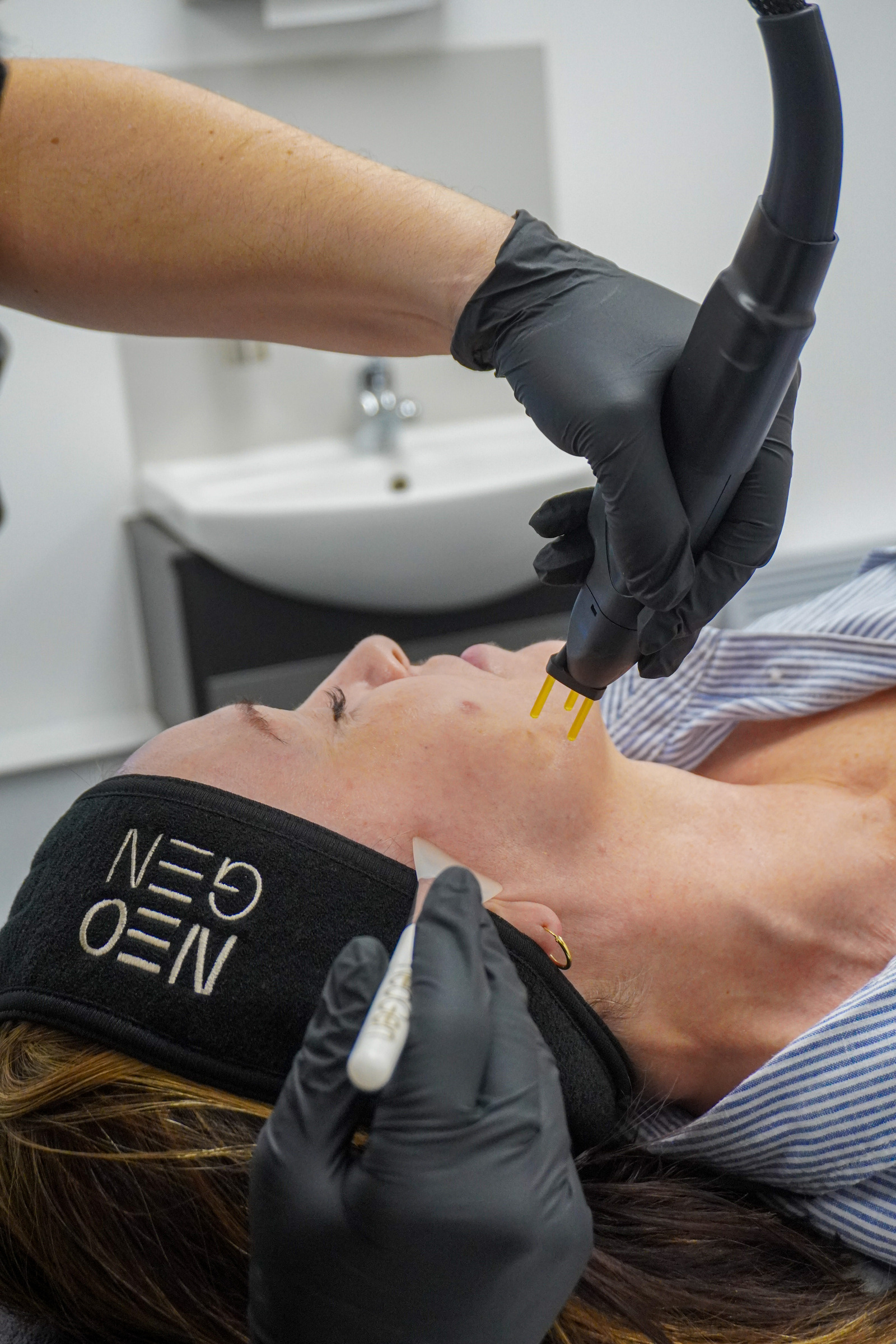
NEOGEN nitrogen plasma: what the energy actually does to skin
NEOGEN plasma at eLIVEate Me in Buckinghamshire. Nitrogen plasma denatures collagen without a laser chromophore. A course of three, then 90 days of rebuild.
More in this desk
50 min · long read · Clinic
CELL STORY liquid microneedling: 50,000 Microspears, no needle gun
CELL STORY at eLIVEate Me in Great Missenden. Sponge Microspears open transient channels, stimulate collagen, and shed in 72 hours — microneedling without a gun.

49 min · long read · Clinic
In-clinic NAD+ and the lyophilised research vial are not the same product
In-clinic NAD+ at eLIVEate Me (£200/month IM) is not Patriot’s 1000 mg lyophilised research vial. Same molecule class, different product, different till.
Essays describe published research. They are not medical advice and they do not authorise human use of any catalogue item.